Embed Size (px)
DESCRIPTION
Validation of the measurement of 3D left ventricular strains using image warping and untagged MRI images. AI Veress, JA Weiss, GC Klein, and GT Gullberg. Introduction: Motivation for Study. - PowerPoint PPT Presentation
Citation preview
Validation of the measurement of 3D left ventricular strains
using image warping and untagged MRI images.
AI Veress, JA Weiss, GC Klein, and GT Gullberg

Introduction: Motivation for Study Assessing regional heart wall
motion (wall motion, thickening, strain, etc.) provides quantitative estimates of local wall function.
Extent of ischemic myocardial disease
Extent of impairment of cardiac function due to hypertrophic and dilated cardiomyopathies.
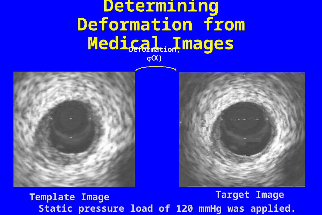
Determining Deformation from Medical Images
Template Image Target Image
Deformation,X)
Static pressure load of 120 mmHg was applied.
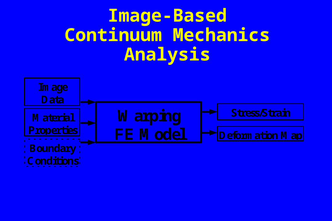
Image-BasedContinuum Mechanics Analysis
WarpingFE Model
ImageData
Stress/StrainMaterialProperties Deformation Map
CCBoundaryConditions
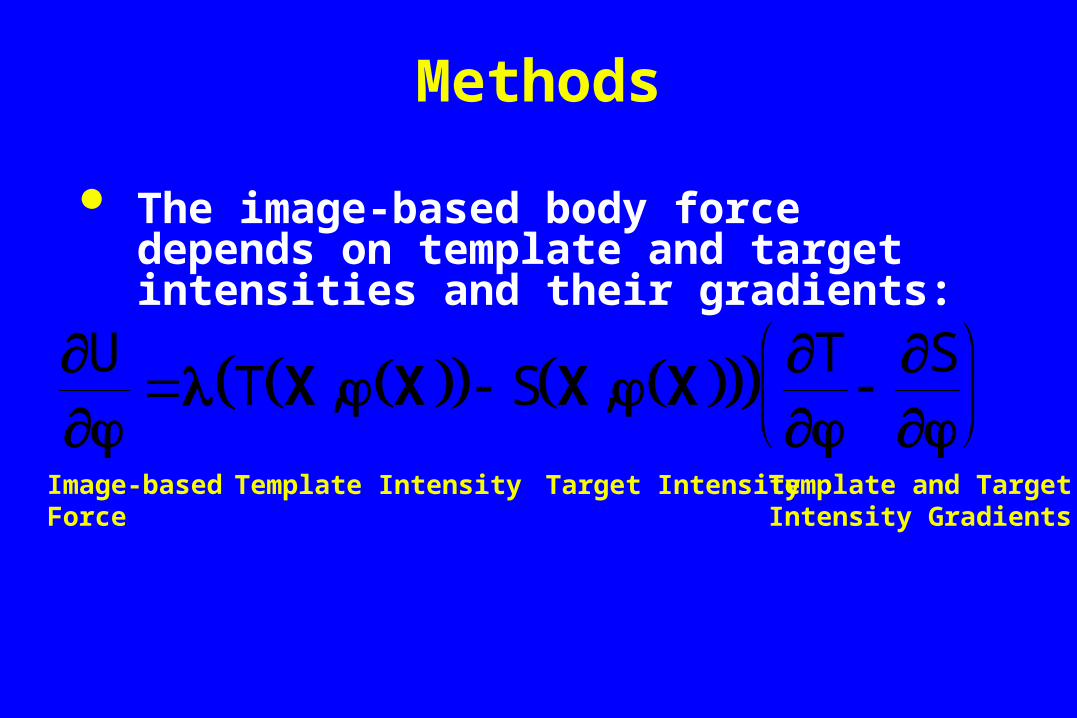
Methods
The image-based body force depends on template and target intensities and their gradients:
UT S
T S
X X X X, ,
Template IntensityImage-basedForce
Target Intensity Template and TargetIntensity Gradients
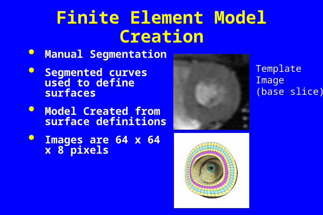
Manual Segmentation
Segmented curves used to define surfaces
Model Created from surface definitions
Images are 64 x 64 x 8 pixels
Finite Element Model Creation
Template Image (base slice)
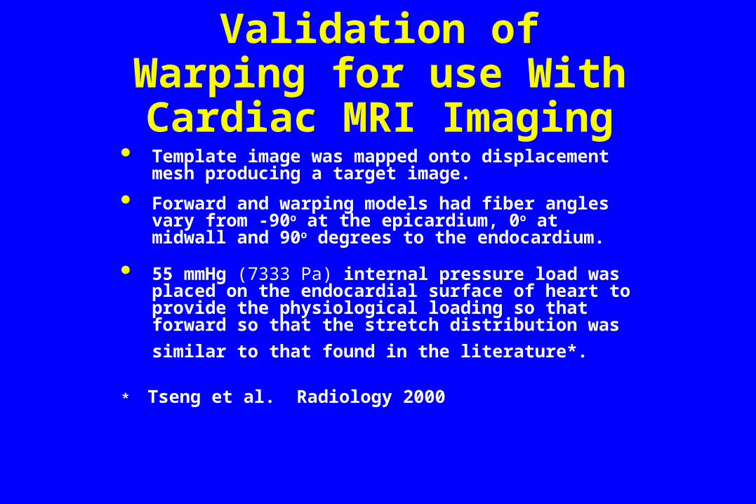
Validation of Warping for use With Cardiac MRI
Imaging Template image was mapped onto displacement mesh
producing a target image.
Forward and warping models had fiber angles vary from -90o at the epicardium, 0o at midwall and 90o degrees to the endocardium.
55 mmHg (7333 Pa) internal pressure load was placed on the endocardial surface of heart to provide the physiological loading so that forward so that the stretch distribution was similar to that found in the
literature*.
* Tseng et al. Radiology 2000
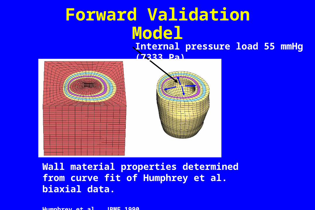
Forward Validation Model
Wall material properties determined from curve fit of Humphrey et al. biaxial data.
Humphrey et al. JBME 1990
Internal pressure load 55 mmHg (7333 Pa)
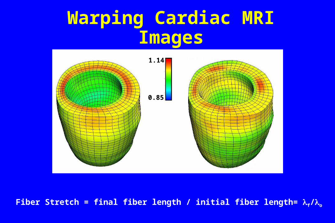
Warping Cardiac MRI Images
Fiber Stretch = final fiber length / initial fiber length= f/o
1.14
0.85
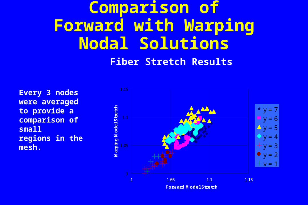
Comparison of Forward with Warping Nodal
Solutions Fiber Stretch Results
1
1.05
1.1
1.15
1 1.05 1.1 1.15
Forward Model Stretch
Wa
rpin
g M
od
el
Str
etc
h y = 7
y = 6
y = 5
y = 4
y = 3
y = 2
y = 1
Every 3 nodes were averagedto provide a comparison of smallregions in the mesh.

Discussion
Using regional average values provides a better method for comparison.
The average of three nodes were used in the present work.
Comparison of the stretches at the nodal locations showed that the nodes were not in the same location in each deformed state.

Conclusion
Initial validation results seems to indicate that reasonably good agreement will be reached.
Strain distributions look reasonable accurate for the resolution of the images.

Acknowledgments
Dr. Grant Gullberg
Dr. Jeffrey Weiss
Dr Gregory Klein
Dr. Richard Rabbitt
Anton Bowden