Embed Size (px)
Citation preview

Nig. J. Biotech. Vol. 38 (1) : 1-13 (June 2021)
ISSN: 0189 1731
Available online at
http://www.ajol.info/index.php/njb/index
and www.biotechsocietynigeria.org
DOI: https://dx.doi.org/10.4314/njb.v38i1.1
Antimicrobial Efficacy of Vitellaria paradoxa fractions and compounds on some wood Fungi and Bacteria
*Ekhuemelo, D. O.1, Anyam, J. V.2, and Ekhuemelo, C.3
1Department of Forest Production and Products, Federal University of Agriculture Makurdi, Nigeria
2Department of Chemistry, Federal University of Agriculture Makurdi, Nigeria 3Department of Crop and Environmental Protection, Federal University of Agriculture Makurdi, Nigeria
Abstract
This study examined antifungal efficacy of Vitellaria paradoxa fractions and compounds in the control of
some wood degrading fungi. Stem bark and heartwood parts of Vitellaria paradoxa were collected, dried, pulverised and macerated sequentially in n- hexane, methanol and ethyl acetate solvents. The mixtures
were filtered, evaporated and the dried samples were mixed and run over silica gel in column chromatography with a mixture of n -hexane and ethyl acetate solvents to obtain fractions. The fractions collected were evaporated and those with white needles were subjected to Magnetic Resonance (NMR) spectroscopic analysis. Spinasterol was isolated and characterised from the heartwood fraction while
the stem bark fractions were fatty. Vitellaria paradoxa fractions were active against Serpula lacrymans, Sclerotium rolfsii, Aspergillus fumigatus, Fomitopsis pinicoca, Phaeolus schweinitzii, Rhizopus sp., Coniophora puteana, Gloeophyllum sepiarium, and Fibroporia vaillantii at zones of inhibition (ZOI) of 18
mm - 24 mm. Although the antibiotics were active (25 – 31 mm), they were found inactive against the Fomitopsis pinicoca fungus which was sensitive to all the V. paradoxa fractions at zones of inhibition of 18
- 24 mm. The minimum inhibitory concentrations (MIC) of the V. paradoxa fractions were active at
50 µg/mL against all test fungi. At minimum fungicidal concentration (MFC) of between 50 - 200 µg/mL, all the test fungi were killed. Based on the ZOI, MIC and MFC, the V. paradoxa stem bark heartwood
fractions have been proven to be very efficient in inhibiting the growth of test wood rot fungi; hence the species could be explored as a potential source of bioactive fungicides.
Keywords: Bacteria, Compound, Fraction, Fungi, Spinasterol, Vitellaria paradoxa *Corresponding author Email: [email protected], Tel: +234-703 133 2803
Introduction
Vitellaria paradoxa (C. F. Gaertn) formally referred to as Butyrospermum parkii and Butyrospermum paradoxa belongs to the family
Sapotaceae. It is commonly known as Shea tree or Shea butter tree (IPGRI, INIA. 2006). In
English, it is called Shea butter tree; Shea nut tree; karate in French (Teketay et al., 2003);
Ka’danya, Kadanya, Mankade in Hausa; Okwuma, Osisi in Igbo (Audu & Awulu, 2017), Ori, Emi in Yoruba (Animasaun et al., 2019), and Chamegh
in Tiv (Shomkegh et al., 2016). The Shea butter tree is of average height of 10 - 15 m with dense
spreading canopy, thick, dark and fissured bark
(ICRAF, 2000). This species is known both as a fruit tree and as an oilseed crop (Ugese et al.,
2008).
The species originated in African and is found in
areas with 400 -1800 mm rainfall per year (IPGRI, INIA. 2006). It either grows naturally or is farmed
as a tree crop in the arid Savanna region of West
Africa countries (Audu & Awulu, 2017). It is believed to have spread from Senegal in West
Africa to Uganda in East Africa till Adamaoua Province in Cameroon which is in North-South

Ekhuemelo et al. / Nig. J. Biotech. Vol. 38 Num. 1 : 1-13 (June 2021)
2
Africa (IPGRI, INIA. 2006). Vitellaria paradoxa grows in the wild area of dry savannah region of West Africa from Senegal in the west to
Sudan in the east to the foothills of Ethiopian highlands. This species also grows in nineteen
countries of Africa. Namely Burkina Faso, Ghana,
Chad, Cameroon, Central African Republic, Ethiopia, Niger, Ivory Coast, Mali, Nigeria, Sierra
Leone, Guinea Bissau, Togo Uganda, Zaire, Senegal, Guinea, Benin and Sudan (Warra,
2011).
Vitellaria paradoxa is multipurpose agroforestry
indigenous tree species that contribute immensely to the livelihood of rural communities
for income generation and as source of raw material for many industries. The ripe fruits are
eaten as a snack and as famine food. Shea butter,
or Shea oil, is a raw material used in factories to manufacture margarine, baking fat, cocoa butter
substitutes as well as different pharmaceutical and moisturising beauty products (IPGRI, INIA.
2006). Shea butter leaves are good forage for animal feeding, soil improvement (Ziblim et al., 2015). The species has the capacity to improve
nutrition, increase healthcare, decrease rural poverty and aid sustainable land care (Moore,
2008).The wooden stem is resistant to termites and a good timber used for various constructing
purposes.
Several authors have reported the use of Shea
butter in medicine (Prescott et al., 2002; El-Mahmoud et al., 2008; Ahmed and Sani, 2013;
Ahmed et al., 2012; Fodouop et al., 2015). It is
used in the treatment of rheumatic and joint pains and healing of wounds, managing swellings
and bruises, dermatitis and other skin (Fodouop et al., 2015). Some people have been reported to
consume extract of V. paradoxa for treatment of various bacterial and fungal infections (Kalgo et
al., 2019). The stem back extract possesses
immunomodulatory properties which have effect on human neutrophils viability and function
(Kalgo et al., 2019) and contain antioxidant constituents with antimicrobial activities
(Olasunkanmi et al., 2017). Shea tree bark, if
purified, could be used to produce an antiseptic agent capable of treating skin infections caused
by groups of fungi (Ahmed et al., 2009).
Vitellaria paradoxa has been researched as an effective medicinal plant (Prescott et al., 2002).
It has proved to be active against bacterial and
fungal diseases (El-Mahmoud et al., 2008; Ahmed and Sani, 2013). The barks, leaves and roots
extracts of V. paradoxa contain phytochemical constituents inhibits growth of some
dermatophytes (Boyejo et al., 2019) and could be
used as a potential source of antibiotic substance for a drug development (Ajijolakewu and Awarun,
2015). Vitellaria paradoxa may be used as potent sources of bioactive substances in the production
of drugs against diseases caused by superficial and enteric organisms (Ajijolakewu and Awarun,
2015). The leaf, stem bark, and seed of shea tree
contain a host of bioactive compounds that can be utilized in the control of infections caused by
T. mentagrophyte, A. fumigatus, E. flocossum, T. rubrum, and M. Audouinii (Ahmed et al., 2012).
Wood is versatile and can be used for a very long time but it is subject to biodeterioration. Wood
deterioration is a very essential process in the environment that recycles complex organic
materials and it is a fundamental component of life (Blanchette, 2000). Primarily, fungi, inserts,
ants and bacteria are some of the agents of
deterioration. Wood decay fungi are classified into two major groups: white- and brown-rot
fungi (Blanchette, 2000). White rot fungi can degrade all cell wall components, including lignin.
They cause bleaching of common wood
colouration, metabolize large amounts of lignin in wood is unique among microorganisms, degrade cells and reduce the strength properties of wood in the late stages of decay. On other hand,
brown-rot fungi depolymerise cellulose quickly
during initial stages of wood colonization. Substantial losses in wood strength start very
early in the decay process, frequently before decay evidence is visually shown (Blanchette,
2000). Bacteria are known to have the ability to decompose wood cellulose although their
influence on wood decay is restricted. Different
bacteria from woodland soil contain enzymes used in the breakdown of cellulose and cellulose
products (Llado´ et al., 2015).
Fungi and bacteria play a major role in the well
being, diversity, and productivity of forest ecosystems. Fungi feed on woody products and
serve to recycle nutrients. As a result, they physically and chemically break down wood
products thereby causing lots of economic hazards (Marcot, 2007). Despite the many studies
Ekhuemelo et al. / Nig. J. Biotech. Vol. 38 Num. 1 : 1-13 (June 2021)
3
that have been done on the medicinal and
antimicrobial properties of V. paradoxa, not much has been done on the effect of V. paradoxa
activity on wood degrading fungi and bacteria. Therefore, this study was undertaken to evaluate
the compounds present in the stem bark and
heartwood fractions of V. paradoxa and assess their effect on selected wood decay fungi and
bacteria.
Materials and Methods Plant parts Collection and preparation
Stem bark of V. paradoxa was collected from the
wild at the Federal University of Agriculture, Makurdi campus, located between longitudes 8°
21ˈ and 9° E and latitudes 7° 21ˈ and 8° N in Benue State, Nigeria, within the southern guinea
savannah ecological zone (Seibert, 2007). The
stem bark was air- dried during harmattan season and pulverised. The heartwood was sawn
at Makurdi Head Bridge Timber Shed to collect its sawdust which was also dried to avoid being
damp.
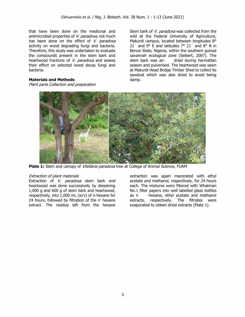
Plate 1: Stem and canopy of Vitellaria paradoxa tree at College of Animal Science, FUAM
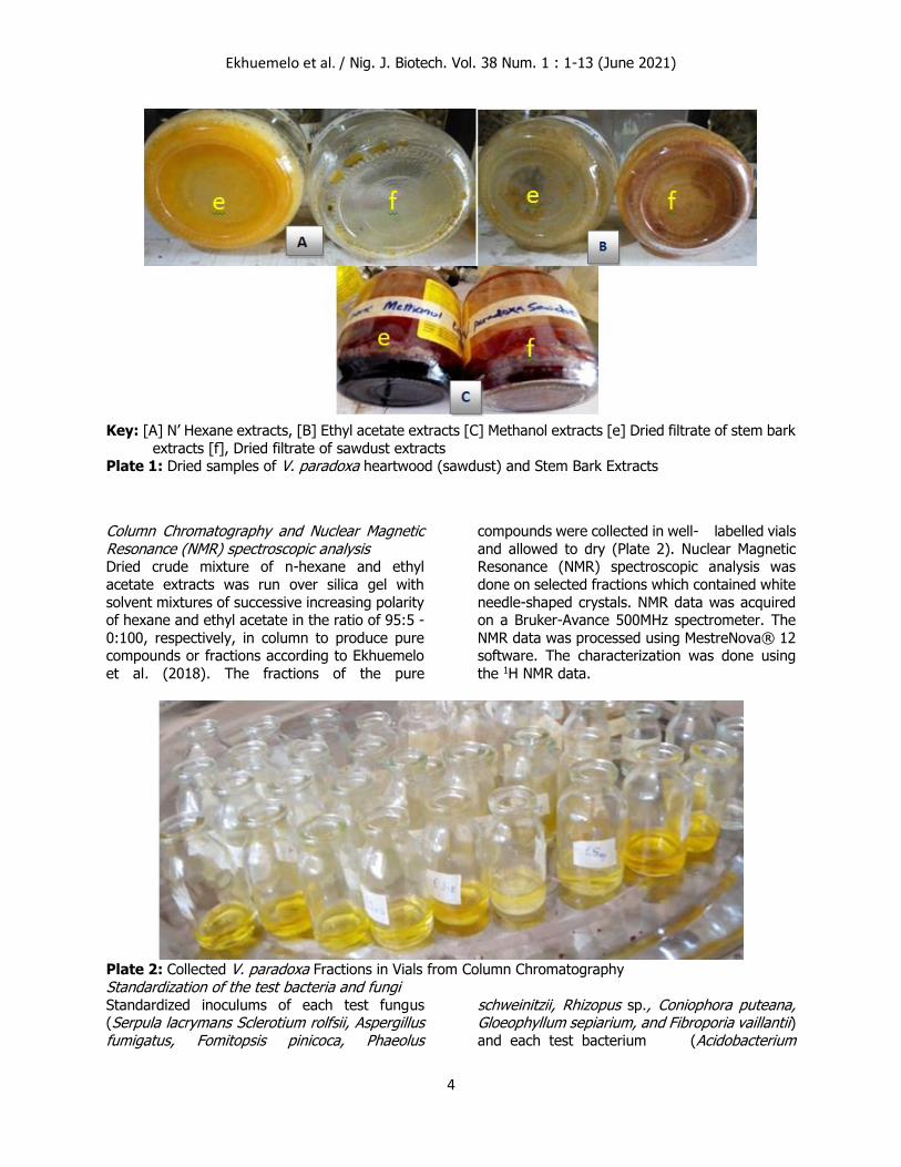
Extraction of plant materials Extraction of V. paradoxa stem bark and
heartwood was done successively by dissolving
1,000 g and 600 g of stem bark and heartwood, respectively, into 1,000 mL (w/v) of n-hexane for
24 hours, followed by filtration of the n’ hexane extract. The residue left from the hexane
extraction was again macerated with ethyl acetate and methanol, respectively, for 24 hours
each. The mixtures were filtered with Whatman
No.1 filter papers into well labelled glass bottles as n -hexane, ethyl acetate and methanol
extracts, respectively. The filtrates were evaporated to obtain dried extracts (Plate 1).
Ekhuemelo et al. / Nig. J. Biotech. Vol. 38 Num. 1 : 1-13 (June 2021)
4
Key: [A] N’ Hexane extracts, [B] Ethyl acetate extracts [C] Methanol extracts [e] Dried filtrate of stem bark
extracts [f], Dried filtrate of sawdust extracts Plate 1: Dried samples of V. paradoxa heartwood (sawdust) and Stem Bark Extracts
Column Chromatography and Nuclear Magnetic Resonance (NMR) spectroscopic analysis Dried crude mixture of n-hexane and ethyl
acetate extracts was run over silica gel with
solvent mixtures of successive increasing polarity of hexane and ethyl acetate in the ratio of 95:5 -
0:100, respectively, in column to produce pure compounds or fractions according to Ekhuemelo
et al. (2018). The fractions of the pure
compounds were collected in well- labelled vials
and allowed to dry (Plate 2). Nuclear Magnetic Resonance (NMR) spectroscopic analysis was
done on selected fractions which contained white
needle-shaped crystals. NMR data was acquired on a Bruker-Avance 500MHz spectrometer. The
NMR data was processed using MestreNova® 12 software. The characterization was done using
the 1H NMR data.
Plate 2: Collected V. paradoxa Fractions in Vials from Column Chromatography
Standardization of the test bacteria and fungi Standardized inoculums of each test fungus (Serpula lacrymans Sclerotium rolfsii, Aspergillus fumigatus, Fomitopsis pinicoca, Phaeolus
schweinitzii, Rhizopus sp., Coniophora puteana, Gloeophyllum sepiarium, and Fibroporia vaillantii) and each test bacterium (Acidobacterium

Ekhuemelo et al. / Nig. J. Biotech. Vol. 38 Num. 1 : 1-13 (June 2021)
5
capsulatum, Actinobacteria sp. Agrobacterium tumefaciens, Bacillus subtilis Ralstonia solanacearum, Enterococcus faecium, Pseudomonas syringae, Escherichia coli, Proteus mirabilis and Pseudomonas aeruginosa) were achieved by preparing their respective
suspensions up to 0.5 McFarland standards. The growth of the actively growing culture in the
broth was altered with sterile saline or broth to obtain turbidity that was clearly comparable to
that of the 0.5 McFarland standards according to Lalitha, (2004); Kuta et al. (2017).
Determination of antimicrobial activity of Vitellaria paradoxa fractions The activity of V. paradoxa fractions on test and bacteria and fungi samples was investigated
using agar diffusion method. Concentrations of
200 μg/mL and 100 μg/mL were prepared from each fraction and antibiotics respectively. The
antibiotics used as control were Ketoconazole, fluconazole and Fulcin for fungi; and
ciprofloxacin, sparfloxacin and cefuroxime for bacteria according to Ekhuemelo et al. (2018).
The Muller-Hinton and Sabouraud Dextrose agar
were used in the culture of bacteria and fungi, respectively. The incubation of test fungi was
made at 30 oC for 7 days and at 37 oC for 24 hours for bacteria. During the period plates of the media
were observed for inhibition of growth. Zones
of inhibition were measured and results recorded in millimetres (mm).
Minimum inhibitory concentration (MIC)/ Minimum Fungicidal concentration (MFC) The MIC of the antifungal fractions and compounds was determined by broth dilution
methods. Different concentrations (200 μg/mL, 100 μg/mL, 50 μg/mL, 25 μg/mL and 12.5
μg/mL) of fractions were added to media in the Petri dishes and inoculated with the test fungi.
The mixture was incubated and examined for
growth according to Ekhuemelo et al. (2018). The
lowest concentration of the fraction that inhibits visible growth of the test fungi was recorded as
the MIC. The MFC of the fraction was determined by sub-
culturing the contents of the Petri dishes that
showed inhibition on Muller-Hinton and Sabouraud Dextrose agar plates for bacteria and
fungi, respectively, while the absence of growth on incubation was recorded as MFC.
Results
Characterization of VPH22 as Spinasterol
Vitellaria paradoxa heartwood fraction (VPH22) was obtained as white needles. It’s proton
nuclear magnetic resonance spectrum (1H-NMR) gave the following data (Figure 1) below: 1H NMR
(500 MHz, Chloroform-d) δ 5.19 – 5.13 (m, 2H), 5.03 (dd, J = 15.1, 8.6 Hz, 1H), 3.60 (tt, J = 10.7,
4.4 Hz, 2H), 2.34 (t, J = 7.5 Hz, 2H), 2.09 – 1.96
(m, 6H), 1.03 (d, J = 6.6 Hz, 3H), 0.88 (s, 2H), 0.86 (s, 3H), 0.84 (s, 3H), 0.82 (s, 1H), 0.81 (s,
2H), 0.80 (s, 8H), 0.55 (s, 2H). The signals at δ 5.19 – 5.13 (m, 2H) and 5.03 (dd, J = 15.1, 8.6
Hz, 1H), indicate the presence of olefinic bonds
(Alexandri et al., 2017), while that at 3.60 (tt, J = 10.7, 4.4 Hz, 1H) indicate an oxymethine
proton (Sun and Yasukawa, 2008); signals at 2.34 (t, J = 7.5 Hz, 2H) and 2.09 – 1.96 (m, 6H)
are representative of methine and methylene protons. The signals at 1.03 (d, J = 6.6 Hz, 3H),
0.88 (s, 2H), 0.86 (s, 3H), 0.84 (s, 3H), 0.81 (s,
3H) and 0.55 (s, 3H) are due to methyl protons (Cheung, and Williamson, 1969). The data
acquired was reminiscent of that for sterols and triterpenes. A literature look up revealed the data
to be unambiguously identical to that of
spinasterol (Villaseñor and Domingo, 2008). Vitellaria paradoxa heartwood fraction (VPH22)
was characterised as Spinasterol (Figure 2).
Ekhuemelo et al. / Nig. J. Biotech. Vol. 38 Num. 1 : 1-13 (June 2021)
6
Figure 1: Proton NMR Spectrum of Vitellaria paradoxa heartwood fraction (VPH22)
Figure 2: Structure of VPH22 (Spinasterol)
Effect of Antifungal activities of V. paradoxa fractions and Antibiotics against test fungi The test fungi were sensitive to all the V. paradoxa fractions (VP17, VP28, VP34, VP47, VP23, VP134, and VPH22) at zones of inhibition (ZOI) of between 18 and
24 mm (Table 1). Aspergillus fumigatus, Coniophora puteana, Fomitopsis pinicoca, Gloeophyllum sepiarium,
Phaeolus schweinitzii, Rhizopus sp. and Serpula lacrymans were sensitive to V. paradoxa stem bark fraction (VP17) with ZOI of between 20 mm to
24 mm. It was also observed that Coniophora puteana, Fibroporia vaillantii, Fomitopsis pinicoca, Gloeophyllum sepiarium, Rhizopus sp., Serpula lacrymans and Sclerotium rolfsii were sensitive to V. paradoxa stem
Ekhuemelo et al. / Nig. J. Biotech. Vol. 38 Num. 1 : 1-13 (June 2021)
7
bark fraction (VP28) with ZOI ranging between
20 mm and 24 mm while the antibiotics were active on all test fungi at ZOI of 25 – 31 mm except F. pinicoca which was resistant.
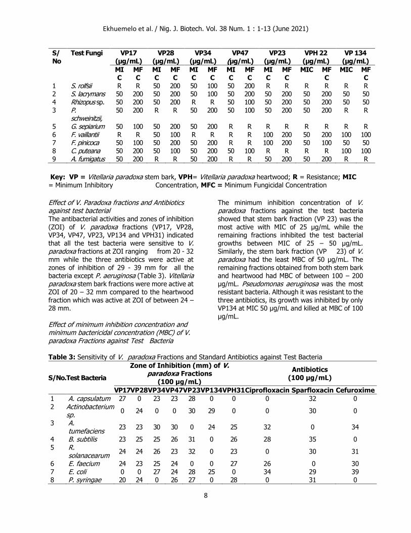
Effect of Minimum inhibition concentration (MIC) of V. paradoxa Fraction against test fungi At the minimum inhibition concentration (MIC) of
50 µg/mL, VP23 fraction prevented the growth of Aspergillus fumigatus, Phaeolus schweinitzii, Rhizopus sp. and Serpula lacrymans test pathogens (Table 2). Also, at 50 µg/mL, V. paradoxa stem bark fraction (VP34) stopped the
growth of Aspergillus fumigatus, Fibroporia vaillantii, Fomitopsis pinicoca, Phaeolus schweinitzii,, Rhizopus sp. and Serpula lacrymans test pathogens while, V. paradoxa heartwood
fraction (VP22) only inhibited Coniophora puteana, Fibroporia vaillantii, Rhizopus sp. and Serpula lacrymans test pathogens. However,
at100 µg/mL, Fibroporia vaillantii and Fomitopsis pinicoca growth was prevented by VP23 isolate while Coniophora puteana and Fibroporia vaillantii growths were inhibited by V. paradoxa stem bark fraction (VP134).
Effect of Minimum Fungicidal concentration of V. paradoxa fractions on test Fungi Table 2 presents the MFC of V. paradoxa fractions of VP17, VP28, VP34, VP47, VP23, VPH22 and
VP134 against test fungi. VP134 fraction was the most active fraction compared to the rest as
it completely inhibited the growths of Fomitopsis pinicoca, Rhizopus sp. and Serpula lacrymans at MFC of 50 µg/mL. At 100 µg/mL, VPH22 fraction killed Fomitopsis pinicoca while VP134 fraction completely stopped the growth of
Coniophora puteana and Fibroporia vaillantii. At
200 µg/mL rest fractions killed all the test fungi.
Table 1: Sensitivity of Vitellaria paradoxa Fractions and Standard Antibiotics against Test Fungi
S/
No
Test fungi
Zone of Inhibition (mm) of Vitellaria paradoxa Fractions
(200 µg/mL)
Zone of Inhibition (mm) of
Antibiotic (100 µg/mL)
VP1
7
VP2
8
VP3
4
VP
47
VP2
3
VP
134
VPH
22
Flucona
zole
Fulci
n
Ketoconazo
le 1 S. rolfsii 0 21 24 21 0 0 0 0 0 25
2 S. lacrymans
21 20 23 20 21 23 20 0 30 30
4 Rhizopus sp.
20 23 0 24 23 21 22 0 29 27
3 P. schweinitzii,
20 0 20 23 20 0 21 0 25 0
5 G. sepiarium
24 21 21 0 0 0 0 29 0 28
6 F. vaillantii 23 20 22 0 19 20 23 0 28 0
7 F. pinicoca 0 24 0 0 18 19 21 0 0 0
8 C. puteana 21 23 20 23 0 18 0 0 31 0 9 A.
fumigatus 20 0
20 0 21 0 20 0 29 25
Key: R = Resistance, VP = Vitellaria paradoxa stem bark, VPH= Vitellaria paradoxa heartwood, ZOI < 10 mm is inactive; 10 -13 mm is partially active; 14 -19 mm is active, and >19 mm is very active.
Table 2: Minimum Inhibition Concentration and Minimum Fungicidal Concentration of Vitellaria paradoxa against Test Fungi
Ekhuemelo et al. / Nig. J. Biotech. Vol. 38 Num. 1 : 1-13 (June 2021)
8
S/
No
Test Fungi VP17 (µg/mL)
VP28
(µg/mL)
VP34
(µg/mL)
VP47 (µg/mL)
VP23
(µg/mL) VPH 22
(µg/mL)
VP 134
(µg/mL)
MIC
MFC
MIC
MFC
MIC
MFC
MIC
MFC
MIC
MFC
MIC MFC
MIC MFC
1 S. rolfsii R R 50 200 50 100 50 200 R R R R R R 2 S. lacrymans 50 200 50 200 50 100 50 200 50 200 50 200 50 50
4 Rhizopus sp. 50 200 50 200 R R 50 100 50 200 50 200 50 50
3 P. schweinitzii,
50 200 R R 50 200 50 100 50 200 50 200 R R
5 G. sepiarium 50 100 50 200 50 200 R R R R R R R R
6 F. vaillantii R R 50 100 R R R R 100 200 50 200 100 100 7 F. pinicoca 50 100 50 200 50 200 R R 100 200 50 100 50 50
8 C. puteana 50 200 50 100 50 200 50 100 R R R R 100 100
9 A. fumigatus 50 200 R R 50 200 R R 50 200 50 200 R R
Key: VP = Vitellaria paradoxa stem bark, VPH= Vitellaria paradoxa heartwood; R = Resistance; MIC
= Minimum Inhibitory Concentration, MFC = Minimum Fungicidal Concentration
Effect of V. Paradoxa fractions and Antibiotics against test bacterial The antibacterial activities and zones of inhibition
(ZOI) of V. paradoxa fractions (VP17, VP28, VP34, VP47, VP23, VP134 and VPH31) indicated
that all the test bacteria were sensitive to V. paradoxa fractions at ZOI ranging from 20 - 32
mm while the three antibiotics were active at
zones of inhibition of 29 - 39 mm for all the bacteria except P. aeruginosa (Table 3). Vitellaria paradoxa stem bark fractions were more active at ZOI of 20 – 32 mm compared to the heartwood
fraction which was active at ZOI of between 24 – 28 mm.
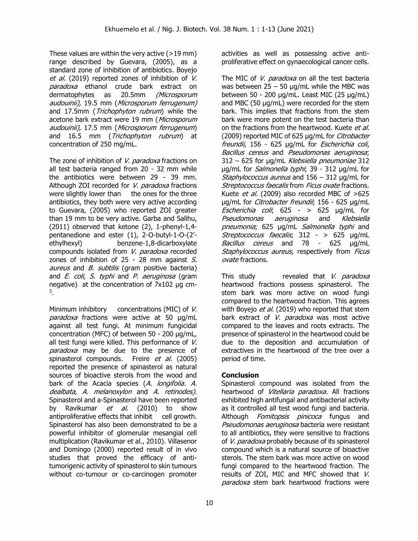
Effect of minimum inhibition concentration and minimum bactericidal concentration (MBC) of V. paradoxa Fractions against Test Bacteria
The minimum inhibition concentration of V. paradoxa fractions against the test bacteria
showed that stem bark fraction (VP 23) was the
most active with MIC of 25 µg/mL while the remaining fractions inhibited the test bacterial
growths between MIC of 25 – 50 µg/mL. Similarly, the stem bark fraction (VP 23) of V. paradoxa had the least MBC of 50 µg/mL. The
remaining fractions obtained from both stem bark and heartwood had MBC of between 100 – 200
µg/mL. Pseudomonas aeruginosa was the most resistant bacteria. Although it was resistant to the
three antibiotics, its growth was inhibited by only VP134 at MIC 50 µg/mL and killed at MBC of 100
µg/mL.
Table 3: Sensitivity of V. paradoxa Fractions and Standard Antibiotics against Test Bacteria
S/No. Test Bacteria
Zone of Inhibition (mm) of V. paradoxa Fractions
(100 µg/mL)
Antibiotics
(100 µg/mL)
VP17 VP28 VP34 VP47 VP23 VP134 VPH31 Ciprofloxacin Sparfloxacin Cefuroxime
1 A. capsulatum 27 0 23 23 28 0 0 0 32 0
2 Actinobacterium sp.
0 24 0 0 30 29 0 0 30 0
3 A. tumefaciens
23 23 30 30 0 24 25 32 0 34
4 B. subtilis 23 25 25 26 31 0 26 28 35 0
5 R. solanacearum
24 24 26 23 32 0 23 0 30 31
6 E. faecium 24 23 25 24 0 0 27 26 0 30
7 E. coli 0 0 27 24 28 25 0 34 29 39 8 P. syringae 20 24 0 26 27 0 28 0 31 0
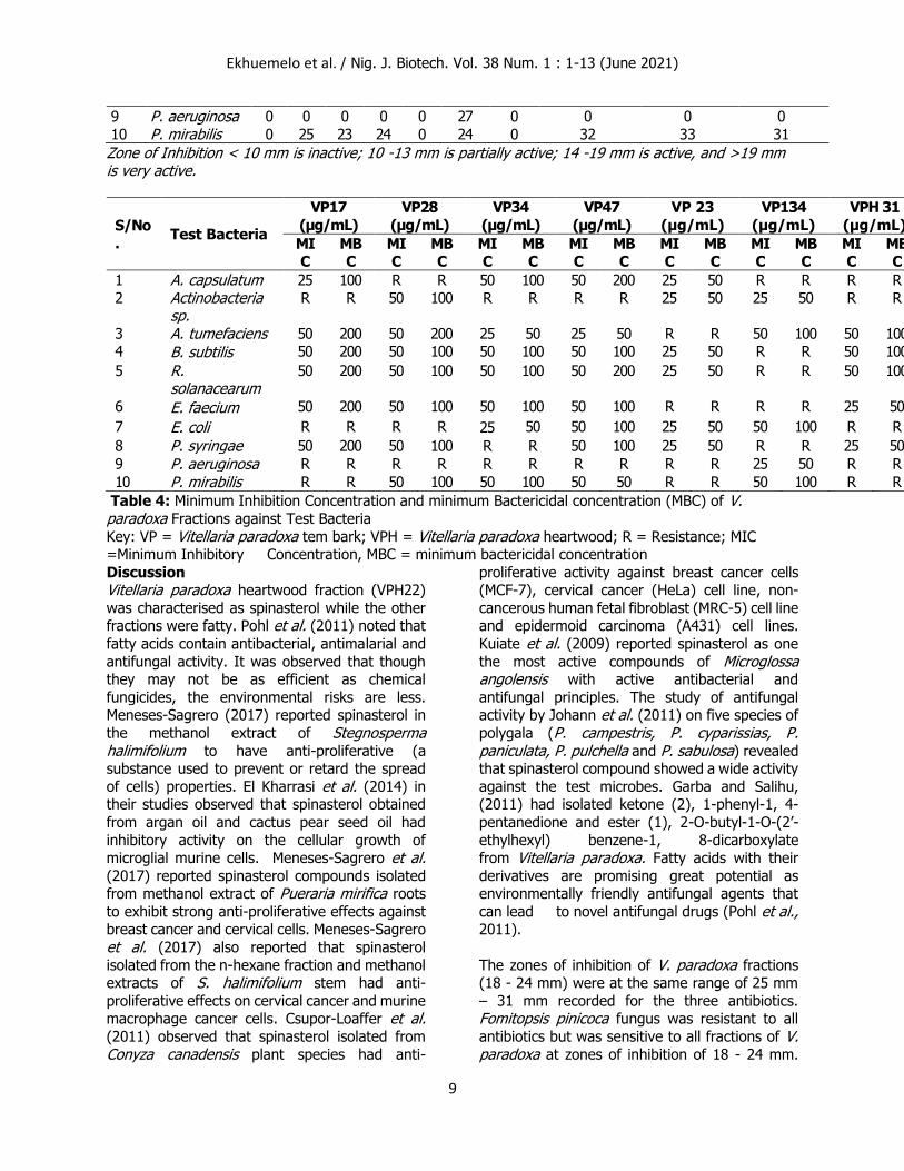
Ekhuemelo et al. / Nig. J. Biotech. Vol. 38 Num. 1 : 1-13 (June 2021)
9
9 P. aeruginosa 0 0 0 0 0 27 0 0 0 0
10 P. mirabilis 0 25 23 24 0 24 0 32 33 31
Zone of Inhibition < 10 mm is inactive; 10 -13 mm is partially active; 14 -19 mm is active, and >19 mm is very active.
S/No
. Test Bacteria
VP17
(µg/mL)
VP28
(µg/mL)
VP34
(µg/mL)
VP47
(µg/mL)
VP 23
(µg/mL)
VP134
(µg/mL)
VPH 31
(µg/mL)
MIC
MBC
MIC
MBC
MIC
MBC
MIC
MBC
MIC
MBC
MIC
MBC
MIC
MBC
1 A. capsulatum 25 100 R R 50 100 50 200 25 50 R R R R
2 Actinobacteria sp.
R R 50 100 R R R R 25 50 25 50 R R
3 A. tumefaciens 50 200 50 200 25 50 25 50 R R 50 100 50 100 4 B. subtilis 50 200 50 100 50 100 50 100 25 50 R R 50 100
5 R. solanacearum
50 200 50 100 50 100 50 200 25 50 R R 50 100
6 E. faecium 50 200 50 100 50 100 50 100 R R R R 25 50
7 E. coli R R R R 25 50 50 100 25 50 50 100 R R
8 P. syringae 50 200 50 100 R R 50 100 25 50 R R 25 50
9 P. aeruginosa R R R R R R R R R R 25 50 R R
10 P. mirabilis R R 50 100 50 100 50 50 R R 50 100 R R
Table 4: Minimum Inhibition Concentration and minimum Bactericidal concentration (MBC) of V. paradoxa Fractions against Test Bacteria
Key: VP = Vitellaria paradoxa tem bark; VPH = Vitellaria paradoxa heartwood; R = Resistance; MIC =Minimum Inhibitory Concentration, MBC = minimum bactericidal concentration
Discussion Vitellaria paradoxa heartwood fraction (VPH22)
was characterised as spinasterol while the other fractions were fatty. Pohl et al. (2011) noted that
fatty acids contain antibacterial, antimalarial and
antifungal activity. It was observed that though they may not be as efficient as chemical
fungicides, the environmental risks are less. Meneses-Sagrero (2017) reported spinasterol in
the methanol extract of Stegnosperma halimifolium to have anti-proliferative (a substance used to prevent or retard the spread
of cells) properties. El Kharrasi et al. (2014) in their studies observed that spinasterol obtained
from argan oil and cactus pear seed oil had
inhibitory activity on the cellular growth of microglial murine cells. Meneses-Sagrero et al. (2017) reported spinasterol compounds isolated from methanol extract of Pueraria mirifica roots
to exhibit strong anti-proliferative effects against breast cancer and cervical cells. Meneses-Sagrero
et al. (2017) also reported that spinasterol
isolated from the n-hexane fraction and methanol extracts of S. halimifolium stem had anti-
proliferative effects on cervical cancer and murine macrophage cancer cells. Csupor-Loaffer et al. (2011) observed that spinasterol isolated from
Conyza canadensis plant species had anti-
proliferative activity against breast cancer cells (MCF-7), cervical cancer (HeLa) cell line, non-
cancerous human fetal fibroblast (MRC-5) cell line and epidermoid carcinoma (A431) cell lines.
Kuiate et al. (2009) reported spinasterol as one
the most active compounds of Microglossa angolensis with active antibacterial and
antifungal principles. The study of antifungal activity by Johann et al. (2011) on five species of
polygala (P. campestris, P. cyparissias, P. paniculata, P. pulchella and P. sabulosa) revealed that spinasterol compound showed a wide activity
against the test microbes. Garba and Salihu, (2011) had isolated ketone (2), 1-phenyl-1, 4-
pentanedione and ester (1), 2-O-butyl-1-O-(2’-
ethylhexyl) benzene-1, 8-dicarboxylate from Vitellaria paradoxa. Fatty acids with their
derivatives are promising great potential as environmentally friendly antifungal agents that
can lead to novel antifungal drugs (Pohl et al., 2011).
The zones of inhibition of V. paradoxa fractions (18 - 24 mm) were at the same range of 25 mm
– 31 mm recorded for the three antibiotics. Fomitopsis pinicoca fungus was resistant to all
antibiotics but was sensitive to all fractions of V. paradoxa at zones of inhibition of 18 - 24 mm.
Ekhuemelo et al. / Nig. J. Biotech. Vol. 38 Num. 1 : 1-13 (June 2021)
10
These values are within the very active (>19 mm)
range described by Guevara, (2005), as a standard zone of inhibition of antibiotics. Boyejo
et al. (2019) reported zones of inhibition of V. paradoxa ethanol crude bark extract on
dermatophytes as 20.5mm (Microsporum audouinii), 19.5 mm (Microsporum ferrugenum) and 17.5mm (Trichophyton rubrum) while the
acetone bark extract were 19 mm (Microsporum audouinii), 17.5 mm (Microsporum ferrugenum)
and 16.5 mm (Trichophyton rubrum) at concentration of 250 mg/mL.
The zone of inhibition of V. paradoxa fractions on all test bacteria ranged from 20 - 32 mm while
the antibiotics were between 29 - 39 mm. Although ZOI recorded for V. paradoxa fractions
were slightly lower than the ones for the three
antibiotics, they both were very active according to Guevara, (2005) who reported ZOI greater
than 19 mm to be very active. Garba and Salihu, (2011) observed that ketone (2), 1-phenyl-1,4-
pentanedione and ester (1), 2-O-butyl-1-O-(2’-ethylhexyl) benzene-1,8-dicarboxylate
compounds isolated from V. paradoxa recorded
zones of inhibition of 25 - 28 mm against S. aureus and B. subtilis (gram positive bacteria)
and E. coli, S. typhi and P. aeruginosa (gram negative) at the concentration of 7x10² μg cm-3.
Minimum inhibitory concentrations (MIC) of V. paradoxa fractions were active at 50 µg/mL against all test fungi. At minimum fungicidal
concentration (MFC) of between 50 - 200 µg/mL, all test fungi were killed. This performance of V. paradoxa may be due to the presence of
spinasterol compounds. Freire et al. (2005) reported the presence of spinasterol as natural
sources of bioactive sterols from the wood and bark of the Acacia species (A. longifolia. A. dealbata, A. melanoxylon and A. retinodes). Spinasterol and a-Spinasterol have been reported by Ravikumar et al. (2010) to show
antiproliferative effects that inhibit cell growth. Spinasterol has also been demonstrated to be a
powerful inhibitor of glomerular mesangial cell
multiplication (Ravikumar et al., 2010). Villasenor and Domingo (2000) reported result of in vivo
studies that proved the efficacy of anti-tumorigenic activity of spinasterol to skin tumours
without co-tumour or co-carcinogen promoter
activities as well as possessing active anti-
proliferative effect on gynaecological cancer cells.
The MIC of V. paradoxa on all the test bacteria was between 25 – 50 µg/mL while the MBC was between 50 - 200 µg/mL. Least MIC (25 µg/mL)
and MBC (50 µg/mL) were recorded for the stem bark. This implies that fractions from the stem
bark were more potent on the test bacteria than on the fractions from the heartwood. Kuete et al. (2009) reported MIC of 625 µg/mL for Citrobacter freundii, 156 - 625 µg/mL for Escherichia coli, Bacillus cereus and Pseudomonas aeruginosa;
312 – 625 for µg/mL Klebsiella pneumoniae 312 µg/mL for Salmonella typhi; 39 - 312 µg/mL for
Staphylococcus aureus and 156 – 312 µg/mL for Streptococcus faecalis from Ficus ovate fractions.
Kuete et al. (2009) also recorded MBC of >625
µg/mL for Citrobacter freundii; 156 - 625 µg/mL Escherichia coli; 625 - > 625 µg/mL for
Pseudomonas aeruginosa and Klebsiella pneumonia; 625 µg/mL Salmonella typhi and
Streptococcus faecalis; 312 - > 625 µg/mL Bacillus cereus and 78 - 625 µg/mL
Staphylococcus aureus, respectively from Ficus ovate fractions.
This study revealed that V. paradoxa heartwood fractions possess spinasterol. The
stem bark was more active on wood fungi
compared to the heartwood fraction. This agrees with Boyejo et al. (2019) who reported that stem
bark extract of V. paradoxa was most active compared to the leaves and roots extracts. The
presence of spinasterol in the heartwood could be
due to the deposition and accumulation of extractives in the heartwood of the tree over a
period of time.
Conclusion Spinasterol compound was isolated from the
heartwood of Vitellaria paradoxa. All fractions
exhibited high antifungal and antibacterial activity as it controlled all test wood fungi and bacteria.
Although Fomitopsis pinicoca fungus and Pseudomonas aeruginosa bacteria were resistant
to all antibiotics, they were sensitive to fractions
of V. paradoxa probably because of its spinasterol compound which is a natural source of bioactive
sterols. The stem bark was more active on wood fungi compared to the heartwood fraction. The
results of ZOI, MIC and MFC showed that V. paradoxa stem bark heartwood fractions were
Ekhuemelo et al. / Nig. J. Biotech. Vol. 38 Num. 1 : 1-13 (June 2021)
11
very efficient in inhibiting the growth of test wood
rot fungi and wood colonising bacteria; hence the species should be explored as a potential source
of bioactive fungicides.
Reference
Ahmed, R.N., Abdulrahaman, A. A. and Sani, A. (2012). In vitro evaluation of antifungal
potentials of methanolic extracts of three organs
of Vitellaria paradoxa (Shea plant). J. Sci. Tech. Math. & Edu. 8(2): 8 – 15.
Ahmed R. N., and Sani A. (2013). Antimycotic
activity and toxicological effects of stem bark extract of Vitellaria paradoxa in wistar rats. Sci.
Inter. (Lahore), 25(1):91-102.
Ahmed R.N., Sani A., and Igunnugbemi O. O.
(2009). Antifungal profiles of extracts of Vitellaria paradoxa (Shea-butter) Bark. Ethnobot Leaflets, 13: 679-688.
Ajijolakewu, K. A. and Awarun F. J. (2015).
Comparative Antibacterial Efficacy of Vitellaria paradoxa (Shea Butter Tree) Extracts Against
Some Clinical Bacterial Isolates. Not. Sci. Biolo. 7(3):264 – 268.
Alexandri, E., Ahmed, R., Siddiqui, H., Choudhary, M. I., Tsiafoulis, C. G., and
Gerothanassis, I. P. (2017). High resolution NMR spectroscopy as a structural and analytical tool
for unsaturated lipids in solution. Molecules, 22(10), 1663.
Animasaun, D. A. Oyedeji, S., Olorunmaiye, K. S., Azeez, M. A., Tijani, I. A. and Morakinyo, J. A.
(2019). Morpho-chemical divergence and fatty acid profile of shea tree seeds (Vitellaria paradoxa) collected from different locations in Kwara State, Nigeria. Act. Bot. Croa. 78 (1), 17–
24
Audu, J. and Awulu, J.O. (2017). Effect of
extraction methods on some food and biodiesel
properties of shea-nut oil (Vitellaria paradoxa). J.
Posthar. Tech. 5(1): 17-26.
Blanchette, R. A. (2000). A review of microbial deterioration found in archaeological wood from
different environments. Inter. Biodeter. &
Biodegra. 46: 189–204.
Boyejo A.O., Azeez I.A., Owolabi S.L., and Issah A.O. (2019). Antifungal and Phytochemical
Screening of Extract from Vitellaria Paradoxa (Shea Butter Tree) Leaves, Barks and Roots on
Dermatophytes. Inter. J. Scient. & Res. Publi.
9(6): 884 – 891.
Cheung, H. T., and Williamson, D. G. (1969). NMR signals of methyl groups of triterpenes with
oxygen functions at positions 2, 3 and 23.
Tetrahedron, 25(1), 119-128. Csupor-Loaffer, B., Hajdu, Z., Zupko, I., Molnar,
J., Forgo, P., Vasas, A., Kele, Z., Hohmann, J., 2011. Antiproliferative constituents of the roots of
Conyza canadensis. Plan. Medi. 77, 1183–1188.
Ekhuemelo, D. O., Agbidye, F. S., Anyam, J. V.
and Ugba, R. B. (2018). Antimicrobial effect of isolated compound of Anadelphia afzeliana (Rendle) Stapf on selected wood fungi and bacteria in Makurdi, Nigeria. Nig. J. Biotech.
35(2): 108-120.
El Kharrasi, Y., Samadi, M., Lopez, T., Nury, T., el
Kebbaj, R., Andreoletti, P., El Hajj, H., Vamecq, J., Moustaid, K., Latruffe, N., El Kebbaj, L.,
Masson, D., Lizard, G., Nasser, B., and Cherkaoui-
Malki, M., (2014). Biological activities of schottenol and spinasterol, two natural
phytosterols present in argan oil and in cactus pear seed oil, on murine miroglial BV2 cells.
Bioch. & Biophy. Res. Comm. 446, 798–804.
El-Mahmood A. M., Doughari J. H., Ladan N.
(2008). Antimicrobial screening of stem bark extract of Vitelleria paradoxa against some
enteric pathogenic microorganisms. Afri. J. Pharm. & Pharmaco. 2(5):089-094.
Freire, C. S. R., Coelho, D. S. C., Santos, N. M., Silvestre, A. J. D., and Pascoal Neto, C.
(2005). Identification of Δ7 phytosterols and phytosteryl glucosides in the wood and bark of
several Acacia species phytosterols and
Ekhuemelo et al. / Nig. J. Biotech. Vol. 38 Num. 1 : 1-13 (June 2021)
12
phytosteryl glucosides in the wood and bark of
several Acacia species. Lipi. 40(3): 317–322. Fodouop, S. P. C., Gatsing, D., Tangue, B. T., Tagne, R. S., Tala, S. D., Tchoumboué, J., and
Kuiate, J. R. (2015). Effect of Salmonella typhimurium infection on rat’s cell oxidation and in vivo antioxidant activity of Vitellaria paradoxa
and Ludwigia abyssinica aqueous extract. Asian Pacif. J. Trop. Dis. 5(1), 38–46.
Garba S. and Salihu L. (2011). Antibacterial
Activities of 2-O-butyl-1-O-(2’-ethylhexyl)
benzene-1,8-dicarboxylate and 1-phenyl-1,4-pentanedione Isolated from Vitellaria paradoxa Root Bark. Asian J. of Scient. Res. 4 (2): 149-157.
Guevara, B. Q. (2005). A Guidebook to Plant Screening: Phytochemical and Biological, Revised
Edition, UST Publishing House, Manila. Pp 156.
ICRAF (2000). International Centre for Research in Agroforestry. Agroforestree Database 2000
IPGRI, INIA (2006). Descriptors for Shea tree
(Vitellaria paradoxa). International Plant Genetic Resources Institute, Rome, Italy; Instituto
Nacional de Investigación y Tecnología Agraria y Alimentaria, Madrid, Spain. P 63.
Johann, S. Mendes, B.G., Missau, F.C., de-Resende, M.A., and Pizzolatti, M.G. (2011).
Antifungal activity of five species of Polygala. Braz. J. Microb. 42: 1065-1075.
Kalgo, M.U., Hamid, K.M., Muhammad, U. A., Balarabe, A., Yeldu, M. H., Yahaya, I.S., Kalgo,
Z.M. Aliyu, B. and Y. G. (2019). Bala Effects of aqueous stem bark extract of Vitellaria paradoxa
on human neutrophil function and viability. Intern. J. Biolog. & Med. Res., 10(3):6782-6787.
Kuete V, Nana F, Ngameni B, Mbaveng AT, Keumedjio F, Ngadjui BT. (2009). Antimicrobial
activity of the crude extract, fractions and compounds from stem bark of Ficus ovata
(Moraceae). J. Ethnopharm., 124,556 - 561.
Kuiate, J., Tene, M., Tane, P., and Tamokou, J.
(2009). Antimicrobial clerodane diterpenoids from Microglossa angolensis Oliv. et Hiern. Ind. J.
Pharmaco. 41(2), 60.
Kuta, F. A., Oyedum U., Garba S. A., Bala, J. D.
and Adedeji, S. A. (2017). Antibacterial Activity of Vitellaria paradoxa on some Enteric Bacteria. Nig. J. Microb. 31(1): 3882-3892.
Lladó, S., Žifčáková, L., Větrovský, T., Eichlerová,
I., and Baldrian, P. (2015). Functional screening of abundant bacteria from acidic forest soil
indicates the metabolic potential of Acidobacteria subdivision 1 for polysaccharide decomposition.
Bio. & Fert. Soi. 52(2), 251–260.
Marcot B. G. (2007). A Review of the Role of
Fungi in Wood Decay of Forest Ecosystems. U.S. Department of Agriculture. Research note PNW-
RN-575. Pp 1- 31.
Meneses-Sagrero, S. E., Navarro-Navarro, M.,
Ruiz-Bustos, E., Del-Toro-Sánchez, C. L., Jiménez-Estrada, M., and Robles-Zepeda, R. E.
(2017). Antiproliferative activity of spinasterol isolated of Stegnosperma halimifolium (Benth,
1844). Sau. Pharm. J., 25(8), 1137–1143.
Moore, S., (2008). The role of Vitellaria paradoxa
in poverty reduction and food security in the Upper East region of Ghana. Ear. & Environ. 3,
209–245.
Olasunkanmi, O.O., Akinpelu, D. A., Adeniyi, P.
O., Ajayi, O. F., Omololu-Aso J. and Olorunmola F. O. (2017).Investigations into Antibacterial,
Phytochemical and Antioxidant Properties of Vitellaria paradoxa (Gaertn.) Stem Bark Extracts.
J. Pharma. Res. Intern. 20(5): 1-17, 2017.
Pohl C. H., Kock J. L. F. and Thibane V. S. (2011).
Antifungal free fatty acids: A Review. Science against microbial pathogens: communicating
current research and technological advances A. Méndez-Vilas (Ed.). Pp61 – 71.
Prescott M. L, Harley P. J. and Klein A. D. (2002). Microbiology. 7th edition. McGraw Hill Inc.
Ravikumar, Y. S., Mahadevan, K. M., Manjunatha,
H., and Satyanarayana, N. D.
(2010). Antiproliferative, apoptotic and antimutagenic activity of isolates from
Polyalthiacerasoides seeds. Phytomed. 17(7), 513–518.
Ekhuemelo et al. / Nig. J. Biotech. Vol. 38 Num. 1 : 1-13 (June 2021)
13
Seibert, U. (2007). Languages of Benue state,
Nigeria, B. Ed project Report, Department of Languages department of languages and
linguistics, University of Jos; 86p.
Shomkegh, S. A., Mbakwe, R. and Sale F. A.
(2016). Ethnobotanical Survey of Wild Plants Utilized for Craft Making and Local Construction
among the Tiv People of Benue State, Nigeria. J. Agric. & Ecol. Res. Intern. 9(3): 1-11.
Sun, Y., and Yasukawa, K. (2008). New anti-
inflammatory ergostane-type ecdysteroids from
the sclerotium of Polyporus umbellatus. Bioorganic & medicinal chemistry letters, 18(11),
3417-3420.
Teketay, D. Gurmu, D. and Bekele, T. (2003).
Vitellaria paradoxa: a multipurpose industrial oilseed tree. Wal. 23: 3-23.
Ugese, F. D., Baiyeri, P. K. and Mbah, B. N.
(2008). Nutritional composition of shea (Vitellaria paradoxa) fruit pulp across its major distribution
zones in Nigeria. Fruits: Th. Intern. J.of Trop. & Subtrop. Horticu. 63 (3) 163 – 170.
Villaseñor, I. M., and Domingo, A. P. (2000). Anticarcinogenicity potential of spinasterol
isolated from squash flowers. Teratogenesis, carcinogenesis, and mutagenesis, 20(3), 99-105.
Warra, A.A. (2011). Cosmetic Potentials of
African Shea Nut (Vitellaria paradoxa) Butter. Cur. Res. Chem. 3: 80-86.
Ziblim, A. I., Abdul-Rasheed, S. and Aikins, T. K. (2015). Forage species used by livestock in the
Kumbungu District of the Northern Region, Ghana. UDS Intern. J. Devel. [UDSIJD], 1(1): 18
– 29.