Embed Size (px)
Citation preview
1
ARTHROGRYPOSIS
Causes, Consequences and Clinical Course in Amyoplasiaand Distal Arthrogryposis
Eva Kimber
Institute of Clinical Sciences
Göteborg 2009


Picture on cover page:Ribera, Jusepe de (lo Spagnoletto) (c.1590-1652) The Club Foot, 1642 (oil on can-vas) Italian C17th oil on canvas Bridgeman Art Library /IBL Bildbyrå

AbstractBackground. Arthrogryposis Multiplex Congenita, AMC, is a heterogeneous con-dition defined as multiple congenital joint contractures in two or more body areas. The pathogenesis is impaired fetal movements. Amyoplasia, the most frequent form, is a sporadically occurring condition with hypoplastic muscles and joint contrac-tures. Distal arthrogryposis (DA) syndromes are often hereditary, and joint involve-ment is predominantly in hands and feet. Arhrogryposis with CNS involvement includes chromosomal and other syndromes.
Aims. The purpose of this study was to investigate patients with arthrogryposis, to classify the different occurring forms, and to investigate causes, muscle and joint involvement, motor function, treatment and outcome.
Methods. Patients were identified via pediatric rehabilitation centers. Family and case history including perinatal findings were recorded. Physical investigation in-cluded joint range of motion, muscle strength and motor function. In patients with DA molecular genetic and, in selected cases, muscle morphologic investigations were carried out.
Results. 131 patients with arthrogryposis were investigated. The most frequent di-agnoses were amyoplasia and DA. In amyoplasia, community ambulators had the best muscle strength, household ambulators had severe contractures in legs but good muscle strength in arms, and non-ambulators had the most severe contractures and muscle weakness. Muscle strength was found to be more important than joint range of motion for motor function.In DA, muscle weakness was present in 44% of investigated patients. Mutations in sarcomeric muscle protein genes were found in seven families with autosomal dominant and in one child with sporadic DA. In one family with a mutation in TNNI2 there were mild myopathic findings, in one family with mutation in TPM2 no obvious myopathy, and in patients from three families with MYH3 mutations mild myopathic findings. Clinical findings were found to be highly variable between families and also within families with DA.
Conclusions. Different forms of arthrogryposis were identified. In amyoplasia, at-tention should be directed at development of muscle strength with early stimulation of active movements. Immobilisation should be minimized. DA syndromes are clin-ically and genetically heterogeneous conditions. Fetal myopathy due to sarcomeric protein dysfunction can cause DA. An early multidisciplinary team evaluation for specific diagnosis and planning of treatment is recommended.
Key words. Arthrogryposis, amyoplasia, distal arthrogryposis, muscle involvement, motor function, contractures, muscle morphology, sarcomeric protein dysfunction.
ISBN 978-91-628-7928-0 Göteborg 2009

ContentsAbstract 5
List of publications 9
Abbreviations 11
Introduction 13 Definition 13 Background 13 Pathogenesis 14 Epidemiology 16 Clinical classification 16 Investigation 18 Treatment 19 Amyoplasia 20 Distal arthrogryposis 21 Gene mutations and sarcomeric muscle proteins in DA 23
Aims of the study 27
Material and methods 29 Patients 29 Clinical investigation 29 Muscle strength 31 Range of motion, ROM 31 Motor function 31 Hand function 32 Blood chemistry and neurophysiology 32 Muscle morphology 32 Molecular genetic investigation 33 Statistical analysis (paper I) 33
Results 35 Survey results 35 Paper 1. Muscle involvement and motor function in amyoplasia 37 Paper 2. A mutation in the fast skeletal muscle troponin I gene causes myopathy and distal arthrogryposis 39
Paper 3. Distal arthrogryposis and muscle weakness associated with a ß-tropomyosin mutation 40 Paper 4. Embryonic myosin heavy chain mutations cause distal arthro- gryposis and developmental myosin myopathy that persists postnatally 41 Paper 5. Distal arthrogryposis: clinical and genetic findings 42
Discussion 45 Amyoplasia (Paper I) 45 Distal artrogryposis (Papers II-V) 46 General discussion 50
Conclusions 55
Acknowledgements 57
References 59
Paper I - V
9
List of publications
I. Kroksmark AK, Kimber E, Jerre R, Beckung E, Tulinius M Muscle involvement and motor function in amyoplasia Am J Med Genet A 2006;140:1757-67
II. Kimber E, Tajsharghi H, Kroksmark AK, Oldfors A, Tulinius M A mutation in the fast skeletal muscle troponin I gene causes myopathy and distal arthrogryposis Neurology 2006;67:597-601
III. Tajsharghi H, Kimber E, Holmgren D, Tulinius M, Oldfors A Distal arthrogryposis and muscle weakness associated with a beta-tropo- myosin mutation Neurology 2007;68:772-5
IV. Tajsharghi H, Kimber E, Kroksmark AK, Jerre R, Tulinius M, Oldfors A Embryonic myosin heavy-chain mutations cause distal arthrogryposis and developmental myopathy that persists postnatally Arch Neurol 2008;65:1083-90
V. Kimber E, Tajsharghi H, Kroksmark AK, Oldfors A, Tulinius M Distal arthrogryposis: Clinical and genetic findings Manuscript
Reprints by permission from respective publishers.

10
11
Abbreviations
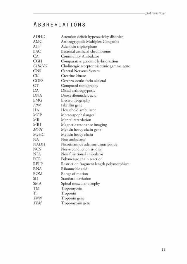
AbbreviationsADHD Attention deficit hyperactivity disorderAMC Arthrogryposis Multiplex CongenitaATP Adenosin triphosphateBAC Bacterial artificial chromosomeCA Community AmbulatorCGH Comparative genomic hybridisationCHRNG Cholinergic receptor nicotinic gamma geneCNS Central Nervous SystemCK Creatine kinaseCOFS Cerebro-oculo-facio-skeletalCT Computed tomographyDA Distal arthrogryposisDNA Deoxyribonucleic acidEMG ElectromyographyFBN Fibrillin geneHA Household ambulatorMCP MetacarpophalangealMR Mental retardationMRI Magnetic resonance imagingMYH Myosin heavy chain geneMyHC Myosin heavy chainNA Non ambulatorNADH Nicotinamide adenine dinucleotideNCS Nerve conduction studiesNFA Non functional ambulator PCR Polymerase chain reaction RFLP Restriction fragment length polymorphismRNA Ribonucleic acidROM Range of motionSD Standard deviationSMA Spinal muscular atrophyTM TropomyosinTn TroponinTNN Troponin geneTPM Tropomyosin gene

12
13
Introduction
introduction
In the field of neuromuscular disorders in children and adolescents, arthrogryposis is found to be a diverse and confusing diagnosis. Parents and professionals are often in doubt regarding specific diagnosis, treatment and prognosis in the child with arthrogryposis. This study was initiated by professionals treating arthrogryposis and by arthrogryposis patient organisations in Scandinavia. The study was conducted as a multicenter study with the investigators travelling to the local child rehabilitation centers in all health care regions of Sweden for personal interviews and investigation of included patients. The overall aims of the study were to clarify causes, clinical consequences and clinical course in arthrogryposis.
DefinitionArthrogryposis Multiplex Congenita (AMC) is defined as congenital, non-progres-sive contractures in more than two joints and in multiple body areas. The term arthrogryposis derives from the Greek words arthron – joint and grypos – curved. The term multiple congenital contractures can be used synonymously with arthro-gryposis. The diagnosis is purely descriptive, and arthrogryposis can be part of a large number of different syndromes, at least 200 1.
Compromised fetal mobility is the main background factor, common to all differ-ent types of arthrogryposis. The cause can be pathology in the peripheral or central nervous system (CNS), in muscles or in connective tissue, defects in neuromuscular transmission, compromised space in utero, maternal disease, external factors like medication or drugs, or compromised vascular supply to the fetus 2.
Arthrogryposis refers to a large and heterogeneous group of conditions, both sporad-ically occurring and hereditary. The literature is confusing regarding types of arthro-gryposis described, as different diagnoses are often lumped together and regarded as one entity. The most frequently occurring form of arthrogryposis is amyoplasia 3, a sporadically occurring condition sometimes referred to as “classical arthrogryposis”. The second most common form is probably distal arthrogryposis, (DA) 4, 5. DA is not one single entity, but a group of syndromes with mainly distal joint contractures 6, 7.
BackgroundHistorically, a patient with AMC is described by Thomas of Monmouth in “The life and Miracles of St William of Norwich” in 1156, as reported by Gordon 8. A boy with arthrogryposis is depicted in a painting by Jusepe de Ribera (“The club foot”) from 1642 9. The first description of arthrogryposis in the medical literature is thought to be by AW Otto, professor of anatomy in Breslau, in a textbook from 1841 10. The term Arthrogryposis was probably first used by Rosencranz in 1905 11, and the term Arthrogryposis Multiplex Congenita by Stern in 1923 12. The term Amyoplasia Congenita is used by Sheldon in 1932 13.
14
Introduction
AMC is discussed and described in orthopedic reviews from 1930 to 1950, and nu-merous specific syndromes with AMC are described in the medical literature from 1950 to 1960. Further research regarding the pathogenesis was published in the fol-lowing decades 12, 14-16. Hall described clinical and genetic evaluation and diagnosis in patients with AMC 17, 18. There are a great number of publications on genetic diag-nostics and further descriptions of mechanisms leading to reduced fetal mobility.
Recently, advances in molecular genetics have made it possible to understand patho-genic mechanisms, especially in distal arthrogryposis (DA) syndromes, where mu-tations in genes that encode for contractile muscle proteins have been found 19-22.
PathogenesisAnimal studies demonstrate that congenital joint contractures can be caused by fetal immobilization. A study from 1962 by Drachman and Coloumbre showed that con-genital joint ankylosis can be produced by relatively short periods of immobilization in chick embryos 16. In 1983, Moessinger (1983) 14 published an animal study where rat fetuses paralysed during part of gestation showed the same anomalies as those described in Pena Shokeir I syndrome: multiple joint contractures, pulmonary hy-poplasia, micrognathia, fetal growth retardation, short umbilical cord, and polyhy-dramnios. These features were previously described by Pena and Shokeir in an early lethal disorder with autosomal recessive inheritance, Pena Shokeir I syndrome 23.
Polyhydramnios is thought to be due to lack of swallowing during fetal life. Oli-gohydramnios deformation sequence includes similar features, also demonstrated in animal studies, possibly caused by fetal immobilization from external factors. However, renal defects are also often present in oligohydramnios 15.
Swinyard 12 discusses the etiology of multiple congenital contractures in animals and in humans and suggests that the joint fixations are caused by a proliferation of capsular connective tissue, a compensatory connective tissue response to lack of movement in utero.
Hall (1986) suggested that the Pena Shokeir syndrome represents a phenotype, fetal akinesia deformation sequence, caused by severely decreased or absent fetal movements 24, including joint contractures, short gut, pulmonary hypoplasia, short umbilical cord, intrauterine growth retardation, and craniofacial abnor-malities. At least 20 familial types of Pena-Shokeir phenotype are now recog-nized, in addition to sporadic cases 25.
Pathologic changes in 96 children with AMC were described in a study by Bank-er in 1986 15. Abnormalities in the neuromuscular system were found in all cases, with the primary alterations in anterior horn cells, roots, peripheral nerves, mo-tor end-plates or muscles. All had onset during fetal development. The vast ma-jority of patients in this study were considered to have a neurogenic cause. Dys-
15
Introduction
genesis or degeneration of the spinal cord, with abnormalities in anterior horn cells, were found in a majority of cases with CNS involvement, while the spinal cord and anterior roots were normal in myopathic cases (e.g. central core disease, congenital muscular dystrophy, nemaline myopathy, myotonic dystrophy). Fiber type predominance or disproportion was also found in some patients, in whom involvement of the spinal cord was not seen. Presumably secondary changes of muscle included fiber type predominance and disproportion, aplasia of muscle (amyoplasia) signifying a primary defect in the anterior horn cells early in the fetal development leading to hypoplasia of muscle and progressive denervation of muscle. CNS involvement included dysgenesis or degeneration of the brain and/or spinal cord in association with cases of trisomy 18, trisomy 21, Möbius syndrome, Zellweger syndrome, spinal muscular atrophy, and others.
In a further pathology study of 83 cases of lethal arthrogryposis in Finland, the majority of cases were also found to have a neurogenic cause, a few myopathic, while a non-neuromuscular cause was present in 10 cases 26.
Investigation of the spinal cord in infants with neurogenic arthrogryposis has demonstrated abnormal histology and unequal distribution of alpha motor neu-rons in anterior horn cells, the latter also predictive of involved muscle groups 27. Several studies report that neurogenic arthrogryposis (of the amyoplasia type) can be caused by vascular compromise in early fetal development, with ischemia of anterior horn cells leading to fetal akinesia and poor or absent muscle develop-ment, but also in some cases to co-existing anomalies with presumed vascular genesis, i.e. gastroschisis, bowel atresia, Möbius syndrome, and focal muscle de-fects 28-31.
Arthrogryposis can be present in several congenital myopathic disorders, e.g. ne-maline myopathy 32, centronuclear myopathy 33, central core disease and others, in congenital muscular dystrophies (Fukuyama, Ullrich) 1, in severe SMA1 with fetal onset 34, and in severe congenital polyneuropathies 35-37.
Several DA syndromes have recently been discovered to be caused by mutations in sarcomeric muscle proteins 19-22. A number of collagen disorders can also ap-pear with arthrogryposis, for example Ehlers-Danlos syndrome 38, Marfan syn-drome 39 and Larsen syndrome. DA9, Beal syndrome, has also been found to be a collagen disorder with a mutation in the FBN2 gene 40.
Disease affecting the neuromuscular junction with resulting weakness can cause arthrogryposis. Congenital myasthenic syndrome with arthrogryposis has been reported 41, congenital myotonic dystrophy 42, and maternal antibodies to fetal neurotransmittors 43 can all cause arthrogryposis.
16
Introduction
Movement restriction in utero caused by e.g. oligohydramniosis, myoma/fibroma of the uterus and bicornate uterus can be associated with arthrogryposis. Ar-throgryposis of the amyoplasia type occurs with increased frequency in one of monozygotic twins, but the cause in these cases is thought to be more related to vascular compromise than to actual crowding 28, 44.
A separate group of arthrogryposis syndromes are those caused by maternal dis-ease during pregnancy, such as maternal multiple sclerosis, maternal myasthenia gravis 1, maternal diabetes mellitus, and maternal hyperthermia 1. Metabolic dis-ease such as phosphofructokinase deficiency can cause arthrogryposis and drugs taken during pregnancy can also be associated with arthrogryposis (e.g. muscle relaxants, misoprostol, cocaine, alcohol)1.
Contractures caused by compromised space in utero have a relatively late ges-tational onset and are relatively mild. These types of congenital contractures regress more easily compared to contractures caused by early immobility of the fetus 45.
EpidemiologySeveral epidemiological surveys of arthrogryposis have been published. The occur-rence has previously been estimated to 1 in 3,300 live births in a Finnish study from 1966 46, 1 in 56,000 live births in a Scottish study of 66 sporadic cases from 1976 47, and 1 in 12,000 live births in a study from Western Australia from 1976 48.
In a retrospective epidemiologic study in western Sweden by Darin et.al., all chil-dren born with multiple congenital contractures between 1979 and 1994 were iden-tified through screening of registers, reviews of medical records and re-examination of children. 68 cases were identified, and the birth prevalence was found to be 1 in 5,100 live births 49. In this study, 39 patients had cerebral or spinal involvement, three patients had mechanical restriction in utero, 12 neuromuscular and nine con-nective tissue involvement.
Clinical classificationSince there is a very large number of disorders with arthrogryposis, differential di-agnosis can be difficult. It is, however, important to make as specific a diagnosis as possible for several reasons:
Treatment can vary depending on the underlying cause in the individual child. For example, stretching should be minimized in diastrophic dysplasia, where too in-tensive mobilisation may damage joint cartilage. The joint contractures can also be more resistant to treatment in certain types of AMC, especially amyoplasia 50 and surgery and splinting may need to be planned accordingly.Risk of re-occurrence varies greatly: amyoplasia occurs sporadically, while several forms of DA have autosomal dominant inheritance with a recurrence rate of 50%.
17
Introduction
The prognosis is also very much dependent on diagnosis, where conditions with CNS involvement may have a poor prognosis, sometimes with early death, while other conditions have a normal lifespan but may need extensive orthopaedic treat-ment and rehabilitation.
An approach to clinical evaluation that has been found to be very useful has been suggested and further developed by Hall 1, 17, 18. According to this, patients can be di-vided into three main groups of disorders: 1 Primarily musculoskeletal involvement; 2 Musculoskeletal involvement plus other system anomalies and, 3 Musculoskeletal in-volvement plus central nervous system dysfunction and/or mental retardation (MR).
Amyoplasia or “classic arthrogryposis” belongs to the first group, as do several camp-todactyly syndromes, distal arthrogryposis type 1, popliteal pterygium syndrome, several symphalangism syndromes, and others.
In the second group, i.e. involvement of limbs and other body areas, several other camptodactyly syndromes, several DA syndromes, myotonic dystrophy, congenital myopathies, myasthenia gravis, connective tissue disorders such as Larsen syndrome and Marfan syndrome with congenital contractures can be included.
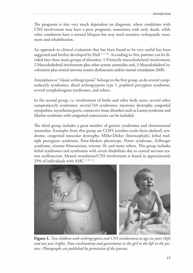
The third group includes a great number of genetic syndromes and chromosomal anomalies. Examples from this group are COFS (cerebro-oculo-facio-skeletal) syn-drome, congenital muscular dystrophy, Miller-Dieker (lissencephaly), lethal mul-tiple pterygium syndrome, Pena-Shokeir phenotype, Potter syndrome, Zellweger syndrome, trisomy 8/mosaicism, trisomy 18, and many others. This group includes lethal syndromes and syndromes with severe disabilities due to central nervous sys-tem malfunction. Mental retardation/CNS involvement is found in approximately 25% of individuals with AMC 2, 51, 52.
Figure 1. Two children with arthrogryposis and CNS involvement at age six years (left) and one year (right). Note tracheostomy and gastrostomy in the girl to the left in the pic-ture. Photographs are published by permission of the parents.
18
Introduction
InvestigationTo clarify the specific diagnosis in a child with arthrogryposis, a careful clinical evaluation including joint mobility, muscle strength, and associated anomalies is important. Family history, pregnancy history including infections, trauma, bleed-ing, drugs and medication, delivery history, developmental milestones in an older child, and associated problems should all be recorded. Evaluation of the mother of a newborn child with arthrogryposis should not be overlooked, regarding e.g. mater-nal myasthenia or myotonic dystrophy.
Possible CNS involvement must be evaluated. Ultrasound/CT scan or MRI of the brain should be performed if there are signs of CNS involvement, and also MRI of the spinal cord if there are signs of spinal involvement. Muscle ultrasound 53, CT-scanning of skeletal muscle 54, and MRI of muscle 55 can all be helpful in evaluating affected muscle involvement in planning muscle biopsy or surgery, and also to follow muscle development over time.
Dysmorphic features, which could indicate chromosomal anomalies, should be looked for and, if present, chromosomal analysis should be performed. Microar-ray based Comparative Genomic hybridisation (arrayCGH), is a method which in recent years has proved to be a reliable diagnostic tool for genome-wide detection of small chromosomal abnormalities, copy number changes, in patients with MR. Studies have demonstrated arrayCGH to detect copy number variations in 10-20% of patients with mental retardation with and without dysmorphic features and/or multiple congenital anomalies 56-59.
If there is any suspicion of myopathic involvement serum muscle enzymes should be analyzed. Other blood or urine chemistry investigations, such as serum lactate and metabolic screening, should be carried out if indicated from clinical findings (such as suspicion of mitochondrial or metabolic disease)
Nerve conduction studies, NCS, can be helpful if there are signs of peripheral nerve involvement/polyneuropathy, and electromyography, EMG, if there are signs of myopathy.
Muscle biopsy for pathological evaluation is essential for a correct diagnosis in myo-pathic conditions 15, and can be of help if the diagnosis is unclear 60. Therefore, if orthopedic surgery is planned a muscle biopsy should also be considered.
In a study of the diagnostic value of NCS/EMG and muscle biopsy in the evaluation of 38 patients with AMC, it was found that when clinical evaluation suggests a spe-cific syndrome, developmental, or exogenous cause, NCS/EMG and muscle biopsy are not helpful, but when the diagnosis is unclear NCS/EMG and muscle biopsy together can help in the diagnostic workup 61.
In two muscle morphologic studies from patients with neurogenic AMC 62 and AMC
19
Introduction
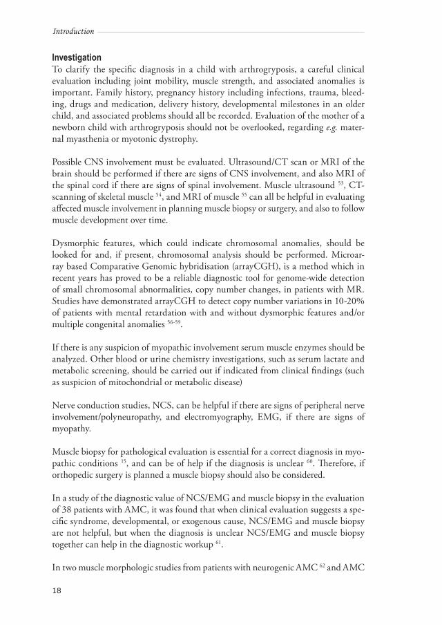
where known myopathic conditions were excluded 63, findings indicating neurogenic cause were found, i.e. variation in fiber size and abnormal fiber type distribution.With careful evaluation and investigation, a specific diagnosis can be reached in at least 30-50% of individuals with arthrogryposis 1. Prenatal diagnosis of AMC by ultrasound is possible, assessing fetal movement 64.
Family history
Clinical investigation
– Joint involvement
• Proximal/distal
• Jaws
• Spine
• ROM, range of motion
– Muscle involvement
• Absence/hypoplasia of muscles
• Muscle weakness
– Other organ involvement
• Eyes, palate, heart, lungs, gastrointestinal, genitourinary system
– Cognitive function
– Developmental history
– Associated problems/disease
Functional assessment
– Gross motor function
– Hand function
– ADL, activities of daily living
Laboratory investigations
– Genetic tests
• Chromosomes,
• DNA-analysis (if signs of specificgenetic syndrome)
– Blood chemistry:
• lactate, muscle enzymes,
– MRI of brain/spinal cord if signs of CNS involvement
– CT/MRI of muscles in amyoplasia
– EMG if signs of myopathy
– NCS if signs of polyneuropathy
Figure 2. Suggested baseline investigations in arthrogryposis.
Treatment Physical therapy, splinting, and orthopedic surgery are the main treatment methods in arthrogryposis 45. A correct genetic diagnosis is important, as treatment needs may vary 65-67.
Early physical therapy is important to avoid further muscle atrophy and, for the same reason, splinting combined with physical therapy is mostly preferable to cast-ing, especially in amyoplasia 1, 5. The first three to four months of life are especially valuable in activating and stimulating muscle function and in stretching contracted joints 66. Daily passive stretching and serial splinting in infants has been found to increase function 68.
Children with arthrogryposis may have major feeding difficulties in infancy, i.e. problems with chewing, sucking, and swallowing, sometimes requiring tube feed-ing. These problems are in many cases related to structural abnormalities in the jaw and tongue. Secondary to this chest infections, constipation, poor growth, and also language problems can be seen 69.
Malignant hyperthermia can occur in some forms of arthrogryposis, which must
20
Introduction
be kept in mind prior to anesthesia for orthopedic and other surgery. Maxillar and mandibular dysplasia and limited mouth opening are common problems, which can make intubation difficult 70, 71.
A multidisciplinary team management is needed, as many aspects need to be taken into account in the planning of treatment to optimize the child s functional out-come. In planning treatment, the child s general development and social activities must be kept in mind. A considerable part of the child s day may be occupied by physical therapy and training 65. The goal of treatment must be to achieve indepen-dence in adult life, and there must also be time for play and other important activities in daily life. Factors of importance to achieve independence are, in order of impor-tance, communication skills, activities of daily living, mobility, and walking 5, 50.
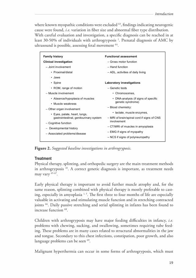
Figure 3. Two boys with amyoplasia at ages three years (left) and 2.5 months (right). Photographs are published by permission of the parents.
Amyoplasia The most common form of arthrogryposis is amyoplasia, which accounts for ap-proximately one third of all cases 3. The word amyoplasia means no muscle forma-tion. Amyoplasia occurs sporadically. Pathogenesis is unknown but thought to be impaired blood circulation to the fetus early in pregnancy with hypotension and hypoxia damaging the anterior horn cells and resulting in lack of or underdevelop-ment of muscle tissue with fatty or connective tissue replacement 3, 72. Clinically, the common morphologic features suggest a genetic syndrome, but occurrence is sporadic and individuals with amyoplasia have unaffected children 1. Amyoplasia occurs in increased frequency in one of monozygotic twins 73.
The diagnostic criteria for amyoplasia are highly specific with decreased muscle mass, typical joint contractures and limb positioning at birth, mostly symmetrical in all four limbs. There may be involvement only of the lower limbs or, less com-monly, only of the upper limbs and asymmetric limb involvement 3, 74. Typically, the shoulders are adducted and internally rotated, and the elbows are extended with the forearms pronated and wrists and fingers flexed. The hips are either in abduction and external rotation with flexed knees, or flexed with extended or flexed knees. Hips
21
Introduction
and knees can be dislocated. The feet are most often in an equinovarus adductus position, although other types of foot deformities occur. Involvement of the spine is also described 3, 74. The contractures can be fixed or flexible.
There is usually dimpling of the skin over affected joints. Common associated find-ings are midline facial hemangiomas and a round facial appearance. Muscle defects in the abdominal wall and inguinal hernias occur in about 10% of children born with amyoplasia, and gastroschisis and bowel atresia have also been recorded 28. Mental development is normal 74, unless there has been a concomitant birth as-phyxia.
Contractures in children with amyoplasia are at their maximum at birth. To increase the joint range of motion (ROM) and to obtain a functional position of the joint, a com-bination of stretching, splinting, and orthopedic surgery is often necessary 65, 66, 68. Early physical therapy is very important, both to mobilize joints and to stimulate muscle growth and to prevent further muscular atrophy 50. In amyoplasia, the joint contrac-tures can be severe and have a tendency to recur after correction 65.
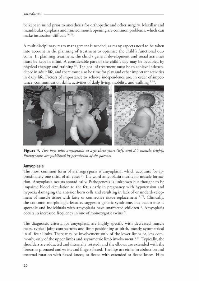
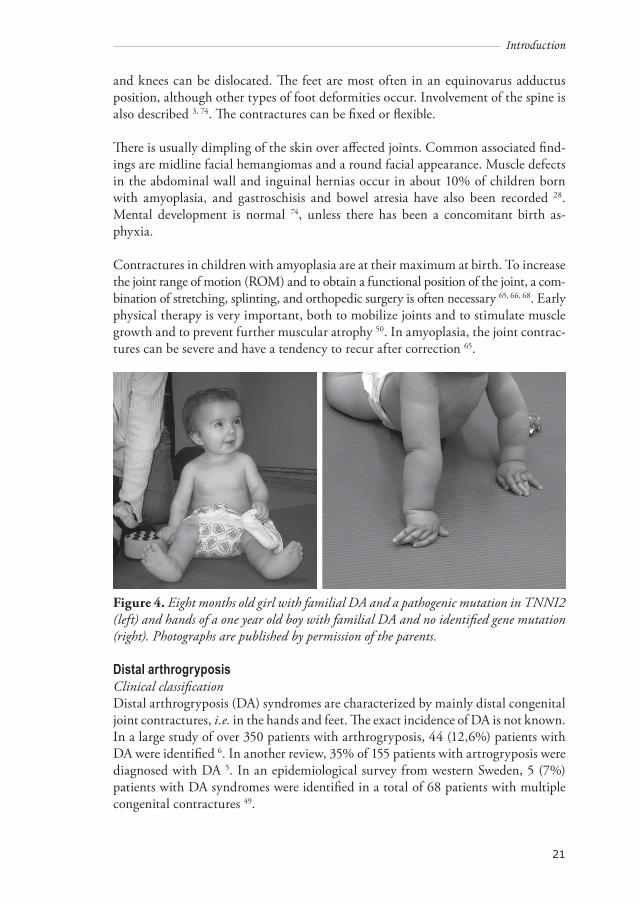
Figure 4. Eight months old girl with familial DA and a pathogenic mutation in TNNI2 (left) and hands of a one year old boy with familial DA and no identified gene mutation (right). Photographs are published by permission of the parents.
Distal arthrogryposisClinical classificationDistal arthrogryposis (DA) syndromes are characterized by mainly distal congenital joint contractures, i.e. in the hands and feet. The exact incidence of DA is not known. In a large study of over 350 patients with arthrogryposis, 44 (12,6%) patients with DA were identified 6. In another review, 35% of 155 patients with artrogryposis were diagnosed with DA 5. In an epidemiological survey from western Sweden, 5 (7%) patients with DA syndromes were identified in a total of 68 patients with multiple congenital contractures 49.
22
Introduction
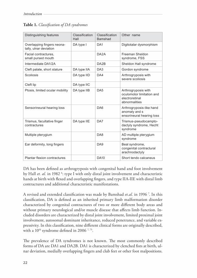
Table 1. Classification of DA syndromes
Distinguishing features Classification Hall
ClassificationBamshad
Other name
Overlapping fingers neona-tally, ulnar deviation
DA type I DA1 Digitotalar dysmorphism
Facial contractures,small pursed mouth
DA2A Freeman Sheldon syndrome, FSS
Intermediate DA1/2A DA2B Sheldon Hall syndrome
Cleft palate, short stature DA type IIA DA3 Gordon syndrome
Scoliosis DA type IID DA4 Arthrogryposis with severe scoliosis
Cleft lip DA type IIC
Ptosis, limited ocular mobility DA type IIB DA5 Arthrogryposis with oculomotor limitation and electroretinal abnormalities
Sensorineural hearing loss DA6 Arthrogryposis-like hand anomaly and sensorineural hearing loss
Trismus, facultative finger contractures
DA type IIE DA7 Trismus-pseudocampto-dactyly syndrome, Hecht syndrome
Multiple pterygium DA8 AD multiple pterygium syndrome
Ear deformity, long fingers DA9 Beal syndrome, congenital contractural arachnodactyly
Plantar flexion contractures DA10 Short tendo calcaneus
DA has been defined as arthrogryposis with congenital hand and foot involvement by Hall et. al. in 1982 6: type I with only distal joint involvement and characteristic hands at birth with flexed and overlapping fingers, and type IIA-IIE with distal limb contractures and additional characteristic manifestations.
A revised and extended classification was made by Bamshad et.al. in 1996 7. In this classification, DA is defined as an inherited primary limb malformation disorder characterized by congenital contractures of two or more different body areas and without primary neurological and/or muscle disease that affects limb function. In-cluded disorders are characterized by distal joint involvement, limited proximal joint involvement, autosomal dominant inheritance, reduced penetrance, and variable ex-pressivity. In this classification, nine different clinical forms are originally described, with a 10th syndrome defined in 2006 7, 75.
The prevalence of DA syndromes is not known. The most commonly described forms of DA are DA1 and DA2B. DA1 is characterized by clenched fists at birth, ul-nar deviation, medially overlapping fingers and club feet or other foot malpositions.
23
Introduction
The hips may be affected, calves small and opening of the mouth mildly limited 76. DA2B, Sheldon Hall syndrome, is similar to but milder than DA2A 4, 77. DA2A, Freeman Sheldon syndrome, is characterized by a small mouth, facial contractures, scoliosis, mainly distal joint contractures and short stature 77. Typical findings in DA2B are vertical talus, ulnar deviation, severe camptodactyly, triangularly shaped face, prominent nasolabial folds, downslanting palpebral fissures, small mouth and prominent chin 4. Foot deformities may be asymmetric 78.
The sarcomer is the functional unit of striated muscle contraction. Mutations in sar-comeric proteins are found in at least 20 different skeletal muscle diseases 79 and can result in DA or in congenital myopathy 33. Mutations in genes encoding sarcomeric muscle proteins are found in several DA syndromes: Sung et.al. described mutations in β tropomyosin (TPM2) in DA1, and mutations in fast troponin I (TNNI2) and fast troponin T (TNNT3) in DA2B 19. Toydemir et. al. describes mutations in em-bryonic myosin heavy chain (MYH3) in DA2A, Freeman Sheldon syndrome, and in DA2B, Sheldon-Hall syndrome 21. Further, a mutation in fetal myosin heavy chain (MYH8) has been described in DA7, trismus-pseudocamptodactyly syndrome 22. A defective function of contractile muscle proteins during fetal life influencing fetal mobility seems to be the common cause of congenital joint contractures in these syndromes 21, 22.
Gene mutations and sarcomeric muscle proteins in DATwo different mutations in the gene encoding an isoform of troponin I (TnI) specific for fast-twitch muscle fibers (TNNI2) have been associated with DA2B. DA2B has also been associated with a mutation in the gene encoding an isoform of troponin T specific for fast twitch contractile proteins (TNNT3) 20. DA1 has, in one affect-ed family, been associated with a mutation in the gene encoding β-tropomyosin (TPM2), 80 which is expressed mainly in slow twitch muscle fibers 81.
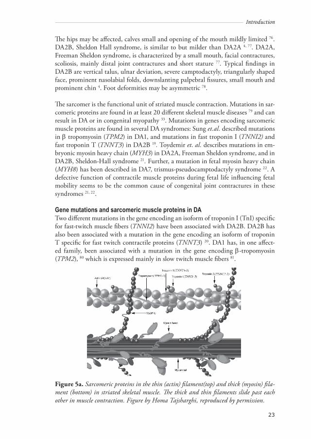
Figure 5a. Sarcomeric proteins in the thin (actin) filament(top) and thick (myosin) fila-ment (bottom) in striated skeletal muscle. The thick and thin filaments slide past each other in muscle contraction. Figure by Homa Tajsharghi, reproduced by permission.

24
Introduction
Figure 5b. The sarcomere is divided into four major components: the Z-disc, I band, A band and M-line, with one sarcomere stretching from one Z-disc to the next. Figure by Homa Tajsharghi, reproduced by permission.
The thin filament in striated muscle contains actin, tropomyosin, nebulin, and the troponin complex (TnI, TnT, and TnC). TM is composed of two α-helical chains, forming a rod-shaped coiled-coil dimer. It is localized head to tail along the length of the actin filament, providing stability, and is essential for myosin-actin interac-tion 81, 82.
There are four TM genes: TPM1, TPM2, TPM3, and TPM4. By alternative splicing, the use of alternative promoters and differential RNA processing, various transcripts are produced, which are specific for striated muscle, smooth muscle, and nonmuscle tissues 81.
There are three primary striated muscle TM isoforms, α-TM, β-TM, and γ-TM, which are highly homologous but are thought to exhibit unique physiologic proper-ties 81. In human striated muscle, α-TM is a product of TPM1, β-TM is encoded from TPM2, and γ-TM is encoded from TPM3. These striated muscle-specific isoforms are expressed in developmental and fiber-type-specific patterns in skeletal muscle and heart 81, 83. In humans, the muscle isoform encoded by TPM1 is predominantly expressed in cardiac muscle and in fast type 2 muscle fibers. TPM2 is mainly ex-pressed in slow type 1, and, to some extent, in fast muscle fibers and cardiac muscle. TPM3 is predominantly expressed in slow muscle fibers and is also expressed in the heart 81. In one family, DA1 was caused by the substitution of a highly conserved amino acid residue (R91G) in TPM2 19.
Myosin is the main component of skeletal muscle sarcomeric thick filaments. It consists of two globular heads attached to a long-helical-coiled coil rod domain. It is a hexamer composed of one pair of myosin heavy chains (MyHCs) and two pairs of myosin light chains. The myosin globular head domain of the myosin motor (myo-sin subfragment 1 [S1]) contains actin and adenosine triphosphate (ATP)-binding regions and is responsible for the force transduction properties of myosin 84.
25
Introduction
Several striated muscle MyHC genes have been described 85. The expression of my-osin isoforms is developmentally regulated 86-88. Myosin myopathies have evolved as a new group of muscle diseases caused by mutations in skeletal muscle myosin heavy-chain (MyHC) genes. The phenotypes of these diseases vary, ranging from prenatal nonprogressive arthrogryposis syndromes to adult-onset progressive muscle weakness. Mutations have been reported in two of three MyHC isoforms expressed in adult limb skeletal muscle. In addition to familial hypertrophic or dilated cardio-myopathy 11 mutations in the slow or cardiac MyHC gene (MYH7) cause skeletal myopathies such as myosin storage myopathy 89-93 and Laing early-onset distal myo-pathy 94, 95. A mutation in the MyHC IIa gene (MYH2) is associated with dominant myopathy characterized by ophthalmoplegia, congenital joint contractures, and rimmed vacuoles in muscle fibers 96-98.
DA2A, Freeman-Sheldon syndrome, and DA2B, Sheldon-Hall syndrome, have been reported as the first disorders associated with mutations in embryonic MyHC (MYH3)21. DA syndromes are associated with missense mutations in various genes coding for sarcomeric proteins. The genes thus far demonstrated to be involved in DA syndromes are TNNI2 (troponin I), TPM2 (β-tropomyosin) 19, TNNT3 (tro-poninT) 20, MYH8 (perinatal MyHC), and MYH3 (embryonic MyHC)21.
These findings indicate that DA syndromes are caused by myopathies with onset during fetal development, but few studies have involved analysis of muscle tissue in these diseases.
In a recent study aiming to investigate the mechanisms of impaired muscle function in two patients with DA2B and a β-tropomyosin mutation (R133W), significant differences in regulation of muscle contraction was demonstrated in type 1 fibers. The found mutation appears to induce alteration in myosin-actin kinetics causing a reduced number of myosin molecules in the strong actin-binding state, resulting in muscle weakness in the absence of muscle wasting 9.

26

27
Aim
aims of the study
The aims of the study were:
• To investigate children and adolescents with arthrogryposis in Sweden and to classify the different occurring forms.
• To describe a group of individuals with amyoplasia, to investigate how muscle strength and joint contractures affect their motor function, and to relate their current functional status to joint position at birth.
• To investigate the clinical, muscle morphologic and genetic findings in families with DA and pathogenic mutations in the sarcomeric muscle protein genes TNNI2, TPM2 and MYH3.
• To describe the clinical findings, clinical course, additional problems and disabilities in 40 individuals with DA and to evaluate genotype-phenotype correlation.
• To study and evaluate given treatment in patients with amyoplasia and DA.

28
29
Methods
material and methodsPatientsIn a national survey in Sweden, 127 children, adolescents and young adults with arthrogryposis were investigated. The index patients were identified through pediat-ric rehabilitation centers or, in a few cases, through the orthopaedic surgeon or the Swedish AMC-association. A further four children with familial DA were added to the study after the initial survey was completed, making the total number 131.
The inclusion criteria were contractures present at birth in more than two joints and in multiple body areas. Children born with myelomeningocele, isolated congenital hip dislocation or pes equinovarus adductus were excluded.
Written informed consent was obtained from adult participating individuals and for participating children, from the parents.
The study was approved by the Ethical Board at the Universities of Gothenburg, Uppsala, Stockholm, Umeå, Örebro, Malmö, Lund and Linköping, and by the heads of Paediatric clinics in the Swedish health-care regions.
INTERVIEW and RECORDS
• Patient data, background factors(family history, pregnancy etc)
• Neonatal findings
• Results from investigations
• Given treatment
• Course, development
• Activities of daily living (ADL)
• Social/school, rehabilitation contacts
• Functional aids
CLINICAL INVESTIGATION
• General physical examination
• Joint mobility (ROM)
• Muscle strength– Manual muscle testing
– Myometry
• Timed functional tests
• Evaluation of – Gross motor function
– Hand function
– Reaching ability
• Language, communication
• Photo and video documentation
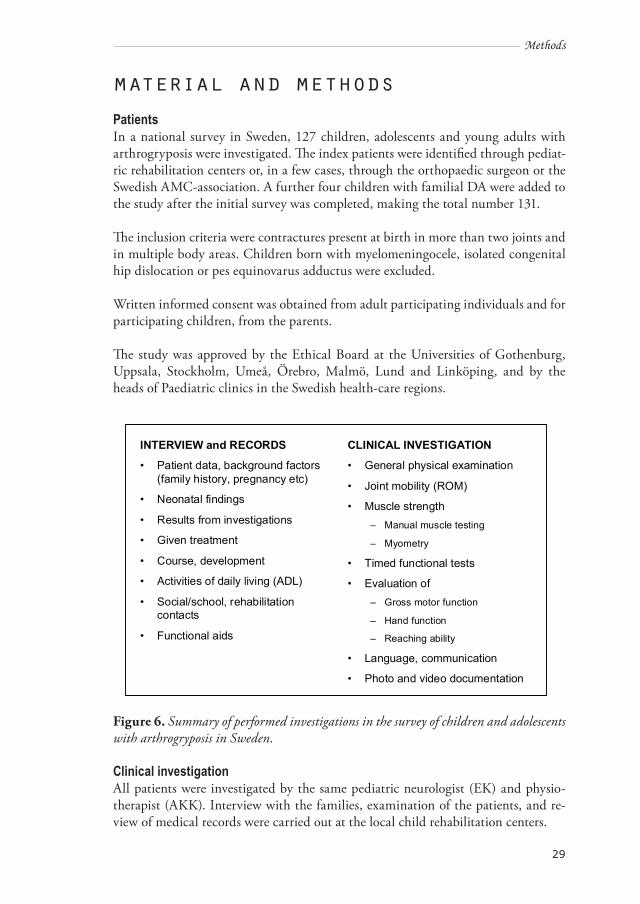
Figure 6. Summary of performed investigations in the survey of children and adolescents with arthrogryposis in Sweden.
Clinical investigationAll patients were investigated by the same pediatric neurologist (EK) and physio-therapist (AKK). Interview with the families, examination of the patients, and re-view of medical records were carried out at the local child rehabilitation centers.
30
Methods
A structured interview including family history, prenatal and perinatal history, neo-natal findings including joint involvement, developmental milestones, associated medical problems, treatment, and outcome was carried out for all index cases. A detailed clinical examination was carried out. Physical examination included eval-uation of facial involvement, other associated signs and symptoms and a neurologi-cal examination.
For each patient, medical records were studied and results from previous investi-gations, including orthopedic procedures and muscle biopsies, were recorded. In patients with amyoplasia (paper I), an orthopedic surgeon (RJ) also reviewed the orthopaedic treatment records.
Affected adult family members were also examined by the same physician and phys-iotherapist, and information on family history was obtained by personal interviews. Extended family members who were found to be asymptomatic carriers of the patho-genic gene mutation in the family were seen and interviewed personally to exclude the presence of previously undiagnosed distal joint involvement.
Diagnosis was determined based on clinical findings and medical records, using known diagnostic criteria. For all patients with dysmorphic signs and/or suspected genetic syndrome, clinical findings and features of the patients were discussed with a clinical geneticist and in some cases a search in a genetic data base, Possum, was performed to aid in the diagnosis. Short stature was defined as below 3 SD. Cardiac investigation was performed in two patients with DA and TPM2 mutation.
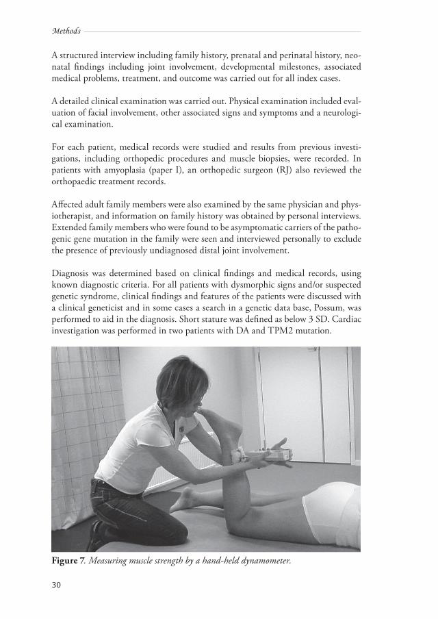
Figure 7. Measuring muscle strength by a hand-held dynamometer.
31
Methods
Muscle strengthIsometric muscle strength was measured with an electronic hand-held dynamometer (adapted Chatillon, Axel Ericson Medical AB, Göteborg, Sweden) with a method standardized by Eek et.al. 100 or, in children too young to participate, by clinical evaluation. Nine muscle groups were measured. An isometric contraction of at least five sec was required and the peak force value in Newton was recorded. The best of three values obtained was compared with the mean value from weight-related refer-ence values for healthy children and adolescents 100. To be able to compare isomet-ric muscle strength independent of age and gender, a percentage of normal muscle strength was calculated.
Range of motion, ROMPassive ROM was measured with an ordinary goniometer. Information on hip dis-location was collected from medical records.
Motor functionMotor function was assessed using a scale designed by Scott et.al. 101. Twenty move-ments were assessed, including head-lifting in the supine position, rolling, sitting up from lying down, sitting, getting off a chair, standing up from lying, standing, standing on heels, standing on toes, standing on one leg, jumping, and climbing up and down stairs. The performance is scored according to a three-point scale: 0 (un-able), 1 (needs self-reinforcement), and 2 (succeeds). Maximum score is 40.
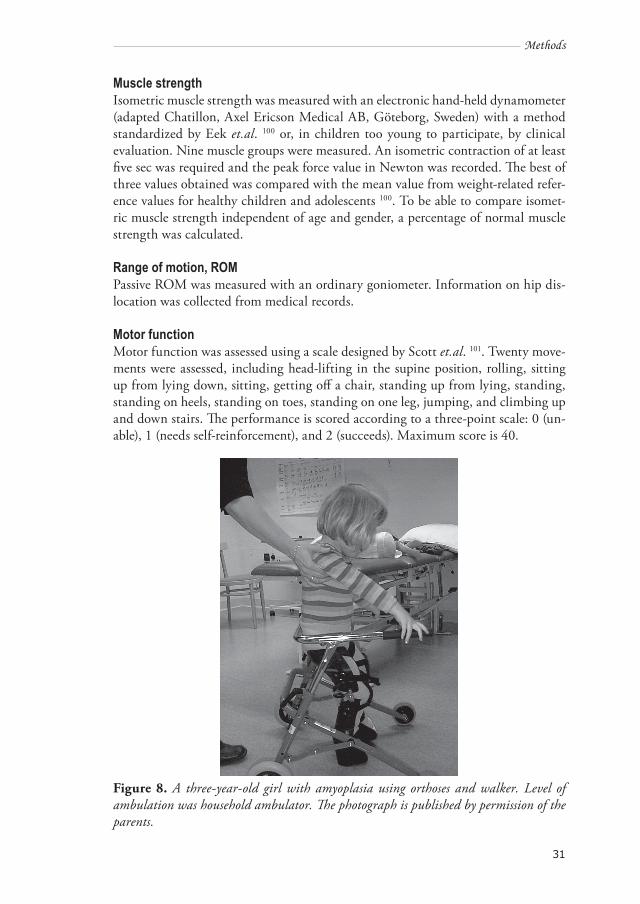
Figure 8. A three-year-old girl with amyoplasia using orthoses and walker. Level of ambulation was household ambulator. The photograph is published by permission of the parents.
32
Methods
According to their level of ambulation the patients were divided into functional groups according to the classification of Hoffer et al 102:1. Community ambulators (CA) walk with aids in the community and do not need a wheelchair;2. Household ambulators (HA) walk with aids in the household and use a wheel chair in the community;3. Non-functional ambulators (NFA) use parallel bars or walkers with support;4. Non-ambulators (NA) do not walk.
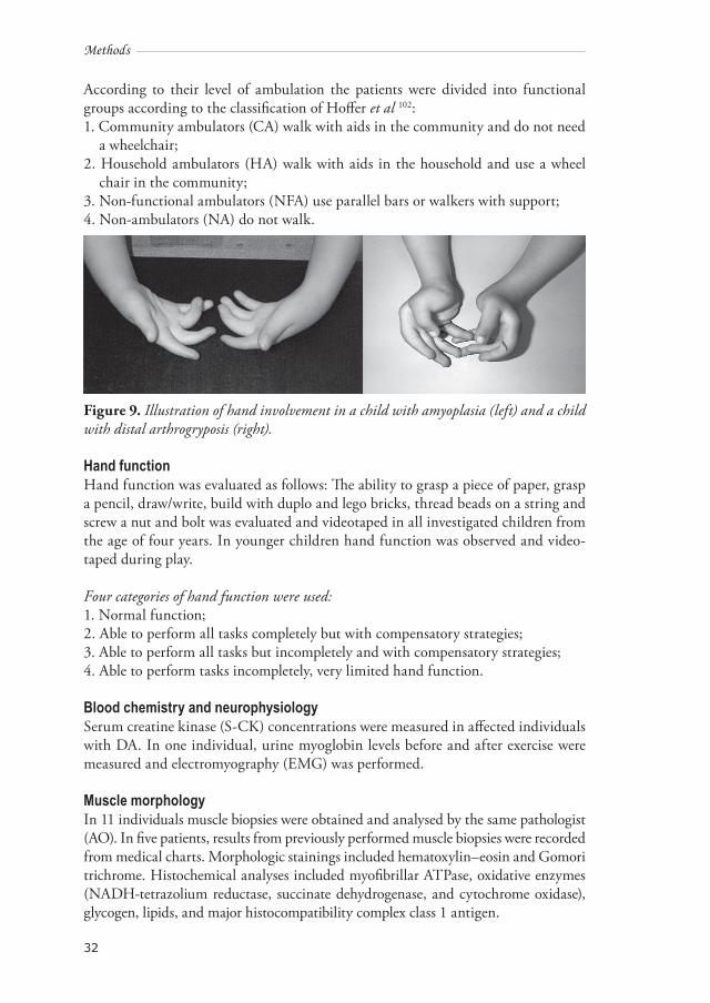
Figure 9. Illustration of hand involvement in a child with amyoplasia (left) and a child with distal arthrogryposis (right).
Hand functionHand function was evaluated as follows: The ability to grasp a piece of paper, grasp a pencil, draw/write, build with duplo and lego bricks, thread beads on a string and screw a nut and bolt was evaluated and videotaped in all investigated children from the age of four years. In younger children hand function was observed and video-taped during play.
Four categories of hand function were used:1. Normal function;2. Able to perform all tasks completely but with compensatory strategies;3. Able to perform all tasks but incompletely and with compensatory strategies;4. Able to perform tasks incompletely, very limited hand function.
Blood chemistry and neurophysiologySerum creatine kinase (S-CK) concentrations were measured in affected individuals with DA. In one individual, urine myoglobin levels before and after exercise were measured and electromyography (EMG) was performed.
Muscle morphologyIn 11 individuals muscle biopsies were obtained and analysed by the same pathologist (AO). In five patients, results from previously performed muscle biopsies were recorded from medical charts. Morphologic stainings included hematoxylin–eosin and Gomori trichrome. Histochemical analyses included myofibrillar ATPase, oxidative enzymes (NADH-tetrazolium reductase, succinate dehydrogenase, and cytochrome oxidase), glycogen, lipids, and major histocompatibility complex class 1 antigen.
33
Methods
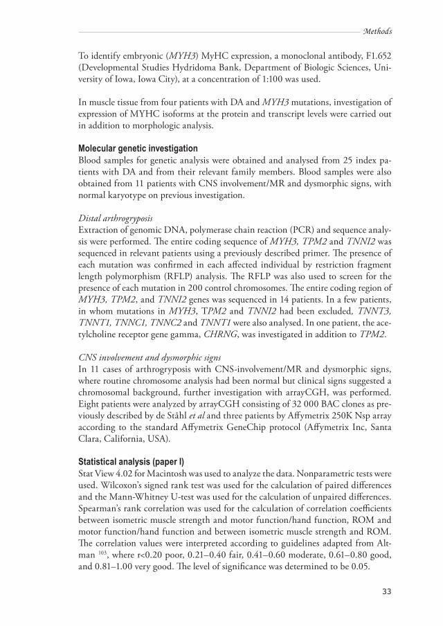
To identify embryonic (MYH3) MyHC expression, a monoclonal antibody, F1.652 (Developmental Studies Hydridoma Bank, Department of Biologic Sciences, Uni-versity of Iowa, Iowa City), at a concentration of 1:100 was used.
In muscle tissue from four patients with DA and MYH3 mutations, investigation of expression of MYHC isoforms at the protein and transcript levels were carried out in addition to morphologic analysis.
Molecular genetic investigationBlood samples for genetic analysis were obtained and analysed from 25 index pa-tients with DA and from their relevant family members. Blood samples were also obtained from 11 patients with CNS involvement/MR and dysmorphic signs, with normal karyotype on previous investigation.
Distal arthrogryposisExtraction of genomic DNA, polymerase chain reaction (PCR) and sequence analy-sis were performed. The entire coding sequence of MYH3, TPM2 and TNNI2 was sequenced in relevant patients using a previously described primer. The presence of each mutation was confirmed in each affected individual by restriction fragment length polymorphism (RFLP) analysis. The RFLP was also used to screen for the presence of each mutation in 200 control chromosomes. The entire coding region of MYH3, TPM2, and TNNI2 genes was sequenced in 14 patients. In a few patients, in whom mutations in MYH3, TPM2 and TNNI2 had been excluded, TNNT3, TNNT1, TNNC1, TNNC2 and TNNT1 were also analysed. In one patient, the ace-tylcholine receptor gene gamma, CHRNG, was investigated in addition to TPM2.
CNS involvement and dysmorphic signsIn 11 cases of arthrogryposis with CNS-involvement/MR and dysmorphic signs, where routine chromosome analysis had been normal but clinical signs suggested a chromosomal background, further investigation with arrayCGH, was performed. Eight patients were analyzed by arrayCGH consisting of 32 000 BAC clones as pre-viously described by de Ståhl et al and three patients by Affymetrix 250K Nsp array according to the standard Affymetrix GeneChip protocol (Affymetrix Inc, Santa Clara, California, USA).
Statistical analysis (paper I) Stat View 4.02 for Macintosh was used to analyze the data. Nonparametric tests were used. Wilcoxon’s signed rank test was used for the calculation of paired differences and the Mann-Whitney U-test was used for the calculation of unpaired differences. Spearman’s rank correlation was used for the calculation of correlation coefficients between isometric muscle strength and motor function/hand function, ROM and motor function/hand function and between isometric muscle strength and ROM. The correlation values were interpreted according to guidelines adapted from Alt-man 103, where r<0.20 poor, 0.21–0.40 fair, 0.41–0.60 moderate, 0.61–0.80 good, and 0.81–1.00 very good. The level of significance was determined to be 0.05.

34
35
Results
Results
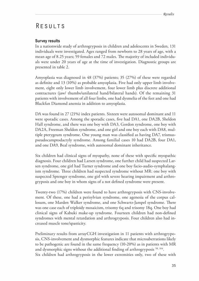
Survey resultsIn a nationwide study of arthrogryposis in children and adolescents in Sweden, 131 individuals were investigated. Ages ranged from newborn to 28 years of age, with a mean age of 8.25 years; 59 females and 72 males. The majority of included individu-als were under 20 years of age at the time of investigation. Diagnostic groups are presented in table 2.
Amyoplasia was diagnosed in 48 (37%) patients; 35 (27%) of these were regarded as definite and 13 (10%) as probable amyoplasia. Five had only upper limb involve-ment, eight only lower limb involvement, four lower limb plus discrete additional contractures (jaw/ thumbs/unilateral hand/bilateral hands). Of the remaining 31 patients with involvement of all four limbs, one had dysmelia of the feet and one had Blackfan Diamond anemia in addition to amyoplasia.
DA was found in 27 (21%) index patients. Sixteen were autosomal dominant and 11 were sporadic cases. Among the sporadic cases, five had DA1, one DA2B, Sheldon Hall syndrome, and there was one boy with DA3, Gordon syndrome, one boy with DA2A, Freeman Sheldon syndrome, and one girl and one boy each with DA8, mul-tiple pterygeum syndrome. One young man was classified as having DA7, trismus-pseudocamptodactyly syndrome. Among familial cases 10 had DA2B, four DA1, and one DA9, Beal syndrome, with autosomal dominant inheritance.
Six children had clinical signs of myopathy, none of these with specific myopathic diagnosis. Four children had Larsen syndrome, one further child had suspected Lar-sen syndrome, one girl had Turner syndrome and one boy facio-audio-symphalang-ism syndrome. Three children had suspected syndrome without MR: one boy with suspected Sprenger syndrome, one girl with severe hearing impairment and arthro-gryposis and one boy in whom signs of a not defined syndrome were present.
Twenty-two (17%) children were found to have arthrogryposis with CNS-involve-ment. Of these, one had a perisylvian syndrome, one agenesia of the corpus cal-losum, one Marden Walker syndrome, and one Schwartz-Jampel syndrome. There was one case each of triploidy mosaicism, trisomy 6q and trisomy 18q. One boy had clinical signs of Kabuki make-up syndrome. Fourteen children had non-defined syndromes with mental retardation and arthrogryposis. Four children also had in-creased muscle tone/spasticity.
Preliminary results from arrayCGH investigation in 11 patients with arthrogrypo-sis, CNS-involvement and dysmorphic features indicate that microaberrations likely to be pathogenic are found in the same frequency (10-20%) as in patients with MR and dysmorphic signs without the additional finding of arthrogryposis 58, 104.Six children had arthrogryposis in the lower extremities only, two of these with
36
Results
vertebral anomalies and caudal regression syndrome. A further 12 children had ar-throgryposis that we could not specify further at present.
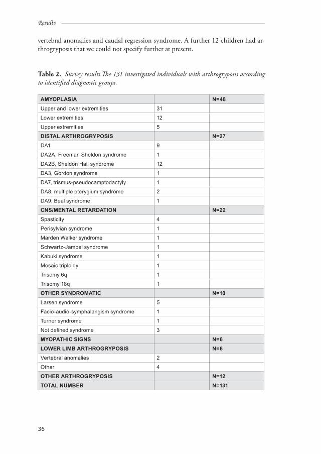
Table 2. Survey results.The 131 investigated individuals with arthrogryposis according to identified diagnostic groups.
AMYOPLASIA N=48Upper and lower extremities 31
Lower extremities 12
Upper extremities 5
DISTAL ARTHROGRYPOSIS N=27DA1 9
DA2A, Freeman Sheldon syndrome 1
DA2B, Sheldon Hall syndrome 12
DA3, Gordon syndrome 1
DA7, trismus-pseudocamptodactyly 1
DA8, multiple pterygium syndrome 2
DA9, Beal syndrome 1
CNS/MENTAL RETARDATION N=22Spasticity 4
Perisylvian syndrome 1
Marden Walker syndrome 1
Schwartz-Jampel syndrome 1
Kabuki syndrome 1
Mosaic triploidy 1
Trisomy 6q 1
Trisomy 18q 1
OTHER SYNDROMATIC N=10Larsen syndrome 5
Facio-audio-symphalangism syndrome 1
Turner syndrome 1
Not defined syndrome 3
MYOPATHIC SIGNS N=6LOWER LIMB ARTHROGRYPOSIS N=6Vertebral anomalies 2
Other 4
OTHER ARTHROGRYPOSIS N=12TOTAL NUMBER N=131
37
Results
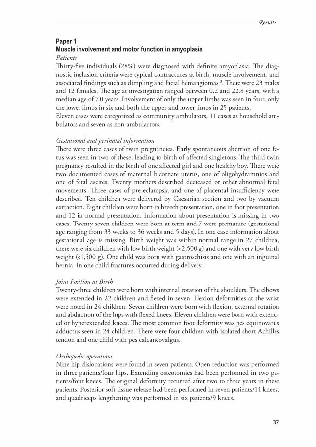
Paper 1Muscle involvement and motor function in amyoplasiaPatientsThirty-five individuals (28%) were diagnosed with definite amyoplasia. The diag-nostic inclusion criteria were typical contractures at birth, muscle involvement, and associated findings such as dimpling and facial hemangiomas 3. There were 23 males and 12 females. The age at investigation ranged between 0.2 and 22.8 years, with a median age of 7.0 years. Involvement of only the upper limbs was seen in four, only the lower limbs in six and both the upper and lower limbs in 25 patients.Eleven cases were categorized as community ambulators, 11 cases as household am-bulators and seven as non-ambulartors.
Gestational and perinatal information There were three cases of twin pregnancies. Early spontaneous abortion of one fe-tus was seen in two of these, leading to birth of affected singletons. The third twin pregnancy resulted in the birth of one affected girl and one healthy boy. There were two documented cases of maternal bicornate uterus, one of oligohydramnios and one of fetal ascites. Twenty mothers described decreased or other abnormal fetal movements. Three cases of pre-eclampsia and one of placental insufficiency were described. Ten children were delivered by Caesarian section and two by vacuum extraction. Eight children were born in breech presentation, one in foot presentation and 12 in normal presentation. Information about presentation is missing in two cases. Twenty-seven children were born at term and 7 were premature (gestational age ranging from 33 weeks to 36 weeks and 5 days). In one case information about gestational age is missing. Birth weight was within normal range in 27 children, there were six children with low birth weight (<2,500 g) and one with very low birth weight (<1,500 g). One child was born with gastroschisis and one with an inguinal hernia. In one child fractures occurred during delivery.
Joint Position at BirthTwenty-three children were born with internal rotation of the shoulders. The elbows were extended in 22 children and flexed in seven. Flexion deformities at the wrist were noted in 24 children. Seven children were born with flexion, external rotation and abduction of the hips with flexed knees. Eleven children were born with extend-ed or hyperextended knees. The most common foot deformity was pes equinovarus adductus seen in 24 children. There were four children with isolated short Achilles tendon and one child with pes calcaneovalgus.
Orthopedic operationsNine hip dislocations were found in seven patients. Open reduction was performed in three patients/four hips. Extending osteotomies had been performed in two pa-tients/four knees. The original deformity recurred after two to three years in these patients. Posterior soft tissue release had been performed in seven patients/14 knees, and quadriceps lengthening was performed in six patients/9 knees.
38
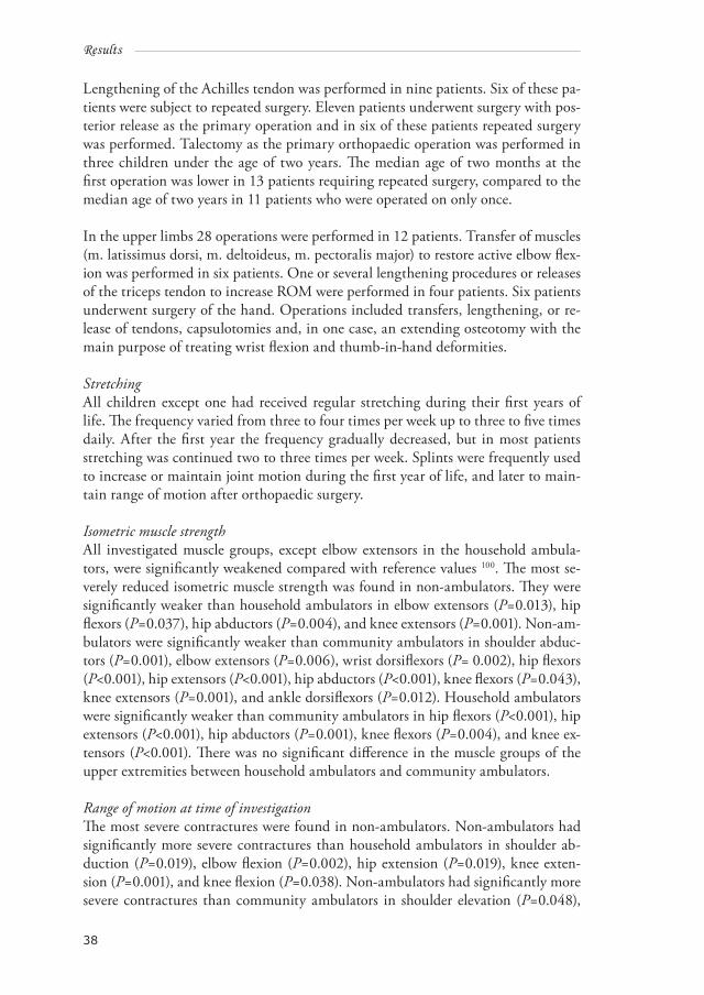
Results
Lengthening of the Achilles tendon was performed in nine patients. Six of these pa-tients were subject to repeated surgery. Eleven patients underwent surgery with pos-terior release as the primary operation and in six of these patients repeated surgery was performed. Talectomy as the primary orthopaedic operation was performed in three children under the age of two years. The median age of two months at the first operation was lower in 13 patients requiring repeated surgery, compared to the median age of two years in 11 patients who were operated on only once.
In the upper limbs 28 operations were performed in 12 patients. Transfer of muscles (m. latissimus dorsi, m. deltoideus, m. pectoralis major) to restore active elbow flex-ion was performed in six patients. One or several lengthening procedures or releases of the triceps tendon to increase ROM were performed in four patients. Six patients underwent surgery of the hand. Operations included transfers, lengthening, or re-lease of tendons, capsulotomies and, in one case, an extending osteotomy with the main purpose of treating wrist flexion and thumb-in-hand deformities.
StretchingAll children except one had received regular stretching during their first years of life. The frequency varied from three to four times per week up to three to five times daily. After the first year the frequency gradually decreased, but in most patients stretching was continued two to three times per week. Splints were frequently used to increase or maintain joint motion during the first year of life, and later to main-tain range of motion after orthopaedic surgery.
Isometric muscle strengthAll investigated muscle groups, except elbow extensors in the household ambula-tors, were significantly weakened compared with reference values 100. The most se-verely reduced isometric muscle strength was found in non-ambulators. They were significantly weaker than household ambulators in elbow extensors (P=0.013), hip flexors (P=0.037), hip abductors (P=0.004), and knee extensors (P=0.001). Non-am-bulators were significantly weaker than community ambulators in shoulder abduc-tors (P=0.001), elbow extensors (P=0.006), wrist dorsiflexors (P= 0.002), hip flexors (P<0.001), hip extensors (P<0.001), hip abductors (P<0.001), knee flexors (P=0.043), knee extensors (P=0.001), and ankle dorsiflexors (P=0.012). Household ambulators were significantly weaker than community ambulators in hip flexors (P<0.001), hip extensors (P<0.001), hip abductors (P=0.001), knee flexors (P=0.004), and knee ex-tensors (P<0.001). There was no significant difference in the muscle groups of the upper extremities between household ambulators and community ambulators.
Range of motion at time of investigationThe most severe contractures were found in non-ambulators. Non-ambulators had significantly more severe contractures than household ambulators in shoulder ab-duction (P=0.019), elbow flexion (P=0.002), hip extension (P=0.019), knee exten-sion (P=0.001), and knee flexion (P=0.038). Non-ambulators had significantly more severe contractures than community ambulators in shoulder elevation (P=0.048),
39
Results
elbow flexion (P=0.001), hip extension (P=0.001), hip abduction (P=0.023), hip ad-duction (P=0.037), hip inward rotation (P=0.003), knee extension (P<0.001), ankle dorsiflexion (P=0.002), and plantar flexion (P=0.001). Household ambulators had significantly more severe contractures than community ambulators in hip extension (P=0.034), hip internal rotation (P=0.006), knee flexion (P=0.008), knee extension (P=0.001), ankle dorsiflexion (P=0.010), and plantar flexion (P=0.001).
Motor FunctionMotor function scores were related to age in all the assessed individuals. The young-est children had poor motor function scores partly due to their age. Community ambulators had the highest scores and none-ambulators the lowest. Community ambulators achieved independent walking at an earlier median age compared to household ambulators.
Hand FunctionNine patients had normal hand function, 13 were able to perform tasks completely but with compensatory strategies, four were able to perform tasks incompletely and with compensatory strategies, and nine patients were able to perform tasks incom-pletely with very limited hand function.
Main correlationsThere were strong correlations between muscle strength and motor function. There were only moderate correlations between range of motion and motor function. Com-munity ambulators had the best muscle strength, and none had knee flexion con-tractures of more than 20 degrees. Household ambulators had severe contractures in the legs, but good muscle strength in the arms. Non-ambulators had the most severe joint contractures and the most pronounced muscle weakness. The majority were born with hips in pronounced abduction, flexion and external rotation.
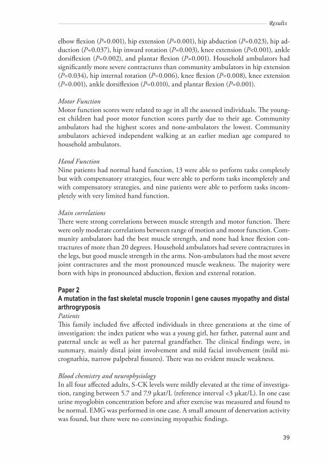
Paper 2A mutation in the fast skeletal muscle troponin I gene causes myopathy and distal arthrogryposisPatientsThis family included five affected individuals in three generations at the time of investigation: the index patient who was a young girl, her father, paternal aunt and paternal uncle as well as her paternal grandfather. The clinical findings were, in summary, mainly distal joint involvement and mild facial involvement (mild mi-crognathia, narrow palpebral fissures). There was no evident muscle weakness.
Blood chemistry and neurophysiologyIn all four affected adults, S-CK levels were mildly elevated at the time of investiga-tion, ranging between 5.7 and 7.9 μkat/L (reference interval <3 μkat/L). In one case urine myoglobin concentration before and after exercise was measured and found to be normal. EMG was performed in one case. A small amount of denervation activity was found, but there were no convincing myopathic findings.
40
Results
Muscle morphologySimilar myopathic changes were found in all four adult cases. There was an in-creased variability of fiber size and frequent muscle fibers with internalized nuclei. Fiber size variability was present among type 1 as well as type 2 fibers but was more pronounced among type 2 fibers, which were generally much larger than the type 1 fibers. Occasional signs of fiber splitting in type 2 fibers were present. The fibers with internalized nuclei were type 2. There was type 1 fiber predominance (which is normal for the tibialis anterior muscle) except in one case where the area occupied by type 2 fibers was equal to or larger than that of type 1 fibers. Regenerating fibers and increased interstitial connective tissue was also found.
Genetic findingsWe performed mutation analysis of TNNI2 in 16 members of the family. The entire coding sequence of TNNI2 was investigated in the index patient. We identified a heterozygous three-base in-frame deletion, 2,918–2,920del, skipping the highly conserved lysine (K) at position 176. The K176del is located in the distal end of the TNNI2 filament, seven residues from the stop codon. The deletion was identified in all five affected family members and not in any of the 11 investigated relatives with-out DA. The mutated residue is highly conserved among species and corresponds to residue K206 in cardiac TnI (cTnI; TNNI3). A mutation of this residue, K206Q in cTnI, has been associated with hypertrophic cardiomyopathy 105.
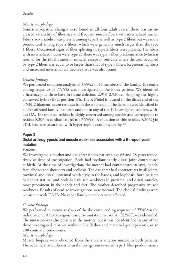
Paper 3Distal arthrogryposis and muscle weakness associated with a ß-tropomyosin mutationPatientsWe investigated a mother and daughter (index patient), age 65 and 28 years respec-tively at time of investigation. Both had predominantly distal joint contractures at birth. At the time of investigation, the mother had contractures in jaws, hands, feet, elbows and shoulders and scoliosis. The daughter had contractures in all joints, proximal and distal, proximal syndactyly in the hands, and kyphosis. Both patients had short stature, and both had muscle weakness in proximal and distal muscles, most prominent in the hands and feet. The mother described progressive muscle weakness. Results of cardiac investigations were normal. The clinical findings were consistent with DA2B. No other family members were affected.
Genetic findingsWe performed mutation analysis of the the entire coding sequence of TPM2 in the index patient. A heterozygous missense mutation in exon 4, C5396T, was identified.The mutation was also present in the mother, but it was not identified in any of the three investigated relatives without DA (father and maternal grandparents), or in 200 control chromosomes. Muscle morphologyMuscle biopsies were obtained from the tibialis anterior muscle in both patients. Histochemical and ultrastructural investigation revealed type 1 fiber predominance
41
Results
but no other major morphologic abnormalities. Immunohistochemistry of myosin isoforms also demonstrated a marked predominance of muscle fibers expressing slow myosin.
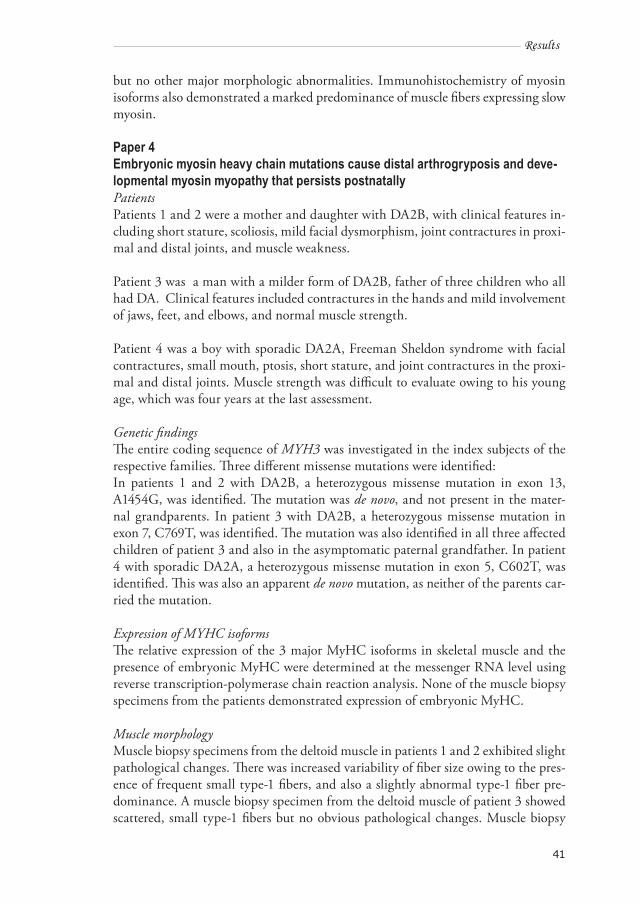
Paper 4Embryonic myosin heavy chain mutations cause distal arthrogryposis and deve-lopmental myosin myopathy that persists postnatallyPatientsPatients 1 and 2 were a mother and daughter with DA2B, with clinical features in-cluding short stature, scoliosis, mild facial dysmorphism, joint contractures in proxi-mal and distal joints, and muscle weakness.
Patient 3 was a man with a milder form of DA2B, father of three children who all had DA. Clinical features included contractures in the hands and mild involvement of jaws, feet, and elbows, and normal muscle strength.
Patient 4 was a boy with sporadic DA2A, Freeman Sheldon syndrome with facial contractures, small mouth, ptosis, short stature, and joint contractures in the proxi-mal and distal joints. Muscle strength was difficult to evaluate owing to his young age, which was four years at the last assessment.
Genetic findingsThe entire coding sequence of MYH3 was investigated in the index subjects of the respective families. Three different missense mutations were identified:In patients 1 and 2 with DA2B, a heterozygous missense mutation in exon 13, A1454G, was identified. The mutation was de novo, and not present in the mater-nal grandparents. In patient 3 with DA2B, a heterozygous missense mutation in exon 7, C769T, was identified. The mutation was also identified in all three affected children of patient 3 and also in the asymptomatic paternal grandfather. In patient 4 with sporadic DA2A, a heterozygous missense mutation in exon 5, C602T, was identified. This was also an apparent de novo mutation, as neither of the parents car-ried the mutation.
Expression of MYHC isoforms The relative expression of the 3 major MyHC isoforms in skeletal muscle and the presence of embryonic MyHC were determined at the messenger RNA level using reverse transcription-polymerase chain reaction analysis. None of the muscle biopsy specimens from the patients demonstrated expression of embryonic MyHC.
Muscle morphology Muscle biopsy specimens from the deltoid muscle in patients 1 and 2 exhibited slight pathological changes. There was increased variability of fiber size owing to the pres-ence of frequent small type-1 fibers, and also a slightly abnormal type-1 fiber pre-dominance. A muscle biopsy specimen from the deltoid muscle of patient 3 showed scattered, small type-1 fibers but no obvious pathological changes. Muscle biopsy
42
Results
specimens from the tibialis anterior muscle of patient 4 at ages 15 months and five years showed slight pathological changes. The major abnormality at age 15 months was numerous fibers expressing the fetal (perinatal) isoform of MyHC (MYH8), and the biopsy specimen obtained at age five years showed marked type-1 fiber predomi-nance and scattered, small type-1 fibers. No fibers expressed fetal MyHC at age five years in patient 4. Expression of embryonic MyHC (MYH3) was not identified in any patients.
Paper 5Distal arthrogryposis: clinical and genetic findingsPatientsIn the original study of 131 patients with arthrogryposis, 27 cases with DA from 21 families were identified. Of the 27 index cases, 11 were sporadic and 16 familial. A further 13 affected relatives were identified and investigated. Including affected parents and extended family members, 40 individuals with familial or sporadic DA were interviewed and examined.
DA classificationFourteen patients, seven familial and seven sporadic cases, were classified as having DA1 and 17 patients, 15 familial and two sporadic cases, were classified as having DA2B. In two familial patients clinical findings were intermediate between DA1 and DA2B. Clinically, dividing lines between these two syndromes where not clear cut.
One child was classified as having DA2A, Freeman Sheldon syndrome, one child as DA3, Gordon syndrome, and one young man as DA7, Trismus-pseudocamptodac-tyly syndrome. These three were all sporadic cases. Multiple pterygium syndrome, DA8, was found in two sporadic cases, and Beal syndrome, DA9, in one family with affected mother and son. We did not identify any patients with DA4 (DA with sco-liosis as the predominant feature), DA5 (DA with limited ocular motility), DA6 (DA with sensorineural hearing loss) or 10 (DA with plantar flexion contractures).
Perinatal dataFourteen of 27 (52%) of the children were born by Caesarean section. Eight of these children were in breech position. Feeding problems in the neonatal period were common (60%).
At birth, hand involvement included ulnar deviation, thumbs in hand, clenched fists, contractures in fingers, contractures in wrists, and overlapping fingers. Foot involvement included pes equinovarus, pes calcaneovalgus, and other foot malposi-tions. Asymmetric foot involvement was found in nine familial cases. Involvement of proximal joints included contractures in hips, knees, shoulders and elbows, dislo-cation of hips, scoliosis/kyphosis, and torticollis.
43
Results
Facial involvementFacial involvement was found more frequently in familial than in sporadic cases. The most frequent findings, in decreasing order, were impaired mouth opening (mild contractures in jaw joints), low set ears, high arched palate, micrognathia, high nose bridge, down-slanting and/or narrow palpebral fissures, facial asymmetry, small mouth, and epicanthal folds. Prominent chin was not a frequent finding
Hand involvementFlexion or, more seldom, extension contractures in the wrist were the most frequently found involvement of hands, followed by ulnar deviation in wrists/fingers. Smooth palms with absent flexion creases and contractures in MCP and finger joints result-ing in camptodactyly were also seen frequently. Thumbs in hands and other thumb malpositioning were also found.
Foot involvementWhere foot malpositioning was present at birth, orthopaedic treatment had most-ly taken place when patients were examined after the first year of life. The most common present types of foot involvement, in order of frequency, were ankle con-tractures, adduction/metatarsus varus, prominent heel pads (also a common find-ing in other types of arthrogryposis), overlapping toes, short Achilles tendons, pes planovalgus and equinus feet. Asymmetric involvement of the feet was a common finding, while asymmetric size of the feet was found in a few cases.
Involvement of proximal jointsProximal joints were more often engaged in the upper limbs than in the lower limbs, with elbow contractures the most common finding. Impaired shoulder mobility was also relatively frequent. Contractures in the hips and knees were found in decreasing frequency. Asymmetric legs (length and/or muscle bulk, especially in calves) were also seen.
Spinal involvementScoliosis was found in nine cases, kyphosis in two. Stiff spine, lumbar or thora-columbar, was found in three cases. Short neck and/or contracted neck muscles were found in 15 cases.
Short statureFive individuals had a short stature, four familial and one sporadic case.
Gross motor function and hand functionAmbulation was affected in 4/38 patients: a mother and daughter with DA2B and MYH3 mutation, one sporadic case with DA2B and no identified mutation, and one sporadic case of DA8.
Hand function was mildly affected in 28/40 patients (able to perform all tasks but with compensatory strategies or aberrant function), and impaired in 5/40 patients.
44
Results
Muscle involvementMuscle weakness, generalised or in hands/feet, was found in 15/36 individuals: 8/29 familial cases, five of these with an identified mutation (two MYH3, two TPM2 and one TNNI2), and 9/11 sporadic cases, one with an identified mutation (MYH3). Calves were thin in 11 familial and five sporadic cases. Muscle pathology had been demonstrated in 10/16 biopsied cases.
Orthopedic surgery The number of performed orthopedic operations varied between none and 22 in different individuals. The majority of operations were in the feet, with only a few in hands, proximal joints or spine.
Pain Pain, either generalised muscle pain or localised pain, was present in 16/39 patients. Pain was a more frequently occurring problem in patients with more severe joint contractures, and in patients who had undergone multiple surgeries. In several of the adult patients, muscle fatigue and pain on exertion was described.
Molecular geneticsGenetic investigation for mutations in genes encoding for sarcomeric proteins iden-tified pathogenic mutations in 21/37 affected cases, 20/29 familial and 1/8 sporadi-cally occurring. In three families, one with mutation in MYH3, one TPM2, and one TNNI2, the mutation was found to be de novo. Four asymptomatic carriers were found. In seven patients with DA2B, five familial cases from two families and two sporadic cases, and six patients with DA1, two familial and four sporadic, no mutation was identified. Clinical findings in these cases did not differ from cases were pathogenic mutations were identified. In one patient with DA8, no pathogenic mutation was identified on investigation.
Clinical vs genetic findingsIn DA1 and DA2B families, symptoms did not comply with the different mutations, or even with whether a pathogenic mutation was found or not. This would indicate firstly that dysfunction of several different sarcomeric proteins during fetal life result in similar clinical symptoms, and secondly that the presence of so far non-identified gene mutations can cause the same clinical symptoms as the identified mutations in MYH3, TPM2 and TNNI2.
The likelihood of identifying a genetic cause of DA was found to be far greater in fa-milial than in sporadic cases. The only sporadic case of DA with an identified patho-genic mutation in our study was a child with DA2A, Freeman Sheldon syndrome. However, in three families with familial DA a de novo mutation could be demon-strated in the oldest affected individual, which demonstrates that pathogenic sarco-meric gene mutations may also be present in other sporadic cases. In two extended families asymptomatic carriers were found, demonstrating varying penetrance.
45
Discussion
discussion
Amyoplasia (Paper I)PatientsThere were twice as many boys as girls in our study of muscle involvement and motor function in 35 children with amyoplasia, and boys were also more severely affected than girls. The reason for this is unclear. In a review of AMC by Hageman et al 106 a sexratio of two boys to one girl is described, referring to a publication by Kite from 1955. As far as we know this relationship has not been found in other studies. How-ever, in our background survey of arthrogryposis in 131 cases in Sweden with a total of 48 patients with definite or possible amyoplasia, the male preponderance was not as high: 28 boys/20 girls.
Joint contracturesThe extent and severity of joint contractures are important predictors of ambula-tory level and motor function outcome in amyoplasia. Patients who turned out to be non-ambulators or household ambulators had a larger number of joints involved at birth compared to community ambulators. The combination of lower limb joint contractures and severe muscle weakness is negative for ambulation development. In non-ambulators, the typical joint positions at birth in lower extremities were flexion, abduction, and external rotation in the hips with knee flexion contractures. However, with good muscle strength in the upper extremities some of the patients with this combination of congenital contractures became household ambulators and used walking aids for support. The importance of good hand function for the ability to use walking aids has been described previously 102, as has the importance of con-tractures in level of ambulatory function 65, 107. Non-ambulators all had severe flexion contractures in the hips and knees. The negative impact on flexion contractures in the lower extremities on the ability to walk has also been demonstrated in previ-ous studies. More than 20 degrees of flexion contractures in the knees make use of orthoses for ambulation impossible108. Flexion contractures in the lower extremities also seem to be more difficult to treat successfully 50.
Orthopedic treatmentThe majority of performed orthopedic operations were in the feet. Achilles tendon lengthening and posterior release often had to be repeated, especially if performed at an early age (during the first four months). No community ambulators had been subject to hip surgery, and only one had knee surgery, while surgery in hips and knees was more often performed in household ambulators and non-ambulators, a consequence of their more severe contractures in the lower extremities. Surgery in the upper limbs mainly included tendon transfers and lengthenings to increase range of motion. Although increased range of motion was often achieved, functional improvement was rare.
46
Discussion
Muscle strength The most important finding in our study was that muscle strength is more impor-tant than joint contractures in the development of motor function. This implies that muscle mass in the infant with amyoplasia is more important than severity of joint contractures in predicting future motor abilities. Muscle mass can be difficult to evaluate on inspection, as the small infant has a relatively large amount of adipose tissue that may disguise lack of muscle bulk. Functional assessment (observation of the baby s spontaneous motor function) and ultrasound/CT scan/MRI to evaluate muscle bulk are ways to get a better evaluation. The importance of muscle strength in development of motor function/level of ambulation in children also has implica-tions in the planning of treatment.
Distal artrogryposis (Papers II-V)Clinical and genetic findingsClinical findings were found to be highly variable in individuals with DA. This was true also between families, and within families with DA. Familial cases were often found to have more severe symptoms than sporadic cases, but this was not true of all patients.
In DA1 predominantly distal joint contractures with no associated anomalies are described, whereas in DA2B, Sheldon Hall syndrome, mild facial involvement (tri-angular face, down-slanting palpebral fissures, prominent nasolabial folds, small mouth), sometimes asymmetric foot involvement and short stature are typical find-ings 76. In DA2A, Freeman Sheldon syndrome, severe facial involvement with small and pursed mouth, joint contractures, and short stature are common findings. The majority of cases in our study were diagnosed with DA1 or DA2B. We found that the clinical division between the two syndromes was unclear, especially in patients where facial involvement was very mild. Both syndromes were found in sporadic and familial patients, although familial involvement was more common in DA2B. The only other familiar patients found were a mother and son with DA9. Other DA forms found were all sporadic cases of DA2A, DA3, DA7 and DA8. We found no cases of DA4, DA5, or DA6. DA1 and DA2B are the most commonly occurring forms of DA 6, 21, while other forms are more rare.
Mutations in genes encoding sarcomeric (contractile) muscle proteins are involved in DA syndromes. Genes initially found to be involved in DA are troponin I (TNNI2), troponin T (TNNT3), and β-tropomyosin (TPM2). Mutations in TPM2 can also be found in nemaline myopathy 109 and cap disease 110. Recently, mutations in MYH3 have also been found in DA syndromes 21. There are several known myosin myopa-thies: familial cardiomyopathy, myosin storage myopathy, Laing early onset myopa-thy and autosomal dominant myopathy with rimmed vacuoles, ophtalmoplegia and congenital joint contractures 111. The pathogenic mechanisms in sarcomeric protein dysfunction leading to myopathy and DA have been unclear. Increased contractility of developing fast-twitch skeletal muscles, causing muscle contraction and secondary joint contractures has been a proposed pathogenic mechanism 112.
47
Discussion
Earlier studies have shown that fetal immobility is the common background for all arthrogryposis syndromes. Defective muscle relaxation may lead to fetal immobility and arthrogryposis. Troponin and tropomyosin are both involved in the regulation of muscle contraction. In a study of R133W β-tropomyosin mutation on regulation of muscle contraction in single human muscle fibers, results indicate that the muta-tion induces alterations in myosin-actin kinetics causing a reduced number of myo-sin molecules in the strong actin-binding state, resulting in overall muscle weakness without simultaneous muscle wasting 99.
Myosin is an essential part of the thick filament in striated muscle (paper IV). The expression of myosin heavy-chain (MyHC) is developmentally regulated. The em-bryonic isoform encoded from MYH3 is expressed during fetal life. In muscle biopsy specimens from patients with DA and MYH3 mutations, the embryonic isoform of MyHC (MYH3) was not detected. This indicates a normal down regulation of MYH3. On morphologic analysis, mild and variable pathologic changes were found, and in one patient also pathologic upgrading of the fetal MyHC isoform (MYH8). These findings indicate that DA syndromes are caused by myopathies with onset during fetal life, and that postnatal muscle manifestations are variable. TNNI2Two families with a pathogenic TNNI2 mutation were identified. In the first fam-ily a novel K176del mutation was found in all affected individuals (paper II). The clinical findings in six affected individuals (five at the time of primary investiga-tion; one more child born during the study) included distal joint contractures but also involvement of proximal joints, short neck muscles, and mild facial involve-ment including mildly narrowed palpebral fissures and a high arched palate. The feet were asymmetrically involved in one individual. Clinical findings were intermediate between DA1 and DA2B. One further family with a different TNNI2 mutation, R174Q, in three affected individuals (mother and two children) was identified in our study. This mutation has previously been reported in DA2B 4. In this family, clinical findings were more consistent with DA2B. Muscle weakness was not appar-ent in the first described family, while muscle weakness was present in one investi-gated individual in the second family. In the first of these two families, the mutation was most likely de novo.
TPM2One family with a novel TPM2 mutation, R133W, was found (paper III). The two patients, mother and daughter, had contractures predominantly in distal joints but also proximal joint involvement, smooth palms, short stature, and spinal involve-ment. Both had muscle weakness in proximal and distal muscles, most prominent in the hands and feet. The mother described progressive muscle weakness. The clinical history and findings were consistent with DA2B. The mutation was found to be de novo.
48
Discussion
MYH3 Three families and one sporadic case with MYH3 mutations were identified. In the first family (patients 1 and 2 in paper IV) with a D462G mutation, the affected mother and daughter both had severe joint involvement in both distal and proximal joints, relatively mild facial involvement, spinal involvement, short stature, asym-metric foot involvement, all consistent with DA2B. Both had weak muscles. The mutation found was de novo.
In the second family, a A234T mutation in MYH3 was found in the affected father (patient 3 in paper IV), in his three affected children, but also in the unaffected paternal grandfather. Clinical signs were consistent with DA2B in the father and in one child, whereas in the other two affected children there was no facial involvement and clinical findings were more like DA1. Clinical features in affected family mem-bers included contractures primarily in the hands and/or feet, mild involvement of the jaws and elbows, and normal muscle strength. In one child with predominantly hand involvement the contractures seemed to be progressive, which is an unusual finding. Incidentally, in the extended family the maternal grandfather was found to have DA1 and a different mutation in MYH3, A1198T. This mutation was also found in his two unaffected sons but not in his also unaffected daughter (mother of the three affected children).
In a third family, an A1752T MYH3-mutation was found in two brothers, father and an unaffected paternal grandmother. The older brother also had a mild mental retardation and the younger additional malformations (cleft palate, bowel atresia and VOC). Both had attention deficit hyperactivity disorder, ADHD. The father in this family had a history of contractures in his feet as a child, but was asymptomatic at the time of investigation. The mutation found in this family was considered to be a possible polymorphism.
Finally, a T178I MYH3-mutation was identified in a boy with DA2A, Freeman-Sheldon syndrome (patient 4 in paper IV). Symptoms included ptosis, blepharophi-mosis, short stature, small and contract mouth, and joint contractures in proximal and distal joints.
DA with no identified mutationWe found two families with DA2B, one with an affected mother and son, and the second family with an affected father and two sons, with typical symptoms in both families including mild facial involvement, predominantly distal joint contractures and asymmetric foot involvement in several patients, and no identified mutation in investigated genes. Symptoms in these two families with DA2B were no less severe compared to those where a pathogenic mutation in genes encoding for sarcomeric proteins were identified. No pathogenic mutation was identified in sporadic cases except the one child with DA2A.
Phenotypic variability in DA is well described previously 78, 113. Clinical features of-
49
Discussion
ten overlap, especially between DA1 and DA2B and between DA2B and DA2A 114. Mutations in MYH3, TNNI2, and TNNT3 have been described in approximately 50% of cases with DA2B, Sheldon-Hall syndrome, in previous studies 4 TPM2 mu-tations in DA2B have also been found recently 114.
In a study of MYH3-mutations in patients with DA2A and DA2B by Toydemir 21, a mutation was found in 58% of investigated cases. According to Bamshad 114, mutations in MYH3 have been found in approximately 90% of DA2A, and 40% of DA2B cases, and mutations in TNNI2, TNNT3, and TPM2 have been found in DA1 and DA2B, suggesting variable expressivity of the same syndrome. Other sarcomeric protein genes involved in DA are MYH8 in DA7, where a single missense mutation has been found in studied families 22, and MYH2 and MYH13 found in two families with DA5 (DA with ophtalmoplegia and ptosis). Both MYH2 and MYH13 are expressed in skeletal and extraocular muscles 114.
We identified mutations in TNNI2 in nine cases with DA1 and DA2B, a TPM2 mutation in two cases with DA2B, and MYH3 mutations in nine DA1, one DA2A and three DA2B cases.
Muscle morphologic findings and muscle weaknessMuscle biopsy performed in four affected adults with a TNNI2-mutation revealed mild myopathic changes. There was an increased variability of fiber size, frequent internalized nuclei, and signs of muscle fiber regeneration and increased interstitial connective tissue in some cases. The structural changes and internalized nuclei were mainly confined to type 2 muscle fibers, which express the mutant TnI isoform. Muscle weakness was not found in these four patients, but in the oldest patient, who also had the most prominent muscle morphologic findings, muscle fatigue was described.
Histochemical and ultrastructural investigation of muscle biopsies in the two af-fected individuals with TPM2-mutation revealed type 1 fiber predominance but no other major morphologic abnormalities. Immunohistochemistry of myosin isoforms also demonstrated a marked predominance of muscle fibers expressing slow myosin. Muscle weakness was found in both patients.
Muscle biopsy specimens in affected patients with MYH3-mutations, patients 1 and 2 (paper IV) exhibited increased variability of fiber size owing to the presence of fre-quent small type-1 fibers, and slightly abnormal type-1 fiber predominance. These two patients both had muscle weakness. A muscle biopsy specimen from patient 3 (paper IV) showed scattered, small, type-1 fibers but no obvious pathologic changes. This patient had no muscle weakness. Muscle biopsy specimens from patient 4 (pa-per IV) showed slight pathological changes; at age 15 months there were numerous fibers (>20% of all fibers) expressing the fetal (perinatal) isoform of MyHC (MYH8), and at age five years there was type-1 fiber predominance and scattered small type-1 fibers. In this patient, muscle weakness was also found.
50
Discussion
Clinical involvement in individuals with DA and MYH3-mutations is highly vari-able, from very mild or no joint involvement to severe contractures. DA associated with MYH3 mutations is secondary to myosin myopathy, and postnatal muscle manifestations are variable.
Mutations in the same gene were found in different DA syndromes, suggesting vary-ing clinical penetrance and expression rather than separate genetic syndromes and, vice versa, different gene mutations were found in the same clinical syndrome sug-gesting multiple gene background. DA has previously been defined as an inherited primary limb malformation disorder characterized by congenital contractures of two or more different body areas and without primary neurologic and/or muscle disease that affects limb function 7.
We suggest that the present classification of DA syndromes could be modified, and DA syndromes re-defined according to pathogenic findings, i. e. sarcomeric protein dysfunction, connective tissue dysfunction or other pathology. DA1, DA2B,Sheldon Hall syndrome, and DA2A, Freeman Sheldon syndrome, may then be regarded as related syndromes with fetal sarcomeric myopathy of increasing severity. At least DA5 and DA7 also belong to this group of DA syndromes caused by sarcomeric protein dysfunction. In DA8, mutations both in TPM2 and in acetylcholinreceptor genes have been described, and DA9 is known to be caused by a mutation in FBN2, and so should rather be regarded as a collagen dysfunction disorder.
Molecular genetic and muscle pathology investigations, in addition to careful clini-cal evaluation including evaluation of muscle strength and joint involvement, are of importance in patients with DA syndromes in order to further define and under-stand these disorders.
General discussionEpidemiologyThe birth prevalence of multiple congenital contractures was found to be 1/5100 live born children in an epidemiological study from western Sweden 49. Based on this study, there should be approximately 1800 individuals with arthrogryposis in Sweden, 400 of which should be individuals of 18 years and younger, with a present total population of 9.3 million and 2.05 million of 18 years and under. However, there is an increased mortality in many syndromes with arthrogryposis, especially those with CNS-involvement, so the actual number of present cases can be assumed to be lower. In our study of arthrogryposis primarily in children and adolescents, we investigated 131 index cases, with a further 13 affected relatives. It can be assumed that we saw a maximum of 1/3 of all present cases in children and adolescents in Sweden. The reasons for “missed” cases are several: first, not all cases are registered at the local rehabilitation center; second, not all cases are registered under the diagnosis of arthrogryposis; third, not all families who had children with arthrogryposis were considered suitable to partake in the study at the present time by the local reha-bilitation professionals and therefore not asked; and fourth, some families declined
51
Discussion
participation in the study. We also assume that some of the cases with mild joint involvement may not have any medical contacts at all, or sporadic contact with an orthopedic/hand surgeon. The children who were not identified as having the diag-nosis of arthrogryposis in spite of the presence of multiple congenital contractures were mainly those who had a diagnosis of congenital myopathy/muscle dystrophy or diagnosed genetic syndromes/malformation syndromes. Children with myelom-eningocele were not included in the study. These children often have arthrogryposis in the lower extremities, but as a group they are well defined and described in other studies, and the causes are well known.
ClassificationThe majority of cases investigated did not have a specific diagnosis other than arthro-gryposis at the time of investigation. In some cases, further medical investigations such as muscle biopsy or radiology investigations were needed to clarify the diagno-sis, but in many cases careful clinical evaluation by professionals with experience in evaluation of children with arthrogryposis was sufficient. Both in amyoplasia and in DA syndromes, as in many types of arthrogryposis, diagnosis is based on clinical evaluation. An evaluation by a clinical geneticist was also invaluable in a number of cases, mainly those with specific malformation syndromes or dysmorphic features. Our conclusion is that a multidisciplinary evaluation by an experienced arthrogry-posis team is needed for a correct diagnosis in the majority of cases.
The relatively high proportion of amyoplasia/probable amyoplasia (37%) and distal arthrogrypos (21%) patients in our study is probably due to the fact that these are the most typical arthrogryposis diagnoses, while other causes may not be as obvious. Intellectual abilities are generally not affected in these syndromes, but in the arthro-gryposis group as a whole mental retardation is present in approximately 25% of all subjects 52. In our study mental retardation/CNS involvement was present in at least 16% (at least two more patients had a suspected mental retardation, but were too young to be evaluated). This is a lower frequency than has previously been reported, probably due to the described bias in reported patients.
Assessment There is a need for expert teams with clinical experience of diagnostics and treat-ment in arthrogryposis. There are many different syndromes, often rare disorders, the clinical picture can be complicated, and arthrogryposis often leads to severe disabilities. A correct diagnosis is important in order to give a relevant prognosis, inform about possible risk of recurrence, and to plan treatment. The first contact should be as early as possible, preferably already in the neonatal ward. Repeated functional assessments are often necessary. Early treatment should be initiated, avoiding immobilisation as far as possible. Treatment must always be planned in close collaboration with the family and the local rehabilitation team. A multidisci-plinary approach is needed, with pediatric neurologist, clinical geneticist, orthope-dic surgeon, hand/plastic surgeon, physical therapist, occupational therapist, and orthopedic technician. Other specialists that are often involved are neonatologist
52
Discussion
for the newborn, dentist, nutritionist, and others. Regional referral teams may be a solution, even though the patient organisation advocates a national competence center for arthrogryposis.
Aspects of treatmentInformation about given treatment was difficult to evaluate and validate. For the majority of cases, a specific diagnosis was not available prior to our investigation, so in planning treatment the underlying cause of arthrogryposis in the individual child was not known. Since joint contractures in amyoplasia have been found to be more resistant to treatment than in DA 50, 65 this information is necessary to compare and evaluate treatment results.
Given treatment was also difficult to recapitulate: The amount of actual stretching (how many times a day, which joints, for how long time, etc) was almost impossible to assess, as documentation and parents recollection could differ considerably. The same was true regarding splinting, were prescribed splints where not always used, at least not as assumed in the medical records. Records of casting and actual orthopae-dic surgical interventions were easier to collect from medical charts, but even then it was sometimes difficult to find the duration of casting and also the exact surgical procedure performed. The results of treatment given was also difficult to evaluate in retrospect, as the opinion of the orthopaedic surgeon, the physiotherapist, the parents and, in cases where the child was old enough, the patient could differ con-siderably.
Functional outcome is the most important aspect of treatment. In our study of amyoplasia, we found that muscle strength is more important than severity of joint contractures for the ability to move/walk. In amyoplasia, muscles are absent or se-verely dysplastic due to anterior cell dysplasia 15. Usually, not all muscle groups are involved, and commonly muscles in the trunk, neck and face are spared. It has been assumed that the underdeveloped muscles do not regenerate, but one recent study has shown increase of muscle mass as demonstrated with MRI studies of muscle in one child in infancy and again at five years of age 55. This finding has important im-plications for treatment and underscores the importance of stimulating muscle use and activity even in very weak muscles in amyoplasia. Ultrasound or MRI investiga-tion of muscle in the neonatal period should be useful to evaluate the possibilites for future muscle strength development and to predict results of treatment.
Early intensive treatment (stretching and splinting) has been found to be especially important in amyoplasia. Casting, especially during the first three or four months, should be minimized 5,50. In planning surgery, it is essential to carefully assess mus-cle bulk and muscle function to make sure the planned intervention does not impair future motor abilities.
Problems with muscle pain and increasing muscle fatigue have been described by adults with amyoplasia and DA. The symptoms described are similar to those found
53
Discussion
in post-polio syndrome, with increasing symptoms in older patients due to lack of reserve capacity in the muscles.
In DA, the pathogenic background is different. In the most commonly occurring DA syndromes, DA1 and DA2B with the present nomenclature, a “new” kind of myopathy with defects in sarcomeric proteins affecting the contractile function in striated skeletal muscles, expressed mainly during fetal life, has been demonstrat-ed. The muscle weakness resulting in congenital, mainly distal joint contractures is presumably at maximum during fetal life. This fetal myopathy can result in mild myopathic findings in adult muscle, and also in muscle weakness in adult life. There are, however, no signs of progressive myopathic symptoms in our investigated pa-tients, other than increasing muscle fatigueability with increasing age. Congenital contractures in DA have been reported to be easier to correct than in amyoplasia 5, even though, in our study, we found individuals with severe joint involvement who had been subject to multiple surgical corrections. The implications for treatment of muscle findings in DA may be that care should be taken not to “over-exercise”, in order to avoid pain and fatigue in muscles with increasing age, and in DA as well as in amyoplasia care should be taken not to cause additional muscle atrophy and weakness by immobilisation or muscle damage due to the surgical procedure when treating joint contractures.
Other aspects of treatment must also be taken into consideration. Jaw involvement is a frequent finding, both in amyoplasia and in DA. Feeding difficulties in infancy are common. Anesthesiological implications are difficulties with intubation. Ma-lign hyperthermia is described in rare cases of arthrogryposis 70. As children with arthrogryposis are often subject to repeated surgical treatment, care must be taken with pain management and to avoid painful procedures as far as possible. Other than postoperative pain, pain found in children with arthrogryposis in our study was related to multiple surgeries, especially in the feet. The general development and wellbeing of the child must also be considered in planning treatment. The following parameters have been found to be important in preparing for an independent adult life in individuals with physical disabilities, in order of importance: 1: Communica-tion skills. 2: Activities of daily living, 3:Mobility, and 4:Walking 50. Consequently, time for play and interaction with peers are at least as important as treatment and training to achieve good motor function in the overall development of the child.
In summaryA correct specific diagnosis should be aimed for. Early physical therapy with stretch-ing of joint and muscle contractures is important, and in the first 3-4 months of life especially rewarding. Active muscle use should be promoted, and immobilisation should be avoided as far as possible. Assessment of muscle strength and motor func-tion should be done before surgery. It is important to care for pain management and avoid painful procedures, since these children are often subject to repeated surgical interventions.

54
55
Conclusions
conclusionsA classification of the investigated patients with arthrogryposis into different specific diagnoses identified the three largest groups, in decreasing order, as amyoplasia, dis-tal arthrogryposis syndromes and arthrogryposis with CNS-involvement.
In amyoplasia, muscle strength was reduced in the majority of patients. The extent of muscle involvement varied between individuals and also between muscle groups in the individual patient. Ultrasound or MRI of muscle tissue in the newborn period would be useful to evaluate prerequisites for future development of muscle strength and motor function, and thereby anticipate response to therapy.
Muscle function in amyoplasia is more important than severity of joint contractures for the prediction of walking ability and functional level. Joint positions at birth are also of importance, especially for the prediction of ambulatory function. We con-clude that more attention should be paid to development of muscle strength with early stimulation of active movements. Immobilisation should be minimized.
In distal arthrogryposis, pathogenic mutations in the same gene were found in dif-ferent clinical syndromes, implying that these syndromes are expressions of vary-ing clinical penetrance and expression of the found mutation rather than separate genetic syndromes. Different gene mutations were also found in the same clinical syndrome, suggesting multiple gene background. We conclude that DA syndromes are clinically and genetically heterogenous conditions.
A pathogenic mutation is more often found in familial than in sporadically occuring cases of DA, but de novo mutations are not uncommon.
In DA1, 2A and 2B pathogenic mutations were found in TNNI2, TPM2 and MYH3. Troponin, tropomyosin and myosin are sarcomeric proteins important for muscle contraction. We conclude that these DA syndromes are caused by fetal myopathy due to sarcomeric protein dysfunction causing muscle weakness and sec-ondary joint contractures, and that DA syndromes may be re-defined according to pathogenic background.
In studying treatment of arthrogryposis, our main conclusion is that a careful clini-cal evaluation by a team with experience in arthrogryposis as early as possible is important for correct diagnosis and planning of treatment, and that care should be taken to minimize immobilisation in order to avoid further muscle atrophy, espe-cially during early life.

56
57
Acknowledgements
AcknowledgementsI would like to express my sincere gratitude and appreciation to:
All the children and families who participated in this study
Professor Már Tulinius, my supervisor, for his always patient and friendly super-vision, scientific guidance, encouragement and support throughout this whole re-search project
Associate Professor Gösta Blennow, co-supervisor, for encouragement, support and inspiration in research and in the clinical practice of pediatric neurology, and for initiating my participation in this research project
Professor Göran Annerén, co-supervisor, for his enthusiasm and support, and for expert guidance in clinical genetics and dysmorphology
Anna-Karin Kroksmark, co-writer and co-investigator, for generously sharing sci-entific and clinical experience and for companionship in all our excursions during the clinical investigation
Homa Tajsharghi, co-writer and scientist, for expert work and friendly collabora-tion in the molecular genetic studies
Professor Anders Oldfors for constructive criticism and expert knowledge in muscle pathology
Associate Professor Ragnar Jerre for collaboration and expert opinions in evalua-tion of orthopaedic treatment
Associate Professor Niklas Darin for his epidemiologic work on multiple congenital contractures and for constructive criticism
Associate Professor Daniel Holmgren for cardiac investigations and evaluation
Associate professor Eva Beckung for collaboration on the work with amyoplasia
Ann-Charlotte Thuresson for collaboration in arrayCGH studies
Professionals at all the local child rehabilitation clinics for their assistance and hospitality
Sonja Högseth for sharing clinical experience and for practical assistance and co-operation
58
Acknowledgements
Ulrika Sterky and Anette Ekberg at ward 334 at The Queen Silvia Childrens Hos-pital for always efficient and cheerful assistance
Karin Marcusson and Tora Hanssen for proof reading and language corrections
Ann-Sofie Petersson and Anna-Karin Larsson for professional assistance with lay-out and illustrations
Barbro Westerberg at the Department of Womens and Childrens Health, Uppsala University for support and computer assistance
Associate Professor Gunnar Ahlsten, my present boss, for letting me have time off from clinical practice for research
Colleagues and staff at the department of Pediatric Neurology, the pediatric neu-rology ward 95B and at Uppsala University Childrens Hospital for daily clinical collaboration, friendship and support
The Swedish AMC-association and especially Ulf Larsson for cooperation and support
All my family and personal friends, none mentioned and none forgotten
Thank you!!!
This study was supported by grants from the National Board of Health and Welfare funding for small and lesser known handicap groups, the Linnéa and Josef Carlsson Foundation, The National Association for Disabled Children and Youths (RBU), the Majblomman Foundation and the Folke Bernadotte Foundation.
59
References
References
1. Hall JG. Arthrogryposes (Multiple Congenital Contractures). In: Rimion RL CJ, Pyeritz RE, et al, ed. Emery and Rimion s Principles and Practise of Medical Genet-ics. 5th ed. Philadelphia: Churchill Livingstone 2007:3785-856.
2. Hall JG. Arthrogryposis multiplex congenita: etiology, genetics, classification, di-agnostic approach, and general aspects. Journal of pediatric orthopaedics. 1997 Jul;6(3):159-66.
3. Hall JG, Reed SD, Driscoll EP. Part I. Amyoplasia: a common, sporadic condition with congenital contractures. Am J Med Genet. 1983 Aug;15(4):571-90.
4. Toydemir RM, Bamshad MJ. Sheldon-Hall syndrome. Orphanet journal of rare dis-eases. 2009;4:11.
5. Sarwark JF, MacEwen GD, Scott CI, Jr. Amyoplasia (a common form of arthrogry-posis). The Journal of bone and joint surgery. 1990 Mar;72(3):465-9.
6. Hall JG, Reed SD, Greene G. The distal arthrogryposes: delineation of new entities--review and nosologic discussion. Am J Med Genet. 1982 Feb;11(2):185-239.
7. Bamshad M, Jorde LB, Carey JC. A revised and extended classification of the distal arthrogryposes. Am J Med Genet. 1996 Nov 11;65(4):277-81.
8. Gordon EC. Arthrogryposis multiplex congenita, AD 1156. Developmental medi-cine and child neurology. 1996 Jan;38(1):80-3.
9. Staheli LT, ed. Arthrogryposis a text atlas: Cambridge University Press 1998.10. Gordon A. The classic. A human monster with inwardly curved extremities. By
Adolph Wilhelm Otto, 1841. Clin Orthop Relat Res. 1985 Apr(194):4-5.11. Rosencranz E. Uber kongenitale Kontrakturen der oberen Extremitäten. Z Orthop
Chir. 1905;14:52-62.12. Swinyard CA, Bleck EE. The etiology of arthrogryposis (multiple congenital con-
tracture). Clin Orthop Relat Res. 1985 Apr(194):15-29.13. Sheldon W. Amyoplasia congenita. Arch Dis Child. 1932;7:117-36.14. Moessinger AC. Fetal akinesia deformation sequence: an animal model. Pediatrics.
1983 Dec;72(6):857-63.15. Banker BQ. Arthrogryposis multiplex congenita: spectrum of pathologic changes.
Human pathology. 1986 Jul;17(7):656-72.16. Drachman DB, Coulombre AJ. Experimental clubfoot and arthrogryposis multiplex
congenita. Lancet. 1962 Sep 15;2(7255):523-6.17. Hall JG. Genetic aspects of arthrogryposis. Clin Orthop Relat Res. 1985
Apr(194):44-53.18. Hall JG. Diagnostic approaches and prognosis in arthrogryposis (congenital contrac-
tures). Pathologica. 1986 Nov-Dec;78(1058):701-8.19. Sung SS, Brassington AM, Grannatt K, Rutherford A, Whitby FG, Krakowiak PA,
et al. Mutations in genes encoding fast-twitch contractile proteins cause distal ar-throgryposis syndromes. Am J Hum Genet. 2003 Mar;72(3):681-90.
20. Sung SS, Brassington AM, Krakowiak PA, Carey JC, Jorde LB, Bamshad M. Muta-tions in TNNT3 cause multiple congenital contractures: a second locus for distal arthrogryposis type 2B. Am J Hum Genet. 2003 Jul;73(1):212-4.
21. Toydemir RM, Rutherford A, Whitby FG, Jorde LB, Carey JC, Bamshad MJ. Muta-tions in embryonic myosin heavy chain (MYH3) cause Freeman-Sheldon syndrome and Sheldon-Hall syndrome. Nat Genet. 2006 May;38(5):561-5.
22. Toydemir RM, Chen H, Proud VK, Martin R, van Bokhoven H, Hamel BC, et al.
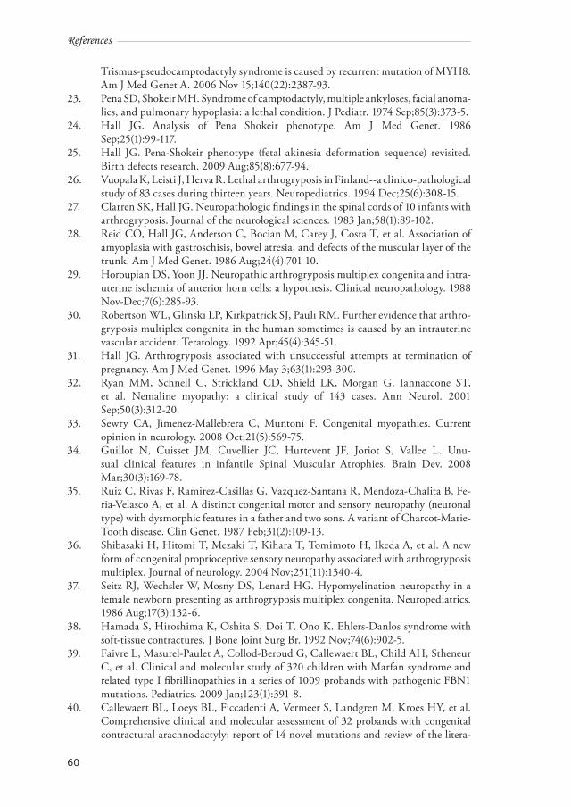
60
References
Trismus-pseudocamptodactyly syndrome is caused by recurrent mutation of MYH8. Am J Med Genet A. 2006 Nov 15;140(22):2387-93.
23. Pena SD, Shokeir MH. Syndrome of camptodactyly, multiple ankyloses, facial anoma-lies, and pulmonary hypoplasia: a lethal condition. J Pediatr. 1974 Sep;85(3):373-5.
24. Hall JG. Analysis of Pena Shokeir phenotype. Am J Med Genet. 1986 Sep;25(1):99-117.
25. Hall JG. Pena-Shokeir phenotype (fetal akinesia deformation sequence) revisited. Birth defects research. 2009 Aug;85(8):677-94.
26. Vuopala K, Leisti J, Herva R. Lethal arthrogryposis in Finland--a clinico-pathological study of 83 cases during thirteen years. Neuropediatrics. 1994 Dec;25(6):308-15.
27. Clarren SK, Hall JG. Neuropathologic findings in the spinal cords of 10 infants with arthrogryposis. Journal of the neurological sciences. 1983 Jan;58(1):89-102.
28. Reid CO, Hall JG, Anderson C, Bocian M, Carey J, Costa T, et al. Association of amyoplasia with gastroschisis, bowel atresia, and defects of the muscular layer of the trunk. Am J Med Genet. 1986 Aug;24(4):701-10.
29. Horoupian DS, Yoon JJ. Neuropathic arthrogryposis multiplex congenita and intra-uterine ischemia of anterior horn cells: a hypothesis. Clinical neuropathology. 1988 Nov-Dec;7(6):285-93.
30. Robertson WL, Glinski LP, Kirkpatrick SJ, Pauli RM. Further evidence that arthro-gryposis multiplex congenita in the human sometimes is caused by an intrauterine vascular accident. Teratology. 1992 Apr;45(4):345-51.
31. Hall JG. Arthrogryposis associated with unsuccessful attempts at termination of pregnancy. Am J Med Genet. 1996 May 3;63(1):293-300.
32. Ryan MM, Schnell C, Strickland CD, Shield LK, Morgan G, Iannaccone ST, et al. Nemaline myopathy: a clinical study of 143 cases. Ann Neurol. 2001 Sep;50(3):312-20.
33. Sewry CA, Jimenez-Mallebrera C, Muntoni F. Congenital myopathies. Current opinion in neurology. 2008 Oct;21(5):569-75.
34. Guillot N, Cuisset JM, Cuvellier JC, Hurtevent JF, Joriot S, Vallee L. Unu-sual clinical features in infantile Spinal Muscular Atrophies. Brain Dev. 2008 Mar;30(3):169-78.
35. Ruiz C, Rivas F, Ramirez-Casillas G, Vazquez-Santana R, Mendoza-Chalita B, Fe-ria-Velasco A, et al. A distinct congenital motor and sensory neuropathy (neuronal type) with dysmorphic features in a father and two sons. A variant of Charcot-Marie-Tooth disease. Clin Genet. 1987 Feb;31(2):109-13.
36. Shibasaki H, Hitomi T, Mezaki T, Kihara T, Tomimoto H, Ikeda A, et al. A new form of congenital proprioceptive sensory neuropathy associated with arthrogryposis multiplex. Journal of neurology. 2004 Nov;251(11):1340-4.
37. Seitz RJ, Wechsler W, Mosny DS, Lenard HG. Hypomyelination neuropathy in a female newborn presenting as arthrogryposis multiplex congenita. Neuropediatrics. 1986 Aug;17(3):132-6.
38. Hamada S, Hiroshima K, Oshita S, Doi T, Ono K. Ehlers-Danlos syndrome with soft-tissue contractures. J Bone Joint Surg Br. 1992 Nov;74(6):902-5.
39. Faivre L, Masurel-Paulet A, Collod-Beroud G, Callewaert BL, Child AH, Stheneur C, et al. Clinical and molecular study of 320 children with Marfan syndrome and related type I fibrillinopathies in a series of 1009 probands with pathogenic FBN1 mutations. Pediatrics. 2009 Jan;123(1):391-8.
40. Callewaert BL, Loeys BL, Ficcadenti A, Vermeer S, Landgren M, Kroes HY, et al. Comprehensive clinical and molecular assessment of 32 probands with congenital contractural arachnodactyly: report of 14 novel mutations and review of the litera-
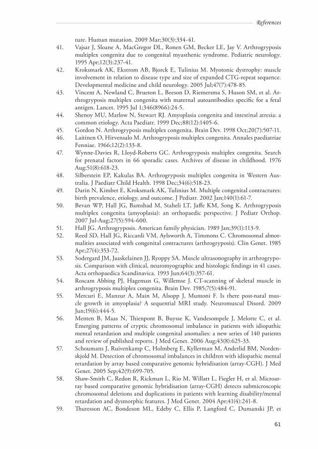
61
References
ture. Human mutation. 2009 Mar;30(3):334-41.41. Vajsar J, Sloane A, MacGregor DL, Ronen GM, Becker LE, Jay V. Arthrogryposis
multiplex congenita due to congenital myasthenic syndrome. Pediatric neurology. 1995 Apr;12(3):237-41.
42. Kroksmark AK, Ekstrom AB, Bjorck E, Tulinius M. Myotonic dystrophy: muscle involvement in relation to disease type and size of expanded CTG-repeat sequence. Developmental medicine and child neurology. 2005 Jul;47(7):478-85.
43. Vincent A, Newland C, Brueton L, Beeson D, Riemersma S, Huson SM, et al. Ar-throgryposis multiplex congenita with maternal autoantibodies specific for a fetal antigen. Lancet. 1995 Jul 1;346(8966):24-5.
44. Shenoy MU, Marlow N, Stewart RJ. Amyoplasia congenita and intestinal atresia: a common etiology. Acta Paediatr. 1999 Dec;88(12):1405-6.
45. Gordon N. Arthrogryposis multiplex congenita. Brain Dev. 1998 Oct;20(7):507-11.46. Laitinen O, Hirvensalo M. Arthrogryposis multiplex congenita. Annales paediatriae
Fenniae. 1966;12(2):133-8.47. Wynne-Davies R, Lloyd-Roberts GC. Arthrogryposis multiplex congenita. Search
for prenatal factors in 66 sporadic cases. Archives of disease in childhood. 1976 Aug;51(8):618-23.
48. Silberstein EP, Kakulas BA. Arthrogryposis multiplex congenita in Western Aus-tralia. J Paediatr Child Health. 1998 Dec;34(6):518-23.
49. Darin N, Kimber E, Kroksmark AK, Tulinius M. Multiple congenital contractures: birth prevalence, etiology, and outcome. J Pediatr. 2002 Jan;140(1):61-7.
50. Bevan WP, Hall JG, Bamshad M, Staheli LT, Jaffe KM, Song K. Arthrogryposis multiplex congenita (amyoplasia): an orthopaedic perspective. J Pediatr Orthop. 2007 Jul-Aug;27(5):594-600.
51. Hall JG. Arthrogryposis. American family physician. 1989 Jan;39(1):113-9.52. Reed SD, Hall JG, Riccardi VM, Aylsworth A, Timmons C. Chromosomal abnor-
malities associated with congenital contractures (arthrogryposis). Clin Genet. 1985 Apr;27(4):353-72.
53. Sodergard JM, Jaaskelainen JJ, Ryoppy SA. Muscle ultrasonography in arthrogrypo-sis. Comparison with clinical, neuromyographic and histologic findings in 41 cases. Acta orthopaedica Scandinavica. 1993 Jun;64(3):357-61.
54. Roscam Abbing PJ, Hageman G, Willemse J. CT-scanning of skeletal muscle in arthrogryposis multiplex congenita. Brain Dev. 1985;7(5):484-91.
55. Mercuri E, Manzur A, Main M, Alsopp J, Muntoni F. Is there post-natal mus-cle growth in amyoplasia? A sequential MRI study. Neuromuscul Disord. 2009 Jun;19(6):444-5.
56. Menten B, Maas N, Thienpont B, Buysse K, Vandesompele J, Melotte C, et al. Emerging patterns of cryptic chromosomal imbalance in patients with idiopathic mental retardation and multiple congenital anomalies: a new series of 140 patients and review of published reports. J Med Genet. 2006 Aug;43(8):625-33.
57. Schoumans J, Ruivenkamp C, Holmberg E, Kyllerman M, Anderlid BM, Norden-skjold M. Detection of chromosomal imbalances in children with idiopathic mental retardation by array based comparative genomic hybridisation (array-CGH). J Med Genet. 2005 Sep;42(9):699-705.
58. Shaw-Smith C, Redon R, Rickman L, Rio M, Willatt L, Fiegler H, et al. Microar-ray based comparative genomic hybridisation (array-CGH) detects submicroscopic chromosomal deletions and duplications in patients with learning disability/mental retardation and dysmorphic features. J Med Genet. 2004 Apr;41(4):241-8.
59. Thuresson AC, Bondeson ML, Edeby C, Ellis P, Langford C, Dumanski JP, et
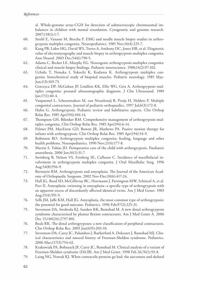
62
References
al. Whole-genome array-CGH for detection of submicroscopic chromosomal im-balances in children with mental retardation. Cytogenetic and genome research. 2007;118(1):1-7.
60. Strehl E, Vanasse M, Brochu P. EMG and needle muscle biopsy studies in arthro-gryposis multiplex congenita. Neuropediatrics. 1985 Nov;16(4):225-7.
61. Kang PB, Lidov HG, David WS, Torres A, Anthony DC, Jones HR, et al. Diagnostic value of electromyography and muscle biopsy in arthrogryposis multiplex congenita. Ann Neurol. 2003 Dec;54(6):790-5.
62. Adams C, Becker LE, Murphy EG. Neurogenic arthrogryposis multiplex congenita: clinical and muscle biopsy findings. Pediatric neuroscience. 1988;14(2):97-102.
63. Uchida T, Nonaka I, Yokochi K, Kodama K. Arthrogryposis multiplex con-genita: histochemical study of biopsied muscles. Pediatric neurology. 1985 May-Jun;1(3):169-73.
64. Gorczyca DP, McGahan JP, Lindfors KK, Ellis WG, Grix A. Arthrogryposis mul-tiplex congenita: prenatal ultrasonographic diagnosis. J Clin Ultrasound. 1989 Jan;17(1):40-4.
65. Vanpaemel L, Schoenmakers M, van Nesselrooij B, Pruijs H, Helders P. Multiple congenital contractures. Journal of pediatric orthopaedics. 1997 Jul;6(3):172-8.
66. Hahn G. Arthrogryposis. Pediatric review and habilitative aspects. Clin Orthop Relat Res. 1985 Apr(194):104-14.
67. Thompson GH, Bilenker RM. Comprehensive management of arthrogryposis mul-tiplex congenita. Clin Orthop Relat Res. 1985 Apr(194):6-14.
68. Palmer PM, MacEwen GD, Bowen JR, Mathews PA. Passive motion therapy for infants with arthrogryposis. Clin Orthop Relat Res. 1985 Apr(194):54-9.
69. Robinson RO. Arthrogryposis multiplex congenita; feeding, language and other health problems. Neuropediatrics. 1990 Nov;21(4):177-8.
70. Martin S, Tobias JD. Perioperative care of the child with arthrogryposis. Paediatric anaesthesia. 2006 Jan;16(1):31-7.
71. Steinberg B, Nelson VS, Feinberg SE, Calhoun C. Incidence of maxillofacial in-volvement in arthrogryposis multiplex congenita. J Oral Maxillofac Surg. 1996 Aug;54(8):956-9.
72. Bernstein RM. Arthrogryposis and amyoplasia. The Journal of the American Acad-emy of Orthopaedic Surgeons. 2002 Nov-Dec;10(6):417-24.
73. Hall JG, Reed SD, McGillivray BC, Herrmann J, Partington MW, Schinzel A, et al. Part II. Amyoplasia: twinning in amyoplasia--a specific type of arthrogryposis with an apparent excess of discordantly affected identical twins. Am J Med Genet. 1983 Aug;15(4):591-9.
74. Sells JM, Jaffe KM, Hall JG. Amyoplasia, the most common type of arthrogryposis: the potential for good outcome. Pediatrics. 1996 Feb;97(2):225-31.
75. Stevenson DA, Swoboda KJ, Sanders RK, Bamshad M. A new distal arthrogryposis syndrome characterized by plantar flexion contractures. Am J Med Genet A. 2006 Dec 15;140(24):2797-801.
76. Beals RK. The distal arthrogryposes: a new classification of peripheral contractures. Clin Orthop Relat Res. 2005 Jun(435):203-10.
77. Stevenson DA, Carey JC, Palumbos J, Rutherford A, Dolcourt J, Bamshad MJ. Clin-ical characteristics and natural history of Freeman-Sheldon syndrome. Pediatrics. 2006 Mar;117(3):754-62.
78. Krakowiak PA, Bohnsack JF, Carey JC, Bamshad M. Clinical analysis of a variant of Freeman-Sheldon syndrome (DA2B). Am J Med Genet. 1998 Feb 26;76(1):93-8.
79. Laing NG, Nowak KJ. When contractile proteins go bad: the sarcomere and skeletal
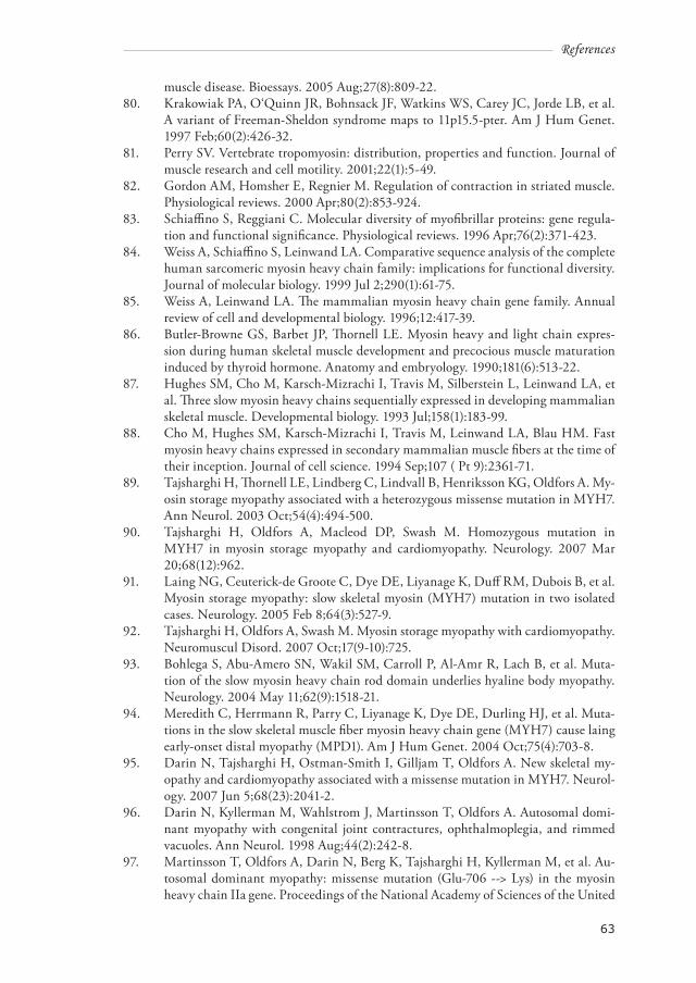
63
References
muscle disease. Bioessays. 2005 Aug;27(8):809-22.80. Krakowiak PA, O‘Quinn JR, Bohnsack JF, Watkins WS, Carey JC, Jorde LB, et al.
A variant of Freeman-Sheldon syndrome maps to 11p15.5-pter. Am J Hum Genet. 1997 Feb;60(2):426-32.
81. Perry SV. Vertebrate tropomyosin: distribution, properties and function. Journal of muscle research and cell motility. 2001;22(1):5-49.
82. Gordon AM, Homsher E, Regnier M. Regulation of contraction in striated muscle. Physiological reviews. 2000 Apr;80(2):853-924.
83. Schiaffino S, Reggiani C. Molecular diversity of myofibrillar proteins: gene regula-tion and functional significance. Physiological reviews. 1996 Apr;76(2):371-423.
84. Weiss A, Schiaffino S, Leinwand LA. Comparative sequence analysis of the complete human sarcomeric myosin heavy chain family: implications for functional diversity. Journal of molecular biology. 1999 Jul 2;290(1):61-75.
85. Weiss A, Leinwand LA. The mammalian myosin heavy chain gene family. Annual review of cell and developmental biology. 1996;12:417-39.
86. Butler-Browne GS, Barbet JP, Thornell LE. Myosin heavy and light chain expres-sion during human skeletal muscle development and precocious muscle maturation induced by thyroid hormone. Anatomy and embryology. 1990;181(6):513-22.
87. Hughes SM, Cho M, Karsch-Mizrachi I, Travis M, Silberstein L, Leinwand LA, et al. Three slow myosin heavy chains sequentially expressed in developing mammalian skeletal muscle. Developmental biology. 1993 Jul;158(1):183-99.
88. Cho M, Hughes SM, Karsch-Mizrachi I, Travis M, Leinwand LA, Blau HM. Fast myosin heavy chains expressed in secondary mammalian muscle fibers at the time of their inception. Journal of cell science. 1994 Sep;107 ( Pt 9):2361-71.
89. Tajsharghi H, Thornell LE, Lindberg C, Lindvall B, Henriksson KG, Oldfors A. My-osin storage myopathy associated with a heterozygous missense mutation in MYH7. Ann Neurol. 2003 Oct;54(4):494-500.
90. Tajsharghi H, Oldfors A, Macleod DP, Swash M. Homozygous mutation in MYH7 in myosin storage myopathy and cardiomyopathy. Neurology. 2007 Mar 20;68(12):962.
91. Laing NG, Ceuterick-de Groote C, Dye DE, Liyanage K, Duff RM, Dubois B, et al. Myosin storage myopathy: slow skeletal myosin (MYH7) mutation in two isolated cases. Neurology. 2005 Feb 8;64(3):527-9.
92. Tajsharghi H, Oldfors A, Swash M. Myosin storage myopathy with cardiomyopathy. Neuromuscul Disord. 2007 Oct;17(9-10):725.
93. Bohlega S, Abu-Amero SN, Wakil SM, Carroll P, Al-Amr R, Lach B, et al. Muta-tion of the slow myosin heavy chain rod domain underlies hyaline body myopathy. Neurology. 2004 May 11;62(9):1518-21.
94. Meredith C, Herrmann R, Parry C, Liyanage K, Dye DE, Durling HJ, et al. Muta-tions in the slow skeletal muscle fiber myosin heavy chain gene (MYH7) cause laing early-onset distal myopathy (MPD1). Am J Hum Genet. 2004 Oct;75(4):703-8.
95. Darin N, Tajsharghi H, Ostman-Smith I, Gilljam T, Oldfors A. New skeletal my-opathy and cardiomyopathy associated with a missense mutation in MYH7. Neurol-ogy. 2007 Jun 5;68(23):2041-2.
96. Darin N, Kyllerman M, Wahlstrom J, Martinsson T, Oldfors A. Autosomal domi-nant myopathy with congenital joint contractures, ophthalmoplegia, and rimmed vacuoles. Ann Neurol. 1998 Aug;44(2):242-8.
97. Martinsson T, Oldfors A, Darin N, Berg K, Tajsharghi H, Kyllerman M, et al. Au-tosomal dominant myopathy: missense mutation (Glu-706 --> Lys) in the myosin heavy chain IIa gene. Proceedings of the National Academy of Sciences of the United
64
References
States of America. 2000 Dec 19;97(26):14614-9.98. Tajsharghi H, Thornell LE, Darin N, Martinsson T, Kyllerman M, Wahlstrom J, et
al. Myosin heavy chain IIa gene mutation E706K is pathogenic and its expression increases with age. Neurology. 2002 Mar 12;58(5):780-6.
99. Ochala J, Li M, Tajsharghi H, Kimber E, Tulinius M, Oldfors A, et al. Effects of a R133W beta-tropomyosin mutation on regulation of muscle contraction in single human muscle fibres. The Journal of physiology. 2007 Jun 15;581(Pt 3):1283-92.
100. Eek MN, Kroksmark AK, Beckung E. Isometric muscle torque in children 5 to 15 years of age: normative data. Archives of physical medicine and rehabilitation. 2006 Aug;87(8):1091-9.
101. Scott OM, Hyde SA, Goddard C, Dubowitz V. Quantitation of muscle function in children: a prospective study in Duchenne muscular dystrophy. Muscle Nerve. 1982 Apr;5(4):291-301.
102. Hoffer MM, Swank S, Eastman F, Clark D, Teitge R. Ambulation in severe arthro-gryposis. J Pediatr Orthop. 1983 Jul;3(3):293-6.
103. Altman D. Practical statistics for medical research. London: Chapman Hall 1991.104. Aradhya S, Manning MA, Splendore A, Cherry AM. Whole-genome array-CGH
identifies novel contiguous gene deletions and duplications associated with devel-opmental delay, mental retardation, and dysmorphic features. Am J Med Genet A. 2007 Jul 1;143A(13):1431-41.
105. Kimura A, Harada H, Park JE, Nishi H, Satoh M, Takahashi M, et al. Mutations in the cardiac troponin I gene associated with hypertrophic cardiomyopathy. Nat Genet. 1997 Aug;16(4):379-82.
106. Hageman G, Willemse J. Arthrogryposis multiplex congenita. Review with com-ment. Neuropediatrics. 1983 Feb;14(1):6-11.
107. Sodergard J, Hakamies-Blomqvist L, Sainio K, Ryoppy S, Vuorinen R. Arthro-gryposis multiplex congenita: perinatal and electromyographic findings, disability, and psychosocial outcome. Journal of pediatric orthopaedics. 1997 Jul;6(3):167-71.
108. Thomas B, Schopler S, Wood W, Oppenheim WL. The knee in arthrogryposis. Clin Orthop Relat Res. 1985 Apr(194):87-92.
109. Donner K, Ollikainen M, Ridanpaa M, Christen HJ, Goebel HH, de Visser M, et al. Mutations in the beta-tropomyosin (TPM2) gene--a rare cause of nemaline my-opathy. Neuromuscul Disord. 2002 Feb;12(2):151-8.
110. Ohlsson M, Quijano-Roy S, Darin N, Brochier G, Lacene E, Avila-Smirnow D, et al. New morphologic and genetic findings in cap disease associated with beta-tropomyosin (TPM2) mutations. Neurology. 2008 Dec 2;71(23):1896-901.
111. Oldfors A. Hereditary myosin myopathies. Neuromuscul Disord. 2007 May;17(5):355-67.
112. Robinson P, Lipscomb S, Preston LC, Altin E, Watkins H, Ashley CC, et al. Muta-tions in fast skeletal troponin I, troponin T, and beta-tropomyosin that cause distal arthrogryposis all increase contractile function. Faseb J. 2007 Mar;21(3):896-905.
113. Klemp P, Hall JG. Dominant distal arthrogryposis in a Maori family with marked variability of expression. Am J Med Genet. 1995 Feb 13;55(4):414-9.
114. Bamshad M, Van Heest AE, Pleasure D. Arthrogryposis: a review and update. The Journal of bone and joint surgery. 2009 Jul;91 Suppl 4:40-6.