Embed Size (px)
Citation preview
CroniconO P E N A C C E S S EC DENTAL SCIENCE
Case Report
Ehler Danlos Syndrome: Oral, Ocular Manifestations and Prophylactic Measures – A Rare Case Report
Raghavendra Havale1*, BS Sheetal2, Kallappa Herakal3, K Rajkumar Chowdary4 and G Anitha5
1Professor in Department of Pedodontics and Preventive Dentistry, AME’s Dental College, Hospital and Research Institute, Raichur, Karnataka, India2District Nodal Officer for National Oral Health Program and Senior Dental Health Officer, Raichur, Karnataka, India 3Professor and Head in Department of Dermatology, NET’s Navodaya Medical College, Raichur, Karnataka, India4Reader in Department of Pedodontics and Preventive Dentistry, Geetanjali Dental College, Udaipur, India5Post Graduate Student in Department of Pedodontics and Preventive Dentistry, AME’s Dental College, Hospital and Research Institute, Raichur, Karnataka, India
Citation: Raghavendra Havale., et al. “Ehler Danlos Syndrome: Oral, Ocular Manifestations and Prophylactic Measures – A Rare Case Report”. EC Dental Science 17.4 (2018): 309-317.
*Corresponding Author: Raghavendra Havale, Professor in Department of Pedodontics and Preventive Dentistry, AME’s Dental College, Hospital and Research Institute, Raichur, Karnataka, India.
Received: December 22, 2018; Published: March 06, 2018
Abstract
Ehler-Danlos type is a rare disorder characterized by soft, hyper extensible skin, abnormal scarring, and easy bruising. Here is a case of classical oral and ocular features of Ehler-Danlos Syndrome (EDS) presented 14 year old blind child casually found at blind school while performing dental screening camp. The boy presented with hyper elasticity of skin, hyper extensibility of joint, hyper pigmented atrophic scars, Ichthyosis Vulgaris and Ocular signs like retinitis pigmentosa, bilateral lenses atrophy, slight keratoconus and keratoglobus. Oral features include periodontitis, gingival recession, bleeding from gums and hyper extensibility of TMJ. Family history was noncontributory for any skin, joint or tooth disorders. The typical clinical signs confirmed the diagnosis of Ehler-Danlos syndrome type VIII associated with ocular features. As there is no specific treatment for the disorder, management is limited to the symptomatic treatment of the dental disease. It is advisable to consider prophylactic measures with Ehler-Danlos type VIII syndrome.
Keywords: Ehler-Danlos Syndrome; Periodontitis; Gingival Recession; Ocular Features; Hyperextensibility of TMJ; Atrophic Scarring; Ichthyosis Vulgaris; Gorlin’s Sign
Introduction
The Ehler-Danlos Syndrome (EDS) syndrome consists of a heterogeneous group of generalized connective tissue disorder. The five cardinal elements of the EDS include hyper extensible doughy skin, dystrophic scar, joint hyper mobility, connective tissue fragility and bruising [1]. Job Janszoon van Meek’ren [2], Dutch surgeon in 1657, was first person to exhibit a young Spanish boy George Albas who was able to stretch the skin of chin reaching chest. Tschernogobow [3], a Russian dermatologist presented the first comprehensive scientific description of multi system features in generalized connective tissue defects, at Venerology and dermatology society meeting at Moscow in 1892. Edward Lauritz Ehlers [4], in 1901 reported the association of hyper elastic skin, hemorrhage and loose jointedness. Henri Alexander Danlos [5], in 1908 recognized cutaneous pseudo tumours and fragility. In 1936 an English physician Fredric Pakes-weber, described the condition as Ehler-Danlos syndrome [6]. This condition is routinely described as “Rubber man” or “Contortionist” [7,8]. Genetic, biochemical and molecular studies have defined more than 10 types of EDS. The estimated prevalence of all types is about 1:5000

310
Ehler Danlos Syndrome: Oral, Ocular Manifestations and Prophylactic Measures – A Rare Case Report
Citation: Raghavendra Havale., et al. “Ehler Danlos Syndrome: Oral, Ocular Manifestations and Prophylactic Measures – A Rare Case Report”. EC Dental Science 17.4 (2018): 309-317.
[9]. Type VII EDS involvement of periodontitis less than 50 cases had been reported in the medical literature to date [10]. We here report a rare case of EDS associated with ocular and oral manifestation, which reminds us of the variable spectrum of EDS and the importance of routine dental evaluation in clinical practice.
Case Report
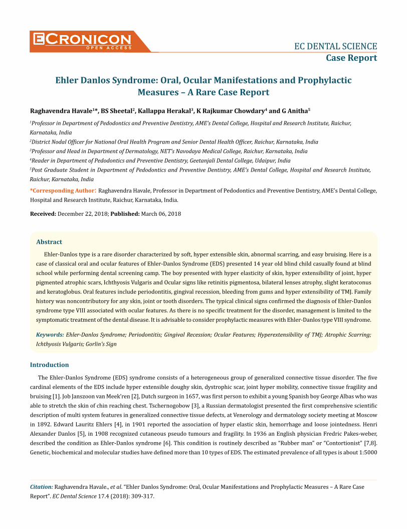
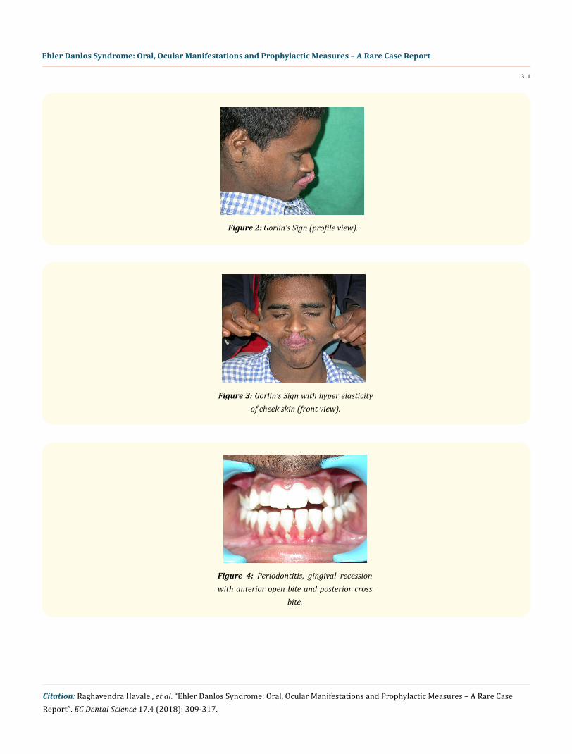
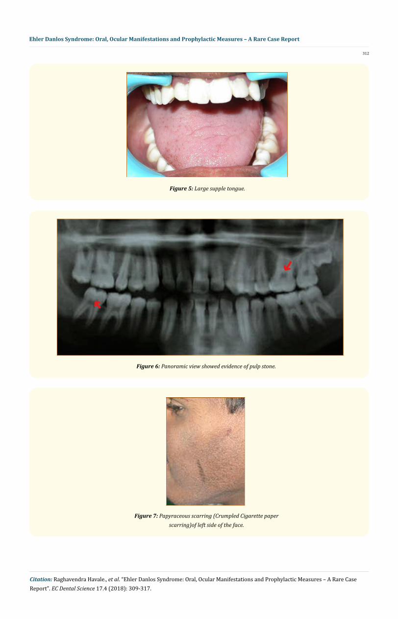
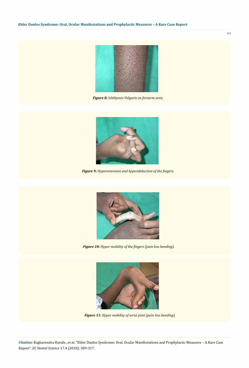
A 14 years old boy who is blind, casually found in dental screening camp at Manik Prabu Academy school for blind, Raichur and brought to the Pedodontics department, AME’s Dental College and Hospital, Raichur for oral prophylaxis. While doing oral prophylaxis he was able to open mouth very wide than usual patient. Face of the patient looks to be abnormal because of widely placed eyes and flat nasal bridge, with abnormal skin. There was no history of seizures/mental retardation/delayed milestone/prolonged bleeding from wounds. There was no history of repeated bone fracture. Personal and family history revealed that socio-economical condition is poor and the child was born to consanguineous married couple. No history of similar complaint noticed in their family. The child was normal in growth and intelligence. Blood investigation was done and coagulate profile was normal. Head and neck examination showed that he has long narrow face, dysplastic ears with prominent pinnae, widely placed eyes with broad nasal bridge. TMJ showed hyper extensibility with maximum inter incisal opening (Figure 1), was measured to 59 mm (normal range is 35 to 45 mm), demonstrated Gorlin sign (tip of tongue touches to tip of nose) and hyper elasticity of skin of the cheeks (Figure 2 and 3). Intraoral examination revealed that gingiva was fragile, loss of gingival attachment, bleeding on probing, anterior open bite, posterior cross bite, mild hypo plastic teeth, with supple large tongue (Fig-ure 4 and 5). Radiographic feature of orthopantomography showed evidence of pulp stones in pulpal chamber of the molars (arrow mark in figure 6) without any other abnormalities. The child was referred to the dermatologist, ophthalmologist, orthopaedician and cardio-pulmonary specialists of Navodaya Medical College and Hospital, Raichur for thorough examination. Dermatologist examination revealed hyperelastic skin with normal recovery type with atrophic scar (cigarette paper scar) at left side of the cheek (Figure 7). Hyper elasticity at neck and fore arm was demonstrated: 1.5 cm without any pain. The skin was affected with Ichthyosis Vulgaris (Figure 8), Keratosis pularis on the extensor aspect of the limbs. Diffuse palmoplantar hyperkeratosis was noticed. Hair and nails are normal. Orthopedic examina-tion revealed that child was able to demonstrate marked hypermobility of elbow, knee, fingers and toes (Hyper extensibility and hyper abduction of fingers and toes without pain) (Figure 9-12). Ophthalmology examination revealed the child was blind because his eyes are affected with retinitis pigmentosa, retinal detachment and bilateral optic lens atrophy with bluish white sclera. The cardiopulmonary examination revealed that the child health was unremarkable. Neurological examination demonstrated slightly diminished reflexes. The oral prophylaxis treatment was performed carefully. Precautions are given to the child to brush the teeth without damaging the gums, to avoid maximum mouth opening to prevent subluxation of TMJ and parents instructed to protect the child from trauma.
Figure 1: Hyper extensibility of TMJ (59 mm Inter incisal opening).
311
Ehler Danlos Syndrome: Oral, Ocular Manifestations and Prophylactic Measures – A Rare Case Report
Citation: Raghavendra Havale., et al. “Ehler Danlos Syndrome: Oral, Ocular Manifestations and Prophylactic Measures – A Rare Case Report”. EC Dental Science 17.4 (2018): 309-317.
Figure 2: Gorlin’s Sign (profile view).
Figure 3: Gorlin’s Sign with hyper elasticity of cheek skin (front view).
Figure 4: Periodontitis, gingival recession with anterior open bite and posterior cross
bite.
312
Ehler Danlos Syndrome: Oral, Ocular Manifestations and Prophylactic Measures – A Rare Case Report
Citation: Raghavendra Havale., et al. “Ehler Danlos Syndrome: Oral, Ocular Manifestations and Prophylactic Measures – A Rare Case Report”. EC Dental Science 17.4 (2018): 309-317.
Figure 5: Large supple tongue.
Figure 6: Panoramic view showed evidence of pulp stone.
Figure 7: Papyraceous scarring (Crumpled Cigarette paper scarring)of left side of the face.
313
Ehler Danlos Syndrome: Oral, Ocular Manifestations and Prophylactic Measures – A Rare Case Report
Citation: Raghavendra Havale., et al. “Ehler Danlos Syndrome: Oral, Ocular Manifestations and Prophylactic Measures – A Rare Case Report”. EC Dental Science 17.4 (2018): 309-317.
Figure 8: Ichthyosis Vulgaris on forearm area.
Figure 9: Hyperextension and hyperabduction of the fingers.
Figure 10: Hyper mobility of the fingers (pain less bending).
Figure 11: Hyper mobility of wrist joint (pain less bending).

314
Ehler Danlos Syndrome: Oral, Ocular Manifestations and Prophylactic Measures – A Rare Case Report
Citation: Raghavendra Havale., et al. “Ehler Danlos Syndrome: Oral, Ocular Manifestations and Prophylactic Measures – A Rare Case Report”. EC Dental Science 17.4 (2018): 309-317.
Figure 12: Double jointedness. Demonstration of excessive extension of digits voluntarily.
Discussion
Syndrome is a group of signs and symptoms that occur together and characterize a disease [11]. Some affects normal active life; some does not affect active life. Ehlers-Danlos Syndromes are a genetically, biochemically and clinically diverse group of heritable connective tissue disorder having joint laxity and dermal features in common [12]. The main clinical presentations are variable joint hypermobility, skin hyperextensibility, and tissue or organ fragility. In 1997 Beighton., et al. [13] revised the past classification based on each distinc-tive clinical manifestation, genetics, biochemical and molecular studies have defined more than 10 types of EDS. The exact aetiology of the syndrome is not yet known. Current molecular biological studies suggests mutation of fibrillar collagen (I, II, III, and V and XI) or enzymes which are responsible for, catalyzing the intracellular or extracellular post translational modifications [14]. This is supposed to result into defective cross linking of various hydroxyproline and hydroxylysine units. There by collagen meshwork will be formed loose. These collagen are distributed in skin, bone, tendons, cartilage, vitreous humor, placenta, chorion and others this distribution explain the pathogenesis and clinical manifestation of certain type of EDS [1]. This syndrome may be inherited as an autosomal dominant (AD), autosomal recessive (AR) or XLR trait. Approximately 80% of affected individuals have type I or type II, around 10% type 3, 4% type IV and about 4% have various other types [15]. In existing case, the classical clinical signs presented are, abnormal “cigarette-paper scars” of poor wound healing over the face, and the positive “Gorlin sign”, ease of bruising in gingiva, gingival recession accompanied with bilat-eral hyperextensive, elbow joints and TM joint, hyper elasticity of the skin over the check, neck, and elbow can confirm the diagnosis of EDS. Skin was presented with Ichthyosis Vulgaris. This condition is autosomal dominant and demonstrated a dry, fine scale that appears “pasted on” over the entire body. Larger and prominent Ichthyosis lesions are seen on lower limbs involving extensor surface [16]. This condition improves in summer because of ambient humidity.
In 1972 McKusciks [17], first identified a form of EDS characterized by periodontal disease, scarring of pretibial skin, accompanied by joint hyper mobility and hyper extensibility of skin. The periodontal disease generally appears after puberty. There is collagen degrada-tion judged by gingival resorption and cutaneous inflammation [18]. Early-onset generalized periodontitis is one of the most significant oral manifestations of this syndrome [19]. Finally it may progress to the premature loss of deciduous and permanent teeth [20]. The hypoplastic enamel [21], deep fissures and long cusps, microdontia, extensive periodontal destruction [23], multiple supernumerary teeth [24] have been reported. Radiographic examination often reveals pulp stones and roots that are short and deformed [22]. Tooth

315
Ehler Danlos Syndrome: Oral, Ocular Manifestations and Prophylactic Measures – A Rare Case Report
Citation: Raghavendra Havale., et al. “Ehler Danlos Syndrome: Oral, Ocular Manifestations and Prophylactic Measures – A Rare Case Report”. EC Dental Science 17.4 (2018): 309-317.
ground sections showed irregular dentin –dentinal tubule into enamel, altered scalloping DEJ [23]. Hyper mobility of TMJ without tooth mobility is observed. Healing of mucous membrane is normal, it does not demonstrate hyper extensibility and does not hold the suture well. Gingival findings show hyperplasia, tissue fragility and bleeding tendency [24,25]. The tongue is very supple. Approximately 50% of those with the syndrome can touch the end of their nose with their tongue (Gorlin’s sign), compared to 8 - 10% of the population [26]. The palate is commonly vaulted [27]. In current case intra oral findings includes fragile gingival tissue, bleeding tendency, gingival recession, with absence of tooth mobility, healing of mucous membrane is normal and does not demonstrate hyper extensibility. Deep palate, mild hypoplastic enamel, hyper extensible joints were seen [28]. The inter incisal distance remained 59 mm without any discomfort (normally 35 - 45 mm). The Panoramic radiographic findings showed presence of pulp stones at molar pulp chamber. The mode of inheritance is considered to be autosomal dominant but some patients were diagnosed without any evidence of familial history [29]. In current case also family history is absent with such symptoms. Due to the lack of any definite biochemical or genetic marker, the diagnosis of EDS-VIII is based on clinical grounds. In 1972 Pinnell., et al. [30] described a form of EDS type VI involving ocular and Scoliosis where deficiency of Lysol Hydroxylase enzyme which helps in formation of collagen. In such case blue sclera (The sclera is the white of the eye or the thick outer coat of the eyeball, bluish appearance is attributed to a thinning of the sclera), keratoconus, (a type of abnormal corneal curvature that occurs when the cornea becomes cone-shaped), angioid streaks (cracks in the Bruch’s membrane, the basement or “anchoring” mem-brane of the retina), keratoglobus (thinning and protrusion of the entire corneal surface), retinal damage and glaucoma (Glaucoma is an increase or change in the intraocular pressure which leads to vision impairment ranging from slight changes to blindness, as well as a pro-gressive loss of peripheral vision) features are normally found [31]. Manifested ocular features in present case were Retinitis pigmentosa (there is damage to the retina. that causes severe vision impairment and blindness) and bilateral optic lens atrophy, retinal detachment, epicanthic fold, mild keratoconus and keratoglobus with bluish white sclera was observed. Child is blind, may be because of Retinitis pig-mentosa, bilateral optic lens atrophy, and retinal detachment. These conditions led to progressive loss of vision to blindness. Besides all the features of skin and joint involvement, and ocular involvement several investigators reported numerous abnormalities of the heart, vascular system, bowel and gut e.g. atrial septal defect, mitral valve Fallot’s tetralogy, aneurysm of the sinus of Valsalva, dissecting aneu-rysm and multiple intracranial aneurysms, gastric atony, rectal prolapse, hiatus hernia and intestinal perforation [32,33]. None of these signs were found in the present case. It is important to recognize and manage functional symptoms and be aware of the complications so certain prophylactic measures to be considered in all children with Connective tissue disorders in Paediatric dental practice. Patients with mitral valve prolapse and regurgitation require antibiotic prophylaxis to prevent bacterial endocarditis [29]. To avoid iatrogenic damage to TMJ, appointments should be as short as possible. The inferior alveolar block injection should be given cautiously otherwise it may lead to hematoma [34]. Light orthodontic forces should be applied to protect periodontal ligament [35]. Longer retention period should be considered to avoid orthodontic relapse [9], special precautions to be taken to avoid injuries of the oral mucosa [36]. It is advisable not to perform maxillofacial surgeries for EDS patients. in case of such surgeries, when absolutely necessary, the blood factors should be carefully evaluated preoperatively. Dermal wounds should be closed without tension, preferably in two layers. Deep stitches should be applied generously. Cutaneous stitches should be left in place twice as long as usual and additional fixation of adjacent skin with adhesive tape can help prevent stretching of the scar. EDS patients are advised to avoid contact sports and heavy exercise to prevent bruising [37] and advised supplementation of ascorbic acid, a cofactor for cross-linking of collagen fibrils, to reduce the tendency of bruising [38].
Conclusion
Oral examinations can be of great help in the diagnosis of EDS and if the classic manifestations of the syndrome are present, immedi-ate consultation is necessary from other specialist to know the other system involvement there by the dental surgeons can perform the dental treatment with care. In special children apart from dental treatment emotional support, behavioral and psychological management is necessary in all types of EDS in order to manage the situation.
Acknowledgement
We thank Staff and Principal of Manik Prabhu Academy School for Blind Raichur, Karnataka, for their cooperation.
Bibliography
1. Raoul CM Hennekam., et al. “Gorlin’s syndromes of the head and neck 5th edition”. oxford university press, Inc (2010): 594-610.

316
Ehler Danlos Syndrome: Oral, Ocular Manifestations and Prophylactic Measures – A Rare Case Report
Citation: Raghavendra Havale., et al. “Ehler Danlos Syndrome: Oral, Ocular Manifestations and Prophylactic Measures – A Rare Case Report”. EC Dental Science 17.4 (2018): 309-317.
2. Beighton P. “The Ehlers-Danlos Syndrome”. William Heinemann Medical Books Ltd (1970): 1-194.
3. Denko CW. “Chernogobuv’s syndrome: A translation of the first modern case report of the EDS”. Journal of Rheumatology (1978): 347-352.
4. Ehlers E. “Cutis laxa Neigung, Zn Hemorrhagien in der Haut, LockerungmahrererArtikulationen”. Dermatology Ztshr 8 (1901): 173.
5. Danlos HA. “Un cas de cutis laxa avec tumeurs par contusion chronique des coudes et des genoux (Xanthome Juvenile pseudo diabe-tique de M. M. Hallopeau et Mace de Lepinav)”. Bull Soc franc dedermat et Syph 19 (1908): 70-72.
6. Enerson OD. “Ehlers-Danlos syndrome.. Oslo, Norway: who named it? (biographical dictionary of medical eponyms) Ehlers Danlos syndrome” (2004).
7. Neville BW., et al. “Oral and Maxillofacial Pathology” . Saunders Elsevier Pennsylvania St. Louis, Missouri (2009): 655-657.
8. Shafer WG., et al. “A Text Book of Oral Pathology”. Saunders publishing, Pennsylvania, fourth edition (2003): 847-848.
9. Pyeritz RE. “Ehlers-Danlos syndrome”. In: L. Goldman and J.C. Bennett. Cecil Textbook of Medicine. 21st edition W.B. Saunders, Phila-delphia, PA. Volume 1 (2000): 1119- 1120.
10. Moore MM., et al. “EDS type VIII: Perodontities, easy brusing, marfanoid habitus, and istinctive facies”. Journal of American Academy of Dermatology 55.2 (2006): S41-S45.
11. Babbush CA., et al. “Mosbys dental dictionary nunez David”. Elsevier St. Louis, Missouri. second edition (2008): 651.
12. Chakraborty AN., et al. “Ehlers-Danlos Syndrome”. Journal of Indian Medical Association 23.8 (1954): 344-345.
13. Beighton P., et al. “EDSs: Revised nosology, Villefranche”. American Journal of Medicine and Genetics 77 (1997): 31-37.
14. Byers PH. “EDS: Recent advances and current of the clinical and genetic Heterogenicity”. Investigation Dermatology 103.5 (1994): 47S-52S.
15. Byers PH. “Disorders of collagen biosynthesis and structure”. In: CR Scriver., et al. editors. The Metabolic and Molecular Bases of In-herited Diseases, 7th- edition. New York, McGraw-Hill (1995): 134, 4029.
16. Al-Hajjaj MS. “Bronchiectasis and Mediastinal Neurofibroma in a Saudi female with Ehlers Danlos syndrome”. Annals of Saudi Medi-cine 20.5-6 (2000): 419-420.
17. Mc Kusick VA. “Editorial-Multiple forma of the Ehlers-Danlos syndrome”. Archives of Surgery 109.4 (1974): 475-476.
18. Oleson BH and Ernst E. “Oral manifestation nerved EDs type VIII”. Taandlaegebladet 91 (1987): 313-315.
19. Hartsfield JK Jr and Kousseff BG. “Phenotypic overlap of Ehlers-Danlos syndrome type IV and VIII”. American Journal of Medicine and Genetics 37.4 (1990): 465-470.
20. Piette E and Douniau R. “Parodontolyse infantile symptomatique d’unsyndrome d’Ehlers-Danlos. Uncassporadique”. Acta stomato-logica Belgica 77.3 (1980): 217-229.
21. Linch DC and Acton CHC. “Ehlers-Danlos syndrome presenting with juvenile destructive periodontitis”. British Dental Journal 147.4 (1979): 95-96.
22. Cohn DH and Byers PH. “Clinical screening for collagen defects in connective tissue diseases”. Clinics in Perinatology 17.4 (1990): 739-809.
23. Cole WG. “Etiology and pathogenesis of heritable connective tissue diseases”. Journal of Pediatric Orthopaedics 13.3 (1993): 392-403.
24. Barabas GM. “The Ehlers-Danlos syndrome. Abnormalities of the enamel, dentine, cementum and the dental pulp: an histological examination of 13 teeth from 6 patients”. British Dental Journal 126.11 (1969): 509-515.

317
Ehler Danlos Syndrome: Oral, Ocular Manifestations and Prophylactic Measures – A Rare Case Report
Citation: Raghavendra Havale., et al. “Ehler Danlos Syndrome: Oral, Ocular Manifestations and Prophylactic Measures – A Rare Case Report”. EC Dental Science 17.4 (2018): 309-317.
25. De Coster PJ., et al. “Oral health in prevalent types of Ehlers-Danlos syndromes”. Journal of Oral Pathology and Medicine 34.5 (2005): 298-307.
26. De Coster PJ., et al. “Generalized joint hypermobility and temporomandibular disorders: inherited connective tissue disease as a model with maximum expression”. Journal of Orofacial Pain 19.1 (2005): 47-57.
27. Letourneau Y., et al. “Oral manifestations of Ehlers-Danlos syndrome”. Journal Canadian Dental Association 67.6 (2001): 330-334.
28. Melamed Y., et al. “Multiple supernumerary teeth(MSNT) and Ehlers-Danlos syndrome”. Journal of Oral Pathology and Medicine 23.2 (1994): 88-91.
29. Fridrich KL., et al. “Dental implications in Ehlers-Danlos syndrome. A case report”. Oral Surgery Oral Medicine and Oral Pathology 69.4 (1990) 431-435.
30. Pinnell SR., et al. “A heritable disorder of connective tissue: hydroxylysine-deficient collagen disease”. New England Journal of Medi-cine 286.19 (1972): 1013-1020.
31. K Stephanie., et al. “Ehlers-Danlos Syndrome -The role of collagen in the eye” (2007): 1-5.
32. Beighton CH., et al. “Gastro-intestinal complications of Ehlers-Danlos syndrome”. Gut 10.12 (1969): 1004-1008.
33. Bruno and Narasimhan P. “The Ehlers Danlos syndrome-A report of 4 cases in 2 generations of a Negro family”. New England Journal of Medicine 264 (1961): 274-277.
34. Sacks H., et al. “Recurrent temporomandibular joint subluxation and facial ecchymosis leading to diagnosis of Ehlers-Danlos syn-drome”. Journal of Oral Maxillofacial Surgery 48.6 (1990): 641-647.
35. Norton LA and Assael LA. “Orthodontic and temporomandibular joint considerations in treatment of patients with Ehlers-Danlos syndrome”. American Journal of Orthodontics and Dentofacial Orthopaedics 111.1 (1997): 75-84.
36. Jones ML. “Orthodontic treatment in Ehlers-Danlos syndrome”. British Journal of Orthodontics 11.3 (1984): 158-162.
37. Pepin M and Byers P. “Ehlers-Danlos syndrome, Vascular type” (2002).
38. Steinmann B., et al. “The Ehlers-Danlos syndrome”. In: Royce PM, Steinmann B, editors. Connective tissue and its heritable disorders: molecular, genetic, and medical aspects. 2nd edition. New York: Wiley-Liss (2002): 431-523.
Volume 17 Issue 4 April 2018©All rights reserved by Raghavendra Havale., et al.

![harlequin ichthyosis and functional recovery by · of ichthyosis, harlequin ichthyosis (HI) (Mendelian Inheritance of Man [MIM] 242500) is the most serious subtype (Figure 1); it](https://img.pdfslide.net/doc/110x75/5d4f332b88c993257d8be9c0/harlequin-ichthyosis-and-functional-recovery-by-of-ichthyosis-harlequin-ichthyosis.jpg)