Embed Size (px)
Citation preview

World Journal of Gastroenterology
World Journal of Gastroenterology
World Journal of G
astroenterology ww
w.w
jgnet.com Volum
e 15 Num
ber 36 Sep 28 2009
Volume 15 Number 36September 28, 2009
ISSN 1007-9327 CN 14-1219/R Local Post Offices Code No. 82-261
ISSN 1007-9327CN 14-1219/R
Baishideng百世登
A Weekly Journal of Gastroenterology and Hepatology
Volume 15 Number 36September 28, 2009
World J Gastroenterol2009 September 28; 15(36): 4481-4608
Online Submissionswjg.wjgnet.com
www.wjgnet.com Printed on Acid-free Paper
Published by The WJG Press and Baishideng Room 903, Building D, Ocean International Center,
No. 62 Dongsihuan Zhonglu, Chaoyang District, Beijing 100025, ChinaTelephone: +86-10-59080039
Fax: +86-10-85381893E-mail: [email protected]://www.wjgnet.com
™©
Indexed and Abstracted in:Current Contents®/Clinical Medicine, Science Citation Index Expanded (also known as SciSearch®), Journal Citation Reports/Science Edition, Index Medicus, MEDLINE, PubMed, Chemical Abstracts, EMBASE/Excerpta Medica, Abstracts Journals, PubMed Central, Digital Object Identifier, CAB Abstracts, and Global Health. ISI, Thomson Reuters, 2008 Impact Factor: 2.081(32/55 Gastroenterology and Hepatology).
I S S N 1 0 0 7 - 9 3 2 7
9 7 7 1 0 07 9 3 2 0 45
3 6

World Journal of Gastroenterology
Editorial Board2007-2009
™©Baishideng百世登
Editorial Office: World Journal of GastroenterologyRoom 903, Building D, Ocean International Center
No. 62 Dongsihuan Zhonglu, Chaoyang District, Beijing 100025, ChinaE-mail: [email protected] http://www.wjgnet.com Telephone: 0086-10-5908-0039 Fax: 0086-10-8538-1893
HONORARY EDITORS-IN-CHIEFMontgomery Bissell, San FranciscoJames L Boyer, New HavenKe-Ji Chen, BeijingJacques Van Dam, StanfordMartin H Floch, New HavenGuadalupe Garcia-Tsao, New HavenZhi-Qiang Huang, Beijing Ira M Jacobson, New YorkDerek Jewell, OxfordEmmet B Keeffe, Palo AltoNicholas F LaRusso, RochesterJie-Shou Li, NanjingGeng-Tao Liu, BeijingBo-Rong Pan, Xi’anFa-Zu Qiu, Wuhan[2]
Eamonn M Quigley, CorkDavid S Rampton, LondonRafiq A Sheikh, SacramentoRudi Schmid, Kentfield[1] Nicholas J Talley, RochesterGuido NJ Tytgat, AmsterdamMeng-Chao Wu, ShanghaiJia-Yu Xu, Shanghai
PRESIDENT AND EDITOR-IN-CHIEFLian-Sheng Ma, Beijing
STRATEGY ASSOCIATE EDITORS-IN-CHIEFPeter Draganov, FloridaRonnie Fass, TucsonHugh J Freeman, VancouverJohn P Geibel, New Haven
Maria Concepción Gutiérrez-Ruiz, MéxicoKazuhiro Hanazaki, KochiAkio Inui, KagoshimaKalpesh Jani, BarodaSanaa M Kamal, CairoIoannis E Koutroubakis, HeraklionJose JG Marin, SalamancaJavier S Martin, Punta del EsteNatalia A Osna, OmahaJose Sahel, MarseilleNed Snyder, GalvestonNathan Subramaniam, BrisbaneWei Tang, TokyoAlan BR Thomson, EdmontonPaul Joseph Thuluvath, BaltimoreJames F Trotter, DenverShingo Tsuji, Osaka Harry HX Xia, HanoverYoshio Yamaoka, HoustonJesus K Yamamoto-Furusho, Mexico
ASSOCIATE EDITORS-IN-CHIEFGianfranco D Alpini, TempleBruno Annibale, RomaRoger W Chapman, OxfordChi-Hin Cho, Hong KongAlexander L Gerbes, MunichShou-Dong Lee, TaipeiWalter E Longo, New HavenYou-Yong Lu, BeijingMasao Omata, Tokyo
BIOSTATISTICAL EDITORLiang-Ping Hu, Beijing
www.wjgnet.com I
The World Journal of Gastroenterology Editorial Board consists of 1126 members, representing a team of worldwide experts in gastroenterology and hepatology. They are from 60 countries, including Albania (1), Argentina (4), Australia (39), Austria (10), Belarus (1), Belgium (15), Brazil (2), Bulgaria (1), Canada (24), Chile (1), China (36), Croatia (2), Cuba (1), Czech (3), Denmark (7), Egypt (3), Estonia (1), Finland (4), France (42), Germany (104), Greece (8), Hungary (2), Iceland (1), India (11), Iran (4), Ireland (3), Israel (8), Italy (95), Japan (164), Lebanon (3), Lithuania (1), Macedonia (1), Malaysia (3), Mexico (5), Monaco (1), Morocco (1), The Netherlands (26), New Zealand (1), Nigeria (1), Norway (3), Pakistan (2), Peru (1), Poland (7), Portugal (1), Russia (3), Saudi Arabia (2), Serbia (1), Singapore (5), Slovakia (2), Slovenia (1), South Africa (2), South Korea (14), Spain (36), Sweden (15), Switzerland (14), Turkey (8), United Arab Emirates (1), United Kingdom (77), United States (290), and Uruguay (1).
GUEST EDITORIAL BOARD MEMBERSChao-Long Chen, KaohsiungLi-Fang Chou, TaipeiKevin Cheng-Wen Hsiao, TaipeiShinn-Jang Hwang, TaipeiIra M Jacobson, New YorkMin-Liang Kuo, TaipeiLein-Ray Mo, TainanSun-Lung Tsai, Young-Kang CityHsiu-Po Wang, TaipeiTa-Sen Yeh, TaoyuanMing-Lung Yu, Kaohsiung
MEMBERS OF THE EDITORIAL BOARD
Bashkim Resuli, Tirana
Julio H Carri, CórdobaCarlos J Pirola, Buenos AiresSilvia Sookoian, Buenos AiresAdriana M Torres, Rosario
Leon Anton Adams, NedlandsMinoti V Apte, Liverpool
Albania
Argentina
Australia

Richard B Banati, LidcombeMichael R Beard, AdelaidePatrick Bertolino, SydneyAndrew V Biankin, SydneyFilip Braet, SydneyAndrew D Clouston, SydneyGraham Cooksley, QueenslandDarrell HG Crawford, BrisbaneAdrian G Cummins, Woodville SouthGuy D Eslick, SydneyMichael A Fink, MelbourneRobert JL Fraser, Daw ParkPeter Raymond Gibson, VictoriaJacob George, WestmeadMark D Gorrell, SydneyYik-Hong Ho, TownsvilleGerald J Holtmann, AdelaideMichael Horowitz, AdelaideJohn E Kellow, SydneyRupert Leong, ConcordGeoffrey W McCaughan, SydneyFinlay A Macrae, VictoriaDaniel Markovich, BrisbanePhillip S Oates, PerthJacqui Richmond, VictoriaStephen M Riordan, SydneyIan C Roberts-Thomson, AdelaideDevanshi Seth, CamperdownArthur Shulkes, MelbourneRoss C Smith, SydneyKevin J Spring, BrisbaneHerbert Tilg, InnsbruckHuy A Tran, New South WalesDebbie Trinder, FremantleMartin J Veysey, GosfordDaniel L Worthley, BedfordDavid Ian Watson, South Australia
Belarus
Rudi Beyaert, GentBart Rik De Geest, LeuvenInge I Depoortere, LeuvenOlivier Detry, LiègeBenedicte Y De Winter, AntwerpKarel Geboes, LeuvenThierry Gustot, BrusselsYves J Horsmans, BrusselsGeert G Leroux-Roels, GhentLouis Libbrecht, LeuvenEtienne M Sokal, BrusselsMarc Peeters, De PintelaanGert A Van Assche, LeuvenYvan Vandenplas, BrusselsEddie Wisse, Keerbergen
Heitor Rosa, GoianiaAna Cristina Simões e Silva, Belo Horizonte
Zahariy Krastev, Sofia
Fernando Alvarez, QuébecDavid Armstrong, OntarioJeffrey P Baker, TorontoOlivier Barbier, QuébecNancy Baxter, TorontoFrank J Burczynski, ManitobaMichael F Byrne,VancouverWang-Xue Chen, OttawaSamuel S Lee, CalgaryGary A Levy, TorontoAndrew L Mason, AlbertaJohn K Marshall, OntarioDonna-Marie McCafferty, CalgaryThomas I Michalak, St. John’sGerald Y Minuk, ManitobaPaul Moayyedi, HamiltonKostas Pantopoulos, QuebecWilliam G Paterson, KingstonEldon Shaffer, CalgaryMartin Storr, CalgaryE F Verdu, OntarioWaliul Khan, OntarioJohn L Wallace, CalgaryEric M Yoshida, Vancouver
Dong-Liang Yang, WuhanWinnie Yeo, Hong KongYuan Yuan, ShenyangMan-Fung Yuen, Hong KongJian-Zhong Zhang, BeijingXin-Xin Zhang, ShanghaiBo-Jian Zheng, Hong KongShu Zheng, HangzhouXiao-Peng Zhang, Beijing
Tamara Cacev, ZagrebMarko Duvnjak, Zagreb
Damian C Rodriguez, Havana
Milan Jirsa, PrahaPavel Trunečka, PragueMarcela Kopacova, Hradec Kralove
Peter Bytzer, CopenhagenAsbjørn M Drewes, AalborgHans Gregersen, AalborgJens H Henriksen, HvidovreClaus P Hovendal, OdenseFin S Larsen, CopenhagenSØren MØller, Hvidovre
Abdel-Rahman El-Zayadi, GizaAmr M Helmy, CairoAyman Yosry, Cairo
Riina Salupere, Tartu
Irma E Jarvela, HelsinkiKatri M Kaukinen, TampereMinna Nyström, HelsinkiPentti Sipponen, Espoo
Bettaieb Ali, DijonAnne Corlu, RennesDenis Ardid, Clermont-FerrandCharles P Balabaud, BordeauxSoumeya Bekri, RouenJacques Belghiti, ClichyJacques Bernuau, Clichy CedexPierre Brissot, RennesPatrice P Cacoub, ParisFranck Carbonnel, BesanconLaurent Castera, PessacBruno Clément, RennesBenoit Coffin, ColombesThomas Decaens, CedexFrancoise L Fabiani, Angers
Peter Ferenci, ViennaValentin Fuhrmann, ViennaAlfred Gangl, ViennaChristoph Gasche, ViennaKurt Lenz, LinzMarkus Peck-Radosavljevic, ViennaRudolf E Stauber, AuenbruggerplatzMichael Trauner, GrazHarald Vogelsang, ViennaGuenter Weiss, Innsbruck
Austria
Yury K Marakhouski, Minsk
Belgium
Brazil
Bulgaria
Canada
Silvana Zanlungo, SantiagoChile
Henry LY Chan, Hong KongXiao-Ping Chen, WuhanZong-Jie Cui, BeijingDa-Jun Deng, BeijingEr-Dan Dong, BeijingSheung-Tat Fan, Hong KongJin Gu, BeijingXin-Yuan Guan, PokfulamDe-Wu Han, TaiyuanMing-Liang He, Hong KongWayne HC Hu, Hong KongChee-Kin Hui, Hong KongChing-Lung Lai, Hong KongKam Chuen Lai, Hong KongJames YW Lau, Hong KongYuk-Tong Lee, Hong KongSuet-Yi Leung, Hong KongWai-Keung Leung, Hong KongJohn M Luk, PokfulamChung-Mau Lo, Hong KongJing-Yun Ma, BeijingRonnie Tung Ping Poon, Hong KongLun-Xiu Qin, ShanghaiYu-Gang Song, GuangzhouQin Su, BeijingWai-Man Wong, Hong KongHong Xiao, Shanghai
China
Croatia
Cuba
Czech
Denmark
Egypt
Estonia
Finland
France
www.wjgnet.comⅡ

www.wjgnet.com Ⅲ
Gérard Feldmann, ParisJean Fioramonti, ToulouseJean-Noël Freund, StrasbourgCatherine Guettier, VillejuifChantal Housset, ParisJuan L Iovanna, MarseilleRene Lambert, LyonPatrick Marcellin, ParisPhilippe Mathurin, LilleTamara Matysiak-Budnik, ParisFrancis Mégraud, BordeauxRichard Moreau, ClichyThierry Piche, NiceRaoul Poupon, ParisJean Rosenbaum, BordeauxDominique Marie Roulot, BobignyThierry Poynard, ParisJean-Philippe Salier, RouenDidier Samuel, VillejuifJean-Yves Scoazec, LyonAlain L Servin, Châtenay-MalabryKhalid A Tazi, ClichyEmmanuel Tiret, ParisBaumert F Thomas, StrasbourgJean-Pierre H Zarski, GrenobleJessica Zucman-Rossi, ParisBoris Guiu, Dijon
Ralf Jakobs, LudwigshafenJutta Keller, HamburgAndrej Khandoga, MunichSibylle Koletzko, MünchenStefan Kubicka, HannoverJoachim Labenz, SiegenFrank Lammert, BonnThomas Langmann, RegensburgChristian Liedtke, AachenMatthias Löhr, StockholmChristian Maaser, MuensterAhmed Madisch, DresdenPeter Malfertheiner, MagdeburgMichael P Manns, HannoverHelmut Messmann, AugsburgStephan Miehlke, DresdenSabine Mihm, GöttingenSilvio Nadalin, TuebingenMarkus F Neurath, MainzJohann Ockenga, BerlinFlorian Obermeier, RegensburgGustav Paumgartner, MunichUlrich KS Peitz, MagdeburgMarkus Reiser, BochumEmil C Reisinger, RostockSteffen Rickes, MagdeburgTilman Sauerbruch, BonnDieter Saur, MunichHans Scherübl, BerlinJoerg Schirra, MunichRoland M Schmid, MünchenVolker Schmitz, BonnAndreas G Schreyer, RegensburgTobias Schroeder, EssenHenning Schulze-Bergkamen, MainzNorbert Senninger, MuensterHans Seifert, OldenburgManfred V Singer, MannheimGisela Sparmann, RostockChristian J Steib, MünchenJurgen M Stein, FrankfurtUlrike S Stein, BerlinManfred Stolte, BayreuthChristian P Strassburg, HannoverWolfgang R Stremmel, HeidelbergHarald F Teutsch, UlmRobert Thimme, FreiburgHans L Tillmann, LeipzigTung-Yu Tsui, RegensburgAxel Ulsenheimer, MunichPatrick Veit-Haibach, EssenClaudia Veltkamp, HeidelbergSiegfried Wagner, DeggendorfHenning Walczak, HeidelbergHeiner Wedemeyer, HannoverFritz von Weizsäcker, BerlinJens Werner, HeidelbergBertram Wiedenmann, BerlinReiner Wiest, RegensburgStefan Wirth, WuppertalStefan JP Zeuzem, Homburg
Alexandra A Alexopoulou, AthensGeorge N Dalekos, LarissaChristos Dervenis, AthensMelanie Maria Deutsch, AthensTsianos Epameinondas, IoanninaElias A Kouroumalis, HeraklionGeorge Papatheodoridis, AthensSpiros Sgouros, Athens
Peter L Lakatos, BudapestZsuzsa Szondy, Debrecen
Hallgrimur Gudjonsson, Reykjavik
Philip Abraham, MumbaiRakesh Aggarwal, LucknowKunissery A Balasubramanian, VelloreDeepak Kumar Bhasin, ChandigarhSujit K Bhattacharya, KolkataYogesh K Chawla, ChandigarhRadha K Dhiman, ChandigarhSri P Misra, AllahabadRamesh Roop Rai, JaipurNageshwar D Reddy, HyderabadRakesh Kumar Tandon, New Delhi
Mohammad Abdollahi, TehranSeyed-Moayed Alavian, TehranReza Malekzadeh, TehranSeyed A Taghavi, Shiraz
Billy Bourke, DublinRonan A Cahill, CorkAnthony P Moran, Galway
Simon Bar-Meir, HashomerAbraham R Eliakim, HaifaZvi Fireman, HaderaYaron Ilan, JerusalemAvidan U Neumann, Ramat-GanYaron Niv, Ran Oren, Tel AvivAmi D Sperber, Beer-Sheva
Hans-Dieter Allescher, G-PartenkirchenMartin Anlauf, KielRudolf Arnold, MarburgMax G Bachem, UlmThomas F Baumert, FreiburgDaniel C Baumgart, BerlinHubert Blum, FreiburgThomas Bock, TuebingenKatja Breitkopf, MannheimDunja Bruder, BraunschweigMarkus W Büchler, HeidelbergChrista Buechler, RegensburgReinhard Buettner, BonnElke Cario, EssenUta Dahmen, EssenChristoph F Dietrich, Bad MergentheimArno J Dormann, KoelnRainer J Duchmann, BerlinVolker F Eckardt, WiesbadenFred Fändrich, KielUlrich R Fölsch, KielHelmut Friess, HeidelbergPeter R Galle, MainzNikolaus Gassler, AachenMarkus Gerhard, MunichWolfram H Gerlich, GiessenDieter Glebe, GiessenBurkhard Göke, MunichFlorian Graepler, TuebingenAxel M Gressner, AachenVeit Gülberg, MunichRainer Haas, MunichEckhart G Hahn, ErlangenStephan Hellmig, KielMartin Hennenberg, BonnJohannes Herkel, HamburgKlaus R Herrlinger, StuttgartEva Herrmann, Homburg/SaarEberhard Hildt, BerlinJörg C Hoffmann, BerlinFerdinand Hofstaedter, RegensburgWerner Hohenberger, ErlangenJörg C Kalff, Bonn
Germany
Greece
Hungary
Iceland
India
Iran
Ireland
Israel
ItalyGiovanni Addolorato, RomaLuigi E Adinolfi, NaplesDomenico Alvaro, RomeVito Annese, San Giovanni RotondFilippo Ansaldi, GenoaAdolfo F Attili, RomaGiovanni Barbara, BolognaClaudio Bassi, VeronaGabrio Bassotti, PerugiaPier M Battezzati, MilanStefano Bellentani, CarpiAntomio Benedetti, AnconaMauro Bernardi, BolognaLivia Biancone, RomeLuigi Bonavina, MilanoFlavia Bortolotti, PadovaGiuseppe Brisinda, RomeElisabetta Buscarini, CremaGiovanni Cammarota, Roma
www.wjgnet.comIV
Kyoichi Adachi, IzumoYasushi Adachi, SapporoTaiji Akamatsu, MatsumotoSk Md Fazle Akbar, EhimeTakafumi Ando, NagoyaAkira Andoh, OtsuTaku Aoki, TokyoMasahiro Arai, TokyoTetsuo Arakawa, OsakaYasuji Arase, TokyoHitoshi Asakura, TokyoTakeshi Azuma, FukuiTakahiro Fujimori, TochigiJiro Fujimoto, HyogoKazuma Fujimoto, SagaMitsuhiro Fujishiro, TokyoYoshihide Fujiyama, OtsuHirokazu Fukui, TochigiHiroyuki Hanai, HamamatsuNaohiko Harada, FukuokaMakoto Hashizume, FukuokaTetsuo Hayakawa, NagoyaToru Hiyama, HigashihiroshimaKazuhide Higuchi, OsakaKeisuke Hino, UbeKeiji Hirata, KitakyushuYuji Iimuro, NishinomiyaKenji Ikeda, TokyoKenichi Ikejima, Bunkyo-kuFumio Imazeki, ChibaYutaka Inagaki, KanagawaYasuhiro Inokuchi, YokohamaHaruhiro Inoue, Yokohama Masayasu Inoue, OsakaHiromi Ishibashi, NagasakiShunji Ishihara, IzumoToru Ishikawa, NiigataKei Ito, SendaiMasayoshi Ito, TokyoToru Ikegami, FukuokaHiroaki Itoh, AkitaRyuichi Iwakiri, SagaYoshiaki Iwasaki, OkayamaTerumi Kamisawa, TokyoHiroshi Kaneko, Aichi-gunShuichi Kaneko, KanazawaTakashi Kanematsu, NagasakiMitsuo Katano, FukuokaMototsugu Kato, SapporoShinzo Kato, TokyoNorifumi Kawada, OsakaSunao Kawano, OsakaMitsuhiro Kida, KanagawaYoshikazu Kinoshita, IzumoTsuneo Kitamura, ChibaSeigo Kitano, OitaKazuhiko Koike, TokyoNorihiro Kokudo, TokyoShoji Kubo, OsakaMasatoshi Kudo, OsakaKatsunori Iijima, SendaiShin Maeda, TokyoShigeru Marubashi, SuitaMasatoshi Makuuchi, TokyoOsamu Matsui, KanazawaYasuhiro Matsumura, KashiwaYasushi Matsuzaki, TsukubaKiyoshi Migita, OmuraKenji Miki, TokyoTetsuya Mine, KanagawaHiroto Miwa, Hyogo
Antonino Cavallari, BolognaGiuseppe Chiarioni, ValeggioMichele Cicala, RomeMassimo Colombo, MilanAmedeo Columbano, CagliariMassimo Conio, SanremoDario Conte, MilanoGino R Corazza, PaviaFrancesco Costa, PisaAntonio Craxi, PalermoSilvio Danese, MilanRoberto de Franchis, MilanoRoberto De Giorgio, BolognaMaria Stella De Mitri, BolognaGiovanni D De Palma, NaplesFabio Farinati, PaduaGiammarco Fava, AnconaFrancesco Feo, SassariFiorucci Stefano, PerugiaAndrea Galli, FirenzeValeria Ghisett, TurinGianluigi Giannelli, BariEdoardo G Giannini, GenoaPaolo Gionchetti, BolognaFabio Grizzi, MilanSalvatore Gruttadauria, PalermoMario Guslandi, MilanoPietro Invernizzi, MilanEzio Laconi, CagliariGiacomo Laffi, FirenzeGiovanni Maconi, MilanLucia Malaguarnera, CataniaEmanuele D Mangoni, NapoliPaolo Manzoni, TorinoGiulio Marchesini, BolognaFabio Marra, FlorenceMarco Marzioni, AnconaRoberto Mazzanti, FlorenceGiuseppe Mazzella, BolognaGiuseppe Montalto, PalermoGiovanni Monteleone, RomeGiovanni Musso, TorinoGerardo Nardone, NapoliValerio Nobili, RomeFabio Pace, MilanoLuisi Pagliaro, PalermoFrancesco Pallone, RomeFabrizio R Parente, MilanMaurizio Parola, TorinoFrancesco Perri, San Giovanni RotondoRaffaele Pezzilli, BolognaAlberto Pilotto, San Giovanni RotondoAlberto Piperno, MonzaMario Pirisi, NovaraAnna C Piscaglia, RomaPaolo Del Poggio, TreviglioGabriele B Porro, MilanoPiero Portincasa, BariCosimo Prantera, RomaBernardino Rampone, SienaOliviero Riggio, RomerClaudio Romano, MessinaMarco Romano, NapoliGerardo Rosati, PotenzaMario Del Tacca, PisaGloria Taliani, RomePier A Testoni, MilanEnrico Roda, BolognaDomenico Sansonno, BariVincenzo Savarino, GenovaVincenzo Stanghellini, BolognaGiovanni Tarantino, NaplesRoberto Testa, GenoaDino Vaira, BolognaRoberto Berni Canani, NaplesGianlorenzo Dionigi, Varese
Japan
Masashi Mizokami, NagoyaYoshiaki Mizuguchi, TokyoMotowo Mizuno, HiroshimaMorito Monden, SuitaHisataka Moriwaki, GifuYasuaki Motomura, IizukaYoshiharu Motoo, KanazawaNaofumi Mukaida, KanazawaKazunari Murakami, OitaKunihiko Murase, TusimaHiroaki Nagano, SuitaMasahito Nagaki, GifuAtsushi Nakajima, YokohamaYuji Naito, KyotoHisato Nakajima, TokyoHiroki Nakamura, YamaguchiShotaro Nakamura, FukuokaMikio Nishioka, NiihamaShuji Nomoto, NagoyaSusumu Ohmada, MaebashiHirohide Ohnishi, AkitaMasayuki Ohta, OitaTetsuo Ohta, KanazawaKazuichi Okazaki, OsakaKatsuhisa Omagari, NagasakiSaburo Onishi, NankokuMorikazu Onji, EhimeSatoshi Osawa, HamamatsuMasanobu Oshima, KanazawaHiromitsu Saisho, ChibaHidetsugu Saito, TokyoYutaka Saito, TokyoMichiie Sakamoto, TokyoYasushi Sano, ChibaHiroki Sasaki, TokyoIwao Sasaki, SendaiMotoko Sasaki, KanazawaChifumi Sato, TokyoShuichi Seki, OsakaHiroshi Shimada, YokohamaMitsuo Shimada, TokushimaTomohiko Shimatan, HiroshimaHiroaki Shimizu, ChibaIchiro Shimizu, TokushimaYukihiro Shimizu, KyotoShinji Shimoda, FukuokaTooru Shimosegawa, SendaiTadashi Shimoyama, HirosakiKen Shirabe, Iizuka CityYoshio Shirai, NiigataKatsuya Shiraki, MieYasushi Shiratori, OkayamaMasayuki Sho, NaraYasuhiko Sugawara, TokyoHidekazu Suzuki, TokyoMinoru Tada, TokyoTadatoshi Takayama, TokyoTadashi Takeda, OsakaKiichi Tamada, TochigiAkira Tanaka, KyotoEiji Tanaka, MatsumotoNoriaki Tanaka, OkayamaShinji Tanaka, HiroshimaHideki Taniguchi, YokohamaKyuichi Tanikawa, KurumeAkira Terano, ShimotsugagunHitoshi Togash, YamagataShinji Togo, YokohamaKazunari Tominaga, OsakaTakuji Torimura, FukuokaMinoru Toyota, SapporoAkihito Tsubota, ChibaTakato Ueno, Kurume

www.wjgnet.com Ⅴ
Shinichi Wada, TochigiHiroyuki Watanabe, KanazawaToshio Watanabe, OsakaYuji Watanabe, EhimeToshiaki Watanabe, TokyoChun-Yang Wen, NagasakiSatoshi Yamagiwa, NiigataKoji Yamaguchi, FukuokaTakayuki Yamamoto, YokkaichiTakashi Yao, FukuokaMasashi Yoneda, TochigiHiroshi Yoshida, TokyoMasashi Yoshida, TokyoNorimasa Yoshida, KyotoHitoshi Yoshiji, NaraKentaro Yoshika, ToyoakeMasahide Yoshikawa, KashiharaKatsutoshi Yoshizato, HigashihiroshimaYoshiaki Murakami, HiroshimaMasahiro Tajika, Nagoya
LebanonBassam N Abboud, BeirutAla I Sharara, BeirutJoseph D Boujaoude, Beirut
LithuaniaLimas Kupcinskas, Kaunas
MacedoniaVladimir C Serafimoski, Skopje
MalaysiaAndrew Seng Boon Chua, IpohKhean-Lee Goh, Kuala LumpurJayaram Menon, Sabah
MexicoDiego Garcia-Compean, MonterreyEduardo R Marín-López, PueblaNahum Méndez-Sánchez, Mexico CitySaúl Villa-Trevio, MéxicoOmar Vergara-Fernandez, México
MonacoPatrick Rampal, Monaco
MoroccoAbdellah Essaid, Rabat
The NetherlandsUlrich Beuers, AmsterdamGerd Bouma, AmsterdamLee Bouwman, LeidenJ Bart A Crusius, AmsterdamNKH de Boer, AmsterdamKoert P de Jong, GroningenHenrike Hamer, MaastrichtFrank Hoentjen, HaarlemJanine K Kruit, Groningen
Ernst J Kuipers, RotterdamCBHW Lamers, LeidenTon Lisman, UtrechtYi Liu, AmsterdamJeroen Maljaars, MaastrichtServaas Morré, AmsterdamChris JJ Mulder, AmsterdamMichael Müller, WageningenAmado S Peña, AmsterdamRobert J Porte, GroningenIngrid B Renes, RotterdamPaul E Sijens, GroningenReinhold W Stockbrugger, MaastrichtLuc JW van der Laan, RotterdamKarel van Erpecum, UtrechtGerard P VanBerge-Henegouwen, UtrechtAlbert Frederik Pull ter Gunne, Tilburg
New ZealandIan D Wallace, Auckland
NigeriaSamuel B Olaleye, Ibadan
Norway Trond Berg, OsloTom H Karlsen, OsloHelge L Waldum, Trondheim
PakistanMuhammad S Khokhar, LahoreSyed MW Jafri, Karachi
PeruHector H Garcia, Lima
PolandTomasz Brzozowski, CracowRobert Flisiak, BialystokHanna Gregorek, WarsawDariusz M Lebensztejn, BialystokWojciech G Polak, WroclawMarek Hartleb, KatowiceBeata Jolanta Jablońska, Katowice
Portugal Miguel C De Moura, Lisbon
Russia Vladimir T Ivashkin, MoscowLeonid Lazebnik, MoscowVasiliy I Reshetnyak, Moscow
Saudi ArabiaIbrahim A Al Mofleh, RiyadhAhmed Helmy, Riyadh
SerbiaDusan M Jovanovic, Sremska Kamenica
Singapore Bow Ho, SingaporeKhek-Yu Ho, SingaporeFock Kwong Ming, SingaporeFrancis Seow-Choen, SingaporeBrian Kim Poh Goh, Singapore
SlovakiaSilvia Pastorekova, BratislavaAnton Vavrecka, Bratislava
SloveniaSasa Markovic, Ljubljana
South AfricaMichael C Kew, Cape TownRosemary Joyce Burnett, Pretoria
South KoreaByung Ihn Choi, SeoulHo Soon Choi, SeoulMarie Yeo, SuwonSun Pyo Hong, Gyeonggi-doJae J Kim, SeoulJin-Hong Kim, SuwonMyung-Hwan Kim, SeoulChang Hong Lee, SeoulJeong Min Lee, SeoulJong Kyun Lee, SeoulEun-Yi Moon, SeoulJae-Gahb Park, SeoulDong Wan Seo, SeoulByung Chul Yoo, Seoul
SpainJuan G Abraldes, BarcelonaAgustin Albillos, MadridRaul J Andrade, MálagaLuis Aparisi, ValenciaFernando Azpiroz, BarcelonaRamon Bataller, BarcelonaJosep M Bordas, BarcelonaJordi Camps, CatalunyaAndres Cardenas, BarcelonaVicente Carreño, MadridJose Castellote, BarcelonaAntoni Castells, BarcelonaVicente Felipo, ValenciaJuan C Garcia-Pagán, BarcelonaJaime B Genover, BarcelonaIgnacio Gil-Bazo, PamplonaJavier P Gisbert, MadridJaime Guardia, BarcelonaIsabel Fabregat, BarcelonaMercedes Fernandez, BarcelonaAngel Lanas, ZaragozaJuan-Ramón Larrubia, GuadalajaraLaura Lladó, BarcelonaMaría IT López, JaénJosé M Mato, DerioJuan F Medina, PamplonaMiguel A Muñoz-Navas, PamplonaJulian Panes, BarcelonaMiguel M Perez, ValenciaMiguel Perez-Mateo, Alicante

Josep M Pique, BarcelonaJesús M Prieto, PamplonaSabino Riestra, Pola De SieroLuis Rodrigo, OviedoManuel Romero-Gómez, SevillaJoan Roselló-Catafau, Barcelona
SwedenEinar S Björnsson, GothenburgCurt Einarsson, HuddingePer M Hellström, StockholmUlf Hindorf, LundElisabeth Hultgren-Hörnquist, ÖrebroAnders Lehmann, MölndalHanns-Ulrich Marschall, StockholmLars C Olbe, MolndalLars A Pahlman, UppsalaMatti Sallberg, StockholmMagnus Simrén, GöteborgXiao-Feng Sun, LinköpingErvin Tóth, MalmöWeimin Ye, StockholmChrister S von Holstein, Lund
SwitzerlandChristoph Beglinger, BaselPierre-Alain Clavien, ZürichJean-Francois Dufour, BernFranco Fortunato, ZürichJean L Frossard, GenevaAndreas Geier, ZürichGerd A Kullak-Ublick, ZurichPierre Michetti, LausanneFrancesco Negro, GenèveBruno Stieger, ZürichRadu Tutuian, ZürichStephan R Vavricka, ZürichGerhard Rogler, ZürichArthur Zimmermann, Berne
TurkeyYusuf Bayraktar, AnkaraFigen Gurakan, AnkaraAydin Karabacakoglu, KonyaSerdar Karakose, KonyaHizir Kurtel, IstanbuOsman C Ozdogan, IstanbulÖzlem Yilmaz, IzmirCihan Yurdaydin, Ankara
United Arab EmiratesSherif M Karam, Al-Ain
United KingdomDavid H Adams, BirminghamSimon Afford, BirminghamNavneet K Ahluwalia, StockportAhmed Alzaraa, ManchesterLesley A Anderson, BelfastCharalambos G Antoniades, LondonAnthony TR Axon, LeedsQasim Aziz, LondonNicholas M Barnes, BirminghamJim D Bell, London
Manal F Abdelmalek, DurhamGary A Abrams, BirminghamMaria T Abreu, New YorkReid B Adams, Virginia
United States
www.wjgnet.comⅥ
Mairi Brittan, LondonAlastair D Burt, NewcastleSimon S Campbell, ManchesterSimon R Carding, LeedsPaul J Ciclitira, LondonEithne Costello, LiverpoolTatjana Crnogorac-Jurcevic, LondonHarry Dalton, TruroAmar P Dhillon, LondonWilliam Dickey, LondonderryJames E East, LondonEmad M El-Omar, AberdeenAhmed M Elsharkawy, Newcastle Upon TyneAnnette Fristscher-Ravens, LondonElizabeth Furrie, DundeeDaniel R Gaya, EdinburghSubrata Ghosh, LondonWilliam Greenhalf, LiverpoolIndra N Guha, SouthamptonGwo-Tzer Ho, EdinburghAnthony R Hobson, SalfordLesley A Houghton, ManchesterStefan G Hübscher, BirminghamRobin Hughes, LondonPali Hungin, StocktonDavid P Hurlstone, SheffieldRajiv Jalan, LondonJanusz AZ Jankowski, OxfordBrian T Johnston, BelfastDavid EJ Jones, NewcastleRoger Jones, LondonMichael A Kamm, HarrowPeter Karayiannis, LondonLaurens Kruidenier, HarlowPatricia F Lalor, BirminghamChee Hooi Lim, MidlandsHong-Xiang Liu, CambridgeYun Ma, LondonK E L McColl, GlasgowStuart A C McDonald, LondonDermot P Mcgovern, OxfordGiorgina Mieli-Vergani, LondonNikolai V Naoumov, LondonJohn P Neoptolemos, LiverpoolJames Neuberger, BirminghamPhilip Noel Newsome, BirminghamMark S Pearce, Newcastle Upon TyneD Mark Pritchard, LiverpoolSakhawat Rahman, EnglandStephen E Roberts, Swansea Marco Senzolo, PadovaSoraya Shirazi-Beechey, LiverpoolRobert Sutton, LiverpoolSimon D Taylor-Robinson, LondonParis P Tekkis, LondonUlrich Thalheimer, LondonDavid G Thompson, SalfordNick P Thompson, NewcastleFrank I Tovey, LondonChris Tselepis, BirminghamDiego Vergani, LondonGeoffrey Warhurst, SalfordAlastair John Watson, LiverpoolPeter J Whorwell, ManchesterRoger Williams, LondonKaren L Wright, BathMin Zhao, Foresterhill
Golo Ahlenstiel, BethesdaBS Anand, HoustonM Ananthanarayanan, New YorkGavin E Arteel, LouisvilleJasmohan S Bajaj, MilwaukeeShashi Bala, WorcesterSubhas Banerjee, Palo AltoJamie S Barkin, Miami BeachKim E Barrett, San DiegoMarc D Basson, LansingAnthony J Bauer, PittsburghWallace F Berman, DurhamTimothy R Billiar, PittsburghEdmund J Bini, New YorkDavid G Binion, MilwaukeeJennifer D Black, BuffaloHerbert L Bonkovsky, CharlotteCarla W Brady, DurhamAndrea D Branch, New YorkRobert S Bresalier, HoustonAlan L Buchman, ChicagoRonald W Busuttil, Los AngelesAlan Cahill, PhiladelphiaJohn M Carethers, San DiegoDavid L Carr-Locke, BostonMaurice A Cerulli, New YorkRavi S Chari, NashvilleAnping Chen, St. LouisJiande Chen, GalvestonXian-Ming Chen, OmahaXin Chen, San FranciscoRamsey Chi-man Cheung, Palo AltoWilliam D Chey, Ann ArborJohn Y Chiang, RootstownParimal Chowdhury, ArkansasRaymond T Chung, BostonJames M Church, ClevelandRam Chuttani, BostonMark G Clemens, CharlotteAna J Coito, Los AngelesVincent Coghlan, BeavertonDavid Cronin II, New HavenJohn Cuppoletti, CincinnatiMark J Czaja, New YorkPeter V Danenberg, Los AngelesKiron M Das, New BrunswickConor P Delaney, ClevelandJose L del Pozo, RochesterSharon DeMorrow, TempleDeborah L Diamond, SeattleDouglas A Drossman, Chapel HillKaterina Dvorak, TucsonBijan Eghtesad, ClevelandHala El-Zimaity, HoustonMichelle Embree-Ku, ProvidenceSukru Emre, New HavenDouglas G Farmer, Los AngelesAlessio Fasano, BaltimoreMark A Feitelson, PhiladelphiaAriel E Feldstein, ClevelandAlessandro Fichera, ChicagoRobert L Fine, New YorkChris E Forsmark, GainesvilleGlenn T Furuta, AuroraChandrashekhar R Gandhi, PittsburghSusan L Gearhart, BaltimoreXupeng Ge, StockholmXin Geng, New BrunswickM Eric Gershwin, SuiteJean-Francois Geschwind, BaltimoreShannon S Glaser, TempleAjay Goel, DallasRichard M Green, ChicagoJulia B Greer, Pittsburgh

James H Grendell, MD, New YorkDavid R Gretch, SeattleStefano Guandalini, ChicagoAnna S Gukovskaya, Los AngelesSanjeev Gupta, BronxDavid J Hackam, PittsburghStephen B Hanauer, ChicagoGavin Harewood, RochesterMargaret M Heitkemper, WashingtonAlan W Hemming, GainesvilleSamuel B Ho, San DiegoPeter R Holt, New YorkColin W Howden, ChicagoHongjin Huang, AlamedaJamal A Ibdah, ColumbiaAtif Iqbal, OmahaHajime Isomoto, RochesterIra M Jacobson, New YorkHartmut Jaeschke, TucsonCheng Ji, Los AngelesLeonard R Johnson, MemphisMichael P Jones, ChicagoPeter J Kahrilas, ChicagoAnthony N cBaltimoreMarshall M Kaplan, BostonNeil Kaplowitz, Los AngelesSerhan Karvar, Los AngelesRashmi Kaul, TulsaJonathan D Kaunitz, Los AngelesAli Keshavarzian, ChicagoMiran Kim, ProvidenceJoseph B Kirsner, ChicagoLeonidas G Koniaris, MiamiBurton I Korelitz, New YorkRobert J Korst, New YorkRichard A Kozarek, SeattleAlyssa M Krasinskas, PittsburghMichael Kremer, Chapel HillShiu-Ming Kuo, BuffaloPaul Y Kwo, IndianapolisDaryl Tan Yeung Lau, GalvestoStephen J Lanspa, OmahaJoel E Lavine, San DiegoBret Lashner, ClevelandDirk J van Leeuwen, LebanonGlen A Lehman, IndianapolisAlex B Lentsch, CincinnatiAndreas Leodolter, La JollaGene LeSage, HoustonJosh Levitsky, ChicagoCynthia Levy, GainesvilleMing Li, New OrleansZhiping Li, BaltimoreZhe-Xiong Lian, DavisLenard M Lichtenberger, HoustonGary R Lichtenstein, PhiladelphiaOtto Schiueh-Tzang Lin, SeattleMartin Lipkin, New YorkChen Liu, GainesvilleRobin G Lorenz, BirminghamMichael R Lucey, MadisonJames D Luketich, PittsburghGuangbin Luo, ChevelandHenry Thomson Lynch, OmahaPatrick M Lynch, HoustonJohn S Macdonald, New YorkBruce V MacFadyen, AugustaWillis C Maddrey, DallasAshok Malani, Los AngelesMercedes Susan Mandell, AuroraPeter J Mannon, BethesdaCharles M Mansbach, TennesseeJohn F Di Mari, TexasJohn M Mariadason, Bronx
Jorge A Marrero, Ann ArborPaul Martin, New YorkPaulo Ney Aguiar Martins, BostonWendy M Mars, PittsburghLaura E Matarese, PittsburghRichard W McCallum, KansasBeth A McCormick, CharlestownLynne V McFarland, WashingtonKevin McGrath, PittsburghHarihara Mehendale, MonroeAli Mencin, New YorkFanyin Meng, OhioStephan Menne, New YorkDidier Merlin, AtlantaHoward Mertz, NashvilleGeorge W Meyer, SacramentoGeorge Michalopoulos, PittsburghJames M Millis, ChicagoAlbert D Min, New YorkPramod K Mistry, New HavenEmiko Mizoguchi, BostonSmruti R Mohanty, ChicagoSatdarshan S Monga, PittsburghTimothy H Moran, BaltimorePeter L Moses, BurlingtonSteven F Moss, ProvidenceAndrew J Muir, DurhamMilton G Mutchnick, DetroitMasaki Nagaya, BostonVictor Navarro, PhiladelphiaLaura E Nagy, ClevelandHiroshi Nakagawa, Philadelphia Douglas B Nelson, MinneaplisJustin H Nguyen, FloridaChristopher O’Brien, MiamiRobert D Odze, BostonBrant K Oelschlager, WashingtonCurtis T Okamoto, Los AngelesStephen JD O’Keefe, PittsburghDimitry Oleynikov, OmahaStephen J Pandol, Los AngelesGeorgios Papachristou, PittsburghPankaj J Pasricha, GalvestonZhiheng Pei, New YorkCS Pitchumoni, New BrunswiucPaul J Pockros, La JollaJay Pravda, GainesvilleMassimo Raimondo, JacksonvilleGS Raju, GalvestonRaymund R Razonable, MinnesotaAdrian Reuben, CharlestonDouglas K Rex, IndianapolisVictor E Reyes, GalvestonBasil Rigas, New YorkYehuda Ringel, Chapel HillRichard A Rippe, Chapel HillMaribel Rodriguez-Torres, SanturceMarcos Rojkind, WashingtonPhilip Rosenthal, San FranciscoBarry Rosser, Jacksonville FloridaHemant K Roy, EvanstonSammy Saab, Los AngelesShawn D Safford, NorfolkDushyant V Sahani, BostonJames M Scheiman, Ann ArborEugene R Schiff, MiamiNicholas J Shaheen, Chapel HillVanessa M Shami, CharlottesvillePrateek Sharma, Kansas CityHarvey L Sharp, MinneapolisStuart Sherman, IndianapolisShivendra Shukla, ColumbiaAlphonse E Sirica, VirginiaShanthi V Sitaraman, Atlanta
Bronislaw L Slomiany, NewarkStuart J Spechler, DallasSubbaramiah Sridhar, AugustaShanthi Srinivasan, AtlantaPeter D Stevens, New YorkCharmaine A Stewart, RochesterChristian D Stone, Saint LouisGary D Stoner, ColumbusR Todd Stravitz, RichmondLiping Su, ChicagoChristina Surawicz, SeattleRobert W Summers, Iowa CityWing-Kin Syn, DurhamGyongyi Szabo, WorcesterYvette Taché, Los AngelesToku Takahashi, MilwaukeeAndrzej S Tarnawski, OrangeK-M Tchou-Wong, New YorkChristopher C Thompson, BostonSwan N Thung, New YorkMichael Torbenson, BaltimoreNatalie J Torok, SacramentoTravagli, Baton RougeGeorge Triadafilopoulos, StanfordChung-Jyi Tsai, LexingtonJanet Elizabeth Tuttle-Newhall, DurhamAndrew Ukleja, FloridaMichael F Vaezi, NashvilleHugo E Vargas, PhoenixArnold Wald, WisconsinScott A Waldman, PhiladelphiaJian-Ying Wang, BaltimoreJunru Wang, Little RockTimothy C Wang, New YorkIrving Waxman, ChicagoSteven A Weinman, GalvestonSteven D Wexner, WestonKeith T Wilson, BaltimoreJacqueline L Wolf, BostonJackie Wood, OhioGeorge Y Wu, FarmingtonJian Wu, SacramentoSamuel Wyllie, HoustonWen Xie, PittsburghVijay Yajnik, BostonVincent W Yang, AtlantaFrancis Y Yao, San FranciscoHal F Yee, San FranciscoXiao-Ming Yin, PittsburghMin You, TampaZobair M Younossi, VirginiaLiqing Yu, Winston-SalemDavid Yule, RochesterRuben Zamora, PittsburghMichael E Zenilman, New YorkZhi Zhong, Chapel HillMichael A Zimmerman, ColoradoStephen D Zucker, CincinnatiRobert CG Martin, LouisvilleImran Hassan, SpringfieldKlaus Thaler, ColumbiaLuca Stocchi, ClevelandKevin Michael Reavis, OrangeMark Bloomston, Columbus
UruguayHenry Cohen, Montevideo
[1]Passed away on October 20, 2007[2]Passed away on June 14, 2008
www.wjgnet.com Ⅶ

4481 Antioxidant therapy in the management of acute, chronic and post-ERCP
pancreatitis: A systematic review
Mohseni Salehi Monfared SS, Vahidi H, Abdolghaffari AH, Nikfar S, Abdollahi M
4491 Adenosine: An immune modulator of inflammatory bowel diseases
Ye JH, Rajendran VM
4499 Iron increases HMOX1 and decreases hepatitis C viral expression in
HCV-expressing cells
Hou WH, Rossi L, Shan Y, Zheng JY, Lambrecht RW, Bonkovsky HL
4511 EGFR and HER2 expression in advanced biliary tract cancer
Harder J, Waiz O, Otto F, Geissler M, Olschewski M, Weinhold B, Blum HE, Schmitt-Graeff A,
Opitz OG
4518 Protein interaction network related to Helicobacter pylori infection response
Kim KK, Kim HB
4529 Attenuation of portal hypertension by natural taurine in rats with liver cirrhosis
Liang J, Deng X, Lin ZX, Zhao LC, Zhang XL
4538 Induction of apoptosis and cell cycle arrest in human HCC MHCC97H cells
with Chrysanthemum indicum extract
Li ZF, Wang ZD, Ji YY, Zhang S, Huang C, Li J, Xia XM
4547 Effects of lysophosphatidic acid on human colon cancer cells and its
mechanisms of action
Sun H, Ren J, Zhu Q, Kong FZ, Wu L, Pan BR
4556 Early graft dysfunction following adult-to-adult living-related liver
transplantation: Predictive factors and outcomes
Gruttadauria S, di Francesco F, Vizzini GB, Luca A, Spada M, Cintorino D, Li Petri S,
Pietrosi G, Pagano D, Gridelli B
4561 COX-2 polymorphisms -765G→C and -1195A→G and colorectal cancer risk
Hoff JH, te Morsche RHM, Roelofs HMJ, van der Logt EMJ, Nagengast FM, Peters WHM
Contents
Weekly Established in October 1995
World Journal ofGastroenterology
Volume 15 Number 36September 28, 2009
www.wjgnet.com
™©Baishideng百世登
EDITORIAL
ORIGINAL ARTICLES
REVIEW
BRIEF ARTICLES

ContentsWorld Journal of Gastroenterology
Volume 15 Number 36 September 28, 2009
4566 Lack of correlation between p53 codon 72 polymorphism and anal cancer risk
Contu SS, Agnes G, Damin AP, Contu PC, Rosito MA, Alexandre CO, Damin DC
4571 Hemoperfusion with polymyxin B-immobilized fiber column improves liver
function after ischemia-reperfusion injury
Sato H, Oshima K, Kobayashi K, Yamazaki H, Suto Y, Takeyoshi I
4576 Epithelioid angiomyolipoma of the liver: Cross-sectional imaging findings of
10 immunohistochemically-verified cases
Xu PJ, Shan Y, Yan FH, Ji Y, Ding Y, Zhou ML
4582 Effect of preoperative transcatheter arterial chemoembolization on
angiogenesis of hepatocellular carcinoma cells
Xiao EH, Guo D, Bian DJ
4587 Pseudolymphoma of the liver associated with primary biliary cirrhosis: A case
report and review of literature
Okada T, Mibayashi H, Hasatani K, Hayashi Y, Tsuji S, Kaneko Y, Yoshimitsu M, Tani T,
Zen Y, Yamagishi M
4593 Repetitive response to gemcitabine that led to curative resection in
cholangiocarcinoma
Kim SH, Kim IH, Kim SW, Lee SO
4596 Post-gastrectomy acute pancreatitis in a patient with gastric carcinoma and
pancreas divisum
Kuo IM, Wang F, Liu KH, Jan YY
4601 Hemorrhagic hepatic cysts mimicking biliary cystadenoma
Zhang YL, Yuan L, Shen F, Wang Y
4604 Acknowledgments to reviewers of World Journal of Gastroenterology
4605 Meetings
4606 Instructions to authors
I-VII Editorial Board
Online Submissions
Online Submissions
www.wjgnet.com
CASE REPORT
FLYLEAF
INSIDE FRONT COVER
INSIDE BACK COVER
ACKNOWLEDGMENTS
APPENDIX

www.wjgnet.com
ContentsWorld Journal of Gastroenterology
Volume 15 Number 36 September 28, 2009
INTRODUCTION World Journal of Gastroenterology is an international, open-access, peer-reviewed, and multi-disciplinary weekly journal that serves gastroenterologists and hepatologists. The biggest advantage of the open access model is that it provides free, full-text articles in PDF and other formats for experts and the public without registration, which eliminates the obstacle that traditional journals possess and usually delays the speed of the propagation and communication of scientific research results. The open access model has been proven to be a true approach that may achieve the ultimate goal of the journals, i.e. the maximization of the values of the readers, the authors and the society.
Maximization of the value of the readers can be comprehended in two ways. First, the journal publishes articles that can be directly read or downloaded free of charge at any time, which attracts more readers. Second, the readers can apply the knowledge in clinical practice without delay after reading and understanding the information in their fields. In addition, the readers are encouraged to propose new ideas based on those of the authors, or to provide viewpoints that are different from those of the authors. Such discussions or debates among different schools of thought will definitely boost advancements and developments in the fields. Maximization of the value of the authors refers to the fact that these journals provide a platform that promotes the speed of propagation and communication to a maximum extent. This is also what the authors really need. Maximization of the value of the society refers to the maximal extent of the social influences and impacts produced by the high quality original articles published in the journal. This is also the main purpose of many journals around the world.
EDITORS FOR THIS ISSUE
Responsible Assistant Editor: Xiao-Fang Liu Responsible Science Editor: Lai-Fu LiResponsible Electronic Editor: Wen-Hua Ma Proofing Editorial Office Director: Jian-Xia ChengProofing Editor-in-Chief: Lian-Sheng Ma
NAME OF JOURNAL World Journal of Gastroenterology
RESPONSIBLE INSTITUTIONDepartment of Science and Technology of Shanxi Province
SPONSOR Taiyuan Research and Treatment Center for Digestive Diseases, 77 Shuangta Xijie, Taiyuan 030001, Shanxi Province, China
EDITINGEditorial Board of World Journal of Gastroenterology, Room 903, Building D, Ocean International Center, No.62 Dongsihuan Zhonglu, Chaoyang District, Beijing 100025, ChinaTelephone: +86-10-59080039Fax: +86-10-85381893E-mail: [email protected]://www.wjgnet.com
PUBLISHINGThe WJG Press and Beijing Baishideng BioMed Scientific Co., Ltd.. Room 903, Building D, Ocean International Center, No.62 Dongsihuan Zhonglu, Chaoyang District, Beijing 100025, ChinaTelephone: +86-10-59080039Fax: +86-10-85381893E-mail: [email protected]://www.wjgnet.com
PRINTINGBeijing Kexin Printing House
OVERSEAS DISTRIBUTORBeijing Bureau for Distribution of Newspapers and Journals (Code No. 82-261)China International Book Trading Corporation PO Box 399, Beijing, China (Code No. M4481)
PUBLICATION DATESeptember 28, 2009
EDITOR-IN-CHIEFLian-Sheng Ma, Beijing
SUBSCRIPTION RMB 50 Yuan for each issue, RMB 2400 Yuan for one year
CSSNISSN 1007-9327CN 14-1219/R
HONORARY EDITORS-IN-CHIEFMontgomery Bissell, San FranciscoJames L Boyer, New HavenChao-Long Chen, KaohsiungKe-Ji Chen, BeijingLi-Fang Chou, TaipeiJacques V Dam, StanfordMartin H Floch, New HavenGuadalupe Garcia-Tsao, New HavenZhi-Qiang Huang, BeijingShinn-Jang Hwang, TaipeiIra M Jacobson, New YorkDerek Jewell, OxfordEmmet B Keeffe, Palo AltoMin-Liang Kuo, TaipeiNicholas F LaRusso, RochesterJie-Shou Li, NanjingGeng-Tao Liu, BeijingLein-Ray Mo, TainanBo-Rong Pan, Xi'anFa-Zu Qiu, WuhanEamonn M Quigley, CorkDavid S Rampton, LondonRafiq A Sheikh, SacramentoRudi Schmid, Kentfield[1]
Nicholas J Talley, RochesterSun-Lung Tsai, Young-Kang CityGuido NJ Tytgat, AmsterdamHsiu-Po Wang, TaipeiJaw-Ching Wu, TaipeiMeng-Chao Wu, ShanghaiMing-Shiang Wu, TaipeiJia-Yu Xu, ShanghaiTa-Sen Yeh, TaoyuanMing-Lung Yu, Kaohsiung
STRATEGY ASSOCIATE EDITORS-IN-CHIEFPeter Draganov, FloridaRonnie Fass, TucsonHugh J Freeman, Vancouver John P Geibel, New Haven Maria C Gutiérrez-Ruiz, México
Kazuhiro Hanazaki, KochiAkio Inui, KagoshimaKalpesh Jani, VadodaraSanaa M Kamal, CairoIoannis E Koutroubakis, HeraklionJose JG Marin, SalamancaJavier S Martin, Punta del EsteNatalia A Osna, OmahaJose Sahel, Marseille Ned Snyder, GalvestonNathan Subramaniam, BrisbaneWei Tang, TokyoAlan BR Thomson, EdmontonPaul Joseph Thuluvath, BaltimoreJames F Trotter, DenverShingo Tsuji, Osaka Harry HX Xia, HanoverYoshio Yamaoka, HoustonJesue K Yamamoto-Furusho, México
ASSOCIATE EDITORS-IN-CHIEFGianfranco D Alpini, TempleBruno Annibale, RomaRoger William Chapman, OxfordChi-Hin Cho, Hong KongAlexander L Gerbes, MunichShou-Dong Lee, TaipeiWalter Edwin Longo, New HavenYou-Yong Lu, BeijingMasao Omata, Tokyo
EDITORIAL OFFICEDirector: Jian-Xia Cheng, BeijingDeputy Director: Jian-Zhong Zhang, Beijing
LANGUAGE EDITORSDirector: Jing-Yun Ma, BeijingDeputy Director: Xian-Lin Wang, Beijing
MEMBERSGianfranco D Alpini, TempleBS Anand, HoustonManoj Kumar, NepalPatricia F Lalor, BirminghamMing Li, New OrleansMargaret Lutze, ChicagoSabine Mihm, GöttingenFrancesco Negro, GenèveBernardino Rampone, SienaRichard A Rippe, Chapel HillStephen E Roberts, Swansea
COPY EDITORSGianfranco D Alpini, TempleSujit Kumar Bhattacharya, KolkataFilip Braet, SydneyKirsteen N Browning, Baton RougeRadha K Dhiman, ChandigarhJohn Frank Di Mari, TexasShannon S Glaser, TempleEberhard Hildt, BerlinPatricia F Lalor, BirminghamMing Li, New OrleansMargaret Lutze, ChicagoMI Torrs, JaénSri Prakash Misra, AllahabadGiovanni Monteleone, RomeGiovanni Musso, TorinoValerio Nobili, RomeOsman Cavit Ozdogan, IstanbulFrancesco Perri, San Giovanni RotondoThierry Piche, NiceBernardino Rampone, SienaRichard A Rippe, Chapel HillRoss C Smith, SydneyDaniel Lindsay Worthley, BedfordGeorge Y Wu, FarmingtonJian Wu, Sacramento
COPYRIGHT© 2009 Published by The WJG Press and Baishideng. All rights reserved; no part of this publication may be reproduced, stored in a retrieval system, or transmitted in any form or by any means, electronic, mechanical, photocopying, recording, or otherwise without the prior permission of WJG. Authors are required to grant WJG an exclusive licence to publish.
SPECIAL STATEMENT All articles published in this journal represent the viewpoints of the authors except where indicated otherwise.
INSTRUCTIONS TO AUTHORSFull instructions are available online at http://www.wjgnet.com/wjg/help/instructions.jsp. If you do not have web access please contact the editorial office.
ONLINE SUBMISSION http://wjg.wjgnet.com

www.wjgnet.com
EDITORIAL
Antioxidant therapy in the management of acute, chronic and post-ERCP pancreatitis: A systematic review
Seyed Sajad Mohseni Salehi Monfared, Hamed Vahidi, Amir Hossein Abdolghaffari, Shekoufeh Nikfar, Mohammad Abdollahi
Seyed Sajad Mohseni Salehi Monfared, Endocrinology and Metabolism Research Centre, Tehran University of Medical Sciences, Tehran 1411413137, IranHamed Vahidi, Student Scientific Research Center, Tehran University of Medical Science, Tehran 1417614411, Iran Amir Hossein Abdolghaffari, Pharmaceutical Sciences Research Center, Tehran University of Medical Sciences, Tehran 1417614411, IranShekoufeh Nikfar, Food & Drug Affairs and Food & Drug Laboratory Research Center, Ministry of Health & Medical Education, Tehran 1314715311, IranMohammad Abdollahi, Faculty of Pharmacy and Pharmaceutical Sciences Research Center, and Endocrinology & Metabolism Research Centre, Tehran University of Medical Sciences, Tehran 1417614411, IranAuthor contributions: Mohseni Salehi Monfared SS reviewed the data and drafted the manuscript; Vahidi H carried out the bibliography and collected the data; Abdolghaffari AH collected the data and prepared tables; Nikfar S conducted the meta-analysis, reviewed the data and the manuscript; Abdollahi M supervised the entire study and edited the manuscript.Correspondence to: Mohammad Abdollahi, Professor, Faculty of Pharmacy and Pharmaceutical Sciences Research Center, and Endocrinology & Metabolism Research Centre, Tehran University of Medical Sciences, Tehran 1417614411, Iran. [email protected]: +98-21-66959104 Fax: +98-21-66959104Received: June 25, 2009 Revised: August 3, 2009Accepted: August 10, 2009Published online: September 28, 2009
AbstractWe systematically reviewed the clinical trials which recruited antioxidants in the therapy of pancreatitis and evaluated whether antioxidants improve the outcome of patients with pancreatitis. Electronic bibliographic databases were searched for any studies which investigated the use of antioxidants in the management of acute pancreatitis (AP) or chronic pancreatitis (CP) and in the prevention of post-endoscopic retrograde cholangio-pancreatography (post-ERCP) pancreatitis (PEP) up to February 2009. Twenty-two randomized, placebo-controlled, clinical trials met our criteria and were included in the review. Except for a cocktail of antioxidants which showed improvement in outcomes in three different clinical trials, the results of the administration of other antioxidants in both AP and CP clinical trials were incongruent and heterogeneous.
Furthermore, antioxidant therapy including allopurinol and N-acetylcysteine failed to prevent the onset of PEP in almost all trials. In conclusion, the present data do not support a benefit of antioxidant therapy alone or in combination with conventional therapy in the management of AP, CP or PEP. Further double blind, randomized, placebo-controlled clinical trials with large sample size need to be conducted.
© 2009 The WJG Press and Baishideng. All rights reserved.
Key words: Antioxidant; Post-endoscopic retrograde cholangio-pancreatography pancreatitis; Oxidative stress; Therapy; Acute pancreatitis; Chronic pancreatitis
Peer reviewer: Dr. Peter Draganov, Division Gastroenterology, Hepatology and Nutrition, University of Florida, Gainesville, 1600 SW Archer Road, PO Box 100214, Florida 32610, United States
Mohseni Salehi Monfared SS, Vahidi H, Abdolghaffari AH, Nikfar S, Abdollahi M. Antioxidant therapy in the management of acute, chronic and post-ERCP pancreatitis: A systematic review. World J Gastroenterol 2009; 15(36): 4481-4490 Available from: URL: http://www.wjgnet.com/1007-9327/15/4481.asp DOI: http://dx.doi.org/10.3748/wjg.15.4481
INTRODUCTIONPancreatitis, both chronic and acute, contributes to thou-sands of annual hospital admissions and consecutive complications[1]. Acute pancreatitis (AP), an acute in-flammatory condition, is thought to be due to activation of enzymes in the pancreatic acinar cells, with inflam-mation spreading into the surrounding tissues[2]. Patients with AP were either treated with strict bowel rest or giv-en parenteral nutrition to allow the pancreas to rest until the serum enzyme levels returned to normal[3]. Chronic pancreatitis (CP) is a progressive inflammatory disorder that is characterized by recurrent episodes of severe abdominal pain. Affected patients typically suffer years of disabling pain, and conventional therapeutic interven-tions are often unable to offer satisfactory analgesia[4].
Oxidative stress caused by short lived intracellular reactive oxygen and nitrogen species, can oxidize lipids in the cell membrane, proteins, depolarize the mitochondrial
Online Submissions: wjg.wjgnet.com World J Gastroenterol 2009 September 28; 15(36): [email protected] World Journal of Gastroenterology ISSN 1007-9327doi:10.3748/wjg.15.4481 © 2009 The WJG Press and Baishideng. All rights reserved.

www.wjgnet.com
membrane, and induce DNA fragmentation. Active free radicals in the body can be produced during diseases or exposure to xenobiotics[5,6].
Basic and clinical evidence suggests that the pathogen-esis of both AP and CP can be associated with oxidative stress seeming independent of the etiology of pancreati-tis, because oxidative stress is observed in different ex-perimental pancreatitis models[7,8]. Findings show that free radical activity and oxidative stress indices such as lipid peroxide levels are higher in the blood and duodenal juice of patients with AP or CP[9,10].
Based on the mentioned findings, the idea of using antioxidant regimens in the management of both AP and CP as a supplement and complementary in combination with its traditional therapy is rational and reasonable. As a result of this hypothesis, antioxidant therapy should improve the inflammatory process that is involved in pancreatitis and therefore ameliorate the recovery rate.
In addition, pancreatitis is the most common serious complication of endoscopic retrograde cholangio-pancreatography (ERCP), occurring in 1%-7% of cases[11]. Although, the exact mechanisms involved in the pathophysiology of post-ERCP pancreatitis (PEP) are not clear, the role of oxidative stress cannot be neglected. Therefore, the use of antioxidants before, during or after this intervention has already been studied in a few clinical trials[12,13]. Although some clinical trials have proved the benefits of using various antioxidants in AP or CP, there are still controversies[14].
To our knowledge, there is no definite consensus on the benefits of antioxidant therapy in the management of AP or CP. Our objective was to systematically review and summarize the literature on antioxidant therapies for AP and CP as well as PEP, to provide recommendations for future research.
METHODS PubMed, Scopus, Google Scholar, Cochrane library database, and Evidence based medicine reviews were searched for any relevant studies that investigated the use of antioxidants in the management of AP or CP and in the prevention of PEP up to February 2009. We also hand-searched references in key articles. The search terms were: AP or CP, pancreatic inflammation, antioxidant, vitamin, superoxide dismutase, manganese, glutamine, butylated hydroxyanisole, taurine, glutathione, curcumin, catalase, peroxidase, lutein, xanthophylls, zeaxanthin, selenium, riboflavin, zinc, carotenoid, cobalamin, retinol, alpha-tocopherol, ascorbic acid, beta-carotene, carotene and all MeSH terms for pharmacologically active antioxidants. Studies were limited to clinical trials and those written in the English language.
To assess the quality of clinical trials, we employed the Jadad score, a previously validated instrument that assesses trials based on appropriate randomization, blinding, and description of study withdrawals or dropouts[15]. The description of this score is as follows: (1) whether randomized (yes = 1 point, no = 0); (2) whether
randomization was described appropriately (yes = 1 point, no = 0); (3) double-blind (yes = 1 point, no = 0); (4) was the double-blinding described appropriately (yes = 1 point, no = 0); (5) whether withdrawals and dropouts were described (yes = 1 point, no = 0). The quality score ranges from 0 to 5 points; a low-quality report score is ≤ 2 and a high-quality report score is at least 3.
Data synthesis was conducted by three reviewers who read the title and abstract of the search results separately to eliminate duplicates, reviews, case studies, and uncontrolled trials. The inclusion criteria were that the studies should be clinical trials which used an antioxidant for the treatment or prevention of pancreatitis. Outcomes of the studies were not the point of selection and all studies that analyzed the effects of an antioxidant on pancreatitis, from pain reduction[16] to changes in plasma cytokines, were included.
Data from selected studies were extracted in the form of 2 × 2 tables. All included studies were weighted and pooled. The data were analyzed using Statsdirect (2.7.3). Relative risk (RR) and 95% confidence intervals (95% CI) were calculated using the Mantel-Haenszel and DerSimonian-Laird methods. The Cochran Q test was used to test heterogeneity. The event rate in the experimental (intervention) group against the event rate in the control group was calculated using L’Abbe plot as an aid to explore the heterogeneity of effect estimates. Funnel plot analysis was used as a publication bias indicator.
RESULTS AND DISCUSSIONA total of 211 potentially relevant papers were identified, of which 22 papers were eligible[4,16-36]. Amongst the 22 papers, 19 (86%) scored 3 and only three studies[17,25,31] scored 2 or lower according to the Jadad score. Table 1 presents controlled clinical trials of antioxidants in patients with AP or CP. Trials that used antioxidants to prevent PEP are summarized in Table 2. To perform a meta-analysis we included only four studies in which allopurinol was used in PEP.
Antioxidants in AP and CPGlutamine: Glutamine is the most abundant amino acid both in plasma and in the intracellular free amino acid pool. It is essential for a wide variety of physiologic processes, in particular, the growth and function of immune cells including lymphocytes and macrophages[17]. Glutamine is normally synthesized de novo by a number of cells and therefore is not an essential amino acid. Although glutamine is an antioxidant, in conditions of excess glutamine utilization such as sepsis, trauma, major surgery or severe AP, endogenous glutamine production may not be adequate and glutamine depletion occurs[23].
In four studies[17,18,22,23] glutamine was supplemented to standard total parenteral nutrition (TPN) in AP patients. In one randomized controlled study (n = 28), glutamine was used in AP in combination with standard TPN and demonstrated a decrease in the duration of TPN therapy and hospitalization without a change in the total
4482 ISSN 1007-9327 CN 14-1219/R World J Gastroenterol September 28, 2009 Volume 15 Number 36

www.wjgnet.com
Tabl
e 1 C
ontr
olle
d cl
inic
al t
rial
s of
ant
ioxi
dant
s in
pat
ient
s w
ith
acut
e or
chr
onic
pan
crea
titis
Stud
y/Ref
.D
rug/
supp
lem
ents
Stud
y de
sign
Jada
d sc
ore
Part
icip
ants
Tre
atm
ent
(int
erve
ntio
n) O
utco
me
(res
ults
)A
dver
se e
ffec
ts/e
vent
s
Cas
eC
ontr
olC
linic
alLa
bora
tory
Bhar
dwaj
et
al[1
6] 2
009
Com
bine
d an
tioxi
dant
(o
rgan
ic s
elen
ium
, vi
tam
in C
, β-c
arot
ene,
α
-toco
pher
ol a
nd
met
hion
ine)
Rand
omiz
ed;
doub
le b
lind;
pl
aceb
o-co
ntro
lled
514
7 pa
tient
s w
ith C
P71
pat
ient
s; c
ombi
ned
antio
xida
nts:
600
µg
orga
nic
sele
nium
, 0.5
4 g
asco
rbic
aci
d, 9
000
IU
β-ca
rote
ne, 2
70 IU
α
-toco
pher
ol a
nd
2 g
met
hion
ine;
pe
r day
; for
6 m
o
76 p
atie
nts;
pl
aceb
oN
umbe
r of p
ainf
ul d
ays
per m
onth
2
Num
bers
of o
ral a
nalg
esic
tabl
ets
and
pare
nter
al a
nalg
esic
inje
ctio
ns
per m
onth
2
Hos
pita
lizat
ion2
Perc
enta
ge o
f pat
ient
s be
com
e pa
in-fr
ee2
Num
ber o
f man
-day
s lo
st p
er m
onth
2
Lipi
d pe
roxi
datio
n (T
BARS
)2
Seru
m S
OD
2
Tota
l ant
ioxi
dant
cap
acity
(FRA
P)1
Seru
m v
itam
in A
1
Seru
m v
itam
in C
1
Seru
m v
itam
in E
1
Eryt
hroc
yte
supe
roxi
de
dism
utas
e2
Hea
dach
e &
co
nstip
atio
n (a
ll du
ring
th
e fir
st m
onth
of
trea
tmen
t)
Xue
et a
l[17]
2008
Glu
tam
ine
Rand
omiz
ed1
80 p
atie
nts
with
se
vere
AP
38 p
atie
nts;
100
mL/
d of
20
% A
GD
intr
aven
ous
infu
sion
; for
10
d;
star
ting
on th
e da
y 1
(Ear
ly tr
eatm
ent)
38 p
atie
nts;
10
0 m
L/d
of 2
0%
AG
D in
trav
enou
s in
fusi
on/f
or 1
0 d
star
ting
on th
e da
y 5
(Lat
e tr
eatm
ent)
Infe
ctio
n ra
te2
Ope
ratio
n ra
te2
Mor
talit
y2
Hos
pita
lizat
ion2
Dur
atio
n of
ARD
S2
Rena
l fai
lure
2
Acu
te h
epat
itis2
Ence
phal
opat
hy2
Ente
ropa
raly
sis2
Dur
atio
n of
sho
ck2
15-d
APA
CH
E Ⅱ
cor
e2
-
Fuen
tes-
Oro
zco
et a
l[18]
2008
Glu
tam
ine
Rand
omiz
ed;
doub
le b
lind;
co
ntro
lled
444
pat
ient
s w
ith A
P22
pat
ient
s; 0
.4 g
/kg
per d
ay o
f L-a
lany
l-L-
Glu
tam
ine
in s
tand
ard
TPN
; 10
d
22 p
atie
nts;
st
anda
rd T
PN;
10 d
Infe
ctio
us m
orbi
dity
2
Hos
pita
l sta
y da
y3
Mor
talit
y3
Seru
m IL
-101
Seru
m IL
-62
CRP
2
Ig A
1
Prot
ein1
Alb
umin
1
Leuc
ocyt
e2
Tota
l lym
phoc
yte1
Nitr
ogen
bal
ance
was
(+) i
n tr
eate
d gr
oup
vs (-
) in
cont
rol g
roup
-
Siri
war
dena
et
al[1
9] 2
007
Com
bine
d an
tioxi
dant
(N
-ace
tylc
yste
ine,
se
leni
um, v
itam
in C
)
Rand
omiz
ed;
doub
le b
lind;
pl
aceb
o-
cont
rolle
d
543
pat
ient
s w
ith
seve
re A
P
22 p
atie
nts;
N
-ace
tylc
yste
ine,
se
leni
um a
nd v
itam
in C
; fo
r 7 d
21 p
atie
nts;
pl
aceb
oO
rgan
dys
func
tion3
APA
CH
E-Ⅱ
3
Hos
pita
lizat
ion3
All
case
mor
talit
y3
Seru
m v
itam
in C
3
Seru
m s
elen
ium
3
GSH
/GSS
G ra
tio3
CRP
3
-
Kir
k et
al[4
] 20
06
Com
bine
d an
tioxi
dant
(s
elen
ium
, β-c
arot
ene,
L-
met
hion
ine,
vi
tam
ins
C a
nd E
)
Rand
omiz
ed;
doub
le-b
lind;
pl
aceb
o-co
ntro
lled;
cr
osso
ver
436
pat
ient
s w
ith C
P36
pat
ient
s; A
ntox
tabl
et:
75 m
g of
sel
eniu
m, 3
mg
β-ca
rote
ne, 4
7 m
g vi
tam
in
E, 1
50 m
g vi
tam
in C
, and
40
0 m
g m
ethi
onon
; fou
r tim
es p
er d
ay; f
or 1
0 w
k
36 p
atie
nts;
pl
aceb
o;
four
tim
es p
er d
ay;
for 1
0 w
k
Qua
lity
of li
fe1
Pain
2
Phys
ical
and
soc
ial f
unct
ioni
ng1
Hea
lth p
erce
ptio
n1
Emot
iona
l fun
ctio
ning
, en
ergy
, men
tal h
ealth
3
Plas
ma
sele
nium
1
Plas
ma
vita
min
C1
Plas
ma
vita
min
E1
Plas
ma
β-ca
rote
ne1
Two
patie
nts
com
plai
ned
of
naus
ea a
nd o
ne o
f an
unp
leas
ant t
aste
du
ring
trea
tmen
t w
ith A
ntox
Dur
gapr
asad
et
al[2
0] 2
005
Cur
cum
inRa
ndom
ized
; si
ngle
blin
d;
plac
ebo-
cont
rolle
d
320
pat
ient
s of
trop
ical
pa
ncre
atiti
s (C
P)
Eigh
t pat
ient
s; ca
psul
e:
500
mg
curc
umin
(95%
) w
ith 5
mg
of p
iper
ine;
thre
e tim
es p
er d
ay; f
or 6
wk
Seve
n pa
tient
s;
plac
ebo
(lact
ose)
Pain
3Er
ythr
ocyt
e M
DA
2
GSH
leve
l3-
Mohseni Salehi Monfared SS et al . Antioxidants in pancreatitis 4483

www.wjgnet.com
Du
et a
l[21]
2003
V
itam
in C
Rand
omiz
ed;
cont
rolle
d3
84 p
atie
nts
with
AP
40 p
atie
nts;
IV
vita
min
C;
10 g
/d; f
or 5
d
44 p
atie
nts;
IV
vita
min
C;
1 g/
d; fo
r 5 d
Hos
pita
lizat
ion2
Det
erio
ratio
n of
dis
ease
2
Impr
ovem
ent o
f dis
ease
1
Cur
e ra
te1
Tnf- α
2
IL-1
2
IL-8
2
CRP
2
Seru
m in
terl
euki
n-2
rece
ptor
2
Plas
ma
vita
min
C1
Plas
ma
lipid
erox
ide1
Plas
ma
vita
min
E1
Plas
ma β-
caro
tene
1
Who
le b
lood
glu
tath
ione
1
Act
ivity
of e
ryth
rocy
te
supe
roxi
de d
ism
utas
e1
Eryt
hroc
yte
cata
lase
1
-
Ock
enga
et
al[2
2] 2
002
Glu
tam
ine
Rand
omiz
ed,
doub
le b
lind;
co
ntro
lled
428
pat
ient
s w
ith A
PSt
anda
rd T
PN w
hich
co
ntai
ns 0
.3 g
/kg
per d
ay
L-al
anin
e-L-
glut
amin
e;
at le
ast 1
wk
Stan
dard
TPN
Hos
pita
lizat
ion2
Dur
atio
n of
TPN
2
Cos
t of T
PN3
Cho
lines
tera
se1
Alb
umin
1
lym
phoc
yte
coun
t1
CRP
2
-
de B
eaux
et
al[2
3] 1
998
Glu
tam
ine
Rand
omiz
ed;
doub
le-b
lind;
co
ntro
lled
514
pat
ient
s w
ith A
PSi
x pa
tient
s;
0.22
g/k
g pe
r day
of
gly
cyl-g
luta
min
e in
sta
ndar
d TP
N;
for 7
d
Seve
n pa
tient
s;
stan
dard
TPN
-Ly
mph
ocyt
ic p
rolif
erat
ion
(by
DN
A s
ynth
esis
)1
TNF3
IL-6
3
IL-8
2
-
Bank
s et
al[2
4] 1
997
Allo
puri
nol
Rand
omiz
ed,
doub
le-b
lind,
tw
o-pe
riod
cr
osso
ver
clin
ical
tria
l
413
pat
ient
s w
ith C
P13
pat
ient
s;
300
mg/
d al
lopu
rino
l; 4
wk
13 p
atie
nts;
pl
aceb
oPa
in3
Uri
c ac
id le
vel2
-
Shar
er
et a
l[25] 1
995
Glu
tath
ione
pr
ecur
sors
(S
-ade
nosy
l m
ethi
onin
e an
d N
-ace
tylc
yste
ine)
Rand
omiz
ed-
79 p
atie
nts
with
AP
SAM
e 43
mg/
kg
and
N-a
cety
lcys
tein
e 30
0 m
g/kg
-A
PAC
HE Ⅱ
sco
re re
duct
ion3
Com
plic
atio
n ra
te3
Day
s in
hos
pita
l3
Mor
talit
y3
--
Bilto
n et
al[2
6] 1
994
S-ad
enos
yl
met
hion
ine
(SA
Me)
Rand
omiz
ed;
doub
le-b
lind;
cr
osso
ver;
plac
ebo-
co
ntro
lled
520
pat
ient
s w
ith A
P or
CP
20 p
atie
nts;
SA
Me
2.4g
/d;
10 w
k
Plac
ebo
Atta
ck ra
te a
nd b
ackg
roun
d pa
in3
Free
radi
cal a
ctiv
ity2
Seru
m s
elen
ium
2
Seru
m β
-car
oten
e2
Seru
m v
itam
in E
2,3
Seru
m v
itam
in C
2
Seru
m S
AM
e1
-
Sele
nium
and
β-
caro
tene
+ S
AM
e20
pat
ient
s; S
AM
e 2.
4 g/
d, S
elen
ium
600
µg
and β-
caro
tene
900
0 IU
; 10
wk
Free
radi
cal a
ctiv
ity2
Seru
m s
elen
ium
2
Seru
m β
-car
oten
e1
Seru
m v
itam
in E
1,3
Seru
m v
itam
in C
2
Seru
m S
AM
e1
Salim
[27] 1
991
Allo
puri
nol;
dim
ethy
l sul
foxi
deRa
ndom
ized
; do
uble
-blin
d;
plac
ebo-
co
ntro
lled
478
pat
ient
s w
ith C
P25
pat
ient
s; al
lopu
rinol
; 50
mg
four
tim
es p
er d
ay,
with
ana
lges
ic re
gim
en (I
M
peth
idin
e hy
droc
hlor
ide;
50
mg
ever
y 4
h, a
nd
IM m
etoc
lopr
amid
e hy
droc
hlor
ide;
10
mg
ever
y 8
h)
27 p
atie
nts;
pl
aceb
o w
ith
anal
gesi
c re
gim
en
Pain
2
Hos
pita
lizat
ion2
Epig
astr
ic te
nder
ness
2
WBC
cou
nt2
Seru
m a
myl
ase2
Seru
m L
DH
2
Alle
rgie
sG
ener
al m
alai
seH
eada
che
Nau
sea
Vom
iting
Dys
peps
iaA
bdom
inal
pai
n
4484 ISSN 1007-9327 CN 14-1219/R World J Gastroenterol September 28, 2009 Volume 15 Number 36

www.wjgnet.com
26 p
atie
nts;
dim
ethy
l su
lfoxi
de; 5
00 m
g fo
ur ti
mes
per
day
; w
ith a
nalg
esic
regi
men
Ude
n et
al[2
8,29
] 19
92, 1
990
Com
bine
d an
tioxi
dant
(s
elen
ium
, β-c
arot
ene,
vi
tam
in C
, vita
min
E,
met
hion
ine)
Rand
omiz
ed;
doub
le-b
lind;
cr
osso
ver;
plac
ebo-
co
ntro
lled
528
pat
ient
s w
ith C
P23
pat
ient
s; d
aily
dos
es
of 6
00 m
g or
gani
c se
leni
um, 9
000
IU
β-ca
rote
ne, 0
.54
g vi
tam
in
C, 2
70 IU
vita
min
E a
nd
2 g
met
hion
ine;
10
wk
23 p
atie
nts;
pl
aceb
oPa
in2
Free
radi
cal a
ctiv
ity2
Seru
m s
elen
ium
1
Seru
m β
-car
oten
e1
Seru
m v
itam
in E
1
Seru
m S
AM
e2
-
1 Sign
ifica
nt in
crea
se a
s co
mpa
red
with
con
trol
; 2 Sign
ifica
nt d
ecre
ase
as c
ompa
red
with
con
trol
; 3 No
sign
ifica
nt d
iffer
ence
bet
wee
n gr
oups
. TBA
RS: T
hiob
arbi
turi
c ac
id r
eact
ive
subs
tanc
es; F
RAP:
Fer
ric
redu
cing
ant
ioxi
dant
pow
er;
SOD
: Sup
erox
ide
dism
utas
e; A
GD
: Ala
nyl-g
luta
min
e di
pept
ide;
CRP
: C-r
eact
ion
prot
ein;
MD
A: M
alon
dial
dehy
de; L
DH
: Lac
tate
deh
ydro
gena
se; A
PAC
HE Ⅱ
: Acu
te P
hysi
olog
y an
d C
hron
ic H
ealth
Eva
luat
ion Ⅱ
; GSH
: Glu
tath
ione
; TP
N: T
otal
par
ente
ral n
utri
tion;
TN
F-α
: Tum
or n
ecro
sis
fact
or- α
; IL:
Inte
rleu
kin.
Tabl
e 2 C
ontr
olle
d cl
inic
al t
rial
s of
ant
ioxi
dant
the
rapy
to
prev
ent
post
-ER
CP
panc
reat
itis
Ref
.D
rug/
supp
lem
ents
Stud
y de
sign
Jada
d sc
ore
n
Tr
eatm
ent
(int
erve
ntio
n)O
utco
me
(res
ults
)A
dver
se
effe
cts/
even
tsO
ther
com
men
ts
Cas
eC
ontr
olPr
imar
yO
ther
Rom
agnu
olo
et a
l[30] 2
008
Allo
puri
nol
Rand
omiz
ed;
doub
le b
lind;
pl
aceb
o-co
ntro
lled
458
629
3 pa
tient
s;
300
mg
oral
allo
puri
nol
60 m
in b
efor
e ER
CP
293
patie
nts;
pl
aceb
oRa
te o
f PEP
3 (5
.5%
vs 4
.1%
)D
isea
se-r
elat
ed
adve
rse
even
ts3
Proc
edur
e-re
late
d co
mpl
icat
ions
3
Hos
pita
lizat
ion3
-In
the
non-
high
-ris
k gr
oup
(n =
520
), th
e cr
ude
PEP
rate
s wer
e 5.
4% fo
r allo
purin
ol a
nd
1.5%
for p
lace
bo (P
= 0
.017
), fa
vorin
g pl
aceb
o,
indi
catin
g ha
rm a
ssoc
iate
d w
ith a
llopu
rinol
, w
here
as in
the
high
-ris
k gr
oup
(n =
66)
, the
PE
P ra
tes w
ere
6.3%
for a
llopu
rinol
and
23.
5%
for p
lace
bo (P
= 0
.050
), fa
vorin
g al
lopu
rinol
Mile
wsk
i et
al[3
1] 2
006
N-a
cety
lcys
tein
eRa
ndom
ized
; pl
aceb
o-co
ntro
lled
210
655
pat
ient
s; 6
00 m
g or
al
N-a
cety
lcys
tein
e 24
and
12
h be
fore
ERC
P an
d 12
00 m
g IV
for
2 d
afte
r the
ERC
P
51 p
atie
nts;
is
oton
ic IV
sal
ine
twic
e fo
r 2 d
afte
r th
e ER
CP
Rate
of P
EP3
(7.3
% v
s 11.
8%)
Uri
ne a
myl
ase
activ
ity3
Seru
m a
myl
ase
activ
ity3
--
Kat
sine
los
et a
l[32] 2
005
Allo
puri
nol
Rand
omiz
ed;
doub
le b
lind;
pl
aceb
o-co
ntro
lled
425
012
5 pa
tient
s; 60
0 m
g or
al
allo
purin
ol 1
5 an
d 3
h be
fore
ERC
P
118
patie
nts;
pl
aceb
oRa
te o
f PEP
2 (3
.2%
vs 1
7.8%
)H
ospi
taliz
atio
n2
Seve
rity
of
panc
reat
itis2
--
Kat
sine
los
et a
l[33] 2
005
N-a
cety
lcys
tein
eRa
ndom
ized
; do
uble
-blin
d;
plac
ebo-
cont
rolle
d
325
612
4 pa
tient
s; 7
0 m
g/kg
2 h
bef
ore
and
35 m
g/kg
at 4
h in
terv
als
for
a to
tal o
f 24
h af
ter t
he p
roce
dure
125
patie
nts;
pl
aceb
o (n
orm
al
salin
e so
lutio
n)
Rate
of P
EP3
Hos
pita
lizat
ion3
-N
ause
a; s
kin
rash
; dia
rrhe
a;
vom
iting
-
Mos
ler
et a
l[34] 2
005
Allo
puri
nol
Rand
omiz
ed;
doub
le b
lind;
pl
aceb
o- c
ontr
olle
d
470
135
5 pa
tient
s; 6
00 m
g 4
h an
d 30
0 m
g 1
h or
al a
llopu
rino
l be
fore
ERC
P
346
patie
nts;
pl
aceb
oRa
te o
f PEP
3 (1
3.0%
vs 1
2.1%
)Se
veri
ty o
f pa
ncre
atiti
s3-
-
Lavy
et a
l[35]
2004
Nat
ural
β-
caro
tene
Rand
omiz
ed;
doub
le-b
lind;
pl
aceb
o-co
ntro
lled
532
114
1 pa
tient
s; 2
g
oral
β-c
arot
ene
12 h
bef
ore
ERC
P
180
patie
nts;
pl
aceb
oRa
te o
f PEP
3 (1
0% v
s 9.4
%)
Seve
re p
ancr
eatit
is2
--
Budz
yńsk
a et
al[3
6] 2
001
Allo
puri
nol
Rand
omiz
ed;
plac
ebo-
cont
rolle
d3
300
99 p
atie
nts;
200
mg
oral
allo
purin
ol
15 a
nd 3
h b
efor
e ER
CP
101
patie
nts;
pl
aceb
oRa
te o
f PEP
3 (1
2.1%
vs 7
.9%
)Se
veri
ty o
f pa
ncre
atiti
s3-
-
1 Sign
ifica
nt in
crea
se a
s co
mpa
red
with
con
trol
; 2 Sign
ifica
nt d
ecre
ase
as c
ompa
red
with
con
trol
; 3 No
sign
ifica
nt d
iffer
ence
bet
wee
n gr
oups
. PEP
: Pos
t end
osco
pic
panc
reat
itis.
Mohseni Salehi Monfared SS et al . Antioxidants in pancreatitis 4485
www.wjgnet.com
cost of parenteral feeding[22]. Another similar study (n = 44) showed that even though TPN therapy containing glutamine reduces infectious morbidity, it has no significant effect on hospitalization and total mortality[18]. However, both studies showed laboratory improvement in AP after administration of glutamine such as an increase in serum albumin or decrease in C-reaction protein (CRP).
Proinflammatory cytokine release was assessed in another study with a small patient number (n = 14). Glutamine supplementation did not significantly influence tumor necrosis factor-α or interleukin (IL)-6 release, but, in contrast, median IL-8 release was reduced by day 7 in the glutamine group while it was increased in the conventional group[23]. Another non-blinded study examined the administration of glutamine in AP for 10 d starting either on the day of admission or 5 d after admission. Investigators reported an improvement in all clinical findings including hospitalization, infection, and mortality rate[17]. No adverse effects were reported in these trials.
Allopurinol: Allopurinol, a xanthine oxidase inhibitor that historically has been effective in preventing attacks of acute gouty arthritis, is an effective anti-oxidant with anti-apoptotic effects. It has been shown that allopurinol is a hydroxyl radical scavenger[37,38]. Two studies used allopurinol to reduce chronic pain in CP[24,27]. In one clinical trial (n = 78), CP patients with chronic pain were admitted to hospital and received an analgesic regimen of pethidine with or without allopurinol. Their results showed that allopurinol could reduce pain and gastric tenderness. Hospitalization was also decreased in allopurinol-treated patients[27]. Another clinical study (n = 13) showed that 4 wk of allopurinol administration did not reduce pain in CP when compared with placebo[24]. Allergy, general malaise, and gastrointestinal disturbances were adverse events of allopurinol.
Vitamin C: Ascorbic acid or vitamin C is a monosaccha-ride antioxidant. This water-soluble vitamin is a reducing agent and can neutralize oxygen species. Vitamin C is an important antioxidant which protects the body from damage caused by inflammation, and high-dose vitamin C can improve immune function[21]. Vitamin C alone was only investigated in one study and other studies used vitamin C in combination with other antioxidants which will be discussed later. In one randomized study (n = 83), 10 g/d of vitamin C was used intravenously compared to 1 g/d of vitamin C in the control group for 5 d in pa-tients with AP. Their results indicated that 10 g vitamin C decreases hospitalization and duration of disease, and increases the cure rate. Proinflammatory cytokines and CRP were also diminished by vitamin C administration[21].
Combined antioxidants (selenium, β-carotene, vitamin C, vitamin E and methionine): A combination of various antioxidants including selenium, β-carotene, vitamin C, vitamin E, and methionine was studied in three controlled clinical trials[4,16,28]. In the first
clinical trial, the efficacy of antioxidant therapy in the management of pancreatitis was determined using the above combination in CP patients (n = 28). Their results showed that this cocktail can reduce the pain which is experienced by patients[28]. Another study with a slightly larger sample size (n = 36) used the above combination at the same doses but with greater bioavailability in CP patients. In this trial, congruent with the previous trial, pain was reduced after 10 wk of the combined antioxidants. Indeed, quality of life, physical and social functioning, and health perception were also enhanced as a result of antioxidant therapy[4]. The latest published controlled clinical trial in the field of antioxidants and pancreatitis has also used this combination at the same doses as the previous studies. In this larger clinical trial (n = 147), the antioxidants were administered for 6 mo, and showed that, similar to the two preceding trials, pain and hospitalization were reduced[16]. All three studies showed that serum concentrations of the above-mentioned antioxidants were higher after a period of intake and laboratory indices of oxidative stress markers such as lipid peroxidation, free radical activity, and total antioxidant capacity improved after therapy. Another cohort study which is not presented in Table 1 examined this combination of antioxidants in 12 CP patients and showed that this combination reduces pain and hospitalization[39]. Headache, nausea, vomiting, and constipation were some of the adverse effects of this combination. A clinical trial studied the effect of selenium, vitamin C and N-acetylcysteine (NAC) combination for 7 d in 43 AP patients. All primary endpoints including hospitalization, Acute Physiology and Chronic Health Evaluation Ⅱ score, and organ dysfunction were statistically similar between the placebo and antioxidant-treated groups[19].
Curcumin: Curcumin is a polyphenolic compound commonly found in the dietary spice turmeric [40]. Curcumin is an inhibitor of nuclear factor-κB and has various biological activities such as anti-inflammatory, antioxidant, antiseptic, and anticancer activity[41]. In the one available pilot study (n = 20), patients with CP received 500 mg of curcumin with 5 mg of piperine or placebo for 6 wk. There was a significant reduction in erythrocyte malondialdehyde levels following curcumin therapy when compared with placebo. A significant increase in glutathione (GSH) levels was also observed. There was no corresponding improvement in pain and no adverse effects were reported[20].
Glutathione precursors [S-adenosyl methionine (SAMe)]: SAMe, a highly bioactive metabolite of methionine is a precursor of glutathione, which is the key defense against reactive species. Of the two clinical trials that examined SAMe in pancreatitis, in one, SAMe was administered to AP patients[25] and in the other, SAMe was administered to CP patients[26]. SAMe did not enhance the clinical outcomes in either AP or CP patients. However, laboratory indices such as free radical activity were better after 10 wk of SAMe administration
4486 ISSN 1007-9327 CN 14-1219/R World J Gastroenterol September 28, 2009 Volume 15 Number 36

www.wjgnet.com
in CP patients. Methionine in combination with other antioxidants was discussed previously under the topic of combined antioxidants.
Antioxidants in PEPNAC: NAC is a free radical scavenger capable of stimulating glutathione synthesis. NAC was used in two clinical trials. In one of these trial (n = 106), 600 mg NAC was given orally 24 h and 12 h before ERCP and 600 mg was given intravenously, twice a day for 2 d after ERCP. Their results showed that the rate of PEP was not significantly reduced. In addition, urine amylase activity, total bilirubin, alanine, aspartate aminotransferases and white blood cells showed no change[31].
In the other double-blind, placebo-controlled trial (n = 256), patients received intravenous NAC at a loading dose of 70 mg/kg 2 h before and 35 mg/kg at 4-h intervals for a total of 24 h after the procedure. Similar to the previous study, there were no statistical differences in the incidence or severity of PEP grades between the groups. The mean duration of hospitalization for pancreatitis was not different in the NAC group as compared to the placebo group[33]. The results of those studies showed the absence of any beneficial effect of NAC on the incidence and the severity of ERCP-induced pancreatitis.
Natural β-Carotene: β-carotene is a natural antioxidant which has been used as a supplement in various conditions. In a double-blind trial, 321 patients were given a single dose of natural β-carotene, 12 h prior to the procedure, and monitored for procedure complications, antioxidant levels, and plasma oxidation for 24 h post-procedure. The overall incidence of AP was not significantly different between the β-carotene and the placebo groups. The rate of severe pancreatitis was lower in the β-carotene-treated group. No reduction in the incidence of PEP was reported but there may be some protective effect of treatment with β-carotene regarding the severity of disease. Adverse events were not reported[35].
Allopurinol: There were four randomized clinical trials which used allopurinol orally before ERCP to prevent
PEP (Table 3). These studies were meta-analyzed for their primary PEP outcome. The summary RR for “prevention of all kinds of pancreatitis” in the four trials[30,32,34,36] was 0.86 with a 95% CI of 0.42-1.77 and a non-significant RR (P = 0.6801, Figure 1A). The Cochrane Q test for heterogeneity indicated that the studies were heterogenous (P = 0.0062, Figure 1B) and could not be combined. Thus the random effect for individual and the summary of RR was applied. Regression of normalized effect vs precision for all included studies for clinical response among allopurinol vs placebo therapy was -1.961983 (95% CI: -14.671469 to 10.747502, P = 0.5749), and Kendall’s test on standardized effect vs variance indicated tau = 0, P = 0.75 (Figure 1C). The summary RR for “prevention of mild pancreatitis” in three trials[30,32,36] was 1.08 with a 95% CI of 0.7-1.67, a non-significant RR (P = 0.7238, Figure 2A). The Cochrane Q test for heterogeneity indicated that the studies were homogenous (P = 0.2255, Figure 2B) and could be combined. Thus the fixed effect for individual and the summary of RR was applied. Regression of normalized effect vs precision for all included studies for clinical response among allopurinol vs placebo therapy could not be calculated because of too few strata. The summary RR for “prevention of moderate pancreatitis” in the three trials[30,32,36] was 0.655 with a 95% CI of 0.388-1.105 and a non-significant RR (P = 0.113, Figure 3A). The Cochrane Q test for heterogeneity indicated that the studies were homogenous (P = 0.0614, Figure 3B) and could be combined. Thus the random effect for individual and the summary of RR was applied. Regression of normalized effect vs precision for all included studies for clinical response among allopurinol vs placebo therapy could not be calculated because of too few strata.
0.18
0.28
0.38
0.48
0.58-2.0 -1.2 -0.4 0.4 1.2
Log (relative risk)
Stan
dard
err
or
Bias assessment plot
0.01 0.1 0.2 0.5 1 2 5Relative risk (95% CI)
Combined (random)
Budzyńska et al [36] 2001
Mosler et al [34] 2005
Katsinelos et al [32] 2005
Romagnuolo et al [30] 2008
Relative risk meta-analysis plot (random effects)
1.33 (0.65, 2.73)
0.18 (0.07, 0.48)
1.07 (0.72, 1.58)
1.53 (0.67, 3.51)
0.86 (0.42, 1.77)
100
80
60
40
20
00 20 40 60 80 100
Control percent
Expe
rimen
tal p
erce
nt
L'Abbe plot (symbol size represents sample size)
A B C
Figure 1 Individual and pooled relative risk (A), heterogeneity indicators (B), and publication bias indicators (C) for the outcome “prevention of all kinds of pancreatitis” in the studies considering allopurinol vs placebo therapy.
Table 3 Studies evaluating post-ERCP pancreatitis after allopurinol administration
Study Allopurinol Placebo
Romagnuolo et al[30] 2008 16/293 12/293Katsinelos et al[32] 2005 4/121 21/118Mosler et al[34] 2005 46/355 42/346Budzyńska et al[36] 2001 12/99 8/101
Mohseni Salehi Monfared SS et al . Antioxidants in pancreatitis 4487

www.wjgnet.com
The summary RR for “prevention of severe pan-creatitis” of allopurinol vs placebo therapy among the three trials[30,32,36] was 0.68 with a 95% CI of 0.2-2.23,
indicating a non-significant RR for allopurinol admin-istration (P = 0.5206, Figure 4A). The Cochrane Q test for heterogeneity indicated that the studies were not sig-nificantly heterogeneous (P = 0.6154, Figure 4B) and the fixed effects for individual and the summary of RR was applied. Regression of normalized effect vs precision for all included studies for any adverse events among allopu-rinol vs placebo therapy could not be calculated because of too few strata.
Oxygen radicals play an essential role in the develop-ment of inflammation in various conditions[42-50]. The involvement of free radicals in the pathogenesis of pancreatitis has been shown in both animal and human studies[51]. Oxidative stress expedites mechanisms which lead to cell damage. It can directly destruct the cell mem-brane, accelerate lipid peroxidation, deplete cell reserves of antioxidants, and change signaling pathways inside the cells[52,53].
Although the pathophysiology of pancreatitis has been studied before, there is no specific therapy for this disastrous disease yet. Enteral or parenteral nutrition, antibiotic therapy, surgical procedures such as removal of abscess and necrosis, and cholecystectomy have been developed to treat AP[14]. In CP, pain management and probably surgical resection of pseudocysts are the goals of treatment. Because these treatments do not target the main problem and are recommended for symptoms and complications, investigators are still looking for new effective approaches in combination with current treatment.
Clinical studies of the evaluation of typical antioxi-dants on AP and CP were performed firstly at Man-
Romagnuolo et al [30] 2008
Combined (fixed)
Mosler et al [34] 2005
Katsinelos et al [32] 2005
2.00 (0.65, 6.19)
1.08 (0.70, 1.67)
1.14 (0.68, 1.91)
0.47 (0.15, 1.43)
Relative risk meta-analysis plot (fixed effects)
0.1 0.2 0.5 1 2 5 10Relative risk (95% CI)
A
B100
80
60
40
20
00 20 40 60 80 100
Control percent
Expe
rimen
tal p
erce
nt
L'Abbe plot (symbol size represents sample size)
Figure 2 Individual and pooled relative risk (A) and heterogeneity indicators (B) for the outcome “prevention of all kinds of pancreatitis” in the studies considering allopurinol vs placebo therapy.
B100
80
60
40
20
00 20 40 60 80 100
Control percent
Expe
rimen
tal p
erce
nt
L'Abbe plot (symbol size represents sample size)
Romagnuolo et al [30] 2008
Combined (fixed)
Mosler et al [34] 2005
Katsinelos et al [32] 2005
1.000 (0.344, 2.911)
0.655 (0.388, 1.105)
0.975 (0.501, 1.895)
0.041 (0.004, 0.397)
Relative risk meta-analysis plot (fixed effects)
0.001 0.01 0.1 0.2 0.5 1 2 5Relative risk (95% CI)
A
Figure 3 Individual and pooled relative risk (A) and heterogeneity indicators (B) for the outcome “prevention of moderate pancreatitis” in the studies considering allopurinol vs placebo therapy.
Romagnuolo et al [30] 2008
Combined (fixed)
Mosler et al [34] 2005
Katsinelos et al [32] 2005
1.00 (0.18, 5.64)
0.68 (0.20, 2.23)
0.97 (0.17, 5.50)
0.19 (0.02, 2.08)
Relative risk meta-analysis plot (fixed effects)
0.01 0.1 0.2 0.5 1 2 5 10Relative risk (95% CI)
A
B100
80
60
40
20
00 20 40 60 80 100
Control percent
Expe
rimen
tal p
erce
nt
L'Abbe plot (symbol size represents sample size)
Figure 4 Individual and pooled relative risk (A) and heterogeneity indicators (B) for the outcome “prevention of severe pancreatitis” in the studies considering allopurinol vs placebo therapy.
4488 ISSN 1007-9327 CN 14-1219/R World J Gastroenterol September 28, 2009 Volume 15 Number 36

www.wjgnet.com
chester Royal Infirmary by Braganza and her colleagues. Two placebo controlled clinical trials[28,29] examining combined antioxidant therapy on recurrent CP showed a significant decrease in pain and an elevation in se-rum antioxidant biomarkers; however, in one study in which SAMe was examined as an antioxidant, alone or in combination with selenium and β-carotene, the re-sults showed that SAMe was ineffective in patients with recurrent pancreatitis. Another two recently published clinical trials[4,16], particularly the latter study with a larger number of subjects (147), which used the same cocktail of antioxidants also showed pain reduction after admin-istration. Therefore, the results of these studies show that such a combination of antioxidants could have a positive effect in the treatment of CP. However, we were unable to meta-analyze these three studies for pain as the primary outcome because pain reduction was assessed in a different way in each study. Except for the mentioned antioxidant cocktail, results of the administration of other antioxidants in both AP and CP clinical trials were incongruent and heterogeneous; and we cannot draw a definite conclusion on the efficacy of such therapy in the management of pancreatitis. We also evaluated the effect of etiology of pancreatitis including alcoholic, gallstone or idiopathic on the results of pain reduction and other outcomes, however, there was no relation between the cause of pancreatitis and clinical outcomes.
Furthermore, antioxidant therapy failed to prevent the onset of PEP in almost all trials (Table 2). Only one clinical trial in which 600 mg of allopurinol was administered twice before ERCP showed a significant decrease in the rate of PEP. However, our meta-analysis revealed that the RR for “prevention of mild, moderate and severe pancreatitis” of allopurinol vs placebo therapy was non-significant for allopurinol administration.
However, the present review indicates that there is insufficient data to support using antioxidants alone or in combination with conventional therapy in the management of AP, CP or PEP. Further double blind, randomized, placebo-controlled clinical trials with a larger sample size need to be conducted.
REFERENCES1 Mofidi R, Madhavan KK, Garden OJ, Parks RW. An audit of
the management of patients with acute pancreatitis against national standards of practice. Br J Surg 2007; 94: 844-848
2 Friedman LS. Liver, biliary tract and pancreas. In: Tierney LM, McPhee SJ, Papadakis MA, eds. Current medical diagnosis and treatment. 45th ed. New York: Lange Medical Books/McGraw-Hill, 2006: 693-701
3 Siow E. Enteral versus parenteral nutrition for acute pancreatitis. Crit Care Nurse 2008; 28: 19-25, 27-31; quiz 32
4 Kirk GR , White JS, McKie L, Stevenson M, Young I, Clements WD, Rowlands BJ. Combined antioxidant therapy reduces pain and improves quality of life in chronic pancreatitis. J Gastrointest Surg 2006; 10: 499-503
5 Rahimi R, Nikfar S, Larijani B, Abdollahi M. A review on the role of antioxidants in the management of diabetes and its complications. Biomed Pharmacother 2005; 59: 365-373
6 Abdollahi M, Ranjbar A, Shadnia S, Nikfar S, Rezaie A. Pesticides and oxidative stress: a review. Med Sci Monit 2004; 10: RA141-RA147
7 Schulz HU, Niederau C, Klonowski-Stumpe H, Halangk W, Luthen R, Lippert H. Oxidative stress in acute pancreatitis. Hepatogastroenterology 1999; 46: 2736-2750
8 Schoenberg MH, Birk D, Beger HG. Oxidative stress in acute and chronic pancreatitis. Am J Clin Nutr 1995; 62: 1306S-1314S
9 Guyan PM, Uden S, Braganza JM. Heightened free radical activity in pancreatitis. Free Radic Biol Med 1990; 8: 347-354
10 Dziurkowska-Marek A, Marek TA, Nowak A, Kacperek-Hartleb T, Sierka E, Nowakowska-Duława E. The dynamics of the oxidant-antioxidant balance in the early phase of human acute biliary pancreatitis. Pancreatology 2004; 4: 215-222
11 Masci E, Toti G, Mariani A, Curioni S, Lomazzi A, Dinelli M, Minoli G, Crosta C, Comin U, Fertitta A, Prada A, Passoni GR, Testoni PA. Complications of diagnostic and therapeutic ERCP: a prospective multicenter study. Am J Gastroenterol 2001; 96: 417-423
12 Kaffes AJ , Bourke MJ, Ding S, Alrubaie A, Kwan V, Williams SJ. A prospective, randomized, placebo-controlled trial of transdermal glyceryl trinitrate in ERCP: effects on technical success and post-ERCP pancreatitis. Gastrointest Endosc 2006; 64: 351-357
13 Moretó M, Zaballa M, Casado I, Merino O, Rueda M, Ramírez K, Urcelay R, Baranda A. Transdermal glyceryl trinitrate for prevention of post-ERCP pancreatitis: A randomized double-blind trial. Gastrointest Endosc 2003; 57: 1-7
14 Leung PS, Chan YC. Role of oxidative stress in pancreatic inflammation. Antioxid Redox Signal 2009; 11: 135-165
15 Moher D, Jadad AR, Nichol G, Penman M, Tugwell P, Walsh S. Assessing the quality of randomized controlled trials: an annotated bibliography of scales and checklists. Control Clin Trials 1995; 16: 62-73
16 Bhardwaj P, Garg PK, Maulik SK, Saraya A, Tandon RK, Acharya SK. A randomized controlled trial of antioxidant supplementation for pain relief in patients with chronic pancreatitis. Gastroenterology 2009; 136: 149-159.e2
17 Xue P, Deng LH, Xia Q, Zhang ZD, Hu WM, Yang XN, Song B, Huang ZW. Impact of alanyl-glutamine dipeptide on severe acute pancreatitis in early stage. World J Gastroenterol 2008; 14: 474-478
18 Fuentes-Orozco C, Cervantes-Guevara G, Muciño-Hernández I, López-Ortega A, Ambriz-González G, Gutiérrez-de-la-Rosa JL, Gómez-Herrera E, Hermosillo-Sandoval JM, González-Ojeda A. L-alanyl-L-glutamine-supplemented parenteral nutrition decreases infectious morbidity rate in patients with severe acute pancreatitis. JPEN J Parenter Enteral Nutr 2008; 32: 403-411
19 Siriwardena AK, Mason JM, Balachandra S, Bagul A, Galloway S, Formela L, Hardman JG, Jamdar S. Randomised, double blind, placebo controlled trial of intravenous antioxidant (n-acetylcysteine, selenium, vitamin C) therapy in severe acute pancreatitis. Gut 2007; 56: 1439-1444
20 Durgaprasad S, Pai CG, Vasanthkumar, Alvres JF, Namitha S. A pilot study of the antioxidant effect of curcumin in tropical pancreatitis. Indian J Med Res 2005; 122: 315-318
21 Du WD, Yuan ZR, Sun J, Tang JX, Cheng AQ, Shen DM, Huang CJ, Song XH, Yu XF, Zheng SB. Therapeutic efficacy of high-dose vitamin C on acute pancreatitis and its potential mechanisms. World J Gastroenterol 2003; 9: 2565-2569
22 Ockenga J, Borchert K, Rifai K, Manns MP, Bischoff SC. Effect of glutamine-enriched total parenteral nutrition in patients with acute pancreatitis. Clin Nutr 2002; 21: 409-416
23 de Beaux AC, O'Riordain MG, Ross JA, Jodozi L, Carter DC, Fearon KC. Glutamine-supplemented total parenteral nutrition reduces blood mononuclear cell interleukin-8 release in severe acute pancreatitis. Nutrition 1998; 14: 261-265
24 Banks PA , Hughes M, Ferrante M, Noordhoek EC, Ramagopal V, Slivka A. Does allopurinol reduce pain of chronic pancreatitis? Int J Pancreatol 1997; 22: 171-176
25 Sharer NM, Scott PD, Deardon DJ, Lee SH, Taylor PM,
Mohseni Salehi Monfared SS et al . Antioxidants in pancreatitis 4489

www.wjgnet.com
Braganza JM. Clinical trial of 24 hours' treatment with glutathione precursors in acute pancreatitis. Clin Drug Investig 1995; 10: 147-157
26 Bilton D, Schofield D, Mei G, Kay PM, Bottiglieri T, Braganza JM. Placebo-controlled trials of antioxidant therapy including S-adenosulmethionine in patients with recurrent non-gallstone pancreatitis. Drug Invest 1994; 8: 10-20
27 Salim AS. Role of oxygen-derived free radical scavengers in the treatment of recurrent pain produced by chronic pancreatitis. A new approach. Arch Surg 1991; 126: 1109-1114
28 Uden S, Schofield D, Miller PF, Day JP, Bottiglier T, Braganza JM. Antioxidant therapy for recurrent pancreatitis: biochemical profiles in a placebo-controlled trial. Aliment Pharmacol Ther 1992; 6: 229-240
29 Uden S, Bilton D, Nathan L, Hunt LP, Main C, Braganza JM. Antioxidant therapy for recurrent pancreatitis: placebo-controlled trial. Aliment Pharmacol Ther 1990; 4: 357-371
30 Romagnuolo J, Hilsden R, Sandha GS, Cole M, Bass S, May G, Love J, Bain VG, McKaigney J, Fedorak RN. Allopurinol to prevent pancreatitis after endoscopic retrograde cholangiopancreatography: a randomized placebo-controlled trial. Clin Gastroenterol Hepatol 2008; 6: 465-471; quiz 371
31 Milewski J, Rydzewska G, Degowska M, Kierzkiewicz M, Rydzewski A. N-acetylcysteine does not prevent post-endoscopic retrograde cholangiopancreatography hyperamylasemia and acute pancreatitis. World J Gastroenterol 2006; 12: 3751-3755
32 Katsinelos P, Kountouras J, Chatzis J, Christodoulou K, Paroutoglou G, Mimidis K, Beltsis A, Zavos C. High-dose allopurinol for prevention of post-ERCP pancreatitis: a prospective randomized double-blind controlled trial. Gastrointest Endosc 2005; 61: 407-415
33 Katsinelos P, Kountouras J, Paroutoglou G, Beltsis A, Mimidis K, Zavos C. Intravenous N-acetylcysteine does not prevent post-ERCP pancreatitis. Gastrointest Endosc 2005; 62: 105-111
34 Mosler P, Sherman S, Marks J, Watkins JL, Geenen JE, Jamidar P, Fogel EL, Lazzell-Pannell L, Temkit M, Tarnasky P, Block KP, Frakes JT, Aziz AA, Malik P, Nickl N, Slivka A, Goff J, Lehman GA. Oral allopurinol does not prevent the frequency or the severity of post-ERCP pancreatitis. Gastrointest Endosc 2005; 62: 245-250
35 Lavy A, Karban A, Suissa A, Yassin K, Hermesh I, Ben-Amotz A. Natural beta-carotene for the prevention of post-ERCP pancreatitis. Pancreas 2004; 29: e45-e50
36 Budzyńska A, Marek T, Nowak A, Kaczor R, Nowakowska-Dulawa E. A prospective, randomized, placebo-controlled trial of prednisone and allopurinol in the prevention of ERCP-induced pancreatitis. Endoscopy 2001; 33: 766-772
37 Afshari M, Larijani B, Rezaie A, Mojtahedi A, Zamani MJ, Astanehi-Asghari F, Mostafalou S, Hosseinnezhad A, Heshmat R, Abdollahi M. Ineffectiveness of allopurinol in reduction of oxidative stress in diabetic patients; a randomized, double-blind placebo-controlled clinical trial. Biomed Pharmacother 2004; 58: 546-550
38 Das DK, Engelman RM, Clement R, Otani H, Prasad MR, Rao PS. Role of xanthine oxidase inhibitor as free radical
scavenger: a novel mechanism of action of allopurinol and oxypurinol in myocardial salvage. Biochem Biophys Res Commun 1987; 148: 314-319
39 De las Heras Castaño G, García de la Paz A, Fernández MD, Fernández Forcelledo JL. Use of antioxidants to treat pain in chronic pancreatitis. Rev Esp Enferm Dig 2000; 92: 375-385
40 Kagan VE, Tyurina YY. Recycling and redox cycling of phenolic antioxidants. Ann N Y Acad Sci 1998; 854: 425-434
41 Amoli MM, Mousavizadeh R, Sorouri R, Rahmani M, Larijani B. Curcumin inhibits in vitro MCP-1 release from mouse pancreatic islets. Transplant Proc 2006; 38: 3035-3038
42 Mashayekhi F, Aghahoseini F, Rezaie A, Zamani MJ, Khorasani R, Abdollahi M. Alteration of cyclic nucleotides levels and oxidative stress in saliva of human subjects with periodontitis. J Contemp Dent Pract 2005; 6: 46-53
43 Vakilian K, Ranjbar A, Zarganjfard A, Mortazavi M, Vosough-Ghanbari S, Mashaiee S, Abdollahi M. On the relation of oxidative stress in delivery mode in pregnant women; A toxicological concern. Toxicol Mech Methods 2009; 19: 94-99
44 Malekirad AA, Ranjbar A, Rahzani K, Kadkhodaee M, Rezaie A, Taghavi B, Abdollahi M. Oxidative stress in operating room personnel: occupational exposure to anesthetic gases. Hum Exp Toxicol 2005; 24: 597-601
45 Rezaie A , Parker RD, Abdollahi M. Oxidative stress and pathogenesis of inflammatory bowel disease: an epiphenomenon or the cause? Dig Dis Sci 2007; 52: 2015-2021
46 Ranjbar A, Solhi H, Mashayekhi FJ, Susanabdi A, Rezaie A, Abdollahi M. Oxidative stress in acute human poisoning with organophosphorus insecticides; a case control study. Environ Toxicol Pharmacol 2005; 20: 88-91
47 Yousefzadeh G, Larijani B, Mohammadirad A, Heshmat R, Dehghan G, Rahimi R, Abdollahi M. Determination of oxidative stress status and concentration of TGF-beta 1 in the blood and saliva of osteoporotic subjects. Ann N Y Acad Sci 2006; 1091: 142-150
48 Mohseni Salehi Monfared SS, Larijani B, Abdollahi M. Islet transplantation and antioxidant management: a comprehensive review. World J Gastroenterol 2009; 15: 1153-1161
49 Jahanshahi G, Motavasel V, Rezaie A, Hashtroudi AA, Daryani NE, Abdollahi M. Alterations in antioxidant power and levels of epidermal growth factor and nitric oxide in saliva of patients with inflammatory bowel diseases. Dig Dis Sci 2004; 49: 1752-1757
50 Malekirad AA, Ranjbar A, Rahzani K, Pilehvarian AA, Rezaie A, Zamani MJ, Abdollahi M. Oxidative stress in radiology staff. Environ Toxicol Pharmacol 2005; 20: 215-218
51 Sanfey H, Sarr MG, Bulkley GB, Cameron JL. Oxygen-derived free radicals and acute pancreatitis: a review. Acta Physiol Scand Suppl 1986; 548: 109-118
52 Valko M, Leibfritz D, Moncol J, Cronin MT, Mazur M, Telser J. Free radicals and antioxidants in normal physiological functions and human disease. Int J Biochem Cell Biol 2007; 39: 44-84
53 Dröge W. Free radicals in the physiological control of cell function. Physiol Rev 2002; 82: 47-95
S- Editor Li LF L- Editor Webster JR E- Editor Zheng XM
4490 ISSN 1007-9327 CN 14-1219/R World J Gastroenterol September 28, 2009 Volume 15 Number 36

www.wjgnet.com
REVIEW
Adenosine: An immune modulator of inflammatory bowel diseases
Jeff Huaqing Ye, Vazhaikkurichi M Rajendran
Online Submissions: wjg.wjgnet.com World J Gastroenterol 2009 September 28; 15(36): [email protected] World Journal of Gastroenterology ISSN 1007-9327doi:10.3748/wjg.15.4491 © 2009 The WJG Press and Baishideng. All rights reserved.
Jeff Huaqing Ye, Digestive Health Center and Southern Tennessee Internal Medicine, Winchester, TN 37398, United StatesVazhaikkurichi M Rajendran, Department of Biochemistry and Microbiology, West Virginia University School of Medicine, Morgantown, WV 26506, United StatesAuthor contributions: Ye JH and Rajendran VM both wrote the paper.Supported by A research grant (DK-018777) from the National Institute of Diabetes and Digestive and Kidney DiseasesCorrespondence to: Jeff Huaqing Ye, MD, PhD, Digestive Health Center and Southern Tennessee Internal Medicine, Winchester, Tennessee 37398, United States. [email protected]: +1-931-9623500 Fax: +1-931-9623545Received: April 10, 2009 Revised: August 25, 2009Accepted: September 1, 2009Published online: September 28, 2009
AbstractInflammatory bowel disease (IBD) is a common and lifelong disabling gastrointestinal disease. Emerging treatments are being developed to target inflammatory cytokines which initiate and perpetuate the immune response. Adenosine is an important modulator of inflammation and its anti-inflammatory effects have been well established in humans as well as in animal models. High extracellular adenosine suppresses and resolves chronic inflammation in IBD models. High extracellular adenosine levels could be achieved by enhanced adenosine absorption and increased de novo synthesis. Increased adenosine concentration leads to activation of the A2a receptor on the cell surface of immune and epithelial cells that would be a potential therapeutic target for chronic intestinal inflammation. Adenosine is transported via concentrative nucleoside transporter and equilibrative nucleoside transporter transporters that are localized in apical and basolateral membranes of intestinal epithelial cells, respectively. Increased extracellular adenosine levels activate the A2a receptor, which would reduce cytokines responsible for chronic inflammation.
© 2009 The WJG Press and Baishideng. All rights reserved.
Key words: Crohn’s disease; Ulcerative colitis; Inflammatory bowel diseases; Epithelial cells; Membrane transporters; Immuno-modulator
Peer reviewers: Akira Andoh, MD, Department of Internal Medicine, Shiga University of Medical Science, Seta Tukinowa, Otsu 520-2192, Japan; Elke Cario, MD, Division of Gastroenterology and Hepatology, University Hospital of Essen, Instituts group I, Virchowstr. 171, Essen D-45147, Germany
Ye JH, Rajendran VM. Adenosine: An immune modulator of inflammatory bowel diseases. World J Gastroenterol 2009; 15(36): 4491-4498 Available from: URL: http://www.wjgnet.com/1007-9327/15/4491.asp DOI: http://dx.doi.org/10.3748/wjg.15.4491
INTRODUCTIONAdenosine is a purine molecule necessary for normal cell metabolism and growth. Recently, adenosine has been recognized as a potential anti-inflammatory molecule. In general, cellular adenosine is produced by both de novo synthesis and by absorption from the diet into the body through transporters in the gastrointestinal tract. It is thought that activation of adenosine receptors deactivates the synthesis of critical components necessary for activation of chronic inflammatory diseases, including inflammatory bowel disease (IBD). Many reviews have focused on the general aspects of adenosine activation of its receptors in inflamed tissues. This review focuses on the identification of the role of intestinal epithelial cell adenosine transporters during IBD.
CURRENT UNDERSTANDING OF INFLAMMATORY BOWEL DISEASESIBD, including Crohn’s disease (CD) and ulcerative colitis (UC), is a common and lifelong disabling gastrointestinal disease[1,2]. It has highest incidence and prevalence in the developed countries. The worldwide incidence varies greatly with that of UC ranging from 0.5-24.5/100 000 and that of CD ranging from 0.1-16/100 000 in different populations. There are more than 2 million IBD patients in the United States[3]. The precise mechanism of IBD is still unknown. CD and UC differ in their histological presentation and cytokine profile. The accumulated data indicate that IBD results from a complex interplay of genetic, environmental, and immunologic factors. The presence of one or more genetically determined
defects leads to an over-reaction of the host mucosal

www.wjgnet.com
immune system to normal constituents of the mucosal microflora. The genetically determined alterations of gut epithelial barrier function enhance exposure of the mucosal immune system to microflora components. The over-reaction causes either a Th1-type T cell-mediated
inflammation (Crohn’s disease) or a Th2-type T cell-mediated inflammation (ulcerative colitis). Multiple cytokines are released in the inflammatory process. The most important factors are tumor necrosis factor (TNF)-a, interleukin (IL)-1, interferon (INF)-g, IL-6, 12, 13 and 17, monocyte chemotactic protein (MCP)-1 and IL-8[4]. These cytokines attract and activate neutrophils, eosinophils, mast/plasma cells and macrophages. These inflammatory cells produce large amounts of unstable chemical species such as reactive oxygen species (ROS) or oxyradicals (i.e. superoxide anions, hydrogen peroxide, hydroxyl radicals, peroxynitrite), resulting in tissue injury[5,6].
Since the cause of IBD is still unknown, currently available treatments for the disease are non-specific and may cause side effects such as osteoporosis and suppression of the immune system. Many patients respond and maintain remission with existing therapy. Thus, at present, there is no cure for IBD. But for some patients, the available therapeutic options for IBD are still inadequate. The conventional treatments use
corticosteroids, mesalamine, and immunosuppressants. These either nonspecifical ly block downstream inflammatory events, such as the secretion of cytokines and activation of immunocytes and neutrophils, or increase tissue adenosine levels, regardless of the nature of the underlying T cell response that generated these events. These agents have been used for treatment of mild and moderate IBD with some success for many years despite shortcomings and toxicities. The newer
therapies using biologics, such as antibodies against TNF-a and a-integrin molecules, eliminate a specific major inflammatory cytokine or act by disrupting accumulation of cells at areas of inflammation. Both strategies have been successful in subsets of IBD patients but have also been associated with significant complications including fatal infections[7-12].
Emerging treatments are being developed to target the hierarchy of the inflammatory cytokine effect including IL-12/IL-23[13], IFN-g[14], IL-6[15], and IL-10 levels[16]. Several antibodies currently on clinic trial include: anti-IL-12p40, an antibody against IL-12 and IL-23, the master cytokines underlying the Th1 response, for Crohn’s disease[13]; anti-IL-23p19, a potentially useful treatment for patients with resistance to anti-TNF therapy which acts by targeting IL-23 and IL-17 rather than IL-12 and IFN-g in experimental colitis. Other approaches to the treatment of IBD currently under investigation are leukocytapheresis to eliminate effector cells[17,18], administration of probiotics, use of GM-CSF to enhance innate immune function[19], administration of microbe-derived agents or intestinal parasites to activate the innate immune system by inducing counter-regulatory immune responses to quell established inflammation[20], administration of anti-CD3 antibodies[21], autologous
hematopoietic stem cell transplant[22], extracorporeal photophoresis to restore immunoregulation[23], and adipose stem cell infusion[24].
ADENOSINE MODULATES CHRONIC INFLAMMATION IN IBDAdenosine exerts broad biologic effects, including smooth muscle contraction, neurotransmission in the peripheral and central nervous systems, platelet aggregation, pain, exocrine and endocrine secretion, lipolysis, glycogenesis, immune system development and response (e.g. severe combined immunodeficiency is due to lack of adenosine-deaminase), cardiac conduction and contractility, and anti-inflammation[25]. It has long been reported that adenosine, a purine nucleoside that is released at injured and inflamed sites, plays a central role in the regulation of inflammatory responses and in limiting inflammatory tissue destruction[26]. Early after the injurious or infectious signal, high concentrations of extracellular adenosine favor a transition from neutrophil infiltration to macrophage recruitment, to facilitate a highly efficient specific immune response carried out by macrophages. At later stages of immune or inflammatory processes, adenosine contributes to the resolution of inflammation, both by down-regulating macrophage activation and by advancing Th2- vs Th1-cell response[26]. Some anti-inflammatory and immunomodulating drugs, such as salicylates, methotrexate and purine analogs like 6-MP and cyclosporine, exert their therapeutic
actions in inflammatory diseases, at least in part, by decreasing intracellular adenosine 5’-triphosphate (ATP) concentrations and increasing extracellular adenosine levels[27].
There are numerous reports that have demonstrated the ability of adenosine to exert anti-inflammatory actions in a variety of animal models. The anti-inflammatory effects can be achieved by increasing intracellular or extracellular adenosine levels through the mechanism of either enhanced production or inhibition of adenosine catabolism. The majority of work has been focusing on inhibition of adenosine catabolism or direct activation of adenosine receptors. A recent article by Antonioli et al[28] reported that inhibition of adenosine deaminase can attenuate mucosal inflammation in experimental colitis through the mechanism of reducing mucosal myeloperoxidase activity, production of malondialdehyde and TNF-a levels as well as plasma TNF-a and interleukin-6 levels. Other studies have demonstrated that adenosine acting on the A2a receptor of T-lymphocytes can selectively suppress the expression of pro-inflammatory cytokines while sparing anti-inflammatory activity mediated by IL-10 and TGF-b[29]. The tissue injury and inflammation in mice with enteritis induced by Clostridium difficile toxin A can be alleviated by a new A2a receptor agonist, ATL 313, through the mechanism of inhibiting neutrophil infiltration, TNF-a production and adenosine deaminase activity[30]. Adenosine can down-regulate neutrophil functions by
4492 ISSN 1007-9327 CN 14-1219/R World J Gastroenterol September 28, 2009 Volume 15 Number 36

www.wjgnet.com
decreasing their adhesion, degranulation, and oxidant activities[25]. An increase in endogenous adenosine levels by inhibition of adenosine kinase ameliorates colitis by suppression of IFN-g in colonic tissue and CD69 expression in splenocytes, as well as maintaining tissue integrity by reducing energy demand, increasing nutrient availability, and modulating the immune system[31]. At a molecular level, adenosine has been demonstrated to be a negative regulator of NF-kB and MAPK signaling in human intestinal epithelial cells[32]. Based on these findings, the adenosine system can represent a very promising target for therapies of inflammatory bowel diseases.
PHYSIOLOGY OF ADENOSINE METABOLISM BY INTESTINAL EPITHELIUM CELLSIntracellular adenosine level is maintained by constant synthesis and degradation, as well as by trans-membrane transport through nucleoside transporters. The intracellular adenosine is produced by de novo synthesis from amino acids, CO2, carbon-1-tetrahydrofolate and ribose-5-phosphate, salvage of endogenous adenine, dephosphorylation of ATP, ADP and AMP, as well as by transportation of exogenous nucleobases and nucleosides through concentrative nucleoside transporters (CNTs) or equilibrative nucleoside transporters (ENTs). Adenosine is metabolized to inosine by adenosine-deaminase, to either an end product uric acid or phosphorylated to ATP by adenosine-kinase or diffused into the extracellular space via ENTs[33,34].
Most tissues and cells have de novo synthesis capacity to produce adenine endogenously from amino acids, CO2 and carbon-1-tetrahydrofolate for their own use. However, some tissues and cells either lack or have very limited de novo synthesis capacity. These tissues and cells rely largely on exogenous nucleoside supply and salvage of endogenous nucleobases and nucleosides. Bone marrow, lymphocytes, leukocytes and intestinal epithelial cells are among them[35-39]. The liver is the major organ supplying the nucleobases to these tissue and cells. The dietary nucleobases and nucleosides are absorbed by intestinal villous epithelial cells and degraded to end products such as uric acid. The uric acid is brought to the liver by blood and is taken up by hepatocytes and transformed into nucleobases. The synthesized nucleobases are released into the blood stream and carried to the tissues.
The intestinal epithelial cells have a very limited capacity for de novo synthesis; the villous cells can directly use the absorbed dietary nucleosides but the cryptal cells depend on blood supply. The impact of lack or limitation of nucleoside supply to cryptal cells on epithelial repair and barrier function during IBD has not been fully investigated[40,41]. It is reasonable to speculate that poor absorption in the intestine or suppression of synthesis in the liver will ultimately result in disruption of epithelial barrier function in chronic bowel inflammatory diseases
like IBD, which in turn will lead to over-exposure of the innate immune system to intraluminal bacterial antigens and cause persistent inflammation or exacerbation of the diseases.
In general, the extracellular adenosine is produced by dephosphorylation of ATP by an enzymatic cascade consisting of Ntpases and ecto-5’-nucleotidase (Ecto 5’NTase), and direct diffusion of intracellular adenosine through ENTs. It is removed by enzymatic degradation by adenosine deaminase to inosine or by adenosine kinase to AMP. It can also be transported back into cells by membranous transporters like CNTs/ENTs. The extracellular adenosine level is believed to be lower than 1 mmol/L in normal tissue but can be as high as 100 mmol/L in inflamed or ischemic tissues[25]. Only a high adenosine level can exert immunomodulatory and immunosuppressive effects. Luminal adenosine level is estimated to be 5 mmol/L in normal intestine, while it is 6 mmol/L in inflamed intestine due to ATP and adenosine secretion in inflammatory and other cell types[42].
It is not entirely clear which cell types are the most important producer of extracellular adenosine, but endothelial cells, neutrophils, nerve terminal and epithelial cells have been identified in the literature[43,44]. Extracellular adenosine binds to adenosine receptors (AR) 1, 2a, 2b and 3, all of which are expressed on the surface of immune cells. Low level expression of A1R is demonstrated in small intestine. A2bR is the only receptor expressed in epithelial cells of cecum and colon. A3R can be detected in jejunum and proximal colon[45,46]. Adenosine receptors are members of the G protein-coupled family of receptors[47]. A1 and A3 receptors are usually coupled with Gi proteins that inhibit adenylate cyclase, whereas the A2aR and A2bR receptors are coupled with Gs proteins that activate adenylate cyclase. Several studies have demonstrated that adenosine attenuates intestinal inflammation predominantly through the effects of the A2aR receptor of neutrophils and T-lymphocytes[29,30,48]. However, Yang et al[49] found that activation of the A2bR can also have anti-inflammatory effects, using a gene knock-out method to delete this gene in order to show a pro-inflammatory phenotype.
MOLECULAR MECHANISM OF ADENOSINE TRANSPORTThe significance of exogenous adenosine transport by intestinal epithelial cells in the treatment of IBD and its impact on epithelial cell barrier function has not been explored. Concentrative nucleoside transporters (CNTs) have been identified as the major transporters for absorption of exogenous nucleosides from the diet. Three distinct CNTs (CNT1, CNT2 and CNT3) that exhibit different substrate specificity have been cloned and characterized from humans, rats and mice[34]. CNT1 predominantly transports pyrimidines. CNT2 transports purine and uridine, while CNT3 transports purines and
Ye JH et al . Role of adenosine in IBD 4493

www.wjgnet.com
pyrimidines. The expression of CNTs and their substrate specificity vary among species. CNT3 was not found in intestinal epithelial cells of human and rat[34,50,51]. CNTs belong to the solute carrier family 28 (SLC-28). CNTs are expressed in the apical membranes of intestinal epithelial cells, as well as in other cell types including hepatocytes, endothelial cells, neutrophils, lymphocytes and macrophages.
CNT2 has been cloned and characterized in humans, mice, rats and rabbits. The rat CNT2 cDNA is 2.9 Kb and encodes a 659 amino acid protein with molecular weight of 72 kDa[52]. The apparent molecular weight on western blot is usually around 60 kDa due to high hydrophobicity of membrane protein. Fourteen putative transmembrane domains were identified by hydropathy analysis. The presence of several consensus sequences for protein kinase-C (PKC) and protein kinase-A (PKA) phosphorylation sites on both N- and C-termini, and an ATP/GTP binding motif in N-terminus, suggest that CNT2 may be regulated by protein kinases and intracellular ATP and GTP. CNT2 may be a glycoprotein as there are five possible N-linked glycosylation sites. Na+-adenosine cotransport in brush-border membranes from rabbit i leum was ident if ied and par t ia l ly characterized in one previous study[53]. In vitro expressed CNT2 in Xenopus laevis oocytes exhibited Na+-dependent adenosine uptake with an apparent Km for adenosine of 6 mmol/L, with substrate selectivity to purine and uridine[54].
REGULATION OF ADENOSINE TRANSPORT AND CNT2 EXPRESSION IN GENERALThe CNT2 expression and adenosine uptake are highly regulated processes and are species and tissue specific[55]. Sub-cellular trafficking (i.e. internalization of membrane transporters to sub-cellular storage vesicles) has been shown to be a regulatory mechanism for CNT2 in several cell lines including adrenal chromaffin cells, reticulocytes and cholangiocytes[56-58]. Although CNT1 has been shown to be up-regulated in intestine and down-regulated in hepatocytes of pyrimidine-free diet fed animals, the dietary effects of adenosine on CNT2 activity and molecular expressions are not known[59,60]. Several studies have shown that nucleoside transport functions and expressions are regulated by hormones[61-66]. Tyrosine and glucagon have been shown to stimulate adenosine transport and CNT2 expression in both in vitro and in vivo models[61-63]. Studies with insulin and glucose have yielded different results regarding adenosine transport and molecular expression. Insulin regulates adenosine transport through different signaling pathways that involve PI3K, MAPK, NO synthase, PKC and MAP kinase[64-66]. One study showed that adenosine transport is up-regulated by activation of the A1 adenosine receptor through ATP-sensitive K-channels in hepatocytes, suggesting some positive feedback regulation among adenosine receptors and the
adenosine transporter[67].The effects of proliferative (EGF and TGF-a)
and differentiating (glucocorticoid) hormones were demonstrated in IEC-6 cells (a rat intestinal cryptal cell line). A four-fold increase of adenosine transport activity and CNT2 molecular expression after treatment with dexamethasone was observed while no significant impact was noted from treatment with proliferative hormones[68]. However, a more recent paper by the same research group found that TGF-b can transcriptionally up-regulate CNT2 gene expression in rat hepatocytes[69]. Adenosine transport and CNT2 expression are altered during cell growth cycle and differentiation. Hepatocarcinogenesis is accompanied by loss of CNT2 expression and increased expression of ENTs. CNT2 mRNA and protein levels were increased right before the peak of incorporation of thymidine into DNA and during liver regeneration after partial hepatectomy[70,71].
CNT2 expression was up-regulated by lipopo-lysaccharide (LPS), NO, INF-a and TNF-a in macrophages and B-lymphocytes in several studies[72-74]. The LPS-induced increase of adenosine transport and molecular expression is TNF-a-dependent but not iNOs-dependent. cNOs is required for maintaining the basal transport activities of adenosine in activated B-cells. The CNT2 is recognized as an important regulator of extracellular adenosine concentrations. CNT2 expression is suppressed in inflamed tissue as a mechanism to maintain high extracellular adenosine concentration. The CNT2 expression is suppressed in neutrophils and macrophages during inflammation[75]. There is controversy between the in vivo and in vitro studies about the functional status of adenosine transport and CNT2 expression during inflammation and the effects of inflammatory mediators.
MECHANISM OF REGULATION OF ADENOSINE TRANSPORT AND CNT2 EXPRESSION IN INTESTINAL EPITHELIAL CELLSA systematic study for the mechanism of regulation of adenosine transport and CNT2 expression in intestinal epithelial cells is not available. Adenosine functions as a nutrient for nucleic acid metabolism, an energy carrier molecule for cell energy metabolism, and a second messenger in autocrine and paracrine hormone regulation. Adenosine transport is regulated differently from other nutrient transporters such as glucose and amino acid transporters. Luminal adenosine and ATP have been reported to regulate glucose and bile acid transport in intestine, proximal renal tubule cells and cholangiocytes[42,76,77]. The role of the activation of purinergic receptors on adenosine uptake was also studied in vascular endothelial cells and chromaffin cells. ATP has been shown to up-regulate adenosine transport and protein expression in vascular endothelial cells and chromaffin cells[78-80]. However, the effect of ATP on
4494 ISSN 1007-9327 CN 14-1219/R World J Gastroenterol September 28, 2009 Volume 15 Number 36

www.wjgnet.com
adenosine transport in normal and inflamed intestine remains to be elucidated.
MECHANISM OF REGULATION OF NA+-CO-TRANSPORT IN INTESTINAL EPITHELIAL CELLS DURING CHRONIC INTESTINAL INFLAMMATIONThe regulation of intestinal epithelial transporters during chronic inflammation is a very complex process, involving many cell types and immune-inflammatory mediators. The cells involved in chronic enteritis include all inflammatory cells (neutrophils, basophils, eosinophils, macrophages, lymphocytes), fibroblasts, vascular endothelial cells, nerve cells and epithelial cells. These cells produce numerous cytokines and immune-inflammatory mediators such as prostaglandins, leukotrienes, reactive oxidative metabolites (ROMs) and nitric oxide[1-6]. So far, there are no perfect animal models for IBD though several different models exist with chemically-induced and genetically-induced enteritis. It is very difficult to fully understand the regulation of nutrient and electrolyte transport in the chronically inflamed intestine.
The mast cell has been implicated as an important player in chronic intestinal inflammation. It releases multiple inflammatory mediators including histamine, serotonin, cytokines, prostaglandins, leukotrienes and ROMs. The released mediators are very important regulators of transporters in epithelial cells. It was consistently demonstrated that blocking mast cell degranulation prevented down-regulation of a number of transporters including Na+-glucose and Na+-amino acid cotransport as well as electrolyte transporters such as Cl-/HCO3
- exchange[81].Nitric oxide has been identified as an important
regulating factor in the intestinal tract. Constitutive nitric oxide synthase (cNOs) plays an important role in the regulation of transporters in normal intestine. It is essential for some transporters to function properly under physiologic conditions. Inducible nitric oxide synthase (iNOS) is activated during chronic inf lammation, ischemia and tissue injury. iNOS produces large amounts of nitric oxide (NO) during chronic inflammation, which is generally considered as detrimental to tissue and cells because of formation of peroxynitrous acid, though controversy continues about its role in inflammation. The mechanism of cNOS and iNOS effects on nutrient co-transporters in epithelial cells is possibly through direct and indirect effects, by stimulating the production and/or release of other inflammatory mediators such as arachidonic acid metabolites (e.g. prostaglandins and leukotrienes)[82-84].
Prostaglandins have been shown to be very important inflammatory mediators in chronic intestinal inflammation. A large amount of prostaglandins are produced in the intestinal tissue during IBD. Prostaglandins inhibit electrolyte absorption and Na-
nutrient co-transporter functions[85-89]. In addition, they also promote mucous secretion and cytoprotection in the gastrointestinal tract during IBD[85,88]. Prostaglandin E2 (PGE2) was also reported to suppress glucose transport in the ovine intestine[90]. Leukotrienes have been demonstrated to inhibit electrolyte transport in a similar pattern to prostaglandins[91-98].
Corticosteroids are the most frequently used broad spectrum immunomodulators in IBD for blocking the production of most major inflammatory mediators. Corticosteroids prevent mast cell degranulation and block the phospholipase A2 (PLA2) enzyme pathway. They also down-regulate arachidonic acid release and production of prostaglandins and leukotrienes. In addition, corticosteroids suppress the production of iNOS and inducible cyclooxygenase (COX-2) during chronic inflammation[99].
It is possible that mast cells, nitric oxide, arachidonic acid metabolites and steroids are all involved in the regulation of adenosine transport and CNT2 expression during chronic enteritis. It should be noted that adenosine itself is an inflammatory modulator. Extracellular adenosine can suppress all these above-mentioned inflammatory cells and mediators. The interplay among adenosine, inflammatory cells and mediators can be very complex and is also very interesting for further study.
Figure 1 Pathways and roles of adenosine. Absorbed and de novo synthe-sized adenosine delivered to extracellular space. Adenosine binds to A2aR of immune cells and activates signaling pathways to inhibit the production of inflammatory mediators. A: Adenosine; APM: Apical membrane; BLM: Basolat-eral membrane; CNT: Concentrative nucleoside transporter; ENT: Equilibrative nucleoside transporter.
Ye JH et al . Role of adenosine in IBD 4495
APM
Intestinal epithelial cells
BLM
A ACNT ENT
A de novo synthesis CO2, CF4, R5-P and salvage from adenosine containing molecules
A
A
CNT
ENT
Inhibition of cytokine transcription
de novo synthesis
cAMP
A2aR
A
A
Extracellularenvironment(lamina propriaand submucosa)
Immune cells
A

www.wjgnet.com
CONCLUSIONThe pathways of adenosine metabolism and transport are fully illustrated in Figure 1. Luminal adenosine is absorbed by intestinal epithelial cells through CNT and ENT transporters and can also be synthesized de novo. The increase in intracellular levels of adenosine leads to extracellular transport along with the release of adenosine from damaged cells during inflammation. Using various therapies during IBD, it may be possible to eventually increase the extracellular levels of adenosine so that the appropriate receptors can be activated to inhibit and reduce the effects of chronic inflammation on the gut. IBD is a common and lifelong disabling gastrointestinal disease. At present, there is no cure for IBD. Emerging treatments are being developed to target cytokines that perpetuate the chronic inflammatory response. Adenosine is an important modulator of inflammation and its anti-inflammatory effects have been well established in humans as well as in animal models. Therapeutic targeting of receptors such as the A2a receptor could reduce cytokine levels and thus reduce the effects of chronic inflammation during IBD.
REFERENCES1 Podolsky DK. Inflammatory bowel disease. N Engl J Med
2002; 347: 417-4292 Lichtenstein GR , Abreu MT, Cohen R, Tremaine W.
American Gastroenterological Association Institute technical review on corticosteroids, immunomodulators, and infliximab in inflammatory bowel disease. Gastroenterology 2006; 130: 940-987
3 Lakatos PL . Recent trends in the epidemiology of inflammatory bowel diseases: up or down? World J Gastroenterol 2006; 12: 6102-6108
4 Strober W, Fuss I, Mannon P. The fundamental basis of inflammatory bowel disease. J Clin Invest 2007; 117: 514-521
5 Oz HS, Chen TS, McClain CJ, de Villiers WJ. Antioxidants as novel therapy in a murine model of colitis. J Nutr Biochem 2005; 16: 297-304
6 Martin GR, Wallace JL. Gastrointestinal inflammation: a central component of mucosal defense and repair. Exp Biol Med (Maywood) 2006; 231: 130-137
7 Adelman B, Sandrock A, Panzara MA. Natalizumab and progressive multifocal leukoencephalopathy. N Engl J Med 2005; 353: 432-433
8 Blonski W, Lichtenstein GR. Complications of biological therapy for inflammatory bowel diseases. Curr Opin Gastroenterol 2006; 22: 30-43
9 Feagan BG, Greenberg GR, Wild G, Fedorak RN, Paré P, McDonald JW, Dubé R, Cohen A, Steinhart AH, Landau S, Aguzzi RA, Fox IH, Vandervoort MK. Treatment of ulcerative colitis with a humanized antibody to the alpha4beta7 integrin. N Engl J Med 2005; 352: 2499-507
10 Hanauer SB, Feagan BG, Lichtenstein GR, Mayer LF, Schreiber S, Colombel JF, Rachmilewitz D, Wolf DC, Olson A, Bao W, Rutgeerts P. Maintenance infliximab for Crohn's disease: the ACCENT I randomised trial. Lancet 2002; 359: 1541-1549
11 Rutgeerts P, Sandborn WJ, Feagan BG, Reinisch W, Olson A, Johanns J, Travers S, Rachmilewitz D, Hanauer SB, Lichtenstein GR, de Villiers WJ, Present D, Sands BE, Colombel JF. Infliximab for induction and maintenance therapy for ulcerative colitis. N Engl J Med 2005; 353: 2462-2476
12 Sandborn WJ, Colombel JF, Enns R, Feagan BG, Hanauer
SB, Lawrance IC, Panaccione R, Sanders M, Schreiber S, Targan S, van Deventer S, Goldblum R, Despain D, Hogge GS, Rutgeerts P. Natalizumab induction and maintenance therapy for Crohn's disease. N Engl J Med 2005; 353 : 1912-1925
13 Mannon PJ, Fuss IJ, Mayer L, Elson CO, Sandborn WJ, Present D, Dolin B, Goodman N, Groden C, Hornung RL, Quezado M, Yang Z, Neurath MF, Salfeld J, Veldman GM, Schwertschlag U, Strober W. Anti-interleukin-12 antibody for active Crohn's disease. N Engl J Med 2004; 351: 2069-2079
14 Hommes DW, Mikhajlova TL, Stoinov S, Stimac D, Vucelic B, Lonovics J, Zákuciová M, D'Haens G, Van Assche G, Ba S, Lee S, Pearce T. Fontolizumab, a humanised anti-interferon gamma antibody, demonstrates safety and clinical activity in patients with moderate to severe Crohn's disease. Gut 2006; 55: 1131-1137
15 Ito H , Takazoe M, Fukuda Y, Hibi T, Kusugami K, An doh A, Ma t su mot o T , Y a ma mu r a T , Az u ma J , Nishimoto N, Yoshizaki K, Shimoyama T, Kishimoto T. A pilot randomized trial of a human anti-interleukin-6 receptor monoclonal antibody in active Crohn's disease. Gastroenterology 2004; 126: 989-996; discussion 947
16 Braat H, Rottiers P, Hommes DW, Huyghebaert N, Remaut E, Remon JP, van Deventer SJ, Neirynck S, Peppelenbosch MP, Steidler L. A phase I trial with transgenic bacteria expressing interleukin-10 in Crohn's disease. Clin Gastroenterol Hepatol 2006; 4: 754-759
17 Nishioka C , Aoyama N, Maekawa S, Shirasaka D, Nakahara T, Tamura T, Fukagawa M, Umezu M, Abe T, Kasuga M. Leukocytapheresis therapy for steroid-naïve patients with active ulcerative colitis: its clinical efficacy and adverse effects compared with those of conventional steroid therapy. J Gastroenterol Hepatol 2005; 20: 1567-1571
18 Sands BE , Sandborn WJ, Wolf DC, Katz S, Safdi M, Schwartz DA, Hanauer SB. Pilot feasibility studies of leukocytapheresis with the Adacolumn Apheresis System in patients with active ulcerative colitis or Crohn disease. J Clin Gastroenterol 2006; 40: 482-489
19 Korzenik JR, Dieckgraefe BK, Valentine JF, Hausman DF, Gilbert MJ. Sargramostim for active Crohn's disease. N Engl J Med 2005; 352: 2193-2201
20 W e i n s t o c k J V . H e l m i n t h s a n d m u c o s a l i m m u n e modulation. Ann N Y Acad Sci 2006; 1072: 356-364
21 Plevy S, Salzberg B, Van Assche G, Regueiro M, Hommes D, Sandborn W, Hanauer S, Targan S, Mayer L, Mahadevan U, Frankel M, Lowder J. A phase I study of visilizumab, a humanized anti-CD3 monoclonal antibody, in severe steroid-refractory ulcerative colitis. Gastroenterology 2007; 133: 1414-1422
22 Oyama Y, Craig RM, Traynor AE, Quigley K, Statkute L, Halverson A, Brush M, Verda L, Kowalska B, Krosnjar N, Kletzel M, Whitington PF, Burt RK. Autologous hematopoietic stem cell transplantation in patients with refractory Crohn's disease. Gastroenterology 2005; 128: 552-563
23 Reinisch W, Nahavandi H, Santella R, Zhang Y, Gasché C, Moser G, Waldhör T, Gangl A, Vogelsang H, Knobler R. Extracorporeal photochemotherapy in patients with steroid-dependent Crohn's disease: a prospective pilot study. Aliment Pharmacol Ther 2001; 15: 1313-1322
24 García-Olmo D, García-Arranz M, Herreros D, Pascual I, Peiro C, Rodríguez-Montes JA. A phase I clinical trial of the treatment of Crohn's fistula by adipose mesenchymal stem cell transplantation. Dis Colon Rectum 2005; 48: 1416-1423
25 Haskó G , Cronstein BN. Adenosine: an endogenous regulator of innate immunity. Trends Immunol 2004; 25: 33-39
26 L a w r e n c e T , W i l l o u g h b y D A , G i l r o y D W . A n t i -inflammatory lipid mediators and insights into the resolution of inflammation. Nat Rev Immunol 2002; 2: 787-795
27 Cronstein BN , Montesinos MC, Weissmann G. Sites of action for future therapy: an adenosine-dependent
4496 ISSN 1007-9327 CN 14-1219/R World J Gastroenterol September 28, 2009 Volume 15 Number 36
www.wjgnet.com
mechanism by which aspirin retains its antiinflammatory activity in cyclooxygenase-2 and NFkappaB knockout mice. Osteoarthritis Cartilage 1999; 7: 361-363
28 Antonioli L, Fornai M, Colucci R, Ghisu N, Da Settimo F, Natale G, Kastsiuchenka O, Duranti E, Virdis A, Vassalle C, La Motta C, Mugnaini L, Breschi MC, Blandizzi C, Del Taca M. Inhibition of adenosine deaminase attenuates inflammation in experimental colitis. J Pharmacol Exp Ther 2007; 322: 435-442
29 Naganuma M, Wiznerowicz EB, Lappas CM, Linden J, Worthington MT, Ernst PB. Cutting edge: Critical role for A2A adenosine receptors in the T cell-mediated regulation of colitis. J Immunol 2006; 177: 2765-2769
30 Cavalcante IC, Castro MV, Barreto AR, Sullivan GW, Vale M, Almeida PR, Linden J, Rieger JM, Cunha FQ, Guerrant RL, Ribeiro RA, Brito GA. Effect of novel A2A adenosine receptor agonist ATL 313 on Clostridium difficile toxin A-induced murine ileal enteritis. Infect Immun 2006; 74: 2606-2612
31 Siegmund B, Rieder F, Albrich S, Wolf K, Bidlingmaier C, Firestein GS, Boyle D, Lehr HA, Loher F, Hartmann G, Endres S, Eigler A. Adenosine kinase inhibitor GP515 improves experimental colitis in mice. J Pharmacol Exp Ther 2001; 296: 99-105
32 Jijon HB, Walker J, Hoentjen F, Diaz H, Ewaschuk J, Jobin C, Madsen KL. Adenosine is a negative regulator of NF-kappaB and MAPK signaling in human intestinal epithelial cells. Cell Immunol 2005; 237: 86-95
33 Baldwin SA, Beal PR, Yao SY, King AE, Cass CE, Young JD. The equilibrative nucleoside transporter family, SLC29. Pflugers Arch 2004; 447: 735-743
34 Gray JH, Owen RP, Giacomini KM. The concentrative nucleoside transporter family, SLC28. Pflugers Arch 2004; 447: 728-734
35 Savaiano DA, Clifford AJ. Adenine, the precursor of nucleic acids in intestinal cells unable to synthesize purines de novo. J Nutr 1981; 111: 1816-1822
36 Savaiano DA, Ho CY, Chu V, Clifford AJ. Metabolism of orally and intravenously administered purines in rats. J Nutr 1980; 110: 1793-1804
37 Ho CY , Miller KV, Savaiano DA, Crane RT, Ericson KA, Clifford AJ. Absorption and metabolism of orally administered purines in fed and fasted rats. J Nutr 1979; 109: 1377-1382
38 Sonoda T, Tatibana M. Metabolic fate of pyrimidines and purines in dietary nucleic acids ingested by mice. Biochim Biophys Acta 1978; 521: 55-66
39 LeLeiko NS, Bronstein AD, Baliga BS, Munro HN. De novo purine nucleotide synthesis in the rat small and large intestine: effect of dietary protein and purines. J Pediatr Gastroenterol Nutr 1983; 2: 313-319
40 Nuñez MC, Ayudarte MV, Morales D, Suarez MD, Gil A. Effect of dietary nucleotides on intestinal repair in rats with experimental chronic diarrhea. JPEN J Parenter Enteral Nutr 1990; 14: 598-604
41 Uauy R, Stringel G, Thomas R, Quan R. Effect of dietary nucleosides on growth and maturation of the developing gut in the rat. J Pediatr Gastroenterol Nutr 1990; 10: 497-503
42 Kimura Y , Turner JR, Braasch DA, Buddington RK. Lumenal adenosine and AMP rapidly increase glucose transport by intact small intestine. Am J Physiol Gastrointest Liver Physiol 2005; 289: G1007-G1014
43 Sperlágh B, Dóda M, Baranyi M, Haskó G. Ischemic-like condition releases norepinephrine and purines from different sources in superfused rat spleen strips. J Neuroimmunol 2000; 111: 45-54
44 Martin C, Leone M, Viviand X, Ayem ML, Guieu R. High adenosine plasma concentration as a prognostic index for outcome in patients with septic shock. Crit Care Med 2000; 28: 3198-3202
45 Yaar R, Jones MR, Chen JF, Ravid K. Animal models for the study of adenosine receptor function. J Cell Physiol 2005; 202:
9-2046 Kolachala VL , Bajaj R, Chalasani M, Sitaraman SV.
Purinergic receptors in gastrointestinal inflammation. Am J Physiol Gastrointest Liver Physiol 2008; 294: G401-G410
47 Fredholm BB, IJzerman AP, Jacobson KA, Klotz KN, Linden J. International Union of Pharmacology. XXV. Nomenclature and classification of adenosine receptors. Pharmacol Rev 2001; 53: 527-552
48 Odashima M, Bamias G, Rivera-Nieves J, Linden J, Nast CC, Moskaluk CA, Marini M, Sugawara K, Kozaiwa K, Otaka M, Watanabe S, Cominelli F. Activation of A2A adenosine receptor attenuates intestinal inflammation in animal models of inflammatory bowel disease. Gastroenterology 2005; 129: 26-33
49 Yang D, Zhang Y, Nguyen HG, Koupenova M, Chauhan AK, Makitalo M, Jones MR, St Hilaire C, Seldin DC, Toselli P, Lamperti E, Schreiber BM, Gavras H, Wagner DD, Ravid K. The A2B adenosine receptor protects against inflammation and excessive vascular adhesion. J Clin Invest 2006; 116: 1913-1923
50 Meier Y, Eloranta JJ, Darimont J, Ismair MG, Hiller C, Fried M, Kullak-Ublick GA, Vavricka SR. Regional distribution of solute carrier mRNA expression along the human intestinal tract. Drug Metab Dispos 2007; 35: 590-594
51 Lu H , Chen C, Klaassen C. Tissue distr ibut ion of concentrative and equilibrative nucleoside transporters in male and female rats and mice. Drug Metab Dispos 2004; 32: 1455-1461
52 Che M , Ortiz DF, Arias IM. Primary structure and functional expression of a cDNA encoding the bile canalicular, purine-specific Na(+)-nucleoside cotransporter. J Biol Chem 1995; 270: 13596-13599
53 Betcher SL, Forrest JN Jr, Knickelbein RG, Dobbins JW. Sodium-adenosine cotransport in brush-border membranes from rabbit ileum. Am J Physiol 1990; 259: G504-G510
54 Gerstin KM, Dresser MJ, Wang J, Giacomini KM. Molecular cloning of a Na+-dependent nucleoside transporter from rabbit intestine. Pharm Res 2000; 17: 906-910
55 Podgorska M, Kocbuch K, Pawelczyk T. Recent advances in studies on biochemical and structural properties of equilibrative and concentrative nucleoside transporters. Acta Biochim Pol 2005; 52: 749-758
56 Torres M, Delicado EG, Fideu MD, Miras-Portugal MT. Down-regulation and recycling of the nitrobenzylthioinosine-sensitive nucleoside transporter in cultured chromaffin cells. Biochim Biophys Acta 1992; 1105: 291-299
57 Liang L, Johnstone RM. Evidence for an internal pool of nucleoside transporters in mammalian reticulocytes. Biochim Biophys Acta 1992; 1106: 189-196
58 Fernández-Veledo S, Huber-Ruano I, Aymerich I, Duflot S, Casado FJ, Pastor-Anglada M. Bile acids alter the subcellular localization of CNT2 (concentrative nucleoside cotransporter) and increase CNT2-related transport activity in liver parenchymal cells. Biochem J 2006; 395: 337-344
59 López-Navarro AT, Ortega MA, Peragón J, Bueno JD, Gil A, Sánchez-Pozo A. Deprivation of dietary nucleotides decreases protein synthesis in the liver and small intestine in rats. Gastroenterology 1996; 110: 1760-1769
60 Valdés R, Ortega MA, Casado FJ, Felipe A, Gil A, Sánchez-Pozo A, Pastor-Anglada M. Nutritional regulation of nucleoside transporter expression in rat small intestine. Gastroenterology 2000; 119: 1623-1630
61 Fideu MD, Miras-Portugal MT. Long term regulation of nucleoside transport by thyroid hormone (T3) in cultured chromaffin cells. Neurochem Res 1992; 17: 1099-1104
62 Fideu MD, Arce A, Esquifino AI, Miras-Portugal MT. Thyroid hormones modulate both adenosine transport and adenosine A1 receptors in rat brain. Am J Physiol 1994; 267: C1651-C1656
63 Gomez-Angelats M, del Santo B, Mercader J, Ferrer-Martinez A, Felipe A, Casado J, Pastor-Anglada M. Hormonal regulation of concentrative nucleoside transport in liver parenchymal cells. Biochem J 1996; 313 (Pt 3): 915-920
Ye JH et al . Role of adenosine in IBD 4497

www.wjgnet.com
64 Sakowicz M, Szutowicz A, Pawelczyk T. Differential effect of insulin and elevated glucose level on adenosine transport in rat B lymphocytes. Int Immunol 2005; 17: 145-154
65 Montecinos VP, Aguayo C, Flores C, Wyatt AW, Pearson JD, Mann GE, Sobrevia L. Regulation of adenosine transport by D-glucose in human fetal endothelial cells: involvement of nitric oxide, protein kinase C and mitogen-activated protein kinase. J Physiol 2000; 529 Pt 3: 777-790
66 Aguayo C, Casado J, González M, Pearson JD, Martín RS, Casanello P, Pastor-Anglada M, Sobrevia L. Equilibrative nucleoside transporter 2 is expressed in human umbilical vein endothelium, but is not involved in the inhibition of adenosine transport induced by hyperglycaemia. Placenta 2005; 26: 641-653
67 Duflot S, Riera B, Fernández-Veledo S, Casadó V, Norman RI, Casado FJ, Lluís C, Franco R, Pastor-Anglada M. ATP-sensitive K(+) channels regulate the concentrative adenosine transporter CNT2 following activation by A(1) adenosine receptors. Mol Cell Biol 2004; 24: 2710-2719
68 Aymerich I, Pastor-Anglada M, Casado FJ. Long term endocrine regulation of nucleoside transporters in rat intestinal epithelial cells. J Gen Physiol 2004; 124: 505-512
69 Valdés R, Fernández-Veledo S, Aymerich I, Casado FJ, Pastor-Anglada M. TGF-beta transcriptionally activates the gene encoding the high-affinity adenosine transporter CNT2 in rat liver parenchymal cells. Cell Mol Life Sci 2006; 63: 2527-2537
70 Dragan Y, Valdés R, Gomez-Angelats M, Felipe A, Javier Casado F, Pitot H, Pastor-Anglada M. Selective loss of nucleoside carrier expression in rat hepatocarcinomas. Hepatology 2000; 32: 239-246
71 del Santo B , Tarafa G, Felipe A, Casado FJ, Pastor-Anglada M. Developmental regulation of the concentrative nucleoside transporters CNT1 and CNT2 in rat liver. J Hepatol 2001; 34: 873-880
72 Soler C, Felipe A, Casado FJ, Celada A, Pastor-Anglada M. Nitric oxide regulates nucleoside transport in activated B lymphocytes. J Leukoc Biol 2000; 67: 345-349
73 Soler C, Valdés R, Garcia-Manteiga J, Xaus J, Comalada M, Casado FJ, Modolell M, Nicholson B, MacLeod C, Felipe A, Celada A, Pastor-Anglada M. Lipopolysaccharide-induced apoptosis of macrophages determines the up-regulation of concentrative nucleoside transporters Cnt1 and Cnt2 through tumor necrosis factor-alpha-dependent and -independent mechanisms. J Biol Chem 2001; 276: 30043-30049
74 Soler C, Felipe A, Mata JF, Casado FJ, Celada A, Pastor-Anglada M. Regulation of nucleoside transport by lipopolysaccharide, phorbol esters, and tumor necrosis factor-alpha in human B-lymphocytes. J Biol Chem 1998; 273: 26939-26945
75 Pastor-Anglada M, Casado FJ, Valdés R, Mata J, García-Manteiga J, Molina M. Complex regulation of nucleoside transporter expression in epithelial and immune system cells. Mol Membr Biol 2001; 18: 81-85
76 Lee YJ, Park SH, Han HJ. ATP stimulates Na+-glucose cotransporter activity via cAMP and p38 MAPK in renal proximal tubule cells. Am J Physiol Cell Physiol 2005; 289: C1268-C1276
77 Minagawa N, Nagata J, Shibao K, Masyuk AI, Gomes DA, Rodrigues MA, Lesage G, Akiba Y, Kaunitz JD, Ehrlich BE, Larusso NF, Nathanson MH. Cyclic AMP regulates bicarbonate secretion in cholangiocytes through release of ATP into bile. Gastroenterology 2007; 133: 1592-1602
78 Casillas T, Delicado EG, Miras-Portugal MT. Adenosine 5'-triphosphate modulation of nitrobenzylthioinosine binding sites in plasma membranes of bovine chromaffin cells. Neurosci Lett 1993; 164: 51-54
79 Parodi J, Flores C, Aguayo C, Rudolph MI, Casanello P, Sobrevia L. Inhibition of nitrobenzylthioinosine-sensitive adenosine transport by elevated D-glucose involves activation of P2Y2 purinoceptors in human umbilical vein endothelial cells. Circ Res 2002; 90: 570-577
80 Delicado EG, Casillas T, Sen RP, Miras-Portugal MT. Evidence that adenine nucleotides modulate nucleoside-transporter function. Characterization of uridine transport in chromaffin cells and plasma membrane vesicles. Eur J Biochem 1994; 225: 355-362
81 Schreiber S, Raedler A, Stenson WF, MacDermott RP. The role of the mucosal immune system in inflammatory bowel disease. Gastroenterol Clin North Am 1992; 21: 451-502
82 Sharma JN, Al-Omran A, Parvathy SS. Role of nitric oxide in inflammatory diseases. Inflammopharmacology 2007; 15: 252-259
83 Persichini T, Cantoni O, Suzuki H, Colasanti M. Cross-talk between constitutive and inducible NO synthase: an update. Antioxid Redox Signal 2006; 8: 949-954
84 Kubes P, McCafferty DM. Nitric oxide and intestinal inflammation. Am J Med 2000; 109: 150-158
85 Racusen LC, Binder HJ. Effect of prostaglandin on ion transport across isolated colonic mucosa. Dig Dis Sci 1980; 25: 900-904
86 Stenson WF . Role of e icosanoids as mediators of inflammation in inflammatory bowel disease. Scand J Gastroenterol Suppl 1990; 172: 13-18
87 Thierauch KH, Dinter H, Stock G. Prostaglandins and their receptors: II. Receptor structure and signal transduction. J Hypertens 1994; 12: 1-5
88 Phillips TE , Stenson WF, Neutra MR. Lipoxygenase metabolites of arachidonic acid do not induce mucus secretion from rabbit intestinal goblet cells in vitro. Prostaglandins Leukot Essent Fatty Acids 1989; 37: 51-55
89 Al-Awqati Q, Greenough WB 3rd. Prostaglandins inhibit intestinal sodium transport. Nat New Biol 1972; 238: 26-27
90 Wilson DE. Role of prostaglandins in gastroduodenal mucosal protection. J Clin Gastroenterol 1991; 13 Suppl 1: S65-S71
91 Grönroos E, Thodeti CK, Sjölander A. Leukotriene D4 induces a rapid increase in cAMP in the human epithelial cell line, Int 407: a potential role for this signal in the regulation of calcium influx through the plasma membrane. Cell Calcium 1998; 24: 9-16
92 Feuerstein G, Hallenbeck JM. Leukotrienes in health and disease. FASEB J 1987; 1: 186-192
93 Henderson WR Jr. The role of leukotrienes in inflammation. Ann Intern Med 1994; 121: 684-697
94 Samuelsson B, Dahlén SE, Lindgren JA, Rouzer CA, Serhan CN. Leukotrienes and lipoxins: structures, biosynthesis, and biological effects. Science 1987; 237: 1171-1176
95 Smith PL, Montzka DP, McCafferty GP, Wasserman MA, Fondacaro JD. Effect of sulfidopeptide leukotrienes D4 and E4 on ileal ion transport in vitro in the rat and rabbit. Am J Physiol 1988; 255: G175-G183
96 Elton E, Chiossone DC, McCafferty GP, Ryan FM, Smith PL. SK&F 104353: selective antagonism of peptidoleukotrine-induced changes in electrolyte transport by rat ileal mucosa in vitro. J Pharmacol Exp Ther 1989; 251: 484-489
97 Smith PL, Chiossone DC, McCafferty GP. Characterization of LTC4 effects on rabbit ileal mucosa in vitro. Naunyn Schmiedebergs Arch Pharmacol 1990; 341: 94-100
98 Wallace JL, Keenan CM. An orally active inhibitor of leukotriene synthesis accelerates healing in a rat model of colitis. Am J Physiol 1990; 258: G527-G534
99 Barnes PJ. How corticosteroids control inflammation: Quintiles Prize Lecture 2005. Br J Pharmacol 2006; 148: 245-254
S- Editor Tian L L- Editor Logan S E- Editor Yin DH
4498 ISSN 1007-9327 CN 14-1219/R World J Gastroenterol September 28, 2009 Volume 15 Number 36

www.wjgnet.com
ORIGINAL ARTICLES
Iron increases HMOX1 and decreases hepatitis C viral expression in HCV-expressing cells
Wei-Hong Hou, Lisa Rossi, Ying Shan, Jian-Yu Zheng, Richard W Lambrecht, Herbert L Bonkovsky
Wei-Hong Hou, Jian-Yu Zheng, The Cannon Research Center and the Liver, Digestive Disease, and Metabolism Laboratory, Carolinas Medical Center, Charlotte, NC 28203, United States; The University of North Carolina, Charlotte, NC 28223, United StatesLisa Rossi, Richard W Lambrecht, Department of Medicin e, University of Connecticut Health Center, Farmington, CT 06030, United StatesYing Shan, Department of Molecular, Microbial & Structural Biology, University of Connecticut Health Center, Farmington, CT 06030, United StatesHerbert L Bonkovsky, The Cannon Research Center and the Liver, Digestive Disease, and Metabolism Laboratory, Carolinas Medical Center, Charlotte, NC 28203, United States; The University of North Carolina, Charlotte 28223, United States; The University of North Carolina, Chapel Hill, NC 27514, United States; Departments of Medicine, and Molecular, Microbial & Structural Biology, University of Connecticut Health Center, Farmington, CT 06030, United States; Department of Medicine of the University of Massachusetts Medical School, Worcester, MA 01655, United StatesAuthor contributions: Hou WH, Shan Y and Rossi L each contributed to the conception, experimentation and drafting of this paper; Zheng JY performed experiments as well as contributing to the drafting of the paper; Lambrecht RW and Bonkovsky HL were instrumental in the conception of the project and the drafting of the paper.Supported by Grant (DK RO1 38825) and contracts (DK NO1 29236 and UO1 DK 06193) from the National Institutes of Health (NIDDK)Correspondence to: Herbert L Bonkovsky, MD, Cannon Research Center, Suite 201, Carolinas Medical Center, 1542 Garden Terrace, Charlotte, NC 28203, United States. [email protected]: +1-704-3553959 Fax: +1-704-3557648Received: May 26, 2009 Revised: September 2, 2009Accepted: September 9, 2009Published online: September 28, 2009
AbstractAIM: To investigate effects of iron on oxidative stress, heme oxygenase-1 (HMOX1) and hepatitis C viral (HCV) expression in human hepatoma cells stably expressing HCV proteins.
METHODS: Effects of iron on oxidative stress, HMOX1, and HCV expression were assessed in CON1 cells. Measurements included mRNA by quantitative reverse transcription-polymerase chain reaction, and protein levels by Western blots.
RESULTS: Iron, in the form of ferric nitrilotriacetate,
increased oxidative stress and up-regulated HMOX1 gene expression. Iron did not affect mRNA or protein levels of Bach1, a repressor of HMOX1. Silencing the up-regulation of HMOX1 nuclear factor-erythroid 2-related factor 2 (Nrf2) by Nrf2-siRNA decreased FeNTA-mediated up-regulation of HMOX1 mRNA levels. These iron effects were completely blocked by deferoxamine (DFO). Iron also significantly decreased levels of HCV core mRNA and protein by 80%-90%, nonstructural 5A mRNA by 90% and protein by about 50% in the Con1 full length HCV replicon cells, whereas DFO increased them.
CONCLUSION: Excess iron up-regulates HMOX1 and down-regulates HCV gene expression in hepatoma cells. This probably mitigates liver injury caused by combined iron overload and HCV infection.
© 2009 The WJG Press and Baishideng. All rights reserved.
Key words: Deferoxamine; Core protein of hepatitis C virus; Hepatitis C; Iron; Heme oxygenase-1; Nuclear factor-erythroid 2-related factor 2; Bach1; Oxidative stress; Nonstructural 5A protein of hepatitis C virus
Peer reviewer: Shashi Bala, PhD, Post doctoral Associate, Department of Medicine, LRB 270L, 364 Plantation street, UMass Medical School, Worcester, MA 01605, United States
Hou WH, Rossi L, Shan Y, Zheng JY, Lambrecht RW, Bonkovsky HL. Iron increases HMOX1 and decreases hepatitis C viral expression in HCV-expressing cells. World J Gastroenterol 2009; 15(36): 4499-4510 Available from: URL: http://www.wjgnet.com/1007-9327/15/4499.asp DOI: http://dx.doi.org/10.3748/wjg.15.4499
INTRODUCTIONIron overload is known to be toxic to many organs, particularly to the liver. The liver is the major site of storage of excess iron. The most common form of iron overload is that related to classic hereditary hemochromatosis, in which, due to mutations in the HFE gene, there is excessive uptake of iron into enterocytes[1-3]. In hemochromatosis, decreased hepatic production and secretion of hepcidin leads to increased ferroportin expression at the plasma membranes,
Online Submissions: wjg.wjgnet.com World J Gastroenterol 2009 September 28; 15(36): [email protected] World Journal of Gastroenterology ISSN 1007-9327doi:10.3748/wjg.15.4499 © 2009 The WJG Press and Baishideng. All rights reserved.

www.wjgnet.com
especially of enterocytes and macrophages. Ferroportin is the only known physiologic iron exporter from cells and its uncontrolled over expression leads to excess uptake of iron from the enterocytes into the portal blood and to increased release of iron from macrophages and other cells of the reticulo-endothelial system, including the Kupffer cells of the liver[4-6]. The excess iron in the portal blood and/or released by Kupffer cells within the liver is taken up by hepatocytes where it is stored, chiefly in the form of holo-ferritin. Iron in ferritin is relatively non-reactive and non-toxic. However, release of tissue ferritin from damaged or dying cells leads to activation of hepatic stellate cells and a cascade of pro-inflammatory and pro-fibrogenic events. This may eventuate in the development of hepatic fibrosis, cirrhosis, and hepatocellular carcinoma, as well as all of the usual complications of advanced chronic liver disease[7-9].
In recent years, it has become increasingly clear that only modest amounts of iron in the liver may play a role as a co-morbid factor in the development and progression of non-hemochromatotic liver diseases[10-15]. The link between iron and non-hemochromatotic liver diseases is particularly strong for steatohepatitis, both non-alcoholic and alcoholic[10,14,15] and viral hepatitis B and C[16-18].
Porphyria cutanea tarda, the most common form of porphyria, is known to be triggered or exacerbated by iron and is often associated with HFE gene mutations, chronic hepatitis C, and alcohol use[19-21]. The treatment of choice for porphyria cutanea tarda involves removal of iron, which leads to remission of the biochemical and clinical features of the disease. Blumberg and colleagues were among the first to stress the importance of iron status in influencing outcomes and progression of acute hepatitis B infection[22,23]. In the case of hepatitis C infection, a number of investigators from throughout the world have noted high prevalences (35%-50%) of elevations of serum ferritin and high, albeit somewhat lower, frequencies of elevations of serum transferrin saturation in patients with chronic hepatitis C[10,24-26]. Despite this, the occurrence of heavy iron overload in chronic hepatitis C is infrequent and is chiefly related to advanced liver disease. Increases in serum measures of iron and stainable iron in the liver have been directly correlated with more severe chronic hepatitis C and with lower likelihoods of response to currently available antiviral therapy, especially type 1 interferons[24,27,28]. In addition, it has been shown repeatedly that reduction of body iron by therapeutic phlebotomy improves the responsiveness of chronic hepatitis C infection to interferon therapy[29].
Heme oxygenase-1 (HMOX1) has emerged as a key cytoprotective gene and enzyme in numerous experimental and clinical contexts (For reviews, see[30-33]). The HMOX1 gene is under complex regulation and can be up-regulated markedly by heme, the physiologic substrate for the HMOX1 protein, by iron and other transition metallic ions, and by oxidative and heat stress and other stressful perturbations. Regulation of HMOX1
gene expression is related in part to alterations in levels of several transcription factors, including Bach1, and nuclear factor-erythroid 2-related factor 2 (Nrf2). Normally, Bach1 in nuclei represses HMOX1 gene expression, whereas Nrf2, in concert with small Maf proteins, up-regulates its expression[34-36].
The study of hepatitis C viral (HCV) infection has been difficult because of the lack of a readily available, inexpensive animal model of acute or chronic hepatitis C infection. The recent development of human hepatoma cell lines, which stably express HCV proteins, and support the replication of HCV RNA or the formation of complete infectious virions of HCV[37,38], has facilitated studies on pathogenesis and the role of potential co-morbid factors, such as iron. We used such lines to investigate the effects of iron on oxidative stress, HMOX1 and HCV expression. Here we report that excess iron results in further increased oxidative stress and up-regulation of HMOX1 via Nrf2, and down-regulation of HCV protein expression in human hepatoma cells in culture (Huh-7) expressing HCV RNA and proteins. These effects are reversed by deferoxamine (DFO), the selective and potent iron chelator.
MATERIALS AND METHODSReagents and materialsMouse anti-HCV nonstructural 5A (NS5A) protein was purchased from Virogen (Plantation, FL). Goat anti-human Bach1, goat anti-human GAPDH polyclonal antibodies, goat anti-mouse IgG, and donkey anti-goat IgG were purchased from Santa Cruz Biotechnology, Inc (Santa Cruz, CA). ECL-Plus was purchased from Amersham Biosciences Corp (Piscataway, NJ). Dimethyl sulfoxide was purchased from FisherBiotech (Fair Lawn, NJ). Dulbecco’s modified Eagle’s medium (DMEM), fetal bovine serum (FBS), zeocin, geneticin, trypsin and TRIzol were from Invitrogen Inc. (Carlsbad, CA). FeCl3, Na3NTA, H2O2, 2',7'-dichlorodihydrofluorescein diacetate (H2DCF-DA) and its oxidation-insensitive analog 2’,7’-dichlorofluorescein diacetate (DCF-DA) were purchased from Sigma-Aldrich (Allentown, PA). DFO mesylate was from Novartis (Cambridge, MA).
Cell cultures The human hepatoma cell line, Huh-7, was purchased from the Japan Health Research Resources Bank (Osaka, Japan). 9-13 and CNS3 cell lines derived from Huh-7 cells, which stably express HCV proteins were gifts from Dr. R Bartenschlager (University of Heidelberg, Heidelberg, Germany). Human hepatoma Huh-7 cells were maintained in DMEM supplemented with 100 U/mL penicillin, 100 µg/mL streptomycin, and 10% (v/v) FBS. 9-13 and CNS3 replicon cells were cultured with additional antibiotics (1 mg/mL geneticin or 10 µg/mL zeocin), respectively. 9-13 replicon cells stably express HCV nonstructural proteins (NS3-5B), and CNS3 cells stably express subgenomic proteins from core to nonstructural protein 3 (core-NS3). The Con1 subgenomic genotype 1b HCV replicon cell line was
4500 ISSN 1007-9327 CN 14-1219/R World J Gastroenterol September 28, 2009 Volume 15 Number 36

www.wjgnet.com
from Apath LLC (St, Louis, MO). The Con1 cell line is a Huh-7.5 cell population containing the full-length HCV genotype 1b replicon. The Con1 cells were maintained in DMEM supplemented with 10% (v/v) FBS and 0.1 mmol/L nonessential amino acids, 100 U/mL penicillin, 100 µg/mL streptomycin, and selection antibiotic 750 µg/mL geneticin. Cells were maintained in a humidified atmosphere of 95% room air and 5% CO2 at 37℃.
siRNA transfection A smart pool of siRNAs targeting four positions of the human Nrf2 mRNA, was purchased from Dharmacon (Lafayette, CO). Transfections of Nrf2-siRNA were performed with Lipofectamine 2000 from Invitrogen (Carlsbad, CA) as described previously[35]. Cells were transfected for 48 h with 20-100 nmol/L Nrf2-siRNA, or an irrelevant control, and subsequently were exposed for 4 h to indicated concentrations of ferric nitrilotriacetate (FeNTA). Cells were harvested and total RNA and proteins were extracted for measurements of mRNA or protein levels by quantitative RT-PCR or Western blots.
Quantitative RT-PCRTotal RNA from treated cells was extracted and cDNA was synthesized and real time quantitative RT-PCR was performed using a MyiQ™ Single Color Real-Time PCR Detection System (BIO-RAD) and iQ™ SYBR Green Supermix Real-Time PCR kit (BIO-RAD, Hercules, CA) as described previously[39,40]. Sequence-specific primers used for HMOX1, HCV core, NS5A and GAPDH were synthesized. We included samples without template and without reverse transcriptase as negative controls, which were expected to produce negligible signals (Ct values > 35). Standard curves of HMOX1, HCV core, NS5A and GAPDH were constructed with results of parallel PCR reactions performed on serial dilutions of a standard DNA (from one of the controls). Fold-change values were calculated by comparative Ct analysis after normalizing for the quantity of GAPDH in the same samples.
Western blottingProtein preparations and Western blots were performed as described previously[39,40]. In brief, total proteins (30-50 µg) were separated on 4%-15% gradient SDS-PAGE gels (Bio-Rad). After electrophoretic transfer onto immunoblot PVDF membrane (Bio-Rad), membranes were blocked for 1 h in PBS containing 5% nonfat dry milk and 0.1% Tween-20, and then incubated overnight with primary antibody at 4℃. The dilutions of the primary antibodies were as follows: 1:500 for anti-NS5A, 1:1000 for anti-Bach1, 1:2000 for anti-HCV core and anti-GAPDH antibodies. The membranes were then incubated for 1 h with horseradish peroxidase-conjugated secondary antibodies (dilution 1:10 000). Finally, the bound antibodies were visualized with the ECL-Plus chemiluminescence system according to the manufacturer’s protocol (Amersham, Piscataway, NJ). A Kodak 1DV3.6
computer-based imaging system (Eastman-Kodak, Rochester, NY) was used to measure the relative optical density of each specific band obtained after western blotting. Data are expressed as percentages of the controls (corresponding to the value obtained with the vehicle-treated cells), which were assigned values of one.
Cellular reactive oxygen species (ROS) production assayLevels of cellular oxidative stress were measured using DCF assay. Briefly, cells were seeded into 24-well plates. The following day, the media were removed, and the cells were washed with PBS (PBS supplemented with 1 mmol/L CaCl2 and 0.5 mmol/L MgCl2), and then incubated with 100 µmol/L 2’,7’-dichlorodihydrofluorescein (H2DCF-DA) or 2’,7’-dichlorofluorescein diacetate (DCF-DA) in DMEM without phenol red for 30 min at 37℃ in the dark. The cells were washed twice with PBS, and then treated with selected concentrations of FeNTA for 1 h. Intracellular ROS levels were measured as an increase in fluorescence of the oxidized product of DCF-DA on a Synergy HT Multi-Detection Microplate Reader (BioTek, Winooski, VT) at the excitation and emission wavelengths of 488 and 525 nm, respectively. The oxidation-insensitive analog of H2DCF-DA served as a control to correct for possible changes in cellular uptake, ester cleavage, and efflux. It showed no changes in fluorescence in these studies.
Statistical analysis Experiments were repeated at least three times with similar results. Except for Western blots, all experiments included at least triplicate samples for each treatment group. Representative results from single experiments are presented. Statistical analyses were performed with JMP 6.0.3 software (SAS Institute, Cary, NC). Initial interpretation of data showed that they were normally distributed. Therefore, appropriate parametric statistical procedures were used: Student’s t-test for comparisons of two means and analysis-of-variance (F statistics) for comparisons of more than two, with pair-wise comparisons by the Kruskal-Wallis test. Values of P < 0.05 were considered significant.
RESULTSIron up-regulates HMOX1 mRNA levels in Huh-7 and cell lines expressing HCV proteinsAs shown in Figure 1, HMOX1 gene expression was significantly increased in CNS3 cells, which express HCV core to NS3, even without exposure to iron or hydrogen peroxide, compared to 9-13 cell lines, which express NS3 to NS5B, or parental Huh-7 cells. Iron, in the form of FeNTA and hydrogen peroxide (another known oxidative stressor), further up-regulated the HMOX1 gene expression in CNS3 cells. Increase of HMOX1 gene expression by iron in Huh-7 (6.7 fold) and 9-13 cells (5.2 fold) was greater than in CNS3 cells (1.9 fold).
Hou WH et al . Iron, heme oxygenase-1, and hepatitis C in human hepatoma cells 4501

www.wjgnet.com
Effects of Iron on Nrf2 and Bach1 protein levels in Huh-7 and cell lines expressing HCV proteinsPrevious studies from our and other laboratories have demonstrated that Bach1 and Nrf2 act as transcriptional factors that regulate HMOX1 gene expression in mammalian cells[34-36], and that Huh-7 cells expressing HCV proteins show significant up-regulation of the HMOX1 gene, and reciprocal down-regulation of the Bach1 gene[41]. To determine whether iron affected the Nrf2 or Bach1 gene expression, parental Huh-7 and cell lines (9-13 and CNS3) expressing HCV proteins were treated with FeNTA, and Nrf2 and Bach1 protein levels were measured by Western blots, as described in Materials and Methods. Cells exposed to 50 and 100 µmol/L FeNTA showed significant accumulation of Nrf2 protein (Figure 2A-C), whereas 50 or 100 µmol/L NaNTA did not affect Nrf2 protein levels (data not shown). In contrast, there were no detectable changes of Bach1 protein levels in either Huh-7 cells or cell lines expressing HCV proteins, suggesting that Bach1 is not involved in up-regulation of the HMOX1 gene expression by iron (Figure 3A-C).
Nrf2-siRNA abrogates up-regulation of the HMOX1 gene expression by iron in 9-13 cellsTo further establish the role of Nrf2 in up-regulation of the HMOX1 gene expression by iron, we silenced Nrf2 gene expression by Nrf2-siRNA as we did previously in Huh-7 cells[35]. In comparison with control, 20 nmol/L
Nrf2-siRNA signif icantly reduced Nrf2 protein expression, and 100 nmol/L Nrf2-siRNA repressed Nrf2 protein expression by 92% (Figure 4A). We also successfully silenced the Nrf2 gene expression in CNS3 cells (data not shown). HMOX1 mRNA levels were significantly induced by iron in cells without Nrf2-siRNA transfection, and this effect was blocked in cells transfected with 100 nmol/L Nrf2-siRNA, indicating that Nrf-2 siRNA plays a key role in up-regulation of the HMOX1 gene expression by iron (Figure 4B).
Increased ROS, induced by iron, in the form of ferric nitrilotriacetate, in the cell lines expressing HCV proteinsOxidative stress is one of the key stressors inducing the HMOX1 gene expression[30,31], occurring due to iron-catalyzed formation of reactive oxygen species (ROS)[42]. We observed that the cells exposed to 50 µmol/L FeNTA exh ib i t ed s i gn i f i can t inc reases in the fluorescence intensity of H2DCF-DA (by 1.4 fold in Huh-7, 1.7 fold in 9-13 and 1.6 fold in CNS3 cells), which are similar to the increases produced by hydrogen peroxide (1 mmol/L). 100 µmol/L FeNTA further increased fluorescence intensity by 2.1 fold in 9-13 and 1.9 folds in CNS3 cells (Figure 5A-C), whereas 50 or 100 µmol/L NaNTA did not affect f luorescence intensity (data not shown). The results of the same experiment done with the oxidation-insensitive analogue of the probe (DCF-DA) in CNS3 (Figure 5D), Huh-7 and 9-13 cells (data not shown) indicated no significant
8
6
4
2
0
HO
-1 m
RN
A (n
orm
aliz
ed t
o G
APD
H)
Control FeNTA (100 µmol/L) H2O2 (1 mmol/L)
a
aB2.0
1.6
1.2
0.8
0.4
0.0
HO
-1 m
RN
A (n
orm
aliz
ed t
o G
APD
H)
Huh7 9-13 CNS3
aA
6
5
4
3
2
1
0
HO
-1 m
RN
A (n
orm
aliz
ed t
o G
APD
H)
Control FeNTA (100 µmol/L) H2O2 (1 mmol/L)
aaC 2.5
2.0
1.5
1.0
0.5
0.0
HO
-1 m
RN
A (n
orm
aliz
ed t
o G
APD
H)
aa
Control FeNTA (100 µmol/L) H2O2 (1 mmol/L)
D
Figure 1 Iron up-regulates HMOX1 mRNA levels in Huh-7, 9-13 and CNS3 cells. A: HMOX1 mRNA levels in Huh-7, 9-13 and CNS3 cells; B: HMOX1 mRNA levels in Huh-7 cells treated with 100 µmol/L FeNTA or 1 mmol/L H2O2 for 6 h; C: HMOX1 mRNA levels in 9-13 cells treated with 100 µmol/L FeNTA or 1 mmol/L H2O2 for 6 h; D: HMOX1 mRNA levels in CNS3 cells treated with 100 µmol/L FeNTA or 1 mmol/L H2O2 for 6 h. Values for cells without any treatment were set equal to 1. HMOX1 mRNA data are presented as means ± SE from triplicate samples, all normalized to GAPDH in the same samples. aP < 0.05 vs control. Huh-7, 9-13 or CNS3 cells were cultured as described in Materials and Methods, and treated with 100 µmol/L FeNTA or 1 mmol/L H2O2 for 6 h. Cells were harvested and total RNA was extracted. Levels of mRNA for HMOX1 and GAPDH were measured by quantitative RT-PCR.
4502 ISSN 1007-9327 CN 14-1219/R World J Gastroenterol September 28, 2009 Volume 15 Number 36

www.wjgnet.com
difference between control cells and cells treated with FeNTA or H2O2. Therefore, the increased fluorescence intensity seen with the oxidation sensitive probe H2DCF-DA (Figure 5A-C) can be directly ascribed to changes in the oxidation of the probe in the cells. We also changed the order of adding the H2DCF-DA and FeNTA or H2O2 and observed the same pattern of results (data not shown).
The iron chelator DFO blocks increased ROS induced by iron in the cell lines expressing HCV proteinsDFO and deferasirox (Exjade) are widely used iron chelators to remove excess iron from the body. They act by binding iron at 1:1 (deferoxamine:iron) and 2:1 (deferasirox:iron) ratios and enhancing its elimination. By removing excess iron, these agents reduce the damage
done by iron to various organs and tissues such as the liver. In this study, DFO was used to examine whether the effects of FeNTA were blocked by DFO chelation. In comparison with 100 µmol/L FeNTA alone, 50 µmol/L DFO (deferoxamine:iron 1:2) significantly decreased DCF fluorescence intensity in 9-13 and CNS3 cells (Figure 6A-C). Indeed, ROS induced by iron were completely blocked by DFO in all three cell lines treated with 50 µmol/L FeNTA and increasing concentrations of DFO (50, 100 and 200 µmol/L) (Figure 6A-C). To confirm we were truly measuring changes in H2DCF-DA oxidation and not changes in its uptake, ester cleavage, or efflux, we repeated experiments shown in Figure 6A-C with the oxidation-insensitive analogue of the probe (DCF-DA). No significant differences between control
Nrf2
GAPDH
Control 20 50 100 FeNTA (µmol/L, 6 h)
Control 20 50 100 FeNTA (µmol/L, 6 h)
Nrf2
GAPDH
Control 20 50 100 FeNTA (µmol/L, 6 h)
Nrf2
GAPDH
C
B
A
Figure 2 Effects of FeNTA on Nrf2 protein levels in Huh-7, 9-13, and CNS3 cells. A: Nrf2 protein levels in Huh-7 cells; B: Nrf2 protein levels in 9-13 cells; C: Nrf2 protein levels in CNS3 cells. aP < 0.05 vs control. Huh-7, 9-13 or CNS3 cells were treated with different concentrations of FeNTA (0, 20, 50, 100 µmol/L) or 100 µmol/L NaNTA for 6 h, after which cells were harvested and total protein was isolated, as described in Materials and Methods. Proteins were separated on 4%-15% SDS-polyacrylamide gel, transferred to a PVDF membrane, and probed with anti-human Nrf2 and GAPDH specific antibodies. The relative amounts of Nrf2 proteins were normalized to GAPDH.
4.0
3.5
3.0
2.5
2.0
1.5
1.0
0.5
0.0
Nrf
2 pr
otei
n (n
orm
aliz
ed t
o G
APD
H)
Control 20 50 100 FeNTA (µmol/L, 6 h)
a
a
4.0
3.5
3.0
2.5
2.0
1.5
1.0
0.5
0.0
Nrf
2 pr
otei
n (n
orm
aliz
ed t
o G
APD
H)
Control 20 50 100 FeNTA (µmol/L, 6 h)
aa
a
4.0
3.5
3.0
2.5
2.0
1.5
1.0
0.5
0.0
Nrf
2 pr
otei
n (n
orm
aliz
ed t
o G
APD
H)
Control 20 50 100 FeNTA (µmol/L, 6 h)
a
a
Hou WH et al . Iron, heme oxygenase-1, and hepatitis C in human hepatoma cells 4503

www.wjgnet.com
and treated cells were found in CNS3 (Figure 6D), Huh-7 or 9-13 cells (data not shown).
Iron decreases HCV protein expression in cell lines expressing HCV proteinsTo evaluate the effect of iron in the form of FeNTA on HCV RNA and protein expression, Con1 full length HCV replicon cells were exposed to FeNTA and with or without DFO. Treatment with FeNTA resulted in a 80%-90% reduction in HCV core mRNA and protein levels (Figure 7A and B), and decreased expression of HCV NS5A mRNA by about 90% and protein by about 50% (Figure 7C and D), whereas no significant effects were produced by NaNTA, establishing that the effects are due to iron and not to the nitrilotriacetate anion
(data not shown). These down-regulatory effects were abrogated by DFO (200 µm).
DISCUSSIONThe major findings of this work are as follows: (1) Iron, in the form of FeNTA, up-regulates HMOX1 gene expression in human Huh-7, and cell lines (9-13 and CNS3) stably expressing HCV proteins (Figure 1); (2) Iron significantly increases Nrf2 protein levels in human hepatoma cells, and silencing the Nrf2 gene with Nrf2-specific siRNA abrogates the up-regulation of HMOX1 by iron (Figures 2 and 4); (3) Iron does not significantly change Bach1 protein levels in human hepatoma cells (Figure 3); (4) Iron increases ROS (Figures 5 and 6) but
1.2
1.0
0.8
0.6
0.4
0.2
0.0
Bac
h1 p
rote
in
(nor
mal
ized
to
GAP
DH
)
None NaNTA FeNTA FeNTA 100 50 100 (µmol/L)
1.2
1.0
0.8
0.6
0.4
0.2
0.0
Bac
h1 p
rote
in
(nor
mal
ized
to
GAP
DH
)
None NaNTA FeNTA FeNTA 100 50 100 (µmol/L)
1.2
1.0
0.8
0.6
0.4
0.2
0.0
Bac
h1 p
rote
in
(nor
mal
ized
to
GAP
DH
)
None NaNTA FeNTA FeNTA 100 50 100 (µmol/L)
Bach1
GAPDH
None NaNTA FeNTA FeNTA 100 50 100 (µmol/L)
Bach1
GAPDH
None NaNTA FeNTA FeNTA 100 50 100 (µmol/L)
Bach1
GAPDH
None NaNTA FeNTA FeNTA 100 50 100 (µmol/L)
C
B
A
Figure 3 Effects of FeNTA on Bach1 protein levels in Huh-7, 9-13, and CNS3 cells. A: Bach1 protein levels in Huh-7 cells; B: Bach1 protein levels in 9-13 cells; C: Bach1 protein levels in CNS3 cells. Huh-7, 9-13 or CNS3 cells were treated with different concentrations of FeNTA (0, 50, 100 µmol/L) or 100 µmol/L NaNTA for 16 h, after which cells were harvested and total protein was isolated, as described in Material and Methods. Proteins were separated on 4%-15% SDS-polyacrylamide gel, transferred to a PVDF membrane, and probed with anti-human Bach1 and GAPDH specific antibodies. The relative amounts of Bach1 were normalized to GAPDH.
4504 ISSN 1007-9327 CN 14-1219/R World J Gastroenterol September 28, 2009 Volume 15 Number 36

www.wjgnet.com
decreases HCV gene expression (Figure 7) in human hepatoma cells; and (5) These effects are blocked by the selective iron chelator DFO (Figures 5-7). However, none of the effects is produced by Na3NTA, establishing that they are due to iron and not to the NTA anion. These results show clearly that iron is capable of acting directly on hepatoma cells and on HCV gene expression in hepatoma cells, without the need for mediation of effects by other tissues, organs, or cell types. Thus, it appears that iron exerts manifold effects on HCV-infected hepatocytes. On the one hand, it increases ROS and oxidative stress, acting in concert with HCV proteins, especially the core protein. On the other hand, it induces HMOX1 by increasing expression and activity of Nrf2 (Figures 1, 2 and 4), and it decreases levels of CNS3 or NS5A mRNA and protein expression (Figure 7). These latter effects are likely mediated by the recently described iron-dependent inactivation of the HCV RNA
polymerase NS5B[43].HMOX1 is a heat shock protein (also known as HSP
32), which can be induced to high levels, not only by heat shock, but also by a large number of physiological or pathological stressors[30-33]. Nrf2 is a basic leucine zipper transcriptional activator[44,45]. It protects cells against oxidative stress through antioxidant response element (ARE)-directed induction of several phase 2 detoxifying and antioxidant enzymes, including HMOX1[35,46]. Nrf2-/- mice displayed a dramatically increased mortality associated with liver failure when fed doses of ethanol that were tolerated by wild type mice, establishing a central role of Nrf2 in the natural defense against ethanol-induced liver injury[47]. Cobalt protoporphyrin (CoPP)-mediated induction of HMOX1 involves increased Nrf2 protein stability in human hepatoma Huh-7 cells[35]. In this study, silencing Nrf2 by Nrf2-siRNA markedly abrogated FeNTA-mediated up-regulation of HMOX1 mRNA levels. Therefore Nrf-2 plays a central role in up-regulation of HMOX1 gene expression by FeNTA (Figure 4B).
Bach1, a member of the basic leucine zipper family of proteins, has been recently shown to be a transcriptional repressor of HMOX1, and to play a critical role in heme-, CoPP-, SnMP- and ZnMP-dependent up-regulation of the HMOX1 gene[35,36,48-53]. Upon exposure to heme, heme binds to Bach1 and forms antagonizing heterodimers with proteins in the Maf-related oncogene family. These heterodimers bind to MAREs, also known as AREs, and suppress expression of genes that respond to Maf-containing heterodimers and other positive transcriptional factors. Surprisingly, ZnMP does not bind to Bach1, but it still produces profound post-transcriptional down-regulation of Bach1 protein levels by increasing proteasomal degradation and transcriptional up-regulation of HMOX1[53]. In contrast, iron does not affect levels of Bach1 mRNA (data not shown) or protein (Figure 3), suggesting that Bach1 is not involved in up-regulation of the HMOX1 gene by iron.
Expression of HMOX1 was recently reported to be decreased in human livers from patients with chronic hepatitis C[54,55] including some with only mild fibrosis. The reasons for this are not known currently. It is known that levels of expression of the HMOX1 gene depend in part upon genetic factors (lengths of GT repeats in the promoter[56-59] and a functional polymorphism (A/T) at position -413 of the promoter[60,61]. Higher expression and/or induction of HMOX1 are probably beneficial to mitigate liver cell injury in HCV infection, as well as in other liver diseases. This may be a therapeutic goal, achieved by treatment with heme or CoPP or with silymarin[62] or other herbal products or compounds that combine anti-oxidant, iron-chelating and HMOX1-inducing effects.
Recently, we showed that HCV expression in CNS3 cells increases the levels of HMOX1 mRNA and protein[41]. This induction is likely in response to oxidative stress. More recently, we showed that micro RNA-122, which is expressed at a high level in hepatocytes, causes down-regulation of Bach1, which, as already described,
Nrf2
GAPDH
0 20 50 100 150 Nrf2-siRNA (nmol/L, 48 h)
A
1.2
1
0.8
0.6
0.4
0.2
0
Nrf
2 pr
otei
n (n
orm
aliz
ed t
o G
APD
H)
a
a
a
a
0 20 50 100 150 Nrf2-siRNA (nmol/L, 48 h)
8
7
6
5
4
3
2
1
0
HO
-1 m
RN
A (n
orm
aliz
ed t
o G
APD
H)
0 50 100 FeNTA (µmol/L)
a
a
FeNTAFeNTA + Nrf2-siRNA
B
Figure 4 Silencing the Nrf2 gene abrogates up-regulation of the HMOX1 gene by iron. A: Dose-response effect of Nrf2-specific siRNA on Nrf2 protein levels; B: Effect of Nrf2-specific siRNA on FeNTA up-regulated levels of HMOX1 mRNA. aP < 0.05 vs control. 9-13 cells were transfected with selected concentrations of Nrf2-siRNA (0, 20, 50, 100, 150 nmol/L). After 48 h of transfection, cells were treated with different concentrations of FeNTA (0, 50, 100 µmol/L) for 6 h, after which cells were harvested and total RNA was isolated. The HMOX1 mRNA levels were measured by quantitative RT-PCR as described in Materials and Methods.
Hou WH et al . Iron, heme oxygenase-1, and hepatitis C in human hepatoma cells 4505

www.wjgnet.com
2.0
1.5
1.0
0.5
0.0
aaA
Control FeNTA FeNTA H2O2
(50 µmol/L) (100 µmol/L) (1 mmol/L)
Fluo
resc
ence
inte
nsity
(per
mill
igra
m t
otal
pro
tein
), ×
104
C 2.5
2.0
1.5
1.0
0.5
0.0
Fluo
resc
ence
inte
nsity
(per
mill
igra
m t
otal
pro
tein
), ×
104 aa
Control FeNTA FeNTA H2O2
(50 µmol/L) (100 µmol/L) (1 mmol/L)
a
6
5
4
3
2
1
0
Fluo
resc
ence
inte
nsity
(per
mill
igra
m t
otal
pro
tein
), ×
104
Control FeNTA FeNTA H2O2
(50 µmol/L) (100 µmol/L) (1 mmol/L)
D
3.0
2.5
2.0
1.5
1.0
0.5
0.0
Fluo
resc
ence
inte
nsity
(per
mill
igra
m t
otal
pro
tein
), ×
104 a
aa
Control FeNTA FeNTA H2O2
(50 µmol/L) (100 µmol/L) (1 mmol/L)
B
Figure 5 Effects of FeNTA on intracellular ROS in Huh-7, 9-13, and CNS3 cells. A: Fluorescence intensity with the H2DCF-DA probe in Huh-7 cells; B: Fluorescence intensity with the H2DCF-DA probe in 9-13 cells; C: Fluorescence intensity with the H2DCF-DA probe in CNS3 cells; D: Fluorescence intensity with the (control) DCF-DA in CNS3 cells. Cells were preincubated with 100 µmol/L H2DCF-DA or DCF-DA for 30 min, and then exposed to selected concentrations of FeNTA (0, 50, 100 µmol/L) for 1 h. Intracellular ROS production was measured as described in Materials and Methods. Data represent fluorescence intensity measured and expressed as relative fluorescence units per milligram total protein (mean ± SE, n = 3 experiments). aP < 0.05 vs control.
A 2.5
2.0
1.5
1.0
0.5
0.0
Fluo
resc
ence
inte
nsity
(per
mill
igra
m t
otal
pro
tein
), ×
104
aa
0 100 100 100 100 50 50 50 µmol/L FeNTA0 0 50 100 200 50 100 200 µmol/L Deferoxamine
a a a
B 2.5
2.0
1.5
1.0
0.5
0.0
Fluo
resc
ence
inte
nsity
(per
mill
igra
m t
otal
pro
tein
), ×
104
aa a aa
a
0 100 100 100 100 50 50 50 µmol/L FeNTA0 0 50 100 200 50 100 200 µmol/L Deferoxamine
C 2.5
2.0
1.5
1.0
0.5
0.0
Fluo
resc
ence
inte
nsity
(per
mill
igra
m t
otal
pro
tein
), ×
104
aa
0 100 100 100 100 50 50 50 µmol/L FeNTA0 0 50 100 200 50 100 200 µmol/L Deferoxamine
a a a
a
D 6
5
4
3
2
1
0
Fluo
resc
ence
inte
nsity
(per
mill
igra
m t
otal
pro
tein
), ×
104
0 100 100 100 100 50 50 50 µmol/L FeNTA0 0 50 100 200 50 100 200 µmol/L Deferoxamine
Figure 6 Effects of deferoxamine on intracellular ROS induced by FeNTA in Huh-7, 9-13, and CNS3 cells. A: Fluorescence intensity with the H2DCF-DA probe in Huh-7 cells; B: Fluorescence intensity with the H2DCF-DA probe in 9-13 cells; C: Fluorescence intensity with the H2DCF-DA probe in CNS3 cells; D: Fluorescence intensity with the (control) DCF-DA in CNS3 cells. Cells were loaded with 100 µmol/L H2DCF-DA for 30 min, treated with different concentrations of deferoxamine (50, 100, 200 µmol/L) for 30 min, and then exposed to different concentrations of FeNTA (50, 100 µmol/L) for 1 h. Intracellular ROS production was measured as described in Materials and Methods.
4506 ISSN 1007-9327 CN 14-1219/R World J Gastroenterol September 28, 2009 Volume 15 Number 36

www.wjgnet.com
tonically down-regulates the HMOX1 gene[40]. In addition, we and others have shown that expression of micro RNA-122 is required for HCV replication in human hepatoma cells[40,63,64]. Whether iron affects levels of micro RNA-122 has not yet been assessed, to our knowledge.
Others recently reported that iron binds to NS5B, the RNA dependent RNA polymerase of HCV, and inhibits its enzymatic activity[43]. The HCV replicon system used in that study showed changes in the gene expression of certain genes involved in iron metabolism, including down-regulation of ceruloplasmin and transferrin receptor 1 but up-regulation of ferroportin thus producing an iron-deficient phenotype[65]. The authors speculated that the HCV genes and proteins somehow produced these changes in order to diminish the effects of iron to inhibit NS5B RNA polymerase activity and to decrease HCV protein expression.
Regardless of these results in cell culture models, the preponderance of clinical evidence[10-15,24-29] supports the view that iron acts as a co-morbid or synergistic factor in chronic hepatitis C infection. Because both iron and HCV infection increase oxidative stress within
hepatocytes, one attractive mechanistic explanation for the additive or synergistic affects of these two perturbations is that they act, at least in part, by increasing oxidative stress in the form of highly reactive oxygen species. These considerations provide additional rationale for the notion that reduction of iron and anti-oxidant therapy[62] may be of benefit in the management of difficult to cure chronic hepatitis C[10-15,24-29,66-68]. Iron reduction has usually been achieved with therapeutic phlebotomies. However, deferasirox (Exjade) recently has been approved in the USA and other countries as oral chelation therapy for iron overload states. Thus, studies of deferasirox for therapy of chronic hepatitis C are timely and important[69], especially because the therapy of chronic hepatitis C currently is fraught with side effects, difficulties of adherence and rates of response that are not better than about 50%[70-72].
In conclusion, iron can cause or exacerbate liver damage, including viral hepatitis. In the work reported in this paper we assessed effects of iron and iron chelators on liver cells, some of which also expressed genes and proteins of the HCV. Iron increased oxidative stress
1.2
1.0
0.8
0.6
0.4
0.2
0.0
HCV
NS5
A m
RN
A (n
orm
aliz
ed t
o G
APD
H)
0 25 50 100 0 25 50 100 µmol/L FeNTA0 0 0 0 200 200 200 200 µmol/L Deferoxamine
a a a
C
1.6
1.4
1.2
1.0
0.8
0.6
0.4
0.2
0.0
HCV
NS5
A m
RN
A (n
orm
aliz
ed t
o G
APD
H)
0 25 50 100 0 25 50 100 µmol/L FeNTA0 0 0 0 200 200 200 200 µmol/L Deferoxamine
a aa
NS5A
GAPDH
0 25 50 100 0 25 50 100 µmol/L FeNTA0 0 0 0 200 200 200 200 µmol/L Deferoxamine
DCore
GAPDH
0 25 50 100 0 25 50 100 µmol/L FeNTA0 0 0 0 200 200 200 200 µmol/L Deferoxamine
1.8
1.6
1.4
1.2
1.0
0.8
0.6
0.4
0.2
0.0
HCV
cor
e m
RN
A (n
orm
aliz
ed t
o G
APD
H)
0 25 50 100 0 25 50 100 µmol/L FeNTA0 0 0 0 200 200 200 200 µmol/L Deferoxamine
a aa
B
A 1.2
1.0
0.8
0.6
0.4
0.2
0.0
HCV
cor
e m
RN
A (n
orm
aliz
ed t
o G
APD
H)
0 25 50 100 0 25 50 100 µmol/L FeNTA0 0 0 0 200 200 200 200 µmol/L Deferoxamine
aa a
Figure 7 Effects of FeNTA on HCV core and NS5A mRNA and protein levels. A: Core mRNA levels in Con1 cells treated with FeNTA and with or without deferoxamine; B: Core protein levels in Con1 cells treated with FeNTA and with or without deferoxamine; C: NS5A mRNA levels in Con1 cells treated with FeNTA and with or without deferoxamine; D: NS5A protein levels in Con1 cells treated with FeNTA and with or without deferoxamine. Data are presented as mean ± SE from triplicate samples, all normalized to GAPDH in the same samples. aP < 0.05 vs control. The Con1 full length HCV replicon cells were treated with indicated concentrations of FeNTA and with or without deferoxamine. After 24 h, cells were harvested and total RNA and proteins were extracted. Levels of mRNA were measured by quantitative RT-PCR, and protein levels were determined by Western blots as described in Materials and Methods. Values for cells without any treatment were set equal to 1.
Hou WH et al . Iron, heme oxygenase-1, and hepatitis C in human hepatoma cells 4507

www.wjgnet.com
and led to up-regulation of the HMOX1 gene, a key cytoprotective gene. A mechanism for this action was to increase expression of the positive transcription factor Nrf-2. In contrast, iron did not affect expression of Bach1. Iron decreased expression of HCV genes and proteins. All the effects of iron were abrogated by DFO. The induction of HMOX1 helps to protect liver cells from the damaging effects of the HCV.
ACKNOWLEDGMENTSWe thank Ralf Bartenschlager (University of Heidelberg, Germany) for kindly supplying the HCV replicon cells used in this paper and Melanie McDermid for help in preparing the manuscript.
COMMENTSBackgroundIron overload is known to be toxic to many organs. The most common form of iron overload is hereditary hemochromatosis. In this disease, iron overload results in damage to many organs including the heart, pancreas and liver. In fact, the main site of iron deposition is in the liver. Recently it has been learned that iron plays a role in non-hemochromatotic liver disease. By insight into the mechanisms of how iron leads to this damage, novel ways to improve outcomes and success in treating these liver diseases may be achieved. One such liver disease is chronic hepatitis C. Research frontiersCurrently chronic hepatitis C affects more than 170 million people worldwide. Standard therapy consists of a combination of pegylated alpha interferon and ribavirin. This is a difficult treatment regimen consisting of almost 1 year of therapy in many cases. Unfortunately, there is only a 50% success rate for treatment overall. There is much ongoing research seeking to improve this success rate. Until recently there were no tissue culture models for investigating hepatitis C, but cell lines have been developed which support hepatitis C viral (HCV) replication. These models allow for a unique and new way to investigate HCV replication and pathogenicity. Innovations and breakthroughsThis article examines the role of iron in inducing heme-oxygenase 1 (HMOX1) in a tissue culture model of hepatitis C. HMOX1 is a heat shock protein that is induced by physiologic and pathologic stressors. Oxidative stress is one such stressor. The authors have shown that HMOX-1 is up regulated in cell lines that express HCV proteins. The addition of iron in the form of ferric nitrilotriacetate (FeNTA) to these cell lines further upregulates HMOX-1 gene expression. This up regulation is independent of Bach1, a protein which functions to suppress HMOX-1. The addition of iron increased oxidative stress in these cell lines as measured by a fluorescence assay and they feel it is this oxidative stress that results in further up regulation of HMOX1 gene expression. Conversely the expression of HCV proteins was down-regulated when HMOX1 was induced. The induction of HMOX1 likely helps to protect liver cells from the damaging effects of the HCV. The iron chelators deferoxamine (Desferal) and deferasirox (Exjade) blocked the effects of FeNTA in generating reactive oxidative stress as measured by fluorescence.ApplicationsClinical evidence supports the view that iron acts as a co-morbid factor in chronic hepatitis C infection. This may be a result of the increased oxidative stress caused by both iron and HCV infection. Therefore the use of anti-oxidant therapy and iron chelators could be of benefit in the treatment of chronic HCV infection. Recently, deferasirox (Exjade) has been approved in the USA and other countries to treat iron overload states. Studies using deferasirox as an adjunct to the treatment of hepatitis C may be an aid to advance the therapy for chronic hepatitis C. Peer reviewThe manuscript is a very well written and well-designed study. In this study authors have shown the critical role of iron on HCV expression and potential use of anti-chelating agents to treat the HCV patients. The study is novel.
REFERENCES1 Franchini M, Veneri D. Hereditary hemochromatosis.
Hematology 2005; 10: 145-1492 Kowdley KV. Iron, hemochromatosis, and hepatocellular
carcinoma. Gastroenterology 2004; 127: S79-S863 Fleming RE, Britton RS, Waheed A, Sly WS, Bacon BR.
Pathophysiology of hereditary hemochromatosis. Semin Liver Dis 2005; 25: 411-419
4 Abboud S, Haile DJ. A novel mammalian iron-regulated protein involved in intracellular iron metabolism. J Biol Chem 2000; 275: 19906-19912
5 Donovan A, Brownlie A, Zhou Y, Shepard J, Pratt SJ, Moynihan J, Paw BH, Drejer A, Barut B, Zapata A, Law TC, Brugnara C, Lux SE, Pinkus GS, Pinkus JL, Kingsley PD, Palis J, Fleming MD, Andrews NC, Zon LI. Positional cloning of zebrafish ferroportin1 identifies a conserved vertebrate iron exporter. Nature 2000; 403: 776-781
6 McKie AT, Marciani P, Rolfs A, Brennan K, Wehr K, Barrow D, Miret S, Bomford A, Peters TJ, Farzaneh F, Hediger MA, Hentze MW, Simpson RJ. A novel duodenal iron-regulated transporter, IREG1, implicated in the basolateral transfer of iron to the circulation. Mol Cell 2000; 5: 299-309
7 Bertino G, Ardiri AM, Boemi PM, Ierna D, Sciuto M, Cilio D, Pulvirenti D, Neri S. [Hepatic iron, iron depletion and response to therapy with peg-Interferon and Ribavirin in chronic hepatitis C. Pilot study] Clin Ter 2007; 158: 391-395
8 Britton RS, Ramm GA, Olynyk J, Singh R, O'Neill R, Bacon BR. Pathophysiology of iron toxicity. Adv Exp Med Biol 1994; 356: 239-253
9 Halliday JW, Ramm GA, Moss D, Powell LW. A new look at ferritin metabolism. Adv Exp Med Biol 1994; 356: 149-156
10 Bonkovsky HL, Banner BF, Lambrecht RW, Rubin RB. Iron in liver diseases other than hemochromatosis. Semin Liver Dis 1996; 16: 65-82
11 Bonkovsky HL, Banner BF, Rothman AL. Iron and chronic viral hepatitis. Hepatology 1997; 25: 759-768
12 Bonkovsky HL, Lambrecht RW. Iron-induced liver injury. Clin Liver Dis 2000; 4: 409-429, vi-vii
13 Bonkovsky HL, Troy N, McNeal K, Banner BF, Sharma A, Obando J, Mehta S, Koff RS, Liu Q, Hsieh CC. Iron and HFE or TfR1 mutations as comorbid factors for development and progression of chronic hepatitis C. J Hepatol 2002; 37: 848-854
14 Bonkovsky HL, Lambrecht RW, Shan Y. Iron as a co-morbid factor in nonhemochromatotic liver disease. Alcohol 2003; 30: 137-144
15 Alla V, Bonkovsky HL. Iron in nonhemochromatotic liver disorders. Semin Liver Dis 2005; 25: 461-472
16 Boucher E, Bourienne A, Adams P, Turlin B, Brissot P, Deugnier Y. Liver iron concentration and distribution in chronic hepatitis C before and after interferon treatment. Gut 1997; 41: 115-120
17 Metwally MA, Zein CO, Zein NN. Clinical significance of hepatic iron deposition and serum iron values in patients with chronic hepatitis C infection. Am J Gastroenterol 2004; 99: 286-291
18 Fujita N, Sugimoto R, Takeo M, Urawa N, Mifuji R, Tanaka H, Kobayashi Y, Iwasa M, Watanabe S, Adachi Y, Kaito M. Hepcidin expression in the liver: relatively low level in patients with chronic hepatitis C. Mol Med 2007; 13: 97-104
19 Bonkovsky H, Lambrecht R. Hemochromatosis, iron overload, and porphyria cutanea tarda. In: Barton JC ECQ, editors. Hemochromatosis: genetics, pathophysiology, diagnosis and treatment. Cambridge: Cambridge University Press, 2000: 453-467
20 Teubner A, Richter M, Schuppan D, Köstler E, Stölzel U. [Hepatitis C, hemochromatosis and porphyria cutanea tarda] Dtsch Med Wochenschr 2006; 131: 691-695
21 Bonkovsky HL, Poh-Fitzpatrick M, Pimstone N, Obando J, Di Bisceglie A, Tattrie C, Tortorelli K, LeClair P, Mercurio MG, Lambrecht RW. Porphyria cutanea tarda, hepatitis C,
COMMENTS
4508 ISSN 1007-9327 CN 14-1219/R World J Gastroenterol September 28, 2009 Volume 15 Number 36

www.wjgnet.com
and HFE gene mutations in North America. Hepatology 1998; 27: 1661-1669
22 Felton C, Lustbader ED, Merten C, Blumberg BS. Serum iron levels and response to hepatitis B virus. Proc Natl Acad Sci USA 1979; 76: 2438-2441
23 Lustbader ED, Hann HW, Blumberg BS. Serum ferritin as a predictor of host response to hepatitis B virus infection. Science 1983; 220: 423-425
24 Bonkovsky HL, Naishadham D, Lambrecht RW, Chung RT, Hoefs JC, Nash SR, Rogers TE, Banner BF, Sterling RK, Donovan JA, Fontana RJ, Di Bisceglie AM, Ghany MG, Morishima C. Roles of iron and HFE mutations on severity and response to therapy during retreatment of advanced chronic hepatitis C. Gastroenterology 2006; 131: 1440-1451
25 Di Bisceglie AM, Axiotis CA, Hoofnagle JH, Bacon BR. Measurements of iron status in patients with chronic hepatitis. Gastroenterology 1992; 102: 2108-2113
26 Arber N, Konikoff FM, Moshkowitz M, Baratz M, Hallak A, Santo M, Halpern Z, Weiss H, Gilat T. Increased serum iron and iron saturation without liver iron accumulation distinguish chronic hepatitis C from other chronic liver diseases. Dig Dis Sci 1994; 39: 2656-2659
27 Van Thiel DH, Friedlander L, Fagiuoli S, Wright HI, Irish W, Gavaler JS. Response to interferon alpha therapy is influenced by the iron content of the liver. J Hepatol 1994; 20: 410-415
28 Olynyk JK , Reddy KR, Di Bisceglie AM, Jeffers LJ, Parker TI, Radick JL, Schiff ER, Bacon BR. Hepatic iron concentration as a predictor of response to interferon alfa therapy in chronic hepatitis C. Gastroenterology 1995; 108: 1104-1109
29 Desai TK , Jamil LH, Balasubramaniam M, Koff R, Bonkovsky HL. Phlebotomy improves therapeutic response to interferon in patients with chronic hepatitis C: a meta-analysis of six prospective randomized controlled trials. Dig Dis Sci 2008; 53: 815-822
30 Bonkovsky HL, Elbirt KK. Heme oxygenase: Its regulation and role. In: Cutler RG, Rodriguez H, editors. Oxidative stress and aging. River Edge, NJ: World Scientific, 2002: 699-706
31 Elbirt KK , Bonkovsky HL. Heme oxygenase: recent advances in understanding its regulation and role. Proc Assoc Am Physicians 1999; 111: 438-447
32 Hill-Kapturczak N , Chang SH, Agarwal A. Heme oxygenase and the kidney. DNA Cell Biol 2002; 21: 307-321
33 Lambrecht RW, Fernandez M, Shan Y, Bonkovsky HL. Heme oxygenase and carbon monoxide in cirrhosis and portal hypertension. Ascites and renal dysfunction in liver disease. 2nd ed. Oxford: Blackwell Science, 2005
34 Igarashi K, Hoshino H, Muto A, Suwabe N, Nishikawa S, Nakauchi H, Yamamoto M. Multivalent DNA binding complex generated by small Maf and Bach1 as a possible biochemical basis for beta-globin locus control region complex. J Biol Chem 1998; 273: 11783-11790
35 Shan Y, Lambrecht RW, Donohue SE, Bonkovsky HL. Role of Bach1 and Nrf2 in up-regulation of the heme oxygenase-1 gene by cobalt protoporphyrin. FASEB J 2006; 20: 2651-2653
36 Sun J, Hoshino H, Takaku K, Nakajima O, Muto A, Suzuki H, Tashiro S, Takahashi S, Shibahara S, Alam J, Taketo MM, Yamamoto M, Igarashi K. Hemoprotein Bach1 regulates enhancer availability of heme oxygenase-1 gene. EMBO J 2002; 21: 5216-5224
37 Lohmann V, Körner F, Koch J, Herian U, Theilmann L, Bartenschlager R. Replication of subgenomic hepatitis C virus RNAs in a hepatoma cell line. Science 1999; 285: 110-113
38 Pietschmann T , Lohmann V, Rutter G, Kurpanek K, Bartenschlager R. Characterization of cell lines carrying self-replicating hepatitis C virus RNAs. J Virol 2001; 75: 1252-1264
39 Schmidt R. Cobalt protoporphyrin as a potential therapeutic agent? FASEB J 2007; 21: 2639; author reply 2640
40 Shan Y , Zheng J , Lambrecht RW, Bonkovsky HL. Reciprocal effects of micro-RNA-122 on expression of heme oxygenase-1 and hepatitis C virus genes in human hepatocytes. Gastroenterology 2007; 133: 1166-1174
41 Ghaziani T , Shan Y, Lambrecht RW, Donohue SE, Pietschmann T, Bartenschlager R, Bonkovsky HL. HCV proteins increase expression of heme oxygenase-1 (HO-1) and decrease expression of Bach1 in human hepatoma cells. J Hepatol 2006; 45: 5-12
42 Nagasawa T, Hatayama T, Watanabe Y, Tanaka M, Niisato Y, Kitts DD. Free radical-mediated effects on skeletal muscle protein in rats treated with Fe-nitrilotriacetate. Biochem Biophys Res Commun 1997; 231: 37-41
43 Fillebeen C, Rivas-Estilla AM, Bisaillon M, Ponka P, Muckenthaler M, Hentze MW, Koromilas AE, Pantopoulos K. Iron inactivates the RNA polymerase NS5B and suppresses subgenomic replication of hepatitis C Virus. J Biol Chem 2005; 280: 9049-9057
44 Andrews NC, Erdjument-Bromage H, Davidson MB, Tempst P, Orkin SH. Erythroid transcription factor NF-E2 is a haematopoietic-specific basic-leucine zipper protein. Nature 1993; 362: 722-728
45 Peters LL, Andrews NC, Eicher EM, Davidson MB, Orkin SH, Lux SE. Mouse microcytic anaemia caused by a defect in the gene encoding the globin enhancer-binding protein NF-E2. Nature 1993; 362: 768-770
46 Chan K, Han XD, Kan YW. An important function of Nrf2 in combating oxidative stress: detoxification of acetaminophen. Proc Natl Acad Sci USA 2001; 98: 4611-4616
47 Lamlé J, Marhenke S, Borlak J, von Wasielewski R, Eriksson CJ, Geffers R, Manns MP, Yamamoto M, Vogel A. Nuclear factor-eythroid 2-related factor 2 prevents alcohol-induced fulminant liver injury. Gastroenterology 2008; 134: 1159-1168
48 Suzuki H, Tashiro S, Hira S, Sun J, Yamazaki C, Zenke Y, Ikeda-Saito M, Yoshida M, Igarashi K. Heme regulates gene expression by triggering Crm1-dependent nuclear export of Bach1. EMBO J 2004; 23: 2544-2553
49 Igarashi K , Sun J . The heme-Bach1 pathway in the regulation of oxidative stress response and erythroid differentiation. Antioxid Redox Signal 2006; 8: 107-118
50 Abate A, Zhao H, Wong RJ, Stevenson DK. The role of Bach1 in the induction of heme oxygenase by tin mesoporphyrin. Biochem Biophys Res Commun 2007; 354: 757-763
51 Igarashi K, Sun H. [Oxidative stress protection by heme] Masui 2002; 51 Suppl: S16-S25
52 Shan Y , Lambrecht RW, Ghaziani T, Donohue SE, Bonkovsky HL. Role of Bach-1 in regulation of heme oxygenase-1 in human liver cells: insights from studies with small interfering RNAS. J Biol Chem 2004; 279: 51769-51774
53 Hou W, Shan Y, Zheng J, Lambrecht RW, Donohue SE, Bonkovsky HL. Zinc mesoporphyrin induces rapid and marked degradation of the transcription factor Bach1 and up-regulates HO-1. Biochim Biophys Acta 2008; 1779: 195-203
54 Abdalla MY, Britigan BE, Wen F, Icardi M, McCormick ML, LaBrecque DR, Voigt M, Brown KE, Schmidt WN. Down-regulation of heme oxygenase-1 by hepatitis C virus infection in vivo and by the in vitro expression of hepatitis C core protein. J Infect Dis 2004; 190: 1109-1118
55 Wen F, Brown KE, Britigan BE, Schmidt WN. Hepatitis C core protein inhibits induction of heme oxygenase-1 and sensitizes hepatocytes to cytotoxicity. Cell Biol Toxicol 2008; 24: 175-188
56 Yamada N, Yamaya M, Okinaga S, Nakayama K, Sekizawa K, Shibahara S, Sasaki H. Microsatellite polymorphism in the heme oxygenase-1 gene promoter is associated with susceptibility to emphysema. Am J Hum Genet 2000; 66: 187-195
57 Chen YH, Lin SJ, Lin MW, Tsai HL, Kuo SS, Chen JW, Charng MJ, Wu TC, Chen LC, Ding YA, Pan WH, Jou YS, Chau LY. Microsatellite polymorphism in promoter of heme oxygenase-1 gene is associated with susceptibility to coronary artery disease in type 2 diabetic patients. Hum
Hou WH et al . Iron, heme oxygenase-1, and hepatitis C in human hepatoma cells 4509

www.wjgnet.com
Genet 2002; 111: 1-858 Kaneda H, Ohno M, Taguchi J, Togo M, Hashimoto H,
Ogasawara K, Aizawa T, Ishizaka N, Nagai R. Heme oxygenase-1 gene promoter polymorphism is associated with coronary artery disease in Japanese patients with coronary risk factors. Arterioscler Thromb Vasc Biol 2002; 22: 1680-1685
59 Exner M, Minar E, Wagner O, Schillinger M. The role of heme oxygenase-1 promoter polymorphisms in human disease. Free Radic Biol Med 2004; 37: 1097-1104
60 Ono K, Goto Y, Takagi S, Baba S, Tago N, Nonogi H, Iwai N. A promoter variant of the heme oxygenase-1 gene may reduce the incidence of ischemic heart disease in Japanese. Atherosclerosis 2004; 173: 315-319
61 Ono K, Mannami T, Iwai N. Association of a promoter variant of the haeme oxygenase-1 gene with hypertension in women. J Hypertens 2003; 21: 1497-1503
62 Bonifaz V , Shan Y, Lambrecht RW, Donohue SE, Moschenross D, Bonkovsky HL. Effects of silymarin on hepatitis C virus and haem oxygenase-1 gene expression in human hepatoma cells. Liver Int 2009; 29: 366-373
63 Jopling CL, Yi M, Lancaster AM, Lemon SM, Sarnow P. Modulation of hepatitis C virus RNA abundance by a liver-specific MicroRNA. Science 2005; 309: 1577-1581
64 Jopling CL, Norman KL, Sarnow P. Positive and negative modulation of viral and cellular mRNAs by liver-specific microRNA miR-122. Cold Spring Harb Symp Quant Biol 2006; 71: 369-376
65 Fillebeen C, Muckenthaler M, Andriopoulos B, Bisaillon M, Mounir Z, Hentze MW, Koromilas AE, Pantopoulos K. Expression of the subgenomic hepatitis C virus replicon alters iron homeostasis in Huh7 cells. J Hepatol 2007; 47: 12-22
66 Hayashi H, Takikawa T, Nishimura N, Yano M, Isomura T, Sakamoto N. Improvement of serum aminotransferase levels after phlebotomy in patients with chronic active hepatitis C and excess hepatic iron. Am J Gastroenterol 1994; 89: 986-988
67 Muretto P, Angelucci E, Lucarelli G. Reversibility of cirrhosis in patients cured of thalassemia by bone marrow transplantation. Ann Intern Med 2002; 136: 667-672
68 Yano M, Hayashi H, Wakusawa S, Sanae F, Takikawa T, Shiono Y, Arao M, Ukai K, Ito H, Watanabe K, Yoshioka K. Long term effects of phlebotomy on biochemical and histological parameters of chronic hepatitis C. Am J Gastroenterol 2002; 97: 133-137
69 Pietrangelo A. Iron chelation beyond transfusion iron overload. Am J Hematol 2007; 82: 1142-1146
70 Hadziyannis SJ, Sette H Jr, Morgan TR, Balan V, Diago M, Marcellin P, Ramadori G, Bodenheimer H Jr, Bernstein D, Rizzetto M, Zeuzem S, Pockros PJ, Lin A, Ackrill AM. Peginterferon-alpha2a and ribavirin combination therapy in chronic hepatitis C: a randomized study of treatment duration and ribavirin dose. Ann Intern Med 2004; 140: 346-355
71 Fried MW, Shiffman ML, Reddy KR, Smith C, Marinos G, Gonçales FL Jr, Häussinger D, Diago M, Carosi G, Dhumeaux D, Craxi A, Lin A, Hoffman J, Yu J. Peginterferon alfa-2a plus ribavirin for chronic hepatitis C virus infection. N Engl J Med 2002; 347: 975-982
72 Manns MP, McHutchison JG, Gordon SC, Rustgi VK, Shiffman M, Reindollar R, Goodman ZD, Koury K, Ling M, Albrecht JK. Peginterferon alfa-2b plus ribavirin compared with interferon alfa-2b plus ribavirin for initial treatment of chronic hepatitis C: a randomised trial. Lancet 2001; 358: 958-965
S- Editor Tian L L- Editor O’Neill M E- Editor Zheng XM
4510 ISSN 1007-9327 CN 14-1219/R World J Gastroenterol September 28, 2009 Volume 15 Number 36

www.wjgnet.com
ORIGINAL ARTICLES
EGFR and HER2 expression in advanced biliary tract cancer
Jan Harder, Oliver Waiz, Florian Otto, Michael Geissler, Manfred Olschewski, Brigitte Weinhold, Hubert E Blum, Annette Schmitt-Graeff, Oliver G Opitz
Jan Harder, Oliver Waiz, Hubert E Blum, Department of Medicine Ⅱ, University Medical Center, D-79106 Freiburg, GermanyJan Harder, Oliver Waiz, Hubert E Blum, Annette Schmitt-Graeff, Oliver G Opitz, Tumorzentrum Ludwig Heilmeyer-Comprehensive Cancer Center Freiburg, D-79106 Freiburg, GermanyFlorian Otto, Tumorzentrum ZeTuP, St. Gallen, Switzerland, and Department of Medicine Ⅰ, University Medical Center, D-79106 Freiburg, GermanyMichael Geissler, Department of Medicine, Gastroenterology and Oncology, Municipal Hospital Esslingen, GermanyManfred Olschewski, Department of Medical Biometry und Statistics, University of Freiburg, D-79106 Freiburg, GermanyBrigitte Weinhold, Annette Schmitt-Graeff, Institute of Pathology, University Medical Center, D-79106 Freiburg, GermanyAuthor contributions: Harder J and Waiz O contributed equally to this work; Harder J, Waiz O, Geissler M and Opitz OG designed research; Harder J, Waiz O, Weinhold B and Schmitt-Graeff A performed research; Harder J, Waiz O, Olschewski M, Otto F and Schmitt-Graeff A analyzed the data; Harder J, Otto F, Blum HE and Opitz OG wrote the paper.Supported by (In part) Hoffmann la Roche AG, Grenzach-Whylen, GermanyCorrespondence to: Dr. Jan Harder, Department of Medicine Ⅱ, University Medical Center, Hugstetter Str. 55, D-79106 Freiburg, Germany. [email protected]: +49-761-2703401 Fax: +49-761-2703550Received: May 1, 2009 Revised: September 2, 2009Accepted: September 9, 2009Published online: September 28, 2009
AbstractAIM: To analyze the pathogenetic role and potential clinical usefulness of the epidermal growth factor re-ceptor (EGFR) and the human epidermal growth factor receptor 2 (HER2) in patients with advanced biliary tract cancer (BTC).
METHODS: EGFR and HER2 expression was studied in biopsy samples from 124 patients (51% women; median age 64.8 years), with advanced BTC diagnosed between 1997 and 2004. Five micrometers sections of paraffin embedded tissue were examined by standard, FDA approved immunohistochemistry. Tumors with scores of 2+ or 3+ for HER2 expression on immuno-chemistry were additionally tested for HER2 gene am-plification by fluorescence in situ hybridisation (FISH).
RESULTS: 34/124 patients (27.4%) had gallblad-der cancer, 47 (37.9%) had intrahepatic BTC and 43 (34.7%) had extrahepatic or perihilar BTC. EGFR expression was examined in a subset of 56 samples. EGFR expression was absent in 22/56 tumors (39.3%). Of the remaining samples expression was scored as 1+ in 12 (21.5%), 2+ in 13 (23.2%) and 3+ in 9 (16%), respectively. HER2 expression was as follows: score 0 73/124 (58.8%), score 1+ 27/124 (21.8%), score 2+ 21/124 (17%) and score 3+ 4/124 (3.2%). HER2 gene amplification was present in 6/124, resulting in an overall amplification rate of 5%.
CONCLUSION: Our data suggest that routine testing and therapeutic targeting of HER2 does not seem to be useful in patients with BTC, while targeting EGFR may be promising.
© 2009 The WJG Press and Baishideng. All rights reserved.
Key words: Cholangiocarcinoma; Gallbladder cancer; Chemotherapy; Targeted therapy
Peer reviewer: Dr. Bart Rik De Geest, Center for Molecular and Vascular Biology, Katholieke Universiteit Leuven, Campus Gasthuisberg, Herestraat 49, Leuven 3000, Belgium
Harder J, Waiz O, Otto F, Geissler M, Olschewski M, Weinhold B, Blum HE, Schmitt-Graeff A, Opitz OG. EGFR and HER2 expression in advanced biliary tract cancer. World J Gastroenterol 2009; 15(36): 4511-4517 Available from: URL: http://www.wjgnet.com/1007-9327/15/4511.asp DOI: http://dx.doi.org/10.3748/wjg.15.4511
INTRODUCTIONBiliary tract cancer (BTC) is a heterogeneous tumor entity consisting of an intrahepatic mass forming type cholangiocarcinoma, perihilar Klatskin tumors, extrahe-patic BTC, also termed intraductal growth type, and gall-bladder cancer. More than 50% of tumors are diagnosed at an advanced stage with these patients having a dismal prognosis with a mean overall survival of 7-8 mo[1,2]. Chemotherapy is widely used but of only little benefit. Therefore, new treatment options are urgently needed. Growth factor inhibitors or antibodies and small mol-ecules such as erlotinib, gefitinib, cetuximab, panitumu-
Online Submissions: wjg.wjgnet.com World J Gastroenterol 2009 September 28; 15(36): [email protected] World Journal of Gastroenterology ISSN 1007-9327doi:10.3748/wjg.15.4511 © 2009 The WJG Press and Baishideng. All rights reserved.
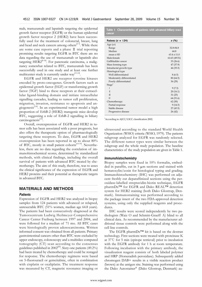
www.wjgnet.com
mab, trastuzumab and lapatinib targeting the epidermal growth factor receptor (EGFR) or the human epidermal growth factor receptor 2 (HER2) have been success-fully used for the treatment of colorectal, breast, lung and head and neck cancers among others[3-7]. While there are some case reports and a phase Ⅱ trial reporting promising results targeting EGFR in BTC there are no data regarding the use of trastuzumab or lapatinib also targeting HER2[8-10]. For pancreatic carcinoma, a malig-nancy somewhat related to BTC, trastuzumab has been successfully used in one study and at least one further multicenter study is currently under way[11,12].
EGFR and HER2 are receptor tyrosine kinases encoded by proto-oncogenes. Growth factors such as epidermal growth factor (EGF) or transforming growth factor (TGF) bind to these receptors at their extracel-lular ligand-binding domain and initiate intracellular signalling cascades, leading to tumor cell proliferation, migration, invasion, resistance to apoptosis and an-giogenesis[3,4]. In an experimental tumor model a high proportion of ErbB-2 (HER2) transgenic mice develop BTC, suggesting a role of ErbB-2 signalling in biliary carcinogenesis[13].
Overall, overexpression of EGFR and HER2 in tu-mor cells has been associated with a poor prognosis, but also offers the therapeutic option of pharmacologically targeting these receptors. To date, EGFR and HER2 overexpression has been reported in up to about 80% of BTC, mostly in small patient cohorts[14-19]. Neverthe-less, there are no data regarding the correlation of im-munohistochemical scores, determined by standardized methods, with clinical findings, including the overall survival of patients with advanced BTC treated by che-motherapy. The aim of this study, therefore, was to asses the clinical significance of the expression of EGFR and HER2 proteins and their potential as therapeutic targets in advanced BTC.
MATERIALS AND METHODSPatientsExpression of EGFR and HER2 was analyzed in biopsy samples from 124 patients with advanced or relapsed, unresectable BTC (51% women, median age 64.8 years). The patients had been consecutively diagnosed at the Tumorzentrum Ludwig Heilmeyer-Comprehensive Cancer Center Freiburg between 1997 and 2004, and were followed for a median of 71 mo. All BTC cases were histologically proven adenocarcinoma. Written informed consent was obtained from all patients. Primary gastrointestinal cancers other than BTC were excluded by upper endoscopy, colonoscopy and a multislice computed tomography (CT) scan according to the consensus guidelines published in 2002[20]. Sixty-one patients (49.2%) had been treated by chemotherapy and could be restaged for response. The chemotherapy regimens were based on 5-fluorouracil or gemcitabine, often in combination with cisplatin or oxaliplatin. The treatment response was measured by CT, magnetic resonance imaging or
ultrasound according to the standard World Health Organization (WHO) criteria (WHO, 1979). The patients subgroup analyzed for EGFR was randomly selected. The different tumor types were similar between the subgroup and the whole study population. The baseline characteristics of the study population are given in Table 1.
ImmunohistochemistryBiopsy samples were fixed in 10% formalin, embed-ded in paraffin, cut in 5 µm sections and stained with hematoxylin/eosin for histological typing and grading. Immunohistochemistry (IHC) was performed on adja-cent freshly cut deparaffinized sections using the per-oxidase-labelled streptavidin-biotin technique, EGFR pharmDx™ for EGFR and Dako REAL™ detection system for HER2 staining (both Dako Glostrup, Den-mark). Immunostaining was performed according to the package insert of the two FDA-approved detection systems, using only the supplied reagents and proce-dures.
IHC results were scored independently by two pa-thologists (Waiz O and Schmitt-Graeff A) blind to all clinical data. As recommended by the manufacturer ad-ditional tissue controls were performed along with the cell line controls.
The EGFR pharmDx™ kit is based on the dextran technology. First sections were treated with proteinase K at 37℃ for 5 min (epitope retrieval) prior to incubation with the EGFR antibody for 1 h at room temperature. Following incubation with the primary antibody, the visualization reagent consists of both labeled polymer and HRP (Horseradish peroxidase). Subsequently added chromogen DAB+ results in a visible reaction product (brown) at the antigen site. Staining was performed using the Dako Autostainer® (Dako Glostrup, Denmark) au-
Table 1 Characteristics of patients with advanced biliary tract cancer
Patients (n = 124) n (%)
Age (yr) Range 32.8-84.8 Median 64.8 mean ± SD 63.4 ± 11.0Male:female 61:63 (49:51)Gallbladder cancer 33 (26.6)Mass forming type 47 (37.9)Intraductal growth type 44 (35.5)Histological type Well differentiated 8 (6.5) Moderately differentiated 80 (64.5) Poorly differentiated 36 (29)Stage1
Ⅰ 9 (7.3) Ⅱ 20 (16.1) Ⅲ 31 (25) Ⅳ 64 (51.6)Chemotherapy 62 (50) Partial response 9 (14.5) Stable disease 27 (43.5) Progressive disease 26 (42)
1According to AJCC/UICC classification 2002.
4512 ISSN 1007-9327 CN 14-1219/R World J Gastroenterol September 28, 2009 Volume 15 Number 36

www.wjgnet.com
tomated system. As recommended for the interpretation of EGFR pharmDx™, both the intensity and percent-age of tumor cells exhibiting membranous and/or cy-toplasmic staining were assessed. If specific membrane staining was observed in less than 1% of tumor cells, the specimen was reported to be EGFR negative. The
four categories negative, weak (1+), moderate (2+) and intense (3+) were used as recommended by the manu-facturer and approved by the FDA (Figure 1).
The detection of HER2 was performed with heat induced epitope retrieval (HIER) in 10 mmol/L citrate buffer (Target Retrieval Solution, pH 6.1, Dako) in a
A B
C D
E F
G H
HER2 EGFR
neg.
1+
2+
3+
Figure 1 Immunohistochemistry of biliary tract cancer specimens for expression of HER2 (A, C, E and G) and EGFR (B, D, F and H). A: HER2 neg.; B: EGFR neg.; C: HER2 1+; D: EGFR 1+; E: HER2 2+; F: EGFR 2+; G: HER2 3+; H: EGFR 3+.
Harder J et al. EGFR and HER2 in biliary tract cancer 4513

www.wjgnet.com
water bath at 98℃ for 45 min and anti-HER2 primary antibody (Polyclonal Rabbit Anti Human c-erbB-2 on-coprotein, Dako Glostrup, Denmark). Following incuba-tion with the primary antibody, the visualization is based on the sequential application of biotinylated link anti-body and streptavidin labeled with alkaline phosphatase antibody. Subsequently added chromogen Fast RED results in a visible reaction product (red) at the antigen site. HER staining was also performed using the Dako Autostainer® (Dako Glostrup, Denmark). Tumors were counted negative if less than 10% of carcinoma cells were stained, irrespective of membrane signal intensity. If 10% or more of carcinoma cells showed membrane staining, the staining intensity was classified as weak (1+), moderate (2+) or intense (3+) as recommended by the manufacturer and approved by the FDA (Figure 1). Cytoplasmic staining was discounted. Images provided by DAKO were used to define these staining categories.
Fluorescence in situ hybridization (FISH)The PathVysion™ detection kit (Abbott, Illinios, USA) was used for FISH analysis in cases of IHC expression scores 2+ or 3+ for HER2. FISH staining was carried out according to the manufacturer’s manual. The pres-ence of gene amplification was determined using the supplied fluorescence-labelled DNA probe for chromo-somal locus 17q11.2-q12 for the detection of HER2. FISH-stained sections were scanned at × 1000 magni-fication and in each of three separate carcinoma areas. Twenty nuclei were analyzed. HER2 and chromosome 17 copy number were counted for all cells and the ratio of HER2 to chromosome 17 was calculated. A normal HER2:17 ratio was defined as ≤ 2; a ratio > 2 was in-terpreted as gene amplification. A normal HER2 copy number was attested at < four signals per cell (Figure 2).
Statistical analysisData are presented as mean ± SD or medians with range for continuous variables and as absolute and relative frequencies for categorical variables. Overall survival estimates and median survival times were calculated by the Kaplan-Meier method followed by Cox models estimating relative risks. All statistical tests were two-sided using a 5% significance level. SASS software was used for the calculations.
RESULTSExpression of EGFR and HER2 proteinFrom the 124 patients examined 34 (27.4%) had gallbladder cancer, 47 (37.9%) had intrahepatic mass forming type BTC (IHCC) and 43 (34.7%) had extrahepatic or perihilar, intraductal growth type BTC (EHCC). Because EGFR expression was expected to be present two fold more frequently than HER2 expression in BTC only a subset of 56 samples was examined. EGFR expression was negative in 39.3% (22/56) of the patients. Weak (1+) positive EGFR staining was found in 21.5% (12/56), moderate (2+) in 23.2% (13/56) and intense (3+) in 16% (9/56) of the tumors. Overall 39.2% of the samples showed EGFR overexpression (scores 2+ and 3+), being significantly (P = 0.028) more frequent in EHCC (57.9%) than in IHCC (25%). HER2 expression was as follows: 72/124 (58%) were negative, 26 (21%) 1+, 22 (18%) 2+ and 4 (3%) 3+. Representative examples of EGFR and HER2 immunohistochemical staining and in situ hybridization are shown in Figures 1 and 2.
A close correlation between treatment response and gene amplification has been shown for HER2 in previous studies[4]. However, unlike the case of trastuzumab and HER2 in breast cancer, EGFR gene amplification detected by FISH has not been approved as being as useful for deciding on an EGFR targeted therapy yet. Therefore, we did not study EGFR gene amplification in our patient cohort. Regarding HER2, based on published data and the manufacturer’s recommendation, tumors with no or 1+ HER2 immunostaining were not further investigated for gene amplification. Of the 124 patients samples tested 25 were examined for HER2 gene amplification. HER2 FISH was performed in 2+ and 3+ samples and was successfully performed in all but one tumor examined. All specimens exhibiting 3+ immunostaining (4/4) showed gene HER2 amplification while amplification was present in 2/21 (10%) of 2+ samples. Taken together, HER2 gene amplification could be detected in 6/124 (5%) tumors.
Correlation of EGFR and HER2 expression with clinicopathological factorsAmong the 124 patients 80 (64.5%) had moderately dif-ferentiated tumors, 36 (29%) had poorly differentiated and
Figure 2 Dual color FISH analysis of HER2 in cholangio-carcinoma specimens. A: Nonamplified tumor with single HER2 gene copy status. The red probe is specific for HER2. The green probe is specific for chromosome 17 centromere; B: Tumor with high amplification of the HER2 gene. There are multiple red HER2 specific signals in this tumor in rela-tion to the green chromosome 17 signals.
BA
4514 ISSN 1007-9327 CN 14-1219/R World J Gastroenterol September 28, 2009 Volume 15 Number 36

www.wjgnet.com
8 (6.5%) had well differentiated tumors. The majority of patients (64/124, 51.6%) had stage Ⅳ disease, 31 (25%) had stage Ⅲ, 20 (16.1%) stage Ⅱ and 9 stage Ⅰ (7.3%). The patients had not undergone surgery because of un-resectability, comorbidity or patients’ wish. Half of the patients (62/124) had been treated with chemotherapy, resulting in tumor control in 59% (14.7% PR, 44.3% SD). Median overall survival was 13 mo with a median OS of 14 mo for patients treated with chemotherapy compared to 9 mo for patients not treated with chemo-therapy. There was no statistical association between protein expression and grade, stage, overall survival and treatment response for EGFR and HER2, respectively. The frequencies of EGFR and HER2 overexpression and clinicopathological variables are summarized in Table 2. In univariate analysis EGFR and HER2 expression could not be shown to be of prognostic relevance for overall survival (P = 0.06 and P = 0.49).
DISCUSSIONExpression of the two ErbB family growth factor receptors EGFR and HER2 has been intensively studied in different tumor entities and led to the use of targeted therapy with specific inhibitors or antibodies of these receptors in colorectal, breast, lung as well as head and neck cancer[4]. To date in other cancers monoclonal antibodies and small molecule tyrosine kinase inhibitors such as cetuximab, trastuzumab, erlotinib, gefitinib and lapatinib are under investigation. Expression of EGFR and HER2 as potential therapeutic targets has been reported in various tumors[4,7,21,22]. For BTC, data for EGFR and HER2 overexpression have been presented in mostly small patient cohorts[14,17,19,23]. Recently Yoshikawa et al[24] described an unselected large cohort of 236 cases of resected BTC. In this study, we investigated EGFR and HER2 expression in a large cohort of patients with advanced, unresectable BTC.
In BTC the percentage of EGFR overexpressing tu-mors in previously reported series ranged from 8.1% to 81%. Yoshikawa et al[24] showed EGFR overexpression in 26.4% of EHCC and 17.7% of IHCC. Similarly, in our study EGFR overexpression was more frequent in EHCC (57.9%) than in IHCC (25%, P = 0.028). In these reports by Ito et al[23] and Yoshikawa et al[24] EGFR over-expression was associated with biological aggressiveness of BTC. In addition, there is increasing evidence that EHCC and IHCC respond differently to chemotherapy suggesting different biological properties of these two tumor subtypes of “cholangiocarcinoma”[25,26].
HER2 overexpression and amplification has been found in a range between 5% and 76% in BTC[14-19,24,27]. This wide range may in par t ref lect the lack of standardized methodologies used within the different studies to assess HER2 status. In our cohort with advanced, unresectable BTC, HER2 overexpression was present in 20% and treatment relevant HER amplification in 5%. Some authors suggest that HER2 overexpression is due to gene deregulation rather than gene amplification because in some reports there is no strict correlation between protein expression and gene amplification[19]. Overall, the highest concordance between HER2 expression and gene amplification was demonstrated for breast cancer[7,28,29]. Our data suggest that in advanced BTC with high HER2 expression there is also a good correlation between overexpression and amplification, since all samples with a 3+ HER2 score showed gene amplification as compared to only 2/21 (10%) with a 2+ HER2 score. Similar correlations have been reported for other malignancies, including breast cancer with a tendency towards lower therapeutic relevant expression rates with improved, standardized detection methods. It has to be assumed that earlier reports overestimated HER2 expression in BTC, explaining the discrepancy between HER2 expression and gene amplification[28,30,31]. Despite the fact that HER2 transgenic mice frequently develop BTC, HER2 does not seem to play a major role in human biliary tract malignancies. This animal tumor model may therefore be of limited value as a model for human BTC.
In recent reports in early stage, resected BTC overall HER2 amplification was about 5%-10%[24,32]. In our cohort of patients with advanced BTC, HER2 amplification was merely 5%. Since targeted therapy with the anti-HER2 antibody trastuzumab in breast cancer is only effective when the HER2/neu receptor is overexpressed via gene amplification HER2 seems not to be a promising target in BTC. Therapeutic trials targeting HER2 should therefore not further be initiated.
Since EGFR expression does not predict i ts therapeutic usefulness, future clinical trials have to evaluate the advantage of Anti-EGFR therapy in comparison with standard treatment in patients with BTC.
In summary, our findings demonstrate that EGFR overexpression is frequent in BTC, especially in EHCC. In contrast, HER2 overexpression and gene amplification is a rare event. While therapeutic targeting
Table 2 EGFR and HER2 expression and clinicopathological factors (Cox’s model)
Parameter EGFR 2+ and 3+ tumors
P HER2 2+ and 3+ tumors
P
Gallbladder cancer 5/13 8/34Mass forming type 6/24 0.088 7/47 0.517Intraductal growth type 11/19 10/43Histological type Well differentiated 2/3 1/8 Moderately differentiated 16/39 0.511 20/80 0.202 Poorly differentiated 4/14 4/36Stage1
Ⅰ 3/4 4/9 Ⅱ 2/6 0.317 4/20 0.059 Ⅲ 8/17 9/31 Ⅳ 9/29 8/64Chemotherapy Partial response 2/3 4/9 Stable disease 3/13 0.296 4/27 0.156 Progressive disease 5/13 5/26
1According to AJCC/UICC classification 2002.
Harder J et al. EGFR and HER2 in biliary tract cancer 4515

www.wjgnet.com
of HER2 seems to be not promising, future clinical trials in patients with BTC should focus on EGFR.
COMMENTSBackgroundThe epidermal growth factor receptor (EGFR) and the human epidermal growth factor receptor 2 (HER2) are involved in the carcinogenesis of many malignancies. Therapeutic molecules targeting EGFR and HER2 have been successfully used for the treatment of colorectal, breast, lung and head and neck cancers among others. It is unknown if EGFR and HER2 are overexpressed in advanced biliary tract cancer (BTC) and therefore may serve as therapeutic targets in these cancers.Research frontiersAs the so called targeted therapies are most effective when the corresponding receptor or signalling pathway is activated, previous studies have focused on EGFR and HER2 in various tumors. There are conflicting data about overexpression of EGFR and HER2 in BTC and about the therapeutic importance of the two growth factor receptors in these tumors. Because of the low incidence, clinical trials on BTC are difficult and mostly performed in small patient cohorts. A possible selection bias might additionally explain these conflicting data. Innovations and breakthroughsWhile EGFR is significantly overexpressed in advanced BTC, HER2 overexpression and amplification is rare and therefore seems not to play a role in the carcinogenesis of BTCs. ApplicationsBecause of these expression data on EGFR and HER2 in BTC, and the correlation of expression and therapeutic effectiveness, especially with HER2 in other tumors, future clinical trials in BTC should focus on EGFR as a therapeutic target.TerminologyEGFR and HER2 are receptor tyrosine kinases encoded by proto-oncogenes. Growth factors such as epidermal growth factor or transforming growth factor bind to these receptors and initiate tumor cell proliferation, migration, invasion, resistance to apoptosis and angiogenesis. BTC is a heterogeneous tumor entity with rising incidence, consisting of intrahepatic mass forming type cholangiocarcinoma, perihilar Klatskin tumors, extrahepatic bile duct tumors, also termed intraductal growth type cholangiocarcinoma, and gallbladder cancer.Peer reviewHarder et al describe low expression and gene amplification of HER2 in biopsy samples of advanced BTC, as assessed by immunohistochemistry and fluorescence in situ hybridisation, respectively. The authors suggest that deviating numbers of some earlier studies may be due to non-standardized techniques. It is convincing that in the present paper results for protein expression and gene amplification correlated well.
REFERENCES1 Malhi H, Gores GJ. Review article: the modern diagnosis
and therapy of cholangiocarcinoma. Aliment Pharmacol Ther 2006; 23: 1287-1296
2 Khan SA, Thomas HC, Davidson BR, Taylor-Robinson SD. Cholangiocarcinoma. Lancet 2005; 366: 1303-1314
3 Ménard S, Casalini P, Campiglio M, Pupa SM, Tagliabue E. Role of HER2/neu in tumor progression and therapy. Cell Mol Life Sci 2004; 61: 2965-2978
4 Hudis CA. Trastuzumab--mechanism of action and use in clinical practice. N Engl J Med 2007; 357: 39-51
5 Hudis CA. Trastuzumab adds to adjuvant chemotherapy for resected HER2-positive breast cancer. Nat Clin Pract Oncol 2006; 3: 12-13
6 Press MF, Lenz HJ. EGFR, HER2 and VEGF pathways: validated targets for cancer treatment. Drugs 2007; 67: 2045-2075
7 Ooi A, Takehana T, Li X, Suzuki S, Kunitomo K, Iino H, Fujii H, Takeda Y, Dobashi Y. Protein overexpression and gene amplification of HER-2 and EGFR in colorectal
cancers: an immunohistochemical and fluorescent in situ hybridization study. Mod Pathol 2004; 17: 895-904
8 Huang TW, Wang CH, Hsieh CB. Effects of the anti-epidermal growth factor receptor antibody cetuximab on cholangiocarcinoma of the liver. Onkologie 2007; 30: 129-131
9 Bralet MP , Bellin MF, Guettier C, Adam R, Paule B. Response to cetuximab and gemcitabine-oxaliplatin in an advanced case of intrahepatic cholangiocarcinoma. Clin Oncol (R Coll Radiol) 2006; 18: 426
10 Sprinzl MF, Schimanski CC, Moehler M, Schadmand-Fischer S, Galle PR, Kanzler S. Gemcitabine in combination with EGF-Receptor antibody (Cetuximab) as a treatment of cholangiocarcinoma: a case report. BMC Cancer 2006; 6: 190
11 Büchler P, Reber HA, Eibl G, Roth MA, Büchler MW, Friess H, Isacoff WH, Hines OJ. Combination therapy for advanced pancreatic cancer using Herceptin plus chemotherapy. Int J Oncol 2005; 27: 1125-1130
12 Büchler P, Reber HA, Büchler MC, Roth MA, Büchler MW, Friess H, Isacoff WH, Hines OJ. Therapy for pancreatic cancer with a recombinant humanized anti-HER2 antibody (herceptin). J Gastrointest Surg 2001; 5: 139-146
13 Kiguchi K, Carbajal S, Chan K, Beltrán L, Ruffino L, Shen J, Matsumoto T, Yoshimi N, DiGiovanni J. Constitutive expression of ErbB-2 in gallbladder epithelium results in development of adenocarcinoma. Cancer Res 2001; 61: 6971-6976
14 Aishima SI , Taguchi KI, Sugimachi K, Shimada M, Sugimachi K, Tsuneyoshi M. c-erbB-2 and c-Met expression relates to cholangiocarcinogenesis and progression of intrahepatic cholangiocarcinoma. Histopathology 2002; 40: 269-278
15 Altimari A, Fiorentino M, Gabusi E, Gruppioni E, Corti B, D'Errico A, Grigioni WF. Investigation of ErbB1 and ErbB2 expression for therapeutic targeting in primary liver tumours. Dig Liver Dis 2003; 35: 332-338
16 Chow NH, Huang SM, Chan SH, Mo LR, Hwang MH, Su WC. Significance of c-erbB-2 expression in normal and neoplastic epithelium of biliary tract. Anticancer Res 1995; 15: 1055-1059
17 Endo K, Yoon BI, Pairojkul C, Demetris AJ, Sirica AE. ERBB-2 overexpression and cyclooxygenase-2 up-regulation in human cholangiocarcinoma and risk conditions. Hepatology 2002; 36: 439-450
18 Terada T, Ashida K, Endo K, Horie S, Maeta H, Matsunaga Y, Takashima K, Ohta T, Kitamura Y. c-erbB-2 protein is expressed in hepatolithiasis and cholangiocarcinoma. Histopathology 1998; 33: 325-331
19 Ukita Y, Kato M, Terada T. Gene amplification and mRNA and protein overexpression of c-erbB-2 (HER-2/neu) in human intrahepatic cholangiocarcinoma as detected by fluorescence in situ hybridization, in situ hybridization, and immunohistochemistry. J Hepatol 2002; 36: 780-785
20 Khan SA, Davidson BR, Goldin R, Pereira SP, Rosenberg WM, Taylor-Robinson SD, Thillainayagam AV, Thomas HC, Thursz MR, Wasan H. Guidelines for the diagnosis and treatment of cholangiocarcinoma: consensus document. Gut 2002; 51 Suppl 6: VI1-VI9
21 Ooi A, Kobayashi M, Mai M, Nakanishi I. Amplification of c-erbB-2 in gastric cancer: detection in formalin-fixed, paraffin-embedded tissue by fluorescence in situ hybridization. Lab Invest 1998; 78: 345-351
22 Takehana T, Kunitomo K, Suzuki S, Kono K, Fujii H, Matsumoto Y, Ooi A. Expression of epidermal growth factor receptor in gastric carcinomas. Clin Gastroenterol Hepatol 2003; 1: 438-445
23 Ito Y, Takeda T, Sasaki Y, Sakon M, Yamada T, Ishiguro S, Imaoka S, Tsujimoto M, Higashiyama S, Monden M, Matsuura N. Expression and clinical significance of the erbB family in intrahepatic cholangiocellular carcinoma. Pathol Res Pract 2001; 197: 95-100
24 Yoshikawa D, Ojima H, Iwasaki M, Hiraoka N, Kosuge T, Kasai S, Hirohashi S, Shibata T. Clinicopathological
COMMENTS
4516 ISSN 1007-9327 CN 14-1219/R World J Gastroenterol September 28, 2009 Volume 15 Number 36

www.wjgnet.com
and prognostic significance of EGFR, VEGF, and HER2 expression in cholangiocarcinoma. Br J Cancer 2008; 98: 418-425
25 Nehls O, Oettle H, Hartmann JT, Hofheinz RD, Hass HG, Horger MS, Koppenhöfer U, Hochhaus A, Stieler J, Trojan J, Gregor M, Klump B. Capecitabine plus oxaliplatin as first-line treatment in patients with advanced biliary system adenocarcinoma: a prospective multicentre phase II trial. Br J Cancer 2008; 98: 309-315
26 Harder J, Riecken B, Kummer O, Lohrmann C, Otto F, Usadel H, Geissler M, Opitz O, Henss H. Outpatient chemotherapy with gemcitabine and oxaliplatin in patients with biliary tract cancer. Br J Cancer 2006; 95: 848-852
27 Voravud N, Foster CS, Gilbertson JA, Sikora K, Waxman J. Oncogene expression in cholangiocarcinoma and in normal hepatic development. Hum Pathol 1989; 20: 1163-1168
28 Yau TK, Sze H, Soong IS, Hioe F, Khoo US, Lee AW. HER2 overexpression of breast cancers in Hong Kong: prevalence and concordance between immunohistochemistry and in-situ hybridisation assays. Hong Kong Med J 2008; 14: 130-135
29 Carlson RW, Moench SJ, Hammond ME, Perez EA, Burstein HJ, Allred DC, Vogel CL, Goldstein LJ, Somlo G, Gradishar WJ, Hudis CA, Jahanzeb M, Stark A, Wolff AC, Press MF, Winer EP, Paik S, Ljung BM. HER2 testing in breast cancer: NCCN Task Force report and recommendations. J Natl Compr Canc Netw 2006; 4 Suppl 3: S1-S22; quiz S23-S24
30 Kobayashi M , Ooi A, Oda Y, Nakanishi I . Protein overexpression and gene amplification of c-erbB-2 in breast carcinomas: a comparative study of immunohistochemistry and fluorescence in situ hybridization of formalin-fixed, paraffin-embedded tissues. Hum Pathol 2002; 33: 21-28
31 Hirashima N, Takahashi W, Yoshii S, Yamane T, Ooi A. Protein overexpression and gene amplification of c-erb B-2 in pulmonary carcinomas: a comparative immunohistochemical and fluorescence in situ hybridization study. Mod Pathol 2001; 14: 556-562
32 Nakazawa K, Dobashi Y, Suzuki S, Fujii H, Takeda Y, Ooi A. Amplification and overexpression of c-erbB-2, epidermal growth factor receptor, and c-met in biliary tract cancers. J Pathol 2005; 206: 356-365
S- Editor Tian L L- Editor O’Neill M E- Editor Zheng XM
Harder J et al. EGFR and HER2 in biliary tract cancer 4517

www.wjgnet.com
ORIGINAL ARTICLES
Protein interaction network related to Helicobacter pylori infection response
Kyu Kwang Kim, Han Bok Kim
Online Submissions: wjg.wjgnet.com World J Gastroenterol 2009 September 28; 15(36): [email protected] World Journal of Gastroenterology ISSN 1007-9327doi:10.3748/wjg.15.4518 © 2009 The WJG Press and Baishideng. All rights reserved.
Kyu Kwang Kim, Daeil Foreign Language High School, Seoul 136-100, South KoreaHan Bok Kim, Department of Biotechnology, The Research Institute for Basic Sciences, Hoseo University, Asan 336-795, South KoreaAuthor contributions: Kim KK performed the experiments and wrote the manuscript; Kim HB edited and reviewed the manuscript and provided financial support for this work.Correspondence to: Dr. Han Bok Kim, Professor, Department of Biotechnology, The Research Institute for Basic Sciences, Hoseo University, Asan 336-795, South Korea. [email protected]: +82-41-5405624 Fax: +82-41-5486231Received: July 13, 2009 Revised: August 12, 2009Accepted: August 19, 2009Published online: September 28, 2009
AbstractAIM: To understand the complex reaction of gastric inflammation induced by Helicobacter pylori (H pylori ) in a systematic manner using a protein interaction network.
METHODS: The expression of genes significantly changed on microarray during H pylori infection was scanned from the web literary database and translated into proteins. A network of protein interactions was constructed by searching the primary interactions of selected proteins. The constructed network was mathematically analyzed and its biological function was examined. In addition, the nodes on the network were checked to determine if they had any further functional importance or relation to other proteins by extending them.
RESULTS: The scale-free network showing the relationship between inflammation and carcinogenesis was constructed. Mathematical analysis showed hub and bottleneck proteins, and these proteins were mostly related to immune response. The network contained pathways and proteins related to H pylori infection, such as the JAK-STAT pathway triggered by interleukins. Activation of nuclear factor (NF)-kB, TLR4, and other proteins known to function as core proteins of immune response were also found. These immune-related proteins interacted on the network with pathways and proteins related to the cell cycle, cell maintenance and proliferation, and
transcription regulators such as BRCA1, FOS, REL, and zinc finger proteins. The extension of nodes showed interactions of the immune proteins with cancer-related proteins. One extended network, the core network, a summarized form of the extended network, and cell pathway model were constructed.
CONCLUSION: Immune-related proteins activated by H pylori infection interact with proto-oncogene proteins. The hub and bottleneck proteins are potential drug targets for gastric inflammation and cancer.
© 2009 The WJG Press and Baishideng. All rights reserved.
Key words: Gastric cancer; Helicobacter pylori ; Inflammation; Pathway; Protein interaction network
Peer reviewer: Hidekazu Suzuki, Assistant Professor, Department of Internal Medicine, Keio University School of Medicine, 35 Shinanomachi, Shinjuku-ku, Tokyo 160-8582, Japan
Kim KK, Kim HB. Protein interaction network related to Helicobacter pylori infection response. World J Gastroenterol 2009; 15(36): 4518-4528 Available from: URL: http://www.wjgnet.com/1007-9327/15/4518.asp DOI: http://dx.doi.org/10.3748/wjg.15.4518
INTRODUCTIONHelicobacter pylori (H pylori) is a gram negative bacterium which infects about 50% of the world population[1-3]. It is known to cause various gastroduodenal diseases such as chronic active gastritis in experimental animals and in humans. In human volunteers, H pylori caused gastritis and hypochlorhydria[4]. Mongolian gerbils infected by H pylori also developed symptoms such as intestinal metaplasia and adenocarcinoma[5-9]. Many scholars have demonstrated a relationship between H pylori and gastric carcinoma[3], and the World Health Organization (WHO) and the International Agency for Research on Cancer consensus group have classified H pylori as a definite biological carcinogen[10].
H pylori colonization causes a strong systemic immune response[11]. It induces the production of interleukins (ILs) (Korean Society for Medical Microbiology, 2004), tumor necrosis factor (TNF)[12,13], and proinflammatory

Kim KK et al . Protein interaction network related to H pylori infection 4519
www.wjgnet.com
cytokines[14]. It also causes activation of nuclear factor kB (NF-kB)[15], activator protein-1 (AP-1), c-Jun, NH2-terminal kinase, mitogen-activated protein kinase/extracellular signal-regulated kinase, and other cell proliferation and survival factors[16]. Bacterial toxins, high levels of superoxides, radicals, and singlet oxygen are known to induce carcinogenesis in gastric cells. Bacterial virulence factors such as CagA and VacA[1,17,18] induce cell hyperproliferation and the expression of oncogenes. However, the exact mechanism between H pylori and gastric carcinoma is unclear[19].
Various tools have been employed to identify the relationship between H pylori and gastric cancer, including c-DNA microarrays[4,20]. However, most of these methods did not consider the systematic interaction of biological components. As an alternative, a network construction and analysis of protein-protein interactions[21] were applied to examine the inflammatory response to H pylori infection in a systematic manner.
MATERIALS AND METHODSThe research method used in this study mainly consisted of three steps. Step one: extraction of the genes which changed significantly during H pylori infection from the database and by querying web databases to gather protein-protein interactions. Step two: construction of a network and summarizing the constructed network. Step three: analysis and extension search of the network. A flow chart showing the data flow is described in Figure 1.
Searching genes related to H pylori infection (Step 1)Genes related to H pylori infection were collected by searching PubMed. The expression of genes significantly changed (P < 0.05) by H pylori infection in the microarray[4,11,13,20] data was examined, and genes related to the immune response were identified and collected. A total of 39 filtered genes (Table 1) were obtained.
Scanning protein interactions and construction of protein interaction networks (Step 2)The protein interaction networks were constructed based on statistical prediction through the analysis of microarray data. Selected genes were queried to the Uniprot database to convert into proteins. The proteins were scanned by a human Protein-protein Interaction Prediction (PIPs) database (http://www.compbio.dundee.ac.uk/www-pips/). Protein links were then extracted from the Human Protein Reference Database reference (HPRD, http://www.hprd.org/index_html). Without HPRD references, any further search of the protein links was stopped. An extended network was constructed by integrating all results extracted from the PIPs server (Figure 2). Pajek (http://vlado.fmf.uni-lj.si/pub/networks/pajek/) was used for the construction of extended networks. Then, a core network showing simplified main pathways, major proteins, and subcellular location information was extracted from the extended network using Cytoscape (http://www.cytoscape.org/).
Analysis of protein interaction network (Step 3)The protein interactions of an extended network were examined whether or not the network contained known pathways related to H pylori infection, inflammation, and carcinogenesis. The core network was not analyzed because it was just the simplified form of the extended network.
Four factors: Shortest paths, degree (connectivity), betweenness centrality (BC), and closeness centrality (CC), were adopted to analyze general mathematical properties of the extended network and to search
Protein/gene name Uniprot ID HPRD reference
ITGB2 P05107LY96 Q9Y6Y9 XTLR10 Q9BXR5TLR2 O60903TLR3 O15455 XVCAM1 P19320HCK P08631MAPK8 P45983RAC2 P15153SOCS2 O14508 XSTAT6 P42226C2 P06681 XC3 P01024C4A P0C0L4 XCCL18 P55774 XCCL19 Q99731 XCCL3 P10147 XCCL4 P13236 XCRP P02741CXCL13 O43927CXCL2 P19875 XCXCL9 Q07325 XHLA-DMA P28067 XHLA-DPB1 Q30154 XHLA-DQB1 P03992 XHLA-DRB5 Q30154 XHSPH1 Q92598C11TA P33076PLAT P00750IFITM1 P13164 XIRF4 Q15306MADCAM1 Q13477ALOX5 P09917 XTLR5 O60602 XCD53 P19397 XTLR6 Q9Y2C9 XSLAMF1 Q13291PTPRC P08575FAIM3 O60667 XCD180 Q99467TLR4 O00206TLR1 Q15399CXCL3 P19876 XCD47 Q08722IFNGR1 P15260IL10RA Q13651 XIL18RAP O95256 XITGAX P20702 XIL8 P10145
Table 1 List of proteins extracted from the literary database showing significant change after H pylori infection
Nodes with no HPRD reference were marked with x. HPRD: Human pro-tein reference database.

www.wjgnet.com
topologically important proteins[21].Degree, the most basic characteristic of a node, is
defined as the number of links the node has with other nodes. Degree distribution is obtained by counting the number of nodes with a fixed degree value, which is variable from minimum to maximum degree, and dividing it by the total number of nodes of a network[22]. Highly concentrated nodes play a major role as a hub in a network. Degree was also used to check if an extended network was scale-free, which is frequently found in cellular networks[22,23]. The scale-free network follows a power-law degree distribution[22]. Power law is defined as:
( ) aP x Cx−=C = ec and ( )P x is a probability that a selected node
has exactly x links (degree value)[23]. a is the degree exponent which determines some properties of the
network. Most of the networks found in nature are known to have degree exponent values between two and three[22]. In this study, cumulative distribution function, a superior method of plotting data[23], was used. The plot of log transformed probability distribution function ( )P x in which x has a degree value greater than or equal to x , was drawn. ( )P x is defined mathematically as[23]:
( ) ( ') 'x
P x p x dx∞
= ∫As the distribution follows power law,
( 1)( ) ' '1
a a
x
CP x C x dx xa
∞ − − −= =−∫
A cumulative plot also follows power law, but the degree exponent of the plot is one less than the original distribution[23]. The degree exponent was calculated by measuring the slope of the regression line and adding one to the exponent value. Other factors such as R
Step 1NCBI
PubMed
Geneset
Queryuniprot DB
Primaryinteraction
dataset
If yes
If no
Primaryinteractions
Step 2
Step 3
Filter valueslower than
two standarddeviation
rangefrom mean
Highdegree value
HighBC
value
If noIf yes
Commonfactor with
high BC set?
Commonfactor with high
degree set?
High-centerednodes
Hubnodes
If no
Bottlenecknodes
CalculateCC value CC value
of two sets
Proteinset
QueryPIPs server
HPRDreference?
If nofiltered?
If yesdiscard data
Importto pajek
Degreevalue
BCvalue
Network
Importto R
package
Summarizenetwork
Extendednetwork
Integrationof data
Cell model
Corenetwork
Extensionsearch of
nodes
Extensioninformation
Figure 1 Flow chart showing overall methods and data flow used in this study.
4520 ISSN 1007-9327 CN 14-1219/R World J Gastroenterol September 28, 2009 Volume 15 Number 36

www.wjgnet.com
square, standard error, and P-value were also computed. BC for node k is defined as:
, ,( ) ( )
ki j
i ji j i j i j
gb k b k
g→
→→
= =∑ ∑jig → is the number of shortest paths from node i to j ,
while kjig → is the number of geodesics among jig → that
passes through node k[21,24]. The BC value of all nodes in the network was examined to check for bottlenecks in the network.
CC is defined as the inverse of the average length of the shortest paths to/from all the other vertices in the graph[25]. It tells us the topological center of the network[25]. CC was calculated by adopting the core algorithm of the R igraph package (http://www.r-project.org/). CC values of the protein set with either large BC value or degree were measured and compared to total CC values to check topological centrality of hubs and bottlenecks in the network.
The shortest path (geodesics) is calculated by measuring the length of all the geodesics from or to the vertices in the network. The average shortest path was measured to see how many average steps were required to link two randomly selected nodes in the network.
After computing BC and the degree of all the nodes, nodes under two standard deviation ranges from the mean were filtered out and CC values of nodes larger than two standard deviation ranges from the mean were measured. As a result, nodes with a large BC value, a large degree, both a large BC and degree, and CC value were obtained. The R package was used to calculate and analyze these values.
The network was constructed by scanning primary interactions of significantly and differentially expressed genes compared to control. Thus, it may not include hidden interactions of protein nodes between the two major nodes. For example, only the primary interaction between node A and B is available by ordinary network analysis, although the two proteins are linked via node C in reality. However, by extending the network, a pathway passing through node C between A and B can be found.
RESULTSProtein interaction networksBy integrating scanned primary interactions of previously selected nodes from the PIPs server, the extended network was constructed. A core network was then derived from the extended network.
The extended network was composed of 604 nodes, connected via 808 edges (Figure 2). One giant network with 599 nodes and 805 edges, and two separate interactions were observed. Examining the shortest paths of the network showed that two randomly selected nodes on the network were connected via 4.89 links. This suggests that the nodes were very closely linked. In addition, a small world effect can be found[26]. The distribution of the shortest paths was plotted using histograms (Figure 3A). The average value (4.89) was similar to other values of human protein networks[21,26].
The cumulative distribution plot showed clear evidence that the extended network follows scale-free distribution (Figure 3B). By measuring the slope of the regression line of the plot drawn on the basis of log transformed cumulative data, the a value of 1.1968 in the power law distribution was determined. As the degree exponent of the cumulative plot is one less than original distribution[23], the true degree exponent value should be 2.1968 (standard error = 0.04, coefficient of determination R square = 0.97, and P-value = nearly zero by the least square fit)[27]. It is known that networks with a degree exponent larger than three do not have features that scale-free networks have[22]. The degree exponent value of the extended network (2.1968) was lower than 3, which was similar to other networks following a scale-free distribution, rather than a random distribution.
Important nodes in the network One of the properties of networks following scale-free distribution is the existence of a small number of highly connected nodes, called hubs which are more important than other less connected nodes[22,28]. The hub nodes are more critical to the survival of cells (Tables 2 and 3). The scale-free networks are prone to breakdown into fragments when nodes are attacked[29]. Other important nodes also have a large BC value. The node with a large BC functions as a bottleneck in the network, even when the node’s degree is low. Nodes with a degree or BC value larger than the mean plus two standard deviations were selected. Sixteen nodes were determined to have a large degree (Table 2) and 19 nodes had a large BC (Table 4). Twelve nodes had both a large degree and a large BC (Table 3). Six nodes: NF-kB3 (Nuclear factor kB p65 subunit), MAPK8 (Mitogen-activated protein kinase 8), NFKBIA (NF-kB inhibitor a), HCK (Hemopoietic cell kinase), PTPRC (Leukocyte common antigen CD45), and ITGB2 (Integrin b-2) were the top six nodes on both degree and BC values.
Protein Degree CC value
RELA/NF-kB3 105 0.047675522MAPK8 68 0.046693511NFKBIA 63 0.047164646HCK 49 0.046599691PTPRC 43 0.046388184ITGB2 40 0.045643782MAP2K1 36 0.045681818PLAT 25 0.045451119STAT6 24 0.046513422HLA-DMA 24 0.04396646TRAF4 24 0.044062843TLR4 24 0.046527778HLA-DRB5 23 0.04573032TLR2 22 0.046083301IL10RA 18 0.046206897ALOX5 18 0.044056404
Table 2 List of proteins with a large degree value and their CC values
CC: Closeness centrality.
Kim KK et al . Protein interaction network related to H pylori infection 4521

www.wjgnet.com
Figu
re 2
The
exte
nded
pro
tein
inte
ract
ion
netw
ork o
f a ce
ll inf
ecte
d by
H p
ylori.
Gre
en no
des (
a lar
ge B
C an
d deg
ree)
, blue
node
s (on
ly a l
arge
degr
ee),
yello
w no
des (
only
a lar
ge B
C).
4522 ISSN 1007-9327 CN 14-1219/R World J Gastroenterol September 28, 2009 Volume 15 Number 36

www.wjgnet.com
STAT6 (Signal transducer and activator of transcription 6), TLRs (Toll-like receptor), TRAF4 (TNF receptor-associated factor 4), and PLAT (TPA, Tissue-type plasminogen activator) also had both a large BC and degree (Table 3). NF-kB3, NFKBIA, PTPRC, TLRs, and HLA-DRB5 are already well-known for having
biological functions related to immune response[2,3,21]. STAT6 is related to the JAK-STAT pathway which sends signals from ILs directly to the nucleus[30]. MAPK8 of the MAPK signaling pathway can be found in the signaling of other inflammatory responses in asthma, and is related to cell proliferation[31]. TRAF4 is involved in tumor necrosis and TPA (PLAT) in plasminogen activation, respectively. Most of these nodes are related to immune response and signal transduction, suggesting that these nodes perform major functions against H pylori infection.
Not only nodes with both a large degree and BC, but also nodes with a large BC and a small degree were considered important in previous research[21], since these nodes function as bottlenecks in the network,
Protein name Function
RELA/NF-kB3 NF-kB is a pleiotropic transcription factor which is present in almost all cell types and is involved in many biological processes such as inflammation, immunity, differentiation, cell growth, tumorigenesis and apoptosis
MAPK8 Responds to activation by environmental stress and pro-inflammatory cytokines by phosphorylating a number of transcription fac-tors, primarily components of AP-1 such as JUN, JDP2 and ATF2 and thus regulates AP-1 transcriptional activity
NFKBIA Inhibits the activity of dimeric NF-kB/REL complexes by trapping REL dimers in the cytoplasm, masking their nuclear localization signals
HCK May serve as part of a signaling pathway coupling the Fc receptor to activation of the respiratory burst. May also contribute to neutro-phil migration and regulate the degranulation process of neutrophils
PTPRC Required for T-cell activation through the antigen receptorITGB2 Receptor for ICAM1, ICAM2, ICAM3 and ICAM4TLR4 Cooperates with LY96 and CD14 to mediate the innate immune response to bacterial lipopolysaccharide (LPS). Acts via MYD88,
TIRAP and TRAF6, leading to NF-kB activation, cytokine secretion and the inflammatory responsePLAT Converts the abundant, but inactive, zymogen plasminogen to plasmin by hydrolyzing a single Arg-Val bond in plasminogen. By
controlling plasmin-mediated proteolysis, it plays an important role in tissue remodeling and degradation, in cell migration and many other physiopathological events
TRAF4 Adapter protein and signal transducer that links members of the tumor necrosis factor receptor family to different signaling pathways by association with the receptor cytoplasmic domain and kinases
MAP2K1 Catalyzes the concomitant phosphorylation of a threonine and a tyrosine residue in a Thr-Glu-Tyr sequence located in MAP kinases. Activates ERK1 and ERK2 MAP kinases
TLR2 Cooperates with LY96 to mediate the innate immune response to bacterial lipoproteins and other microbial cell wall componentsSTAT6 Carries out a dual function: signal transduction and activation of transcription. Involved in interleukin-4 signaling
Table 3 List of proteins with both a large BC and degree, and their functions
0 2 4 6 8 10 12Shortest path
Freq
uenc
y
100 000
80 000
60 000
40 000
20 000
0
A
0.0 0.5 1.0 1.5 2.0K
0.0
-0.5
-1.0
-1.5
-2.0
-2.5
PK
B
Figure 3 Properties of the extended network. A: Histogram showing distribu-tion of the shortest path. Two randomly selected nodes were connected via 4.9 links; B: Cumulative degree distribution plot of the extended network showing that degree distribution follows the power law. Line indicates the degree expo-nent of 1.2, which is one lower than the true degree exponent of 2.2.
Protein BC CC
RELA/NF-kB3 62240.75685 0.047675522MAPK8 37123.82691 0.046693511PTPRC 34614.23746 0.046388184HCK 33484.99898 0.046599691NFKBIA 28771.33132 0.047164646ITGB2 27901.91607 0.045643782TLR4 19587.75501 0.046527778PLAT 19339.65216 0.045451119CCL4 18441.86414 0.043654528HMGB1 15263.85652 0.046466826MAP2K1 14047.72141 0.045681818STAT6 13376.61128 0.046513422CXCR4 11471.71166 0.044769471CSNK2A1 11279.76894 0.046574496HLA-DRB5 11088.56681 0.045730320BTK 11016.20767 0.046621308TLR2 10824.15886 0.046083301RAC2 10692.38019 0.044872749TRAF4 10472.80579 0.044062843
Table 4 List of proteins with a large BC value and their CC values
BC: Betweenness centrality.
Kim KK et al . Protein interaction network related to H pylori infection 4523

www.wjgnet.com
even without the role of hubs. Six nodes: HMGB1 (High mobility group protein B), BTK (Bruton tyrosine kinase), CSNK2A1 (CSK2A1, Casein kinase Ⅱ subunit alpha), RAC2 (Ras-related C3 botulinum toxin substrate 2), CCL4 (C-C motif chemokine 4), and CXCR4 (C-X-C chemokine receptor type 4) had a large BC but a low degree. Large BC nodes such as CXCR4, CCL4, BTK, CSNK2A1, and RAC2 with the exception of HMGB1 are related to immune response and signal transduction (Table 5). HMGB1 unwinds double-stranded DNA and binds preferentially to single-stranded DNA, which may be related to the gene regulation of immune response. As expected these large BC nodes were linked to important nodes, such as hubs. HMGB1 was linked to NF-kB3, TLR4, TLR2, and PLAT, which have a large BC degree (Figure 2). BTK interacted with NFKBIA, TLR4, HCK, and IL10RA. NFKBIA, TLR4, and HCK had a large BC and degree, while IL10RA had a large degree only. CSNK2A1 was linked to NF-kB3, NFKBIA, PTPRC, and HSPH1. RAC2 interacted with NFKBIA, HCK, and ALOX5. Lastly, CCL4 and CXCR4 were linked to PTPRC and PLAT. Thus, it was demonstrated that the nodes with large BC play important roles in the connection and communication of nodes including hubs.
The CC values of nodes with a large degree or BC were checked to see if these proteins were near to the topological center of the network. The larger the CC value is, the closer the node is to the center of the network[21]. NF-kB3 was closest to the topological center, and NFKBIA was the second closest in the network (Tables 2 and 4).
Biological functions of pathways and nodes in the network Pathways related to immune response and other biological phenomena were observed in the network (Figures 4 and 5). The network contained previously known pathways which were involved in H pylori infection and inflammation.
The network (Figures 2 and 4) showed interactions of IL 1, 4, 8, 10, 13, 17, and 18 receptors with JAKs and STATs that send signals from cell-surface receptors to the nucleus[30]. IL 8 increases significantly during H pylori infection, thus it was used as a standard to determine the pathogenicity of different H pylori strains[19]. IL 1, 10, and 18 changed significantly, which was demonstrated by microarray analysis or Western blotting data[11,13,32].
IL 4 and 13 are proinflammatory cytokines. While IL 4 induces eosinophilic inflammation and differentiation of Th2 cells, IL 13 produces immunoglobulin E (IgE)[33].
Interactions of Toll-like receptors (TLRs), also known to be immune-related, were observed. The TLR4 signaling pathway is associated with an immune response by interacting with MYD88 and IRAK1[34,35] in the network (Figure 4). They were linked to proteins in the nucleus through MAPKs.
Another pathway in the network was found among the MAPKs. Interactions among MAPK 1, 3, and 8 in the network were observed. In immune-related diseases such as asthma, the activation of MAPK due to infection has also been reported[21,36,37].
Besides full pathways, the presence of single or a few interactions having biological functions were informative. NF-kB and AP-1 are two key regulatory factors of inflammation[38-40]. NF-kB1-NF-kB3 linkage and JAK-NFKBIA-STAT linkage were found (Figure 5). The regulation of NF-kB by AP-1(JUN) and NFKBIA was also observed (Figure 4).
Although activation of TNFa[13] was not found in the network, TNFSF11 (Tumor necrosis factor ligand superfamily member 11) and TRAFs (TNF receptor associated factor), related to TNF, were found. Tumor necrosis factors induce cell proliferation by activating anti-apoptosis[16]. Cell proliferation and carcinogenesis are one of the well-known characteristics of cells infected by H pylori[19]. In addition, BRCA1 (Breast cancer type 1 susceptibility protein), FOS (c-fos, Proto-oncogene protein)[41], REL (C-Rel proto-oncogene protein), and VAV1 (Proto-oncogene vav), which are oncogenes, were found. The presence of TNF and the oncogenes in the network suggests that H pylori infection may be related to carcinogenesis.
SRC (Proto-oncogene tyrosine-protein kinase) in the network is involved in cell maintenance and communication[21]. CDK5 (Cyclin-dependent kinase 5), RASA1 (Ras GTPase-activating protein 1) and RASA3 are related to cell growth effect[30].
Not only protein nodes related to inflammation and carcinogenesis, but also proteins related to stress resistance were found. Infection of H pylori increases levels of superoxide and singlet oxygen. The stress-resistance protein, HSPH1 (Heat shock protein 105 kDa), HSPA8 (Heat shock cognate 71 kDa protein), and HSPB1 (Heat shock protein b-1) were found.
Generally, stimulation and regulation of the immune
Protein name Function
HMGB1 Binds to preferentially single-stranded DNA and unwinds double-stranded DNABTK Plays a crucial role in B-cell ontogenyCSNK2A1 Casein kinases are operationally defined by their preferential utilization of acidic proteins such as caseins as substratesCXCR4 Transduces a signal by increasing the intracellular calcium ion levelCCL4 Monokine with inflammatory and chemokinetic propertiesRAC2 Plasma membrane-associated small GTPase which cycles between an active GTP-bound and inactive GDP-bound state. In active state
binds to a variety of effector proteins to regulate cellular responses, such as secretory processes, phagocytosis of apoptotic cells and epithelial cell polarization. Seems to be involved in the regulation of NADPH oxidase
Table 5 List of proteins with only a large BC and their functions
4524 ISSN 1007-9327 CN 14-1219/R World J Gastroenterol September 28, 2009 Volume 15 Number 36

www.wjgnet.com
system through their receptors were found in the network. Activation of cell signaling, cell proliferation, cell survival, proto-oncogenes, and stress resistance were observed. These functions are reminiscent of the observed response of cells infected by H pylori. The virtual network analysis in this study reflects the real protein-interaction-network in the cell.
Extension search of the network A biologically important protein can be missed, as the network is constructed by searching only the primary interactions of selected genes. To overcome this problem, further interactions of nodes regardless of their degree or BC were examined. The extension of SRC led to BCAR1 (Figure 5). Thus, the role of cell maintenance[42]
RAC1IFNGR1
IL8RB
TLR4
SIGIRRPRKCZ
MAP2K1
MAPK8IP3
CDK5HSP90B
IL18RAP MAP3K7
TNFSF11
MAPK8
KBKAP
MYD88
PIK3R1
NR3C1
NFKBIA
NF-kB1
IL1A
TLR2
TLR6
IL1RAPL1 TLR10
TLR1 L17RD
TLR9 TLR5TLR3
BTK
TLR7IL4R
CD14
ITGB2
ITGAD
VCAM1 IL13
SYKSTAT1
STAT2STAT6
BRCA1CDK9
NF-kB3
FOS
MAP3K2
REL JUN
HMGB1
ARRB1
MYC
JAK2
STAT3
IRS1PTPN6
TRAF6
TRAF4
IRAK1TICAM1
LYN
PTPRC
LCP2
JAK1
VAV1
RASA1
VIL2
CXCR4
IL8
CCL4
IL8RA
SERPING1
HCK
STAT5A
MAPK3
BLNK
SOCS2 STAT5B
IL10RA
DOK1
PLAT
Figure 4 Core network showing simplified interactions and major pathways. Color of the nodes indicates subcellular location. Red (nucleus), orange (nucleus and cytoplasm), yellow (cytoplasm), blue (membrane), green (extracellular), and white (unknown).
Kim KK et al . Protein interaction network related to H pylori infection 4525
TLR4 IL4R
HMGB1
FOSNFKBIA
NF-kB3
NF-kB1BRCA1
Cell survival
Pro-inflammatory signal Nucleus
STAT
JAK
BTK
STAT
JAK
JAK-STAT signaling
CDK5
Cell cycle
PIK3R1
HCK
RASA1
SRC
BCAR1
Cell maintenancecytoplasm
PTPRC
VAV1
TNFSF11
Cell proliferation
MAPK signaling
MAPK1
MAPK3
MAPK8
AVPR2
GNAQ
Figure 5 Cell model showing major interactions after H pylori infection.

www.wjgnet.com
was connected with that of carcinogenesis in BCAR1. ADRB2 (b-2 adrenergic receptor) extension was linked to PRKCE, PRKACA, MAPK1, and MAPK3 (Figure 2). This pathway has not previously been reported in H pylori infection, but has been found in immune-related diseases such as asthma[21]. BRCA1 was further linked to CDK2, 4, 7, and CDC2 (Cell division control protein 2 homolog) (Figure 5). CDKs are activated proceeding to the cell cycle. The extension of BRCA1 was linked to JUND (transcription factor jun D), which binds to an AP-1 site and stimulates its promoter activity. BRCA1 extension led to ZNF467 (Zinc finger protein 467), a transcription regulator which has a possible relationship with cancer (Figure 2). The extension of MAPK1 led to GNAQ (Guanine nucleotide-binding protein G(q) subunit a) via GNAS (Guanine nucleotide-binding protein G(s) subunit a isoforms short) and AVPR2 (Vasopressin V2 receptor)[21] (Figure 5). The proteins in this pathway contribute to cell proliferation, a well-known characteristic of cells infected by H pylori[19]. STAT1-CREBBP (CREB-binding protein) linkage was related to G1 arrest of a cell[21] (Figure 5).
DISCUSSIONThe correlation between inflammation caused by H pylori infection and gastric cancer has been studied and supported by many researchers. It is important to understand the relationship between inflammation and the carcinogenesis mechanism. Microarray data were used to determine the global gene expression of infected cells. Microarray data showed up/down regulation of gene expression related to immune response, cell cycle, cell growth, and signal transduction, which may support the hypothesis that H pylori infection causes cancer development[4,11,13,20]. However, the data did not present a clear mechanism of carcinogenesis in a systematic manner. In this study, network analysis methods were applied to integrate previous data and construct the network model which shows the relationship between inflammation and cancer development.
The extended network showing primary interactions of significantly expressed genes (proteins in the network) was constructed. The network contained many protein nodes related to immune response and signal transduction induced by extracellular signals such as cytokines. The important nodes selected based on large BC and degree values were mostly involved in immune response and signal transduction. For example, the p65 subunit of NF-kB (NF-kB3), one of the most important regulatory factors of inflammation, was the node with the largest degree and BC value. Large BC nodes, the bottlenecks in the network, were linked to important nodes with a large degree, a large BC, or both. Like large BC and degree nodes, a large BC node was mostly related to immune response and signal transduction, with the exception of HMGB1. The constructed network also contained many pathways related to immune response and signal transduction. TLR4, JAK-STAT, and MAPK8 pathways are major pathways found in the network. Not
only the pathways, but important nodes such as NF-kB and AP-1 (JUN) were also found in the network.
The network also showed many nodes related to carcinogenesis. Tumor related proteins such as BRCA1, FOS, REL, VAV1, TNFSF11, and TRAFs were found. The extension search of nodes was also linked to pathways related to cell proliferation, cell survival, and the cell cycle. The extracellular signal from ILs and TLRs goes to NF-kB, NFKBIA, and AP-1 in the nucleus via the JAK-STAT and MAPK signaling pathways. The signal then goes to proteins in the cytoplasm via the JAK-STAT pathway and BTK, promoting cell proliferation and proceeding to the cell cycle. These activated processes are one of the characteristics of cells infected by H pylori. In addition, H pylori infection is known to increase levels of radicals and oxides. Radicals and oxides are widely thought to be possible mutagens. Oxidative stress may be an additional mechanism of carcinogenesis.
Another important factor of hub and bottleneck protein nodes is that they are potential drug targets. By inhibiting the functions of hubs and bottlenecks by small molecules, the function of the network can be shut down, meaning that the inflammatory and carcinogenesis processes can be stopped, theoretically. Traditionally, antibiotics have been used to treat gastric inflammation caused by H pylori infection[14]. However, this treatment has the potential problem of antibiotic resistance in the bacteria. As a potential alternative, this study presented the hub and bottleneck nodes as a drug target of gastric inflammation, cancer, and other diseases caused by H pylori infection.
The analysis of protein network interactions showed immune response and carcinogenesis-related cell responses in a bigger picture. The extension search of nodes also demonstrated key signal transductions linking inflammatory response and carcinogenesis. This study showed how a systematic approach such as the network construction produces meaningful information. It also offered a relatively easy and simple framework to understand the complexity of cellular interactions having functional importance. Therefore, the application of this tool may be an alternative to find important genes and drug targets in other diseases and in complex biological systems.
ACKNOWLEDGMENTSThe authors thank Sohyun Hwang, Seung-woo Son, and Doheon Lee of Korea Advanced Institute of Science and Technology (KAIST) for their kind advice and aid.
COMMENTSBackgroundThe correlation between inflammation caused by Helicobacter pylori (H pylori) infection and gastric cancer has been studied and supported by many researchers.Research frontiersTo explain the relationship between H pylori infection and cancer development,
COMMENTS
4526 ISSN 1007-9327 CN 14-1219/R World J Gastroenterol September 28, 2009 Volume 15 Number 36

www.wjgnet.com
microarray analysis was used. Microarray data showed the regulatory patterns of gene expression related to immune response, cell cycle, cell growth, and signal transduction. However, the data obtained did not show the mechanism of carcinogenesis in a systematic manner.Innovations and breakthroughsIn this study, protein network analysis, one of the bioinformatic tools, was applied to integrate previous microarray data, and a network model was constructed showing the relationship between inflammation and cancer development. The network contained many proteins related to immune response and signal transduction induced by extracellular cytokines. Some tumor-related proteins (BRCA1, FOS, REL, VAV1, TNFSF11, TRAF) were found.ApplicationsThis article offered a relatively easy and simple framework to understand the complexity of cellular interactions having functional importance. This tool may be used as an alternative to find important genes and drug targets in gastric inflammation and cancer and in complex biological systems.Peer reviewThis study about protein interaction network in H pylori infection is potentially interesting and informative.
REFERENCES1 Suzuki H, Hibi T, Marshall BJ. Helicobacter pylori: present
status and future prospects in Japan. J Gastroenterol 2007; 42: 1-15
2 Brooks GF, Janet SB, Ornston LN. Medical microbiology. 20th ed. East Norwalk: Appleton & Lange, 1995
3 Martinon F, Holler N, Richard C, Tschopp J. Activation of a pro-apoptotic amplification loop through inhibition of NF-kappaB-dependent survival signals by caspase-mediated inactivation of RIP. FEBS Lett 2000; 468: 134-136
4 Kitadai Y, Sasaki A, Ito M, Tanaka S, Oue N, Yasui W, Aihara M, Imagawa K, Haruma K, Chayama K. Helicobacter pylori infection influences expression of genes related to angiogenesis and invasion in human gastric carcinoma cells. Biochem Biophys Res Commun 2003; 311: 809-814
5 Parsonnet J, Friedman GD, Vandersteen DP, Chang Y, Vogelman JH, Orentreich N, Sibley RK. Helicobacter pylori infection and the risk of gastric carcinoma. N Engl J Med 1991; 325: 1127-1131
6 P e e k R M J r , B l a s e r M J . H e l i c o b a c t e r p y l o r i a n d gastrointestinal tract adenocarcinomas. Nat Rev Cancer 2002; 2: 28-37
7 Sugiyama A, Maruta F, Ikeno T, Ishida K, Kawasaki S, Katsuyama T, Shimizu N, Tatematsu M. Helicobacter pylori infection enhances N-methyl-N-nitrosourea-induced stomach carcinogenesis in the Mongolian gerbil. Cancer Res 1998; 58: 2067-2069
8 Talley NJ, Zinsmeister AR, Weaver A, DiMagno EP, Carpenter HA, Perez-Perez GI, Blaser MJ. Gastric adenocarcinoma and Helicobacter pylori infection. J Natl Cancer Inst 1991; 83: 1734-1739
9 Watanabe T , Tada M, Nagai H, Sasaki S, Nakao M. Helicobacter pylori infection induces gastric cancer in mongolian gerbils. Gastroenterology 1998; 115: 642-648
10 Schistosomes, liver flukes and Helicobacter pylori. IARC Working Group on the Evaluation of Carcinogenic Risks to Humans. Lyon, 7-14 June 1994. IARC Monogr Eval Carcinog Risks Hum 1994; 61: 1-241
11 Wen S, Felley CP, Bouzourene H, Reimers M, Michetti P, Pan-Hammarström Q. Inflammatory gene profiles in gastric mucosa during Helicobacter pylori infection in humans. J Immunol 2004; 172: 2595-2606
12 Noach LA, Bosma NB, Jansen J, Hoek FJ, van Deventer SJ, Tytgat GN. Mucosal tumor necrosis factor-alpha, interleukin-1 beta, and interleukin-8 production in patients with Helicobacter pylori infection. Scand J Gastroenterol 1994; 29: 425-429
13 Shibata W, Hirata Y, Yoshida H, Otsuka M, Hoshida Y,
Ogura K, Maeda S, Ohmae T, Yanai A, Mitsuno Y, Seki N, Kawabe T, Omata M. NF-kappaB and ERK-signaling pathways contribute to the gene expression induced by cag PAI-positive-Helicobacter pylori infection. World J Gastroenterol 2005; 11: 6134-6143
14 Korea Soc ie ty for Medica l Microb io loy . Medica l Microbiology. 3rd ed. Seoul: Hyunmoon, 2004
15 Keates S, Hitti YS, Upton M, Kelly CP. Helicobacter pylori infection activates NF-kappa B in gastric epithelial cells. Gastroenterology 1997; 113: 1099-1109
16 Yanai A , Hirata Y, Mitsuno Y, Maeda S, Shibata W, Akanuma M, Yoshida H, Kawabe T, Omata M. Helicobacter pylori induces antiapoptosis through buclear factor-kappaB activation. J Infect Dis 2003; 188: 1741-1751
17 Hirata Y , Maeda S, Mitsuno Y, Tateishi K, Yanai A, Akanuma M, Yoshida H, Kawabe T, Shiratori Y, Omata M. Helicobacter pylori CagA protein activates serum response element-driven transcription independently of tyrosine phosphorylation. Gastroenterology 2002; 123: 1962-1971
18 Prinz C , Schöniger M, Rad R, Becker I, Keiditsch E, Wagenpfeil S, Classen M, Rösch T, Schepp W, Gerhard M. Key importance of the Helicobacter pylori adherence factor blood group antigen binding adhesin during chronic gastric inflammation. Cancer Res 2001; 61: 1903-1909
19 Yoon YJ. PhD thesis. Study on the correlation of oncogenic expression with Helicobactor pylori virulence factors based on RpoB polymorphism. Seoul National University, 2007
20 Hofman VJ, Moreilhon C, Brest PD, Lassalle S, Le Brigand K, Sicard D, Raymond J, Lamarque D, Hébuterne XA, Mari B, Barbry PJ, Hofman PM. Gene expression profiling in human gastric mucosa infected with Helicobacter pylori. Mod Pathol 2007; 20: 974-989
21 Hwang S, Son SW, Kim SC, Kim YJ, Jeong H, Lee D. A protein interaction network associated with asthma. J Theor Biol 2008; 252: 722-731
22 Barabási AL, Oltvai ZN. Network biology: understanding the cell's functional organization. Nat Rev Genet 2004; 5: 101-113
23 Newman MEJ. Power laws, Pareto distributions and Zipf’s law. Contemporary Physics 2005; 46: 323-351
24 Brandes U. A faster algorithm for betweenness centrality. J Math Sociol 2001; 25: 163-177
25 Freeman LC. A set of measures of centrality based on betweenness. Sociometry 1997; 40: 35-41
26 Stelzl U, Worm U, Lalowski M, Haenig C, Brembeck FH, Goehler H, Stroedicke M, Zenkner M, Schoenherr A, Koeppen S, Timm J, Mintzlaff S, Abraham C, Bock N, Kietzmann S, Goedde A, Toksöz E, Droege A, Krobitsch S, Korn B, Birchmeier W, Lehrach H, Wanker EE. A human protein-protein interaction network: a resource for annotating the proteome. Cell 2005; 122: 957-968
27 Son SW, Jeong H. Reconstruction of a genetic network from gene perturbation data. J Kor Phys Soc 2006; 48: S208
28 Jeong H, Mason SP, Barabási AL, Oltvai ZN. Lethality and centrality in protein networks. Nature 2001; 411: 41-42
29 Albert R, Jeong H, Barabasi AL. Error and attack tolerance of complex networks. Nature 2000; 406: 378-382
30 Alberts B, Johnson A, Lewis J, Raff M, Roberts K, Walter P, editors. Molecular Biology of the Cell. 4th ed. New York: Garland Science, 2002
31 Han J, Lee JD, Bibbs L, Ulevitch RJ. A MAP kinase targeted by endotoxin and hyperosmolarity in mammalian cells. Science 1994; 265: 808-811
32 Tomita T, Jackson AM, Hida N, Hayat M, Dixon MF, Shimoyama T, Axon AT, Robinson PA, Crabtree JE. Expression of Interleukin-18, a Th1 cytokine, in human gastric mucosa is increased in Helicobacter pylori infection. J Infect Dis 2001; 183: 620-627
33 Wills-Karp M, Luyimbazi J, Xu X, Schofield B, Neben TY, Karp CL, Donaldson DD. Interleukin-13: central mediator of allergic asthma. Science 1998; 282: 2258-2261
34 Jiang H, Harris MB, Rothman P. IL-4/IL-13 signaling
Kim KK et al . Protein interaction network related to H pylori infection 4527

www.wjgnet.com
beyond JAK/STAT. J Allergy Clin Immunol 2000; 105: 1063-1070
35 Nakashima K, Hirota T, Obara K, Shimizu M, Jodo A, Kameda M, Doi S, Fujita K, Shirakawa T, Enomoto T, Kishi F, Yoshihara S, Matsumoto K, Saito H, Suzuki Y, Nakamura Y, Tamari M. An association study of asthma and related phenotypes with polymorphisms in negative regulator molecules of the TLR signaling pathway. J Hum Genet 2006; 51: 284-291
36 Lee JC, Laydon JT, McDonnell PC, Gallagher TF, Kumar S, Green D, McNulty D, Blumenthal MJ, Heys JR, Landvatter SW. A protein kinase involved in the regulation of inflammatory cytokine biosynthesis. Nature 1994; 372: 739-746
37 Keates S, Keates AC, Warny M, Peek RM Jr, Murray PG, Kelly CP. Differential activation of mitogen-activated protein kinases in AGS gastric epithelial cells by cag+ and cag- Helicobacter pylori. J Immunol 1999; 163: 5552-5559
38 Baeuerle PA . IkappaB-NF-kappaB structures: at the interface of inflammation control. Cell 1998; 95: 729-731
39 Barnes PJ, Karin M. Nuclear factor-kappaB: a pivotal transcription factor in chronic inflammatory diseases. N Engl J Med 1997; 336: 1066-1071
40 Kim T, Yoon J, Cho H, Lee WB, Kim J, Song YH, Kim SN, Yoon JH, Kim-Ha J, Kim YJ. Downregulation of lipopolysaccharide response in Drosophila by negative crosstalk between the AP1 and NF-kappaB signaling modules. Nat Immunol 2005; 6: 211-218
41 Mitsuno Y, Maeda S, Yoshida H, Hirata Y, Ogura K, Akanuma M, Kawabe T, Shiratori Y, Omata M. Helicobacter pylori activates the proto-oncogene c-fos through SRE transactivation. Biochem Biophys Res Commun 2002; 291: 868-874
42 Mitra SK, Hanson DA, Schlaepfer DD. Focal adhesion kinase: in command and control of cell motility. Nat Rev Mol Cell Biol 2005; 6: 56-68
S- Editor Cheng JX L- Editor Webster JR E- Editor Yin DH
4528 ISSN 1007-9327 CN 14-1219/R World J Gastroenterol September 28, 2009 Volume 15 Number 36

Jian Liang, Xin Deng, Li-Chun Zhao, The Affiliated Ruikang Hospital of Guangxi Traditional Chinese Medical College, Nanning 530011, Guangxi Zhuang Autonomous Region, ChinaZhi-Xiu Lin, School of Chinese Medicine, The Chinese University of Hong Kong, Shatin, N.T., Hong Kong, ChinaXi-Liu Zhang, The First Affiliated Hospital of Guangxi University of Chinese Medicine, Nanning 530021, Guangxi Zhuang Autonomous Region, ChinaAuthor contributions: Liang J and Deng X designed research; Deng X and Zhang XL performed research; Deng X and Lin ZX analyzed data; Liang J, Lin ZX and Zhao LC wrote and revised the paper. Supported by The National Natural Science Foundation of China, Grant, No. 30660235; Guangxi Science Foundation for Youths, Grant, No. 0728080; National “11th 5-year” Support Plan of China, Grant, No. 2006BAI0802-07Correspondence to: Xin Deng, MD, PhD, The Affiliated Ruikang Hospital of Guangxi Traditional Chinese Medical College, Nanning 530011, Guangxi Zhuang Autonomous Region, China. [email protected]: +86-771-2321919 Fax: +86-771-2411156 Received: June 15, 2009 Revised: August 18, 2009Accepted: August 25, 2009Published online: September 28, 2009
AbstractAIM: To investigate the inhibitory effect of natural tau-rine (NTau) on portal hypertension (PHT) in rats with experimentally-induced liver cirrhosis (LC).
METHODS: Experimentally-induced LC Wistar rats (20 rats/group) were treated with either oral saline or oral NTau for 6 consecutive weeks. Evaluation parameters included portal venous pressure (PVP), portal venous resistance (PVR), portal venous flow (PVF), splanchnic vascular resistance (SVR) and mean arterial pressure (MAP). Vasoactive substance levels including nitric ox-ide (NO), nitric oxide synthase (NOS) and cyclic gua-nosine monophosphate (cGMP) were also measured. Histological investigation of type Ⅰ and Ⅲ collagen (COL Ⅰ and Ⅲ) and transforming growth factor-β1 (TGF-β1) was also performed.
RESULTS: Treatment with NTau (1) significantly de-creased PVP, PVR and PVF, and increased MAP and SVP; (2) markedly increased the vascular compliance and reduced the zero-stress of the portal vein; (3) markedly decreased the amount of NO and cGMP and activity of NOS; and (4) improved the pathological sta-
tus of the liver tissue and reduced the expression of COL Ⅰ, COL Ⅲ and TGF-β1.
CONCLUSION: NTau inhibited the LC-induced PHT by improving hyperdynamic circulation, morphology of liver and biomechanical properties of the portal vein in experimentally-induced LC rats.
© 2009 The WJG Press and Baishideng. All rights reserved.
Key words: Taurine; Liver cirrhosis; Portal hyperten-sion; Rat
Peer reviewer: Alastair D Burt, Professor, Dean of Clinical Medicine, Faculty of Medical Sciences, Newcastle University, Room 13, Peacock Hall, Royal Victoria Infirmary, Newcastle upon Tyne NE1 4LP, United Kingdom
Liang J, Deng X, Lin ZX, Zhao LC, Zhang XL. Attenuation of portal hypertension by natural taurine in rats with liver cirrhosis. World J Gastroenterol 2009; 15(36): 4529-4537 Available from: URL: http://www.wjgnet.com/1007-9327/15/4529.asp DOI: http://dx.doi.org/10.3748/wjg.15.4529
INTRODUCTIONLiver cirrhosis (LC)-induced portal hypertension (PHT), also referred to as hepatocirrhotic portal hypertension, is highly susceptible to life-threatening complications such as esophageal and fundus ventriculi variceal bleeding, as well as ascites and hepatic encephalopathy (HE), resulting in high mortality among this group of patients. Currently, several treatment modalities are commonly employed for the management of LC-induced PHT, including systemic drug treatment, surgical intervention, endoscopic ligation, and liver transplantation. Although endoscopic ligation is useful in preventing initial hemorrhage in the upper digestive tract, it is less effective when dealing with recurring bleeding[1]. Transjugular intrahepatic portosystemic stent shunt (TIPSS) can, to a certain extent, reduce various complications of PHT. It may, however, augment the incidence of HE. Therefore, TIPSS is not generally the first therapeutic option for patients with PHT owing to its inability to improve the survival rate[2]. On the other hand, liver transplantation is regarded as the last option to combat advanced liver cirrhosis. However, the inherent risk
Online Submissions: wjg.wjgnet.com World J Gastroenterol 2009 September 28; 15(36): [email protected] World Journal of Gastroenterology ISSN 1007-9327doi:10.3748/wjg.15.4529 © 2009 The WJG Press and Baishideng. All rights reserved.
Attenuation of portal hypertension by natural taurine in rats with liver cirrhosis
Jian Liang, Xin Deng, Zhi-Xiu Lin, Li-Chun Zhao, Xi-Liu Zhang
ORIGINAL ARTICLES
www.wjgnet.com

associated with this radical surgical procedure and the inevitable high costs of organ transplantation, together with the long-standing disturbance of the systemic hemodynamics after transplantation, practically prevents it from being a routinely used method. Presently, long-term pharmacological treatment is still the mainstay for LC-induced PHT. To date, to develop an effective therapeutic approach for the management of LC-induced PHT remains a formidable challenge to many researchers in the field.
Our previous study showed that natural taurine (NTau) markedly inhibited the contraction and collapse of hepatic stellate cells (HSCs)[3]. It has been well established that contraction of HSCs significantly contributes to the initiation and progression of hepatocirrhotic portal hypertension[4,5]. As a continuation of our previous efforts to evaluate the therapeutic potential of NTau for LC-induced PHT, the present study aims at investigating the inhibitory effects of NTau on PHT, specifically focusing on the perspectives of hepatic “forward” and “backward” flow theories as well as on portal venous biomechanics and hemodynamics.
MATERIALS AND METHODSAnimals and reagentsMale specific-pathogen-free (SPF) Wistar rats weighing 215 ± 18 g were provided by the Centre of Experi-mental Animals, Guangxi Medical University, Nanning, Guangxi Province, China. Immunohistochemical staining kits for type Ⅰ collagen (COL Ⅰ), type Ⅲ collagen (COL Ⅲ) and transforming growth factor-β1 (TGF-β1) were purchased from the Wuhan Boster Biological Technol-ogy Ltd (Wuhan, Hubei Province, China). Assay kits for nitric oxide (NO), nitric oxide synthase (NOS) and cyclic guanosine monophosphate (cGMP) were obtained from the Nanjing Jiangcheng Bioengineering Institute (Nan-jing, Jiangsu Province, China). The instruments used in the current project included an electromagnetic flow-meter (MFV-3200; Nihon Kohden, Japan), an 8-channel physiology recorder (RM-6000; Nihon Kohden, Japan), an ultraviolet spectrophotometer (80-2106-20; Pharma-cia, UK), a UV-spectrophotometer (9100; Beijing Ray-leigh Analytical Instrument Corporation, Beijing, China), and a light microscope (BX51, Olympus, Japan).
Extraction of NTau The natural taurine (2-aminoethyl-sulfonic acid) used in the present study was extracted from black clams (Meretrix meretrix L.). Briefly, after the clam meat was cleaned and weighed (500 g), it was then minced in an electrical blender (4000 r/min) ten times, with each process lasting for about 10 s. Distilled water (1 L) was then added into the mince and further homogenized for 30 min. The mixture was then boiled in a water bath at 100℃ for 30 min, followed by filtering the mixture through four layers of gauze. The residue on the top of the gauze was discarded and the filtrate was then centrifuged to obtain the supernatant, which was then de-acidified with HCl
(HCl:H2O = 3). After centrifugation, the proteins were adjusted to pH 10 with NaOH (20%) aqueous solution to yield the de-alkalined proteins. After the pH value was adjusted to 5, the supernatant was further condensed. The other unwanted amino acids and pigments were removed by column chromatography using strong acid cation exchange resin as the solid phase and eluting with distilled water. The resultant NTau was quantitatively measured by high performance liquid chromatography (HPLC) and the purity of the NTau was determined to be 98.8%.
Establishment of animal model and treatment protocolAn animal model of LC was established following a previously described protocol[6]. In brief, rats were fed with animal chow consisting of 80% corn flour, 19.5% animal fat and 0.5% cholesterol. The animals were only allowed to drink 15% aqueous alcohol. After an initial subcutaneous injection of a 40% CCl4-olive oil solution at a dose of 5 mL/kg, the subcutaneous injections were repeated once every 3 d at a dose of 3 mL/kg for a total duration of 42 d. All rats were kept at room temperature under 12-h dark/light cycles and received humane care in accordance with the Guidelines of the Guangxi University of Chinese Medicine for the Care of Laboratory Animals.
Forty Wistar rats were randomized into 2 groups, with 20 rats in each group: a model group without NTau treatment (LC - NTau), and a model group treated with NTau (LC + NTau). During establishment of the model, rats in the LC + NTau group were concomitantly administered with 600 mg/kg NTau by gavage once daily, while LC - NTau group received only saline. Another 20 Wistar rats which received only normal animal chow and no CCl4-olive oil solution injection were also used as the normal healthy control (NML) in the experimental design.
Influence of NTau on the “forward flow” theory of LCMeasurement of hemodynamic parameters: Measurement of the hemodynamic parameters was conducted according to the methods described previously[7] and was performed by an experienced technician affiliated to the first author’s research group. Briefly, the animals were fasted for 8 h prior to measurement. Pentobarbital sodium (30 mg/kg) was injected through an ear vein to induce anesthesia. Then, a median epigastric incision was made in each animal which was placed in a supine position. The main portal vein (about 2 cm in length) was dissociated and exposed by blunt dissection. An incision was made followed by placement of an electromagnetic probe with an appropriate caliber connected with the electromagnetic flowmeter into the portal vein to measure the portal venous flow (PVF). Similar catheterizations were made into the portal vein and carotid artery for the measurement of the portal venous pressure (PVP) and mean arterial pressure (MAP) with the aid of a physiology recorder. All data were recorded after the hemodynamic parameters were stabilized. Portal venous resistance
www.wjgnet.com
4530 ISSN 1007-9327 CN 14-1219/R World J Gastroenterol September 28, 2009 Volume 15 Number 36

(PVR) and splanchnic vascular resistance (SVR) were also calculated separately using the following equations PVR = PVP/PVF and SVR = MAP/PVF, respectively.
Determination of NO, NOS and cGMP in portal venous blood: Heparinized portal venous blood (5 mL) was obtained and centrifuged at 3000 r/min for 10 min. The serum was extracted for determination of the con-centrations of NO, NOS and cGMP using the nitrate reductase method, chemical colorimetry and radioim-munoassay respectively according to the manufacturers’ instructions.
Influence of NTau on the “backward flow” theory of LCHistological investigation of the liver tissue: Twenty-four hours after the last dosing of the experiment, the rats were anesthetized with ethyl ether, and the livers were quickly removed from the etherized animals. Tis-sue mass of a size measuring about 1 cm × 1 cm × 1 cm was collected from a site about 0.5 cm distant from the hepatic margin of the left lobe and then placed in a 4% paraformaldehyde solution for fixation for 24 h. The tis-sue mass was dehydrated in increasing concentrations of ethanol. After hydration, wax-impregnation, embedding and sectioning, HE and Masson staining were sequen-tially performed. Morphological changes and the degree of fiber hyperplasia of the liver tissue in rats with LC were observed under a light microscope. The grading of LC was performed according to the Knodell HAI, Scheuer, METAVIR, modified Ishak HAI and Chevallier grading systems[8,9].
Quantitative detection of COL Ⅰ, COL Ⅲ and TGF-β1 in liver tissue: Liver tissue was fixed with 10% formalin and embedded in paraffin. Then, serial sections (4 μm) were cut. Immunohistochemical staining was performed by the Streptavidin-biotin complex (SABC) method. Briefly, the paraffin sections were baked in an oven at 60℃ for 1 h and then placed into a pure xylene solution for deparaffinage twice, with each lasting for 15 min. The sections were then placed into a 3% hydrogen peroxide solution at room temperature for 30 min to inactivate endogenous peroxidase, followed by boiling in a 0.01 mol/L citrate buffer under high temperature and pressure conditions for 2 min. After this, the tissues were covered with a normal goat serum blocking buffer and placed in an incubator at 37℃ for 30 min. Subse-quently, anti-COL Ⅰ, COL Ⅲ and TGF-β1 primary an-tibodies were separately added at a dilution of 1:100 to the tissues. The sections were incubated at 37℃ for 30 min. After being kept at 4℃ overnight, the sections were washed thoroughly in PBS (5 min × 3 times), and then biotin secondary antibody was added before incubation at 37℃ for 30 min. The sections were washed again in PBS (5 min × 3 times) and in distilled water (3 min × 3 times), followed by incubation with avidin-peroxidase at 37℃ for 20 min. 3,3′-diaminobenzidine (DAB) was add-ed after smearing. The color developing was monitored under a light microscope. The staining was stopped by washing the sections in distilled water. Following stain-
ing with hematoxylin for 1 min and washing in distilled water, the sections were sequentially dehydrated in 95% and 100% ethanol for 5 min each. After air drying, the sections were sealed by neutral gum and observed under a light microscope. Mias-2000 image analysis software (Institute of Image and Graphics, Sichuan, China) was used for quantitative measurement. The ratio of the pos-itive area of COL Ⅰ, COL Ⅲ and TGF-β1 to the overall visual field area was calculated.
Determination of biomechanics of the portal vein: After sacrifice of the rats, the main portal vein was im-mediately removed and connected to a three-way baro-ceptor. The pressure was amplified by a dynamic electric resistor which was linked to a computer. The biome-chanics of the portal vein were evaluated by measuring the corresponding pressure when the relative volume of blood vessels was changed. The vascular compliance, denoted as C, was calculated using the following for-mula: C = 2πR·ΔR/ΔP, where R represents the radius of the blood vessel, ΔR and ΔP are the change of the radius and portal vein pressure respectively.
To measure the zero-stress state of the blood vessels, cross-sections were made across the portal vein and arterial rings were obtained. A cut along the ventral margin of the arterial ring was made and the expanded angle of the arteriae aorta was observed under an anatomical microscope. The photographic recording was carried out, followed by printing out on paper. The included angle, i.e. the opening angle, which was formed from the midpoint of the inner lining of the arterial ring to the two broken ends of the inner lining, was measured and used to represent changes in the zero-stress state of the blood vessels. In each rat, 5 open angles of the arterial ring were measured and their mean value was calculated.
Statistical analysisData were expressed as mean ± SE. Stat is t ica l comparisons among NTau-treated animals, non-treated model controls, and healthy controls were carried out using one-way analysis of variance (ANOVA), followed by post-hoc Dunnett’s test using the appropriate group as the control. Comparison of degrees of LC between groups was conducted by rank sum test. The analysis was performed on the SPSS for Windows (version 14.0). Differences were considered significant at P < 0.05 or P < 0.01.
RESULTSEffect of NTau on hemodynamic alterationsTable 1 summarizes the hemodynamic data of the normal healthy animals, those having undergone LC induction and LC animals treated with NTau for six consecutive weeks. Compared with the normal healthy control group, the LC rats had very significantly higher PVP (P < 0.01), and the PVF and PVR were also markedly elevated in the experimentally-induced LC animals, while the MAP and SVR were considerably lower in these model animals
www.wjgnet.com
Liang J et al . Natural taurine attenuates portal hypertension 4531

(P < 0.05). The treatment with NTau significantly attenuated PVP (P < 0.01), PVF and PVR (P < 0.05) when compared with the non-treated model group. Accordingly, the NTau treatment enhanced the MAP and SVR (P < 0.05). These experimental data suggest that NTau was able to improve the hemodynamic conditions in animals with LC.
Effects of NTau on the concentrations of NO and cGMP and activity of NOS in the portal venous bloodThe effects of NTau on the concentrations of NO and cGMP and NOS activity are summarized in Table 2. When compared with the healthy control group, the amount of NO and cGMP and the activity of NOS were significantly increased in animals with LC. Treatment with NTau caused a significant reduction in NO (P < 0.01) and cGMP (P < 0.05) content in the blood and in the activity of NOS (P < 0.05) when compared with that of the non-treated LC animals. The data derived from this experiment indicate that treatment with NTau significantly mitigates the release and activation of vasoactive substances such as NO, cGMP and NOS.
Histological investigation of the liver tissue Histological observation of normal rat liver under HE staining was characterized by intact and distinct structure of liver tissue, normal structure of hepatic lobules, and radial distribution of a cord-like arrangement of hepatocytes around the central vein (Figure 1A). In experimentally-induced LC rats, abnormal histological features were observed such as the destruction of the normal structure of hepatic lobules; extensive fibroplasia of interstitial tissue of the liver, which divided the hepatic lobules into different sizes of hepatocellular mass (i.e. formation of pseudo-lobules); extensive fatty degeneration of hepatocytes with some necrosis; and infiltration of many inflammatory cells into the portal areas and hepatic lobules. In the NTau-treated
group, the damage of the structure of hepatic lobules was still observed, but with milder fibroplasia, fewer infiltrations of inflammatory cells and only modest foamy degeneration of hepatocytes when compared with the non-treated LC rats. Under Masson staining, only a small amount of collagen was observed in the normal healthy group. In contrast, a large amount of collagen fibrils were found in the LC model rats; however, only thin collagen fibrils were observed in the NTau-treated animals.
The degree of LC in all three animal groups is presented in Table 3. It is evident that the experimental procedure involved in establishing the model, essentially the high fat diet and injection of CCl4, succeeded in inducing LC in all rats, and the degree of LC ranged from SIV to SVI. The treatment with NTau succeeded in ameliorating the extent of LC.
COL Ⅰ, COL Ⅲ and TGF-β1 changes in the liver tissueFigure 2 is a bar chart presentation of the ratio of the positive area of COL Ⅰ, COL Ⅲ and TGF-β1 in the different animal groups. It is evident that the induction of LC in the model rats significantly increased the amount of COL Ⅰ, COL Ⅲ and TGF-β1, with COL Ⅰ increasing from 9.41 ± 1.36 to 30.63 ± 8.25, COL Ⅲ from 10.55 ± 2.46 to 33.65 ± 7.52, and TGF-β1
from 15.37 ± 4.62 to 31.28 ± 7.85, respectively. The
www.wjgnet.com
Table 1 Comparison of the portal hemodynamics in different groups of animals
MAP (mmHg) PVP (mmHg) PVF (mL/min per kg) SVR (mmHg/mL per min/kg) PVR (mmHg/mL per min/kg)
NML 105.12 ± 8.56 5.26 ± 0.72 43.15 ± 1.23 2.1 ± 0.23 0.07 ± 0.03LC - NTau 90.23 ± 4.51a 7.85 ± 0.91a 59.65 ± 4.32a 1.61 ± 0.26b 0.10 ± 0.04a
LC + NTau 93.42 ± 3.25c 5.38 ± 0.65d 48.21 ± 2.25c 1.81 ± 0.35c 0.07 ± 0.05c
aP < 0.05, bP < 0.01 vs the normal healthy control group; cP < 0.05, dP < 0.01 vs the non-treatment model group.
Table 2 Effects of NTau on the concentrations of NO, cGMP and activity of NOS in portal venous blood
n NO (μmol/L) cGMP (nmol/L) NOS (U/L)
NML 20 21.41 ± 2.32 0.21 ± 0.02 2.88 ± 1.35LC - NTau 20 48.56 ± 3.61a 0.34 ± 0.04a 6.45 ± 0.42a
LC + NTau 20 33.14 ± 5.33b 0.26 ± 0.01c 3.81 ± 1.21c
aP < 0.05 vs the control group; bP < 0.01, cP < 0.05 vs the non-treated model group.
Table 3 Degrees of liver cirrhosis of rats in different experimental groups
n Grading of LC1
S0 SⅠ SⅡ SⅢ SⅣ SⅤ SⅥ
NML 20 0 0 0 0 0 0 0LC - NTaub 20 0 0 0 0 5 12 3LC + NTaud 20 0 0 2 6 7 5 0
1 Grading criteria: S0, normal structure of liver tissue, which is characterized by an absence of deposition of abnormal collagen fibers; SⅠ, occurrence of fibrous septa (FS) due to hyperplasia of collagen fibers in portal areas (P) or areas surrounding the central vein (C); extension of FS within 50% of the distance between two portal areas (P-P) or between portal and central vein areas (P-C); SⅡ, extension of fibrous septa beyond 50% of the P-P or P-C; connection of fibrous septa is either incomplete or complete; absence of completely enclosed or separated hepatic lobules; SⅢ, presence of completely enclosed hepatic lobules by FS; SⅣ, separation of hepatic lobules by extensive hyperplasia of collagen fibers; destruction of the normal lobular structure; pseudo-lobules formed are mainly large-square-shaped (> 50%); SⅤ, concomitant presence of large-square- and small-round-shaped pseudo-lobules (both less than 50%); and SⅥ, intrahepatic pseudo-lobules are mainly small-round-shaped (> 50%). bP < 0.01 vs the normal healthy control; dP < 0.01 vs the non-treated LC group.
4532 ISSN 1007-9327 CN 14-1219/R World J Gastroenterol September 28, 2009 Volume 15 Number 36

intervention with NTau was capable of reducing the amount of all these parameters in the LC rats, with COL Ⅰ decreasing from 30.63 ± 8.25 to 17.58 ± 3.6, COL Ⅲ from 33.65 ± 7.52 to 20.34 ± 6.41 and TGF-β1 from 31.28 ± 7.85 to 22.17 ± 5.43, respectively. Figures 3-5 present the immunohistochemical analysis data of the expression of COL Ⅰ, COL Ⅲ and TGF-β1 in the liver tissue of the different experimental groups.
COL Ⅰ was only slightly expressed in the basement membrane of the central vein of the hepatic lobules in the normal healthy rats. Its expression was significantly elevated in the LC rats with thicker collagen fibrils clearly detectable in the connective tissue surrounding the hepatocytes. Compared with the non-treated model group, the rats in the NTau treatment group had conspicuously reduced expression of COL Ⅰ and thinner collagen fibri ls in the connective t issue surrounding the hepatocytes. There was mild expression of COL Ⅲ in the interst i t ia l connect ive t i ssue surrounding hepatocytes in the normal healthy rats. However, significantly increased and thickened COL Ⅲ fibrils were observed in the interstitial connective tissue surrounding hepatocytes in the model group. Treatment with NTau was capable of markedly attenuating the expression of COL Ⅲ, with thinner collagen fibrils being observed. Similarly, TGF-β1 was only expressed mildly in the hepatocellular cytoplasm in normal rats, and there was a strongly positive expression of TGF-β1
in hepatocellular cytoplasm in the LC rats. Similarly to expression of COL Ⅰ and Ⅲ, treatment with NTau showed a significant reduction in the expression of TGF-β1 in hepatocellular cytoplasm.
Biomechanical and biodynamic changes in the portal veinExperiments on portal vein biomechanics showed that, in general, the portal compliance decreased as the pres-sure in the portal vein increased (Figure 6). Among the different groups of rats, the portal compliance in the model group was significantly lower than that of the normal control group (P < 0.05), while the NTau treat-ment was able to markedly improve the portal compli-ance when compared with the non-treated model group (P < 0.05).
In addition, there was a substantial difference (P < 0.01) in the opening angle of the portal ring between the model group (89.23 ± 10.47 degree) and the healthy control group (76.25 ± 9.45 degree). The NTau treat-ment significantly reduced the opening angle of the portal ring (82.61 ± 6.31) when compared to the animals in the model group (P < 0.05). The direct proportion of zero-stress to opening angle indicated that the LC rats had a very significant increase in zero-stress level, and the NTau treatment was able to mitigate this abnormal elevation.
DISCUSSIONTaurine, a sulfur-containing β-amino acid [H2N-(CH2)2-SO2OH], is ubiquitously distributed in tissues of mammalian and marine organisms. Taurine was once thought to be a non-functional terminal metabolite of sulfur-containing amino acids in the body. However, recent studies have confirmed that taurine has a wide variety of biological functions including maintaining homeostasis and regulating physiological functions of different systems. Taurine also possesses a wide spectrum of pharmacological activities such as antipyretic, anticonvulsant, antiplatelet-aggregation, hypotensive, immunity-enhancing, liver-protecting and angiotasis-regulating effects[10-17]. It is known that the liver is the main
www.wjgnet.com
A B C
Figure 1 Histological fingdings of liver tissue of the rats in different experimental groups. A: Normal healthy rats; B: LC rats; C: NTau-treated groups. (magnification, 10 × 20).
Figure 2 Bar chart presentation of the amount of COL Ⅰ, COL Ⅲ and TGF-β1 in the liver tissue of rats in different experimental groups.
50
45
40
35
30
25
20
15
10
5
0
Ratio
of
posi
tive
area
(%
)
CIL ⅠCOL ⅢTGF-B1
Healthy control Model group NTau treatment
Liang J et al . Natural taurine attenuates portal hypertension 4533

organ for taurine biosynthesis and also an important target organ for taurine’s many biological activities. Interestingly, the amount of taurine in the liver tissue of rats with chronic liver disease falls below the normal range[18].
As a free amino acid, taurine can either be synthe-sized through chemical reactions or extracted from natural sources. Studies in China and other countries have demonstrated that synthesized taurine may inhibit hepatic fibrosis (HF) by inhibiting collagenation and proliferation of HSCs[19,20]. However, the role of taurine in inhibiting PHT has hitherto not been systematically investigated. Our previous study found that NTau pro-moted apoptosis of HSCs in a more marked manner
than that of synthesized product[21], and that NTau could lower PHT of LC through inhibiting contraction of HSCs[22].
Two seemingly contradictory theories, i.e. the “back-ward flow” theory and the “forward flow” theory, have been put forward to explain the development of PHT in LC. In the “backward flow” theory, an increase in intrahepatic resistance is thought to be the main reason for the occurrence of PHT. The “forward flow” theory, on the other hand, considers systemic hyperdynamic circulation (SHC) as the primary cause for PHT. Recent studies have shown that both mechanisms are involved in the pathogenesis of PHT[23]. In fact, patients with LC
www.wjgnet.com
A B C
Figure 3 Immunohistochemical analysis of the expression of COL I in liver tissue of rats in different experimental groups. A: Normal healthy rats; B: LC Rats. Note the expression of a large amount of COL Ⅰ; C: NTau-treated rats. Note the reduction of the expression of COL Ⅰ. (magnification, 10 × 20).
Figure 4 Immunohistochemical analysis of the expression of COL Ⅲ in liver tissue of rats in different experimental groups. A: Normal healthy rats; B: LC rats. Note the abundance in the expression of COL Ⅲ; C: NTau-treated rats. The expression of COL Ⅲ was decreased. (magnification, 10 × 20).
A B C
Figure 5 Immunohistochemical analysis of the expression of TGF-β1 in liver tissue of rats in different experimental groups. A: Normal healthy rats; B: LC rats. Note the increased expression of TGF-β1; C: NTau-treated rats. Note that the expression of TGF-β1 was reduced. (magnification, 10 × 20).
A B C
4534 ISSN 1007-9327 CN 14-1219/R World J Gastroenterol September 28, 2009 Volume 15 Number 36

may present both disturbances of blood circulation (i.e. increased resistance of “backward flow” theory) in liver tissue and abnormality in vasoactive substances (i.e. hy-perdynamic circulation of “forward flow” theory).
By combining the “backward flow” and the “forward flow” theory together, this would furnish a better understanding of the pathogenesis of PHT[24,25]. In the “forward flow” theory, the pathogenesis of PHT may be primarily attributed to initiation factors, including dilation of peripheral vessels, decrease in peripheral vascular resistance and mean arterial pressure, increase in blood volume, splanchnic blood flow and cardiac output, and development of systemic hyperdynamic circulatory syndrome (HCS)[23,26]. HCS plays an important role in the maintenance of PHT and is also a primary cause for the development of sodium-water retention, ascites, hepato-renal and hepato-pulmonary syndromes. Our experimental study showed that NTau was capable of decreasing portal blood flow and improving systemic HCS by reducing PVF and increasing SVR and MAP. All these experimental observations indicate that NTau is able to reduce portal pressure through acting on the “forward flow” mechanism.
An increase in endogenous vasodilators is regarded as the most significant factor for peripheral arterial vasodilatation. Consequently, an increase of vasodilators may elicit a wide spectrum of pathophysiological alterations including vasodilatation of peripheral and splanchnic tertiary arterioles, decreased resistance, deficiency of effective arterial blood volume, activation of neurohumoral systems for pressure and water-sodium retention, compensatory expansion of plasma volume, increase in returned blood volume and cardiac output, and splanchnic active hyperemia. All these alterations can lead to hyperdynamic systemic and splanchnic circulation. Among the common vasodilators, NO, glicentin, prostacyclin and calcitonin gene-related peptides are the most important ones. NO is produced by L-arginine in the presence of NOS and may exert biological effects by increasing cGMP via activation of guanylate cyclase. It is also an important signaling molecule involved in various physiological processes such as vasodilatation. NO has also been found to be involved in the development
of a hyperdynamic circulatory state in LC[27,28]. In our experiments, decreased activity of NOS and reduced amounts of NO and cGMP were observed after the NTau treatment, suggesting that NTau attenuates PHT of LC most probably via regulating the NO system.
In the “backward flow” theory, the most critical initiating determinants of portal pressure include the progression of HF, passive structural disorder in the liver, formation of pseudo-lobules and interrupted supply of blood circulation to hepatocytes, which further contribute to hepatocellular necrosis, proliferation of collagen, and formation of regenerative nodules. Consequently, compression and traction of peripheral vessels and bile duct by regenerative nodules and proliferated collagen fibrils may lead to increased resistance to blood flow of portal and hepatic veins, and subsequently result in PHT[25,29,30]. In our study, the treatment with NTau led to the improvement of the structure of liver tissue and significantly lowered the amounts of COLI and COL Ⅲ, indicating that NTau can decrease portal pressure by acting on the “backward flow” mechanism, namely by improving the pathological structure of liver tissue and inhibiting HF.
TGF-β1 is a multifunctional peptide with a wide range of potential influences on the growth and differentiation of cells, aggregation of extracellular matrix (ECM), and immune response. It is also one of the mediators that is most closely associated with the production of fibrils. In the occurrence and development of HF, TGF-β1 has an ability to activate HSCs and promote the gene expression of collagen as well as the synthesis and deposition of ECM[31,32]. In our experiments, the level of TGF-β1 in the NTau treatment group was significantly lower than that of the LC model group, suggesting that NTau inhibits the development of HF, possibly through inhibiting the expression of TGF-β1.
Increased resistance to PVF may elicit biomechanical changes in the por ta l ve in by caus ing vascular reconstruction characterized by thickening of vessel walls, narrowing of vessel lumen and proliferation of smooth muscle cells, resulting in further maintenance and exacerbation of PHT. Such alterations in biomechanics subsequently make treatment of LC-induced PHT ever more difficult[33,34]. Therefore, to achieve a favorable therapeutic response in the treatment of PHT of LC, terminating this vicious circle becomes particularly important, and lowering the portal pressure should not be the sole target for intervention. Vessel wall is a viscoelastic tissue with unique biomechanical properties including creep, stress-relaxation and hysteresis. Compliance and zero-stress state are usually used to describe the biomechanical properties of vessels. Indeed, reduced portal compliance and enlarged opening angle were found in rats with LC. The NTau treatment, on the other hand, was able to improve the portal compliance and decrease the opening angle, indicating that NTau could inhibit the development of LC-induced PHT by improving the biomechanical properties of the portal vein.
The pathomechanism of LC-induced PHT is complex,
www.wjgnet.com
Figure 6 Comparison of the portal ring compliance among rats in different experimental groups.
Healthy control
Model group
NTau treatment
1.8
1.6
1.4
1.2
1.0
0.8
0.6
0.4
0.2
0.0
Com
plia
nce
(102 m
2 kPa-1
)
1 2 3 4 5 6
p (kPa)
Liang J et al . Natural taurine attenuates portal hypertension 4535

for it involves SHC, structural alterations in the liver, increased resistance to PVF and biomechanical changes in the portal vein. Our present experimental data derived from the LC rat model unequivocally demonstrate that NTau can inhibit PHT of LC by improving hyperdynamic circulation, structure of liver tissue, and the biomechanical properties of the portal vein by delaying the progression of HF. Given that taurine is an important nutrient in the body with a deficiency in chronic liver diseases, the supplementation of an adequate amount of taurine may improve the functional status of the body. In view of its pharmacological and nutritional values, we believe that treatment with taurine may provide an additional dimension for the management of portal hypertension associated with liver cirrhosis.
COMMENTSBackgroundPortal hypertension which acts as the main manifestation of patients in the compensatory stage of cirrhosis of the liver is the material cause of death. Treatment of portal hypertension caused by liver cirrhosis can not only enhance the prognosis of the disease but also improve the patients’ quality of life. Hence, the concept of implementing treatment of portal hypertension is gradually attracting more and more attention, changing the passive state of the past which practised symptomatic treatment when complications developed.Research frontiersIntensive literature review shows that taurine can suppress the course of liver cirrhosis. This project investigates the function of taurine in inhibiting the course of liver cirrhosis and portal hypertension caused by liver cirrhosis from different aspects.Innovations and breakthroughsThis study proposes the concept of the integration of the three theories for the formation of liver cirrhosis: the backward flow theory, the forward flow theory and the biomechanics of portal vein theory, then systematically explores the inhibiting effect and possible mechanism of action of taurine on portal hypertension caused by liver cirrhosis.ApplicationsThe treatment of portal hypertension mainly embodies medication treatment, surgical treatment, interventional treatment, liver transplant etc. At present, long-term medication treatment takes the center stage. Though there are plenty of medicines available for portal hypertension caused by liver cirrhosis, there is still a lack of effective medication. As a result, there is sound justification for investigating the therapeutic functions of taurine in portal hypertension caused by liver cirrhosis. Pure natural taurine widely exists in marine fauna. The ocean, which occupies 70.8% of the total earth’s surface, is a natural medicinal resource with huge potential. It makes great sense to explore new areas of marine life’s application in the search for new medicinal resources.TerminologyForward flow theory is one of the theories of the formation of portal hypertension caused by liver cirrhosis. The pathogenesis of PHT may be primarily attributed to initiation factors, including dilation of peripheral vessels, decrease in peripheral vascular resistance and mean arterial pressure, increase in blood volume, splanchnic blood flow and cardiac output, and development of systemic hyperdynamic circulatory syndrome. For backward flow theory, the main cause of portal hypertension caused by liver cirrhosis is the formation of diffuse fibrous septa and regenerative nodules in liver which is followed by hepatic sinus narrowing. Then intra-liver vessels constrict and the resistance of blood flow in the portal system increases which results in passive congestion of the portal system. Finally, portal hypertension develops. As vasodilatation increases, the reactivity of vessels to endogenous vascular-constriction substances falls, then functional arteriovenous fistula and portal-systemic shunting develop and result in hyperdynamic circulation over the whole body while blood flow of the portal vein increases.Peer reviewThis is a small but reasonably designed experimental study to examine the impact of natural taurine in a rat model of cirrhosis. The paper particularly
concentrates on the effects of this agent on the level of portal hypertension and clearly shows that this is attenuated following repeat dosage by gavage.
REFERENCES1 Bruha R, Petrtyl J, Urbanek P, Svestka T, Kalab M, Marecek
Z. [Long-term pharmacological treatment of portal hypertension] Cas Lek Cesk 2005; 144 Suppl 1: 63-66
2 Hassoun Z , Pomier-Layrargues G. The transjugular intrahepatic portosystemic shunt in the treatment of portal hypertension. Eur J Gastroenterol Hepatol 2004; 16: 1-4
3 Liang J, Deng X, WU JY, Yang GY, Huang RB. The effect of natural taurine on hepatic stellate cell of rat. Guangxi Yixue 2006; 28: 35-37
4 Reynaert H, Thompson MG, Thomas T, Geerts A. Hepatic stellate cells: role in microcirculation and pathophysiology of portal hypertension. Gut 2002; 50: 571-581
5 Safadi R, Friedman SL. Hepatic fibrosis--role of hepatic stellate cell activation. MedGenMed 2002; 4: 27
6 Wang YK, Chi BR, Sun B, Wang ZC. Establishment and stability of hepatic cirrhosis rat models. Jilin Daxue Xuebao (Yixue ban) 2005; 31: 893-895
7 Shi B, Zhu L, Zhang ZB, Xie WF, Wu GQ, Liu BY, Chao YX. The changes of biomechanical properties of the portal veins in the rats during the pathogenesis of intrahepatic portal hypertension. J Med Biomech 2004; 19: 228-232
8 Brunt EM. Grading and staging the histological lesions of chronic hepatitis: the Knodell histology activity index and beyond. Hepatol 2000; 31: 241-246
9 Hunt N, Fleming K. Reproducibility of liver biopsy grading and staging. Liver 1999; 19: 169-170
10 Marie CB, Mathieu O, Guillaume Q, Maxim M. Taurine-deficient dilated cardiomyopathy in a family of golden retrievers. J Am Anim Hosp Assoc 2005; 41: 284-291
11 Robert CB, Kwang SK, Andrea JF, Mark DK, Kittleson KA, MacDonald DJ, Maggs JB, Quinton RR. Low plasma taurine concentration in Newfoundland dog is associated with low plasma methionine and cyst(e)ine concentrations and low taurine synthesis. J Nutr 2006; 136: 2525-2533
12 Yildirim Z, Kilic N, Ozer C, Babul A, Take G, Erdogan D. Effects of taurine in cellular responses to oxidative stress in young and middle-aged rat liver. Ann N Y Acad Sci 2007; 1100: 553-561
13 Morales I, Dopico JG, Sabate M, Gonzalez-Hernandez T, Rodriguez M. Substantia nigra osmoregulation: taurine and ATP involvement. Am J Physiol Cell Physiol 2007; 292: C1934-C1941
14 Hosoi M, Takeuchi K, Sawada H, Toyohara H. Expression and functional analysis of mussel taurine transporter, as a key molecule in cellular osmoconforming. J Exp Biol 2005; 208: 4203-4211
15 Tabassum H, Parvez S, Rehman H, Dev Banerjee B, Siemen D, Raisuddin S. Nephrotoxicity and its prevention by taurine in tamoxifen induced oxidative stress in mice. Hum Exp Toxicol 2007; 26: 509-518
16 Bianchi L, Colivicchi MA, Ballini C, Fattori M, Venturi C, Giovannini MG, Healy J, Tipton KF, Della Corte L. Taurine, taurine analogues, and taurine functions: overview. Adv Exp Med Biol 2006; 583: 443-448
17 Razvodovskii IuE , Doroshenko EM, Prokopchik NI, Smirnov VIu, Ostrovskii SIu. [Hepatoprotective effects of amino acids with branched hydrocarbon chains and taurine in experimental subchronic alcohol intoxication and ethanol withdrawal] Biomed Khim 2004; 50: 64-72
18 Warskulat U, Borsch E, Reinehr R, Heller-Stilb B, Monnighoff I, Buchczyk D, Donner M, Flogel U, Kappert G, Soboll S, Beer S, Pfeffer K, Marschall HU, Gabrielsen M, Amiry-Moghaddam M, Ottersen OP, Dienes HP, Haussinger D. Chronic liver disease is triggered by taurine transporter knockout in the mouse. FASEB J 2006; 20: 574-576
19 Chen YX, Zhang XR, Xie WF, Li S. Effects of taurine on
www.wjgnet.com
4536 ISSN 1007-9327 CN 14-1219/R World J Gastroenterol September 28, 2009 Volume 15 Number 36
COMMENTS

proliferation and apoptosis of hepatic stellate cells in vitro. Hepatobiliary Pancreat Dis Int 2004; 3: 106-109
20 Miyazaki T, Karube M, Matsuzaki Y, Ikegami T, Doy M, Tanaka N, Bouscarel B. Taurine inhibits oxidative damage and prevents fibrosis in carbon tetrachloride-induced hepatic fibrosis. J Hepatol 2005; 43: 117-125
21 Liang J, Deng X, Yang GY, Huang RB, Pang YS. Study on the effect of natural taurine in hepatic fibrosis of tissue and serum. Guangxi Zhongyi Xueyuan Xuebao 2006; 9: 2-4
22 Liang J, Deng X, WU JY, Yang GY, Huang RB. Effect of taurine at hepatic stellate cell on portal hypertension. Guangxi Yixue 2006; 28: 5-7
23 Montano-Loza A, Meza-Junco J. [Pathogenesis of portal hypertension] Rev Invest Clin 2005; 57: 596-607
24 Moreau R, Lebrec D. Molecular and structural basis of portal hypertension. Clin Liver Dis 2006; 10: 445-457, vii
25 Groszmann RJ, Abraldes JG. Portal hypertension: from bedside to bench. J Clin Gastroenterol 2005; 39: S125-S130
26 Reichen J , Lebrec D. The future treatment of portal hypertension. Best Pract Res Clin Gastroenterol 2007; 21: 191-202
27 Shams V, Erkan T, Gumustas MK, Cullu F, Kutlu T, Kaya H, Aydin S, Tumay G. The role of nitric oxide in pediatric
patients with portal hypertension. J Trop Pediatr 2003; 49: 33-36
28 Shah V. Cellular and molecular basis of portal hypertension. Clin Liver Dis 2001; 5: 629-644
29 Rockey DC. Hepatic fibrosis, stellate cells, and portal hypertension. Clin Liver Dis 2006; 10: 459-479, vii-viii
30 Li J , Niu JZ, Wang JF, Li Y, Tao XH. Pathological mechanisms of alcohol-induced hepatic portal hypertension in early stage fibrosis rat model. World J Gastroenterol 2005; 11: 6483-6488
31 Gressner AM, Weiskirchen R, Breitkopf K, Dooley S. Roles of TGF-beta in hepatic fibrosis. Front Biosci 2002; 7: d793-d807
32 Gressner AM , Weiskirchen R. Modern pathogenetic concepts of liver fibrosis suggest stellate cells and TGF-beta as major players and therapeutic targets. J Cell Mol Med 2006; 10: 76-99
33 Shi B, Zhu L, Zhang ZB, Xie WF, Wu GQ, Liu BY, Chao YX. The changes of biomechanical properties of the portal veins in the rats during the pathogenesis of intrahepatic portal hypertension. J Med Biomech 2004; 19: 228-233
34 Li T, Yang Z. Research progress of vasculopathy in portal hypertension. World J Gastroenterol 2005; 11: 6079-6084
S- Editor Tian L L- Editor Logan S E- Editor Ma WH
www.wjgnet.com
Liang J et al . Natural taurine attenuates portal hypertension 4537

Zong-Fang Li, Zhi-Dong Wang, Shu Zhang, Xian-Ming Xia, Department of General Surgery, The Second Affiliated Hospital, School of Medicine, Xi’an Jiaotong University, Xi’an 710004, Shaanxi Province, China Yuan-Yuan Ji, Jun Li, Scientific Research and Experiment Center, The Second Affiliated Hospital, School of Medicine, Xi’an Jiaotong University, Xi’an 710004, Shaanxi Province, ChinaChen Huang, Key Laboratory of Environment and Genes Related to Diseases of the Education Ministry, School of Medicine, Xi’an Jiaotong University, Xi’an710061, Shaanxi Province, ChinaAuthor contributions: Li ZF and Wang ZD designed the research and contributed equally to this work; Li ZF, Wang ZD, Ji YY, Zhang S, Huang C, performed the research; Wang ZD, Ji YY, Li J, Xia XM contributed to the materials, methods and analysis; Wang ZD and Zhang S analyzed the data; Li ZF and Wang ZD wrote the paper.Supported by Grants From the National Natural Science Foundation of China, No. 30672766 and Science and Technology Developing Foundation of Shaanxi Province, China, No. 2006 K16-G4 (1) Correspondence to: Zong-Fang Li, Professor, Department of General Surgery, The Second Affiliated Hospital, School of Medicine, Xi’an Jiaotong University, No. 157, West 5th Road, Xi’an 710004, Shaanxi Province, China. [email protected]: +86-29-87678006 Fax: +86-29-87678634 Received: June 7, 2009 Revised: August 1, 2009 Accepted: August 8, 2009Published online: September 28, 2009
AbstractAIM: To investigate the effects of Chrysanthemum indicum extract (CIE) on inhibition of proliferation and on apoptosis, and the underlying mechanisms, in a human hepatocellular carcinoma (HCC) MHCC97H cell line.
METHODS: Viable rat hepatocytes and human endothelial ECV304 cells were examined by trypan blue exclusion and MTT assay, respectively, as normal controls. The proliferation of MHCC97H cells was determined by MTT assay. The cellular morphology of MHCC97H cells was observed by phase contrast microscopy. Flow cytometry was performed to analyze cell apoptosis with annexin V/propidium iodide (PI), mitochondrial membrane potential with rhodamine 123 and cell cycle with PI in MHCC97H cells. Apoptotic proteins such as cytochrome C, caspase-9, caspase-3
and cell cycle proteins, including P21 and CDK4, were measured by Western blotting.
RESULTS: CIE inhibited proliferation of MHCC97H cells in a time- and dose-dependent manner without cytotoxicity in rat hepatocytes and human endothelial cells. CIE induced apoptosis of MHCC97H cells in a concentration-dependent manner, as determined by flow cytometry. The apoptosis was accompanied by a decrease in mitochondrial membrane potential, release of cytochrome C and activation of caspase-9 and caspase-3. CIE arrested the cell cycle in the S phase by increasing P21 and decreasing CDK4 protein expression.
CONCLUSION: CIE exerted a significant apoptotic effect through a mitochondrial pathway and arrested the cell cycle by regulation of cell cycle-related proteins in MHCC97H cells without an effect on normal cells. The cancer-specific selectivity shown in this study suggests that the plant extract could be a promising novel treatment for human cancer.
© 2009 The WJG Press and Baishideng. All rights reserved.
Key words: Apoptosis; Cell cycle; Chinese traditional medicine; Chrysanthemum indicum ; Hepatocellular carcinoma; Herbal medicine
Peer reviewer: Dr. Yukihiro Shimizu, Kyoto Katsura Hospital, 17 Yamada-Hirao, Nishikyo, Kyoto 615-8256, Japan
Li ZF, Wang ZD, Ji YY, Zhang S, Huang C, Li J, Xia XM. Induction of apoptosis and cell cycle arrest in human HCC MHCC97H cells with Chrysanthemum indicum extract. World J Gastroenterol 2009; 15(36): 4538-4546 Available from: URL: http://www.wjgnet.com/1007-9327/15/4538.asp DOI: http://dx.doi.org/10.3748/wjg.15.4538
INTRODUCTIONHepatocellular carcinoma (HCC) is known as a common and aggressive malignant tumor worldwide. In China, HCC accounts for 90% of primary liver cancer, which is the second most common cause of death[1]. Chemotherapy plays an important role in the treatment of cancer, but it is limited to a significant extent by its toxicities, significant
Online Submissions: wjg.wjgnet.com World J Gastroenterol 2009 September 28; 15 (36): [email protected] World Journal of Gastroenterology ISSN 1007-9327doi:10.3748/wjg.15.4538 © 2009 The WJG Press and Baishideng. All rights reserved.
Induction of apoptosis and cell cycle arrest in human HCC MHCC97H cells with Chrysanthemum indicum extract
Zong-Fang Li, Zhi-Dong Wang, Yuan-Yuan Ji, Shu Zhang, Chen Huang, Jun Li, Xian-Ming Xia
ORIGINAL ARTICLES
www.wjgnet.com

resistance to available chemotherapeutic agents and side effects, including myelosuppression, neutropenia and thrombocytopenia[2,3]. One possible way to increase the efficacy of anticancer drugs and to decrease toxicities or side effects is to develop traditional medicines, especially from medicinal plants[4-7].
Natural products have become increasingly important for new pharmaceutical discoveries, and among all the uses for natural products in biomedical science, traditional Chinese herbology has been a pioneering specialty[8]. This is particularly evident in the treatment of cancers, in which more than 60% of drugs are of natural origin[9]. Hence a new medicinal plant with anticancer activities could be a valuable substance in cancer treatment. The flowers of Chrysanthemum indicum (Chrysanthemi Indici Flos), a Compositae plant, is a traditional Chinese medicine and medicinal plant distributed widely in China. Oriental Chrysanthemum indicum traditional medicine has been used to treat vertigo, hypertensive symptoms and several infectious diseases such as pneumonia, colitis, stomatitis and carbuncles[10]. A series of studies have demonstrated that Chrysanthemum indicum possesses antimicrobial[11], antiinflammatory[11-13], immunomodulatory[12], and neuroprotective effects[14]. Recently, much attention has been devoted to the anticancer activity of Chrysanthemum indicum on human PC3, HL 60 and HeLa cancer cells in a dose- and time-dependent manner[15-17]. However, its anticancer mechanism of action is still not clear and needs further investigation.
Apoptosis induced by herbs has become a principal mechanism by which anticancer therapy exerts its effect[7,18]. Upstream initiator caspases including caspase-9 activate downstream effector caspases such as caspase-3, playing a pivotal role in the induction of apoptosis. Caspase-9, triggered by chemotherapeutic drugs, is the apical caspase in the mitochondria-initiated apoptosis pathway, which requires the release of cytochrome C from the mitochondria as well as interaction with Apaf-1[19]. This pathway, associated with changes in the permeability of the outer mitochondrial membrane and the collapse of the membrane potential (Δψm), results in release of cytotoxic proteins and caspase activation[19].
Cell cycle regulation, a fundamental mechanism determining cell proliferation, is tightly mediated through a complex network of positive factors, such as cyclin-dependent kinases (CDKs) and cyclin, and negative factors, including CDK-inhibitor (CDKI) regulatory molecules. The activated CDK4-cyclin complexes are inactivated by binding to P21, a CDKI[20]. Plant extracts which arrest the cell cycle in cancer cells via regulation of CDK and CDKI proteins also can be used for therapeutic intervention[21-23].
The aim of the present study was to examine the anticancer activities of Chrysanthemum indicum and related mechanisms in MHCC97H cell lines, typical human HCC cell lines, which are commonly used in the study of antitumor cells[24]. In order to compare the actions of Chrysanthemum indicum on normal hepatocytes and endothelial cells, the effects of the extract were examined in rat hepatocytes and a human umbilical
vein endothelial cell ECV304 cell line. Furthermore, we investigated the effect of the extract in human MHCC97H cells and the mechanisms underlying its effect on inhibition of proliferation.
MATERIALS AND METHODSPlant material and extractionFresh, ripe fruits of high quality flowers of Chrysanthemum indicum were procured from Xi’an traditional medicine g roup (Shaanxi , China) in March 2006 and the characteristics were consistent with that described in the Pharmacopeia of the People’s Republic of China. Moreover, Chrysanthemum indicum was also authenticated by Professor Wang Jun-Xian, a taxonomist in the Department of Pharmacy in Xi’an Jiongtong University. The plant materials were air dried at room temperature and then powdered. The dried and powdered fruit of Chrysanthemum indicum (500 g) were extracted with 95% ethanol (EtOH) twice under refluxed temperature. After evaporation of organic solvent under reduced pressure, the resultant Chrysanthemum indicum EtOH extract (CIE) was concentrated under reduced pressure to give 66.5 g (13.3%) EtOH extract. The dry extract was stored in a refrigerator at -20℃ until use in the experiments. CIE was dissolved in phosphate buffered solution (PBS) and diluted in cultured medium before use, and the control group was made up of medium, PBS and the cells.
Reagents, antibodies, cells, and culture medium A human MHCC97H HCC cell line was purchased from the Liver Cancer Institute of Fudan University (Shang Hai, China). A human ECV304 cell line was obtained from the Cell Bank of Academia Sinica (Shang Hai, China). MHCC97H cells and ECV304 cells were cultured in DMEM medium supplemented with 10% fetal bovine serum (FBS) in a humidified incubator containing 5% CO2 in air at 37℃ before use and subcultured with 0.25% trypsin-0.02% EDTA. 3-(4,5-dimethylthiazol-2-yl)-2,5-diphenyltetrazolium bromide (MTT), rhodamine 123 (Rh123), annexin V and propidium iodide (PI) were purchased from Sigma Corporation (Sigma, St.Louis, MO, USA). DMEM, FBS and trypsin were obtained from GibcoBRL, Grand Island, NY, USA. Anti-caspase-3, anti-caspase-9, anti-cytochrome C, and anti-β-actin were purchased from Santa Cruz Company. Anti-CDK4 and P21 were obtained from eBioscience Corporation.
Hepatocyte preparation, culture and viability assayHepatocytes were isolated by the collagenase perfusion method[9] from 10-wk-old male Sprague-Dawley rats anesthetized with intraperitoneal administration of ketamine. The viability of the isolated hepatocytes was over 90% as determined by 0.2% trypan blue exclusion. The cells were plated in 35 mm plastic dishes at a density of 3 × 10⁵ cells/mL in 2 mL of Williams’ Medium E supplemented with 10% FBS, and were cultured in a humidified atmosphere of 5% CO2 and 95% air at 37℃ overnight. After overnight incubation, the culture medium was changed to fresh medium, and
Li ZF et al . HCC cells and Chrysanthemum indicum 4539
www.wjgnet.com

cultures were incubated with varying concentrations of CIE 400, 800 and 1200 μg/mL for 24 h. According to a previous report[25], the cells were then trypan blue stained and a hemocytometer was used to determine the total cell count and viable cell number. Viability of cells were determined as follows: viability (%) = viable cell number/total cell count × 100%.
Cell viability assay ECV304 cells were seeded in a 96-well plate (5 × 10⁴
cells/well). After 24 h seeding, cells were treated with CIE (400, 800 and 1200 μg/mL) for 24 h, in 3 parallel wells each, with untreated cells serving as a control, then the MTT assay as described by Xiao et al[26] was performed. At 24 h, 20 μL MTT solution (5 mg/mL) was added to each well and incubated for a further 4 h. The medium was removed and 200 μL DMSO was added to each well. Absorbance (A) at 570 nm was measured using a microculture reader. The percentage of viable cells was calculated as follows: (A of experimental group/A of control group) × 100%.
MHCC97H cells were seeded into 96-well plates at a density of 5 × 10⁴/well and were then incubated with different concentrations of CIE (400, 800 and 1200 μg/mL) for 24, 48 and 72 h, then the MTT assay was performed. At 24, 48 and 72 h, 20 μL MTT solution (5 mg/mL) was added to each well, and the cells were further incubated at 37℃ for 4 h. The MTT assay and calculation of the percentage of viable cells were the same as described above.
Apoptosis assays In accordance with the study of Chen et al[27], the apop-totic rates were analyzed by flow cytometry using an an-nexin V-FITC/PI kit. Staining was performed according to the manufacturer’s instructions, and flow cytometry was conducted on a FACS Caliber (Becton Dickinson, Mountain View, NJ, USA). Cells that were annexin V (-) and PI (-) were considered viable cells. Cells that were annexin V (+) and PI (-) were considered early apoptotic cells. Cells that were annexin V (+) and PI (+) were con-sidered late apoptotic cells.
Cell cycle assays Cell cycle analyses were carried out by the method of Vinodhkumar et al[28]. Briefly, cells were incubated in culture media alone or culture media containing 400-1200 μg/mL of CIE, at 37℃ for 48 h. Cells were harvested in cold PBS, fixed in 70% EtOH, and stored at 4℃. Fixed cells were washed with PBS once and suspended in 1 mL of PI staining reagent 50 mg/mL containing 100 μg/mL Rnase, and were then incubated in the dark for 30 min. The distribution of the cell cycle was measured by a Becton Dickinson FACS analysis system and quantitation of cell cycle distribution was carried out using Multicycle Software.
Detection of mitochondrial membrane potential (MMPΔψm)Loss of MMPΔψm was assessed by flow cytometry,
using a fluorescent indicator Rh123, as described by Tang et al[29] and Li et al[30]. Briefly, cells were treated with different concentrations of CIE. Then, Rh123 working solution was added to the culture at a final concentration of 2 μg/mL and then incubated in the dark at 37℃ for 30 min. Cells were then washed with PBS, and fluorescence of Rh123 was detected immediately using a FACS Caliber, at an excitation wavelength of 488 nm and emission wavelength of 525 nm.
Western blotting Cancer cells (2.5 × 10⁷/well) were treated with different concentrations of CIE for 24 or 48 h. To extract cytoplasmic protein as by the method of Li et al[31], cells were collected by centrifugation at 200 r/min for 10 min at 4℃. The cells were washed twice with ice-cold PBS, followed by centrifugation at 200 r/min for 5 min. The cell pellet was then suspended in ice-cold cell extraction buffer for 30 min on ice. In addition, as described by Hsu et al[32], cells were then lysed in a sample buffer, followed by sonication and denaturation. Protein concentrations were measured using DC Protein Assay (Bio-Rad, Hercules, CA) and equal amounts of protein (50 μg) were subjected to SDS-PAGE on 12% gel. The proteins were then electrophoretically transferred to nitrocellulose membranes and processed for immunoblotting. Membranes were first blocked with 5% non-fat dry milk overnight at 37℃ and immunolabeled using primary antibodies. Goat anti-rabbit horseradish peroxidase-conjugated antibodies (Cell Signaling Technology, Beverly, MA) were used as secondary antibodies and detected with enhanced chemiluminescence (Amersham, USA). Equal loading of each lane was evaluated by immunoblotting using the same membranes with β-actin antibodies after detachment of previous primary antibodies. The band density for the target protein in each sample was measured with image analysis software (Gene Genus, Gene Company) and normalized to β-actin expression.
Statistical analysis All data were expressed as mean ± SE. Statistical analysis was performed with analysis of variance (ANOVA) using the statistical software SPSS 11.0. P-values < 0.05 were regarded as statistically significantly.
RESULTSEffect of CIE on numbers of viable rat hepatocytes and ECV304 cellsIn order to compare the effects of CIE on rat hepatocytes and human ECV304 cells, the numbers of viable cells were measured. As shown in Figure 1A, CIE did not decrease the number of viable rat hepatocytes, used as a normal cell model. To confirm the activity of CIE in human cells, we measured the number of viable cells treated with varying concentrations of CIE in human endothelial cells (ECV304). CIE did not reduce the number of viable ECV304 cells at any dose (Figure 1B). Therefore, the effect of CIE in inhibiting
www.wjgnet.com
4540 ISSN 1007-9327 CN 14-1219/R World J Gastroenterol September 28, 2009 Volume 15 Number 36

proliferation of MHCC97H cells and its mechanism were examined in subsequent experiments.
Cytotoxic activities of CIE against human HCC cells When MHCC97H cells were incubated with 400-1200 μg/mL CIE for 24, 48, 72 h, as shown in Figure 2, there was a significant dose-dependent reduction in cell viability. The IC50 value at 24 h was 1009 ± 130 μg. When 1200 μg/mL CIE was incubated with cancer cells for 72 h, viable cells amounted to only 25% of control. These findings indicated that CIE significantly decreased
proliferation of MHCC97H cells in a dose- and time-dependent manner. Hence, the proliferation inhibitory effect of CIE on MHCC97H and its mechanisms were tested in the following experiments.
CIE induces MHCC97H cell apoptosis MHCC97H cells were incubated with different CIE concentrations (400, 800 and 1200 μg/mL) for 24 h and were analyzed by flow cytometry. Pretreatment of MHCC97H cells with various concentrations of CIE induced significant apoptosis (Figure 3A). The numbers of early and late apoptotic cells were significantly increased compared with the control group (Figure 3B). The proportion of early and late apoptotic cells in the 1200 μg/mL treatment group was more than 10 times higher than in the drug-free cells.
Effects of CIE on cell morphology After incubation with CIE at different concentrations (400, 800, 1200 μg/mL), the cells were examined by phase contrast microscopy for evidence of morphological apoptosis induced by CIE (Figure 4). The control cells
www.wjgnet.com
100
80
60
40
20
0
Cell
viab
ility
(%
)
0 400 800 1200 1600
Concentration (μg/mL)
120
100
80
60
40
20
0
Cell
viab
ility
(%
)
0 400 800 1200 1600
Concentration (μg/mL)
Figure 1 Effects of Chrysanthemum indicum extract (CIE) on cell viability of normal cells. A: Rat hepatocytes; B: Human umbilical vein endothelial cell line ECV304. Various concentrations of CIE were added, and the cells were incubated for 24 h. Cell viability was measured by 0.2% trypan blue exclusion (A) and MTT assay (B) respectively. Results presented are representative of 3 independent experiments.
A
B
100
80
60
40
20
0
Cell
viab
ility
(%
)
0 400 800 1200 1600
Concentration (μg/mL)
24 h
48 h
72 h
Figure 2 Dose-response curves for CIE in MHCC97H cells, following 24 h, 48 h and 72 h incubation, as assessed by MTT assay. CIE produced a concentration- and time-dependent decrease in cellular proliferation. Results presented are representative of 3 independent experiments.
104
103
102
101
100
100 101 102 103 104
Annexin Y FITC
PI
Control104
103
102
101
100
100 101 102 103 104
Annexin Y FITC
PI
400 μg
104
103
102
101
100
100 101 102 103 104
Annexin Y FITCPI
800 μg104
103
102
101
100
100 101 102 103 104
Annexin Y FITC
PI
1200 μg
A
Early apoptotic cells
Late apoptotic cells
30
25
20
15
10
5
0
Apop
totic
cel
ls (
%)
Control 400 800 1200
Concentration (μg/mL)
Figure 3 Effect of CIE on the induction of MHCC97H cell apoptosis. A: The apoptosis of MHCC97H cells induced by CIE were determined by flow cytometry at 24 h; B: CIE induced a concentration-dependent increase in early and late cellular apoptosis. Results presented are representative of 3 independent experiments. aP < 0.05, bP < 0.01 vs control group.
B
ba
bb
b
b
Li ZF et al . HCC cells and Chrysanthemum indicum 4541

showed a typical polygonal and intact appearance (control), whereas the CIE-treated cells displayed morphological changes with preapoptotic characteristics, such as cellular shrinkage (400, 800 μg/mL), rounding (800 μg/mL), and poor adherence (1200 μg/mL), as well as round floating shapes (1200 μg/mL).
CIE causes loss of MMPΔψm
To explore whether CIE-induced apoptosis involved the MMPΔψm, we used a fluorescent indicator, Rh123 to detect the MMPΔψm when MHCC97H cells were treated with 400-1200 μg/mL of CIE for 24 h. As shown in Figure 5A and B, after exposure to different CIE doses, cells exhibited much lower Rh123 staining (236.7 ± 9.3, 170.7 ± 13.9, 105 ± 10.5) than controls (275 ± 14.5: P < 0.05 or P < 0.01), indicating that CIE can significantly decrease MMPΔψm associated with cancer cell apoptosis.
CIE-induced apoptosis is caspase-dependentTo determine whether apoptosis induced by CIE was a mitochondrial-dependent caspase pathway, we further tested whether cytochrome C could be released from the mitochondria into the cytoplasm. As shown in Figure 6A, although there was no detectable cytochrome C in the cytosolic fraction of continuously growing MHCC97H cells, the level of cytochrome C released from the mitochondria increased dose-dependently in the presence of CIE concentrations ranging from 400 to 1200 μg/mL. In accordance with mitochondrial cytochrome C release into the cytoplasm, caspase-9 protein expression was increasingly detected. Accordingly, caspase-3 protein expression was also increased dose-dependently on exposure to CIE (Figure 6A and B). Taken together, these findings suggest that CIE exerted a significant apoptotic effect on MHCC97H cells in a concentration-dependent manner through the mitochondrial pathway, and was accompanied by a decrease in MMPΔψm, release of cytochrome C, and activation of caspase-9 and caspase-3.
Cell cycle analysis The cell cycle of cancer cells was also determined by flow cytometry. MHCC97H cells treated with CIE 400, 800, 1200 μg/mL for 48 h showed an accumulation of cells in the S phase of the cell cycle. In contrast, the population of cells in G0-G1and G2/M phases was
significantly decreased, especially at 400 μg/mL CIE (Figure 7A-C). In addition, as shown in Figure 7A, cancer cells incubated with higher doses of CIE for 48 h also showed a sub-G1 peak indicating apoptosis. These observations suggest that a small number of cancer cells escape from the S phase and undergo apoptosis, particularly at the 1200 μg/mL CIE concentration. Therefore, with the dose-dependent increase in cancer cell apoptosis, the proportion of cells arrested in the
www.wjgnet.com
Figure 4 Morphological changes in MHCC97H cells with CIE. Cells were observed by phase contrast microscopy in controls and after treatment with 400, 800, 1200 μg/mL CIE (× 200).
Control 400 800 1200 μg/mL
4542 ISSN 1007-9327 CN 14-1219/R World J Gastroenterol September 28, 2009 Volume 15 Number 36
300
250
200
150
100
50
0
Rela
tive
fluor
esce
nce
unit
of
Rh1
23 s
tain
ing
Control 400 800 1200
Concentration (μg/mL)
100
80
60
40
20
0100 101 102 103 104
Rho 123
Coun
ts
Control 100
80
60
40
20
0100 101 102 103 104
Rho 123
Coun
ts
400 μg
100
80
60
40
20
0100 101 102 103 104
Rho 123
Coun
ts
800 μg 100
80
60
40
20
0100 101 102 103 104
Rho 123
Coun
ts
1200 μg
Figure 5 Effect of CIE on MHCC97H cellular mitochondrial membrane potential (MMPΔψm). A: The MMPΔψm of MHCC97H cells were determined by flow cytometry at 24 h after CIE; B: Results presented are representative of 3 independent experiments. aP < 0.05, bP < 0.01 vs control group.
A
Ba
b
b
M1 M1
M1M1

S phase by CIE decreased as the CIE concentration increased from 400 to 1200 μg/mL.
The mechanism of action of CIE on the cell cycle To determine the mechanism by which CIE arrested the cell cycle in the S phase, Western blotting was used to determine the expression levels of cell cycle-regulating proteins including P21 and CDK4. P21 protein expression was markedly higher than that of the control group at all CIE doses tested. In contrast, CDK4 levels were significantly lower than that of the control group, as shown in Figure 8A and B. The results suggested that CIE could arrest the cell cycle via upregulation of P21 and downregulation of CDK4.
DISCUSSIONSo far, the underlying mechanisms of the pharmacological effect of Chrysanthemum indicum in cancer therapy have been unclear, and this study examined the effect of CIE and its underlying mechanisms on inhibition of tumor cell proliferation. In the present study, we have demonstrated that CIE potently inhibits the proliferation of MHCC97H cells by inducing apoptosis (Figures 3 and 4) and arresting the cell cycle (Figure 7) but has no cytotoxicity in rat hepatocytes and human endothelial cells (ECV304) that were used as representatives of normal cells (Figure 1).
Morphological changes in apoptotic characteristics, such as cellular shrinkage, rounding, poor adherence, and round floating shapes in CIE-treated cells were also observed by phase-contrast microscopy (Figure 4). The
induction of cancer cell apoptosis without side effects is recognized as an important target in cancer therapy. Apoptosis triggered by activation of the mitochondrial-dependent caspase pathway represents the main progra-mmed cell death mechanism[19]. The mitochondrial-dependent apoptosis pathway is activated by various intracellular stresses that induce permeabilization of the mitochondrial membrane, leading to cytochrome C release[33]. Flow cytometry with Rh123 staining showed
www.wjgnet.com
100
80
60
40
20
0Prop
ortio
n of
cel
l cyc
le (
%)
Control 400 800 1200
Concentration (μg/mL)
B
1009080706050403020100
Cell
cyle
dis
trib
utio
n (%
)
Control 400 800 1200
Concentration (μg/mL)
C
Figure 7 Effect of CIE on MHCC97H cell cycle. A: The cell cycle of MHCC97H cells in the presence of CIE was determined by flow cytometry at 48 h; B and C: Results presented are representative of 3 independent experiments. aP < 0.05, bP < 0.01 vs control group.
G0-G1 S G2-M
G0-G1 S G2-M
b
b b
600
400
200
0 0 50 100 150 Channels (FL2-A)
Num
ber
600
500
400
300
200
100
0 0 50 100 150 Channels (FL2-A)
Num
ber
500
400
300
200
100
0 0 50 100 150 Channels (FL2-A)
Num
ber
600
500
400
300
200
100
0 0 50 100 150 Channels (FL2-A)
Num
ber
AControl 400 μg
800 μg 1200 μg
Li ZF et al . HCC cells and Chrysanthemum indicum 4543
40
30
20
10
0Arbi
trar
y de
nsito
met
ry u
nits
Control 400 800 1200
Concentration (μg/mL)
B
Control 400 800 1200 μg/mL
Cytochrome C
Caspase-9
Caspase-3
β-actin
A
Figure 6 Effect of CIE on apoptosis-related protein expression. A: The expressions of cytochrome C, caspase-9 and caspase-3 were assessed by Western blotting; B: Results presented are representative of 3 independent experiments. aP < 0.05, bP < 0.01 vs control group.
Cytochrome C
Caspase-9
Caspase-3
aa
bb
bb
b b
b

disruption of MMPΔψm in the CIE-treated cells (Figure 5), indicating that the mitochondrial apoptotic pathway played a pivotal role in CIE-induced apoptosis of MHCC97H cells. Furthermore, cytosolic cytochrome C activates pro-caspase-9 by binding to Apaf-1 and in the presence of dATP, and contributes to activation of caspase-9 and caspase-3, thus triggering apoptosis[34,35]. In this study, caspase-9 and caspase-3 levels reached a maximum at about 24 h in the cells after exposure to CIE 1200 μg/mL (Figure 6). These results indicated that CIE-induced apoptosis of MHCC97H cells was mediated by loss of MMPΔψm, increased cytosolic translocation of cytochrome C, and activation of caspase-9 and caspase-3.
The inhibition of tumor cell growth without toxicity in normal cells has attracted attention as an important target in cancer therapy. Dysregulation of the cell cycle mechanism has also been shown to play an important role in various cancer cell growths, including HCC. In this study, CIE inhibited MHCC97H cell proliferation partly as a result of accumulation of cells in the S phase of the cell cycle. The present study, to the best of our knowledge, is also the first to demonstrate that CIE induced arrest of the cell cycle in the S phase in HCC cells (Figure 7). The S phase is associated with DNA synthesis and plays a crucial role in cell cycle progression. Recently, a series of S phase chemotherapeutic agents such as Smilax glabra Roxb[36], baicalein from Scutellariae radix roots[37] and others[38] have been found to inhibit cancer cells, including HCC. Furthermore, in accordance with these results, CIE upregulated P21 and downregulated CDK4 (Figure 8), indicating that cell cycle-related proteins were involved in the CIE-induced cell cycle arrest in MHCC97H cells. One of the CDKI proteins, P21 can perform a key function in controlling cell cycle progression by negatively regulating
CDK4 activity[38]. Inappropriate expression of cell cycle-related proteins, such as CDK4 and P21, could be one of the major factors contributing to HCC development[39]. Moreover, CDK4 and P21 play important roles in regulation of the S phase of the cell cycle[37,38,40,41]. These findings, taken together with the present study, suggest that upregulation of P21 and downregulation of CDK4 are likely to be involved in the S phase arrest induced by CIE in HCC cells.
Additionally, in clinical studies, Chrysanthemum indicum can be used in combination with other chemotherapeutic agents or traditional Chinese medicines in treatment of other cancers. Xiang et al [42] found that patients with metastatic breast cancer postoperatively receiving Chrysanthemum indicum as one of the main components, in combination with other traditional Chinese medicines, had a 5-year overall survival rate of 70% and a complete response rate of 60%, and in combination with chemotherapeutic agents, had a 5-year overall survival rate of 77% and a complete remission rate of 80%, without adverse effects. Bi et al[43] demonstrated that Chrysanthemum indicum, in combination with traditional Chinese medicines, achieved a response rate of 67% in advanced stage esophageal carcinoma patients, without myelosuppression or toxicities of the liver and kidney.
In conclusion, different effects of CIE treatment were observed in cancer and normal cells. CIE exerted a significant apoptotic effect on MHCC97H cells through the mitochondrial-dependent caspase-3 pathway. It arrested the cell cycle of cancer cells in the S phase by upregulation of P21 and downregulation of CDK4. In addition, the cancer-specific selectivity shown in this study suggests that the herb could be a promising novel plant with potential in the treatment of human cancer without side effects.
COMMENTSBackgroundEthnopharmacology used in folk medicine continues to be an important source of discovery and development of novel therapeutic agents in cancer. The flowers of Chrysanthemum indicum, a Compositae plant, is common in ethnopharmacology, and has long had wide spread use in the treatment of hypertension, colitis, pneumonia and carbuncles by traditional Chinese practitioners. Recently, much attention has been devoted to the anticancer activity of Chrysanthemum indicum, especially in hepatocellular carcinoma (HCC). However, the underlying mechanisms of the pharmacological effect of the plant extract in cancer therapy have been largely undetermined.Research frontiersInduction of apoptosis and arrest of the cell cycle by plant extracts has become a principal mechanism by which anticancer therapy is effective. Apoptosis triggered by the activation of the mitochondrial-dependent caspase pathway represents the main programmed cell death mechanism. Permeabilization of the outside mitochondrial membrane plays a vital role in cell apoptosis, during which loss of the mitochondrial membrane potential and release of cytochrome C into the cytosol, followed by caspase-9-dependent activation of caspase-3 occurs, resulting in apoptosis. Dysregulation of the cell cycle mechanism has also been shown to perform an important function in growth of various cancer cells, including HCC. The S phase is associated with DNA synthesis and plays a crucial role in cell cycle progression. One of the CDKIs, P21, can influence key functions in the control of the cell cycle by negatively regulating CDK4 activity, and plays an important role in regulation of the S phase of the cell cycle.Innovations and breakthroughsSo far, there has been no evidence found to show that the mitochondrial
www.wjgnet.com
Figure 8 Effect of CIE on cell cycle-related protein expression. A: The expression of CDK4 and P21 was assessed by Western blotting; B: Results presented are representative of 3 independent experiments. bP < 0.01 vs control group.
Control 400 800 1200 μg/mLA
CDK4
P21
β-actin
40
30
20
10
0
Arbi
try
dens
itom
etry
uni
ts
P21
and
CDK4
Control 400 800 1200
Concentration (μg/mL)
B
b bb
b
b
b
P21
CDK4
4544 ISSN 1007-9327 CN 14-1219/R World J Gastroenterol September 28, 2009 Volume 15 Number 36
COMMENTS

pathway is involved in induction of apoptosis and cell cycle arrest by Chrysanthemum indicum extract (CIE) in human HCC cells. Therefore, the present study examined the anticancer activities of CIE and related mechanisms in MHCC97H cell lines. The data showed that CIE could induce apoptosis and arrest the cell cycle of MHCC97H cells. CIE exerted a significant apoptotic effect on MHCC97H cells through the mitochondrial-dependent caspase-3 pathway, and arrested the cell cycle in the S phase in cancer cells by upregulation of P21 and downregulation of CDK4.ApplicationsThis study suggests that Chrysanthemum indicum could be a promising plant with potential in the novel treatment of human cancer, particularly HCC.Peer reviewThe manuscript written by Li ZF et al describes that Chrysanthemum indicum extract can induce apoptosis and cell cycle arrest in a hepatoma cell line. Many patients with HCC still die each year, and novel therapeutic strategies are needed. The data are encouraging and promising.
REFERENCES1 Tang ZY, Ye SL, Liu YK, Qin LX, Sun HC, Ye QH, Wang
L, Zhou J, Qiu SJ, Li Y, Ji XN, Liu H, Xia JL, Wu ZQ, Fan J, Ma ZC, Zhou XD, Lin ZY, Liu KD. A decade’s studies on metastasis of hepatocellular carcinoma. J Cancer Res Clin Oncol 2004; 130: 187-196
2 Chau GY, Lui WY, Tsay SH, Chao Y, King KL, Wu CW. Postresectional adjuvant intraportal chemotherapy in patients with hepatocellular carcinoma: a case-control study. Ann Surg Oncol 2006; 13: 1329-1337
3 Ono T, Yamanoi A, Nazmy El Assal O, Kohno H, Nagasue N. Adjuvant chemotherapy after resection of hepatocellular carcinoma causes deterioration of long-term prognosis in cirrhotic patients: metaanalysis of three randomized controlled trials. Cancer 2001; 91: 2378-2385
4 Dai ZJ , Wang XJ, Li ZF, Ji ZZ, Ren HT, Tang W, Liu XX, Kang HF, Guan HT, Song LQ. Scutellaria barbate extract induces apoptosis of hepatoma H22 cells via the mitochondrial pathway involving caspase-3. World J Gastroenterol 2008; 14: 7321-7328
5 Johnson IT. Phytochemicals and cancer. Proc Nutr Soc 2007; 66: 207-215
6 Greenwald P. Cancer chemoprevention. BMJ 2002; 324: 714-718
7 Li H, Wang LJ, Qiu GF, Yu JQ, Liang SC, Hu XM. Apoptosis of Hela cells induced by extract from Cremanthodium humile. Food Chem Toxicol 2007; 45: 2040-2046
8 Li WY, Chiu LC, Lam WS, Wong WY, Chan YT, Ho YP, Wong EY, Wong YS, Ooi VE. Ethyl acetate extract of Chinese medicinal herb Sarcandra glabra induces growth inhibition on human leukemic HL-60 cells, associated with cell cycle arrest and up-regulation of pro-apoptotic Bax/Bcl-2 ratio. Oncol Rep 2007; 17: 425-431
9 Norikura T , Kojima-Yuasa A, Shimizu M, Huang X, Xu S, Kametani S, Rho SN, Kennedy DO, Matsui-Yuasa I. Mechanism of growth inhibitory effect of Blumea balsamifera extract in hepatocellular carcinoma. Biosci Biotechnol Biochem 2008; 72: 1183-1189
10 Shunying Z , Yang Y, Huaidong Y, Yue Y, Guolin Z. Chemical composition and antimicrobial activity of the essential oils of Chrysanthemum indicum. J Ethnopharmacol 2005; 96: 151-158
11 Cheng W, Li J, You T, Hu C. Anti-inflammatory and immunomodulatory activities of the extracts from the inflorescence of Chrysanthemum indicum Linne. J Ethnopharmacol 2005; 101: 334-337
12 Chen XY, Li J, Cheng WM, Jiang H, Xie XF, Hu R. Effect of total flavonoids of Chrysanthemum indicum on the apoptosis of synoviocytes in joint of adjuvant arthritis rats. Am J Chin Med 2008; 36: 695-704
13 Lee do Y, Choi G, Yoon T, Cheon MS, Choo BK, Kim HK. Anti-inflammatory activity of Chrysanthemum indicum
extract in acute and chronic cutaneous inflammation. J Ethnopharmacol 2009; 123: 149-154
14 Chun HS, Kim JM, Choi EH, Chang N. Neuroprotective effects of several korean medicinal plants traditionally used for stroke remedy. J Med Food 2008; 11: 246-251
15 Cai HF. The research progression of Flos Chrysanthemum indicum on chemical constituent and medical application. Zhongguo Yiliao Qianyan 2007; 2: 118-120
16 Jin SR, Zhu BD, Qin XH. The effect of Chrysanthemum indicum on SMMC7721, PC3 and HL60 cell lines. Zhongyao Yaoli Yu Linchuang 2005; 21: 39-41
17 Wu DH, Yang LW, SuWW. The research progression of Chrysanthemum indicum on chemical constituent and pharmacology. Zhongyaocai 2004; 27: 142-144
18 Nunez G, Benedict MA, Hu Y, Inohara N. Caspases: the proteases of the apoptotic pathway. Oncogene 1998; 17: 3237-3245
19 Kim JH , Go HY, Jin DH, Kim HP, Hong MH, Chung WY, Park JH, Jang JB, Jung H, Shin YC, Kim SH, Ko SG. Inhibition of the PI3K-Akt/PKB survival pathway enhanced an ethanol extract of Rhus verniciflua Stokes-induced apoptosis via a mitochondrial pathway in AGS gastric cancer cell lines. Cancer Lett 2008; 265: 197-205
20 Choi EJ, Kim GH. Daidzein causes cell cycle arrest at the G1 and G2/M phases in human breast cancer MCF-7 and MDA-MB-453 cells. Phytomedicine 2008; 15: 683-690
21 Tsai SL , Suk FM, Wang CI , Liu DZ, Hou WC, Lin P J , H u n g LF , L i an g Y C. Ant i - t u mor p ot e n t i a l o f 15,16-dihydrotanshinone I against breast adenocarcinoma through inducing G1 arrest and apoptosis. Biochem Pharmacol 2007; 74: 1575-1586
22 Sun J, Hai Liu R. Cranberry phytochemical extracts induce cell cycle arrest and apoptosis in human MCF-7 breast cancer cells. Cancer Lett 2006; 241: 124-134
23 Mishra KP, Padwad YS, Dutta A, Ganju L, Sairam M, Banerjee PK, Sawhney RC. Aqueous extract of Rhodiola imbricata rhizome inhibits proliferation of an erythroleukemic cell line K-562 by inducing apoptosis and cell cycle arrest at G2/M phase. Immunobiology 2008; 213: 125-131
24 Wang Z, Zhou J, Fan J, Tan CJ, Qiu SJ, Yu Y, Huang XW, Tang ZY. Sirolimus inhibits the growth and metastatic progression of hepatocellular carcinoma. J Cancer Res Clin Oncol 2009; 135: 715-722
25 Yan ZC, Chen D, Wu XZ, Xie GR, Ba Y, Yan Z. Effects of aqueous extracts of Aconitum carmichaeli, Rhizoma bolbostemmatis, Phytolacca acinosa, Panax notoginseng and Gekko swinhonis Guenther on Bel-7402 cells. World J Gastroenterol 2007; 13: 2743-2746
26 Xiao YF, Wu DD, Liu SX, Chen X, Ren LF. Effect of arsenic trioxide on vascular endothelial cell proliferation and expression of vascular endothelial growth factor receptors Flt-1 and KDR in gastric cancer in nude mice. World J Gastroenterol 2007; 13: 6498-6505
27 Chen NY, Lai HH, Hsu TH, Lin FY, Chen JZ, Lo HC. Induction of apoptosis in human lung carcinoma A549 epithelial cells with an ethanol extract of Tremella mesenterica. Biosci Biotechnol Biochem 2008; 72: 1283-1289
28 Vinodhkumar R , Song YS, Devaki T. Romidepsin (depsipeptide) induced cell cycle arrest, apoptosis and histone hyperacetylation in lung carcinoma cells (A549) are associated with increase in p21 and hypophosphorylated retinoblastoma proteins expression. Biomed Pharmacother 2008; 62: 85-93
29 Tang W, Liu JW, Zhao WM, Wei DZ, Zhong JJ. Ganoderic acid T from Ganoderma lucidum mycel ia induces mitochondria mediated apoptosis in lung cancer cells. Life Sci 2006; 80: 205-211
30 Li L , Lu Q, Shen Y, Hu X. Schisandrin B enhances doxorubicin-induced apoptosis of cancer cells but not normal cells. Biochem Pharmacol 2006; 71: 584-595
31 Li H, Wang LJ, Qiu GF, Yu JQ, Liang SC, Hu XM. Apoptosis of Hela cells induced by extract from Cremanthodium
www.wjgnet.com
Li ZF et al . HCC cells and Chrysanthemum indicum 4545

humile. Food Chem Toxicol 2007; 45: 2040-204632 Hsu YL, Kuo PL, Cho CY, Ni WC, Tzeng TF, Ng LT, Kuo
YH, Lin CC. Antrodia cinnamomea fruiting bodies extract suppresses the invasive potential of human liver cancer cell line PLC/PRF/5 through inhibition of nuclear factor kappaB pathway. Food Chem Toxicol 2007; 45: 1249-1257
33 Won HJ, Han CH, Kim YH, Kwon HJ, Kim BW, Choi JS, Kim KH. Induction of apoptosis in human acute leukemia Jurkat T cells by Albizzia julibrissin extract is mediated via mitochondria-dependent caspase-3 activation. J Ethnopharmacol 2006; 106: 383-389
34 Li P, Nijhawan D, Budihardjo I, Srinivasula SM, Ahmad M, Alnemri ES, Wang X. Cytochrome c and dATP-dependent formation of Apaf-1/caspase-9 complex initiates an apoptotic protease cascade. Cell 1997; 91: 479-489
35 Cheng AC, Jian CB, Huang YT, Lai CS, Hsu PC, Pan MH. Induction of apoptosis by Uncaria tomentosa through reactive oxygen species production, cytochrome C release, and caspases activation in human leukemia cells. Food Chem Toxicol 2007; 45: 2206-2218
36 Sa F, Gao JL, Fung KP, Zheng Y, Lee SM, Wang YT. Anti-proliferative and pro-apoptotic effect of Smilax glabra Roxb. extract on hepatoma cell lines. Chem Biol Interact 2008; 171: 1-14
37 Lee HZ, Leung HW, Lai MY, Wu CH. Baicalein induced cell cycle arrest and apoptosis in human lung squamous
carcinoma CH27 cells. Anticancer Res 2005; 25: 959-96438 Han YH, Kim SH, Kim SZ, Park WH. Antimycin A as a
mitochondria damage agent induces an S phase arrest of the cell cycle in HeLa cells. Life Sci 2008; 83: 346-355
39 Masaki T, Shiratori Y, Rengifo W, Igarashi K, Yamagata M, Kurokohchi K, Uchida N, Miyauchi Y, Yoshiji H, Watanabe S, Omata M, Kuriyama S. Cyclins and cyclin-dependent kinases: comparative study of hepatocellular carcinoma versus cirrhosis. Hepatology 2003; 37: 534-543
40 Shishodia S, Sethi G, Ahn KS, Aggarwal BB. Guggulsterone inhibits tumor cell proliferation, induces S-phase arrest, and promotes apoptosis through activation of c-Jun N-terminal kinase, suppression of Akt pathway, and downregulation of antiapoptotic gene products. Biochem Pharmacol 2007; 74: 118-130
41 Song G, Chen GG, Chau DK, Miao J, Lai PB. Bid exhibits S phase checkpoint activation and plays a pro-apoptotic role in response to etoposide-induced DNA damage in hepatocellular carcinoma cells. Apoptosis 2008; 13: 693-701
42 Xiang LP, Ouyang H, Xiao YL. The clinical observation of Juzao pill antitumor postoperative breast cancer in recurrence and metastasis. Zhongguo Linchuang Yaoli Yu Zhiliaoxue 2002; 7: 63-64
43 Bi X, Song XL, Zhang JZ. Analysis of xiaoliu formula treatment of esophageal carcinoma patients with advanced stage. Zhongchengyao 2008; 30: 1266-1268
S- Editor Li LF L- Editor Cant MR E- Editor Ma WH
www.wjgnet.com
4546 ISSN 1007-9327 CN 14-1219/R World J Gastroenterol September 28, 2009 Volume 15 Number 36

www.wjgnet.com
ORIGINAL ARTICLES
Effects of lysophosphatidic acid on human colon cancer cells and its mechanisms of action
Hong Sun, Juan Ren, Qing Zhu, Fan-Zhong Kong, Lei Wu, Bo-Rong Pan
Hong Sun, Juan Ren, Qing Zhu, Fan-Zhong Kong, Lei Wu, Cancer Center, First Affiliated Hospital of Xi’an Jiaotong University, Xi’an 710061, Shaanxi Province, ChinaBo-Rong Pan, Cancer Institute and Clinic, Fourth Military Medical University, Xi’an 710032, Shaanxi Province, ChinaAuthor contributions: Sun H, Ren J and Zhu Q contributed equally to this work; Ren J designed research; Ren J, Kong FZ and Wu L performed research; Sun H and Zhu Q contributed new reagents/analytical tools; Sun H, Ren J and Zhu Q analyzed data; Sun H, Ren J and Pan BR wrote the paper.Correspondence to: Hong Sun, Associate Professor, Cancer Center, First Affiliated Hospital of Xi’an Jiaotong University, Xi’an 710061, Shaanxi Province, China. [email protected]: +86-29-85324029 Fax: +86-29-84333065Received: June 26, 2009 Revised: August 1, 2009Accepted: August 8, 2009Published online: September 28, 2009
AbstractAIM: To study the effects of lysophosphatidic acid (LPA) on proliferation, adhesion, migration, and apoptosis in the human colon cancer cell line, SW480, and its mechanisms of action.
METHODS: Methyl tetrazolium assay was used to as-sess cell proliferation. Flow cytometry was employed to detect cell apoptosis. Cell migration was measured by using a Boyden transwell migration chamber. Cell ad-hesion assay was performed in 96-well plates accord-ing to protocol.
RESULTS: LPA significantly stimulated SW480 cell proliferation in a dose-dependent and time-dependent manner compared with the control group (P < 0.05) while the mitogen-activated protein kinase (MAPK) in-hibitor, PD98059, significantly blocked the LPA stimula-tion effect on proliferation. LPA also significantly stimu-lated adhesion and migration of SW480 cells in a dose-dependent manner (P < 0.05). Rho kinase inhibitor, Y-27632, significantly inhibited the up-regulatory effect of LPA on adhesion and migration (P < 0.05). LPA sig-nificantly protected cells from apoptosis induced by the chemotherapeutic drugs, cisplatin and 5-FU (P < 0.05), but the phosphoinositide 3-kinase (PI3K) inhibitor, LY294002, significantly blocked the protective effect of LPA on apoptosis.
CONCLUSION: LPA stimulated proliferation, adhesion,
migration of SW480 cells, and protected from apopto-sis. The Ras/Raf-MAPK, G12/13-Rho-RhoA and PI3K-AKT/PKB signal pathways may be involved.
© 2009 The WJG Press and Baishideng. All rights reserved.
Key words: Lysophosphatidic acid; Colon cancer; Prolif-eration; Apoptosis; Adhesion; Migration; Signal pathway
Peer reviewer: Shingo Tsuji, MD, PhD, AGAF, Professor, Department of Internal Medicine and Therapeutics, Osaka University Graduate School of Medicine (A8), 2-2 Yamadaoka, Suita, Osaka 565-0871, Japan
Sun H, Ren J, Zhu Q, Kong FZ, Wu L, Pan BR. Effects of lysophosphatidic acid on human colon cancer cells and its mechanisms of action. World J Gastroenterol 2009; 15(36): 4547-4555 Available from: URL: http://www.wjgnet.com/1007-9327/15/4547.asp DOI: http://dx.doi.org/10.3748/wjg.15.4547
INTRODUCTIONColorectal cancer (CRC) is a common form of cancer and a major cause of cancer death. The incidence of CRC has been rapidly increasing in recent years. Although the incidence of CRC was substantially lower in Asia than in the USA in the mid-twentieth century, the incidence in Japan and China has been rapidly increasing[1,2]. Thus, CRC is now a leading cancer killer worldwide.
Lysophosphatidic acid (LPA) was first found in the ascitic fluid from ovarian cancer patients. It is a bioactive glycerophospholipid generated and released by platelets, macrophages, epithelial cells, and some tumor cells. Studies have shown the presence of high levels of LPA in the ascitic fluid of patients with ovarian cancer[3] and LPA is known to be an “ovarian cancer activating factor”, which exerts growth factor-like effects through four specific G protein-coupled receptors (LPA1-4).
LPA is a potent mediator with a broad range of cellular responses, including regulation of cell proliferation, protection from apoptosis, modulation of chemotaxis and transcellular migration[4,5], which mediates survival of ovarian cancer cells, macrophages, fibroblasts, and neonatal cardiac myocytes. Some of these cellular responses indicate that LPA is a mediator of tumor progression.
Online Submissions: wjg.wjgnet.com World J Gastroenterol 2009 September 28; 15(36): [email protected] World Journal of Gastroenterology ISSN 1007-9327doi:10.3748/wjg.15.4547 © 2009 The WJG Press and Baishideng. All rights reserved.

www.wjgnet.com
In a recent study we found that plasma levels of several LPAs, including 18:1-LPA and 18:2-LPA, were significantly increased in CRC patients compared with controls[6]. This is the first report of high levels of LPA in plasma of CRC patients. It implies that LPA may play roles in CRC development. In order to clarify these roles of LPA in CRC development, the LPA effect on the CRC cell line, SW480, was studied in vitro.
LPA was found firstly to be increased in the body fluids of ovarian cancer patients, so the roles of LPA in ovarian cancer have been widely studied. A few preliminary studies of LPA in CRC have been reported, but not in the cell line SW480. Our previous study revealed a high expression of LPA receptors on SW480 cells, especially LPA receptor 2[7,8]. This study aimed to investigate the effect of LPA on proliferation, migration, adhesion, and apoptosis in the CRC cell line, SW480.
MATERIALS AND METHODS1-Oleoyl LPA (18:1 LPA) was purchased from Avanti Polar Lipids (Birmingham, Al, USA). Inhibitor of phosphoinositide 3-kinase (PI3K), LY290042, and inhibitor of mitogen-activated protein kinase (MAPK), PD98059, are from Cell Signaling (Beverly, MA, USA). Rho kinase inhibitor, Y-27632, was from Biomol (Beverly, MA, USA). Boyden transwell migration chambers and 24-well plates were from Corning Costar Corporation (Cambridge, MA).
SW480 cells were cultured in Dulbecco’s modified Eagle’s medium (DMEM) supplemented with 100 mL/L fetal bovine serum, streptomycin (100 mg/L), and penicillin (100 kU/L) at 37℃ in 50 mL/L CO2 incubator. Cells were serum starved for 12 h before LPA treatment.
Cell proliferation assayMethyl tetrazolium (MTT) colorimetry assay was employed to measure cell proliferation. SW480 cells (2 × 10³/well) were seeded in 96-well plates. After cells were starved for 12 h, DMEM containing LPA supplemented with 1 g/L bovine serum albumin was put into the wells. After 24, 48, 72 and 96 h of culture, 20 µL of MTT solution (5 g/L) was added to each well. Four hours later, the medium was removed and 150 µL of dimethyl sulfoxide was added to each well. Absorbance value was measured at 490 nm on a Microplate Reader (EXL800). Each assay was performed in quintuplicate.
Annexin V stainingAfter treating with cisplatin or 5-FU, LPA and/or inhibitors, the cells were resuspended in binding buffer (10 mmol/L HEPES/NaOH, pH 7.4, 140 mmol/L NaCl, 2.5 mmol/L CaCl2). Then the cells were stained with 5 µL of annexin-FITC and 5 mg/L propidium iodide (PI), and then analyzed by flow cytometry (FACSCalibur cytometer, BD Biosciences), and CellQuest (BD Biosciences) was used to quantify the apoptotic cells. Experiments were performed in triplicate.
Cell adhesion assayFlat-bottom 96-well plates were coated with 2 µg of collagen Ⅰ (0.04 g/L) (Sigma Chemical Co.) in phosphate-buffered saline overnight at 4℃. Plates were blocked with 2 g/L BSA for 2 h at room temperature followed by washing three times with DMEM. SW480 cells (4 × 10⁴/well) were added to each well. Four hours later, unbound cells were removed, washing twice with DMEM. Bound cells were fixed by methanol and stained with crystal blue. Stained cells were counted with a phase contrast microscope. Experiments were performed in triplicate.
Cell migration assayMigration assays were performed in Costar transwell cell culture chamber inserts (coated with collagen Ⅰ; Corning Costar Corporation, Cambridge, MA) with an 8 µm pore size as described as Figure 1. Briefly, SW480 cells (5 × 10⁴ cells in 100 µL of starvation medium) were used for cell migration, which was conducted for 4 h at 37℃. Migrated cells were fixed, stained and counted in five randomly (× 200) selected fields with a phase contrast microscope, and the average numbers of cells per field were counted.
Administration of LPA and inhibitorsSW480 cells were starved in serum-free DMEM for 12 h and treated with LPA at different doses, and then reconstituted in DMEM containing 10 g/L BSA. All inhibitors including LY294002 (50 µmol/L), PD98059 (10 µmol/L), and Y-27632 (10 µmol/L) were applied to cells 30 min before the action of LPA.
Statistical analysisStatist ical significance was assessed by one-way ANOVA using SPSS software. Data are presented as the mean ± SE.
RESULTSLPA stimulation of proliferation of SW480 cellsSW480 cells were starved in serum-free DMEM for 12 h and treated with LPA at different doses. After different time periods, MTT assay was performed to evaluate the
Cell culture insert
Collagen Ⅰ matrix
SW480 cells
24-well plates18:1 LPA
8 µm PET membrane
Figure 1 Costar transwell cell culture chamber inserts.
4548 ISSN 1007-9327 CN 14-1219/R World J Gastroenterol September 28, 2009 Volume 15 Number 36

www.wjgnet.com
activity of cell growth. It was found that LPA signifi-cantly stimulated the proliferation of SW480 cells in a dose- and time-dependent manner (P < 0.001, Figure 2A). LPA, especially when the concentration was ≥ 10 µmol/L, remarkably stimulated cell growth compared with the control group.
In order to investigate the signal pathways which mediated the stimulation effect of LPA on SW480 cells, inhibitors against key molecules of several signal transduction pathways were applied to the LPA-treated group. Three inhibitors were employed including PI3K inhibitor (LY290042), MAPK inhibitor (PD98059), and Rho kinase inhibitor (Y-27632). It was found that after applying the inhibitors, the stimulation effect of LPA on cell growth was significantly blocked by PD98059 and LY290042 (P < 0.001, Figure 2B); especially PD98059. This indicated that the Ras/Raf-MAPK signal pathway and the PI3K-AKT/PKB signal pathway may be in-volved in the LPA stimulation effect on proliferation of SW480 cells.
LPA induction of migration of SW480 cellsSW480 cells (1 × 10⁵ cells in 100 µL of starvation me-dium) were seeded on the transwell inserts with an 8 µm pore size. Different doses of LPA in DMEM were added to the lower chamber of the transwell. Cells were then incubated at 37℃ for 4 h. Cells migrated to the lower surface of inserts were fixed, stained, and quantified. It was found that LPA significantly enhanced SW480 cell migration toward the lower chamber of the transwell in a dose-dependent manner compared with the control (P < 0.001, Figure 3A and B). This indicates that LPA has a significant chemotactic effect on SW480 cells.
In order to investigate the signal pathways which mediated the chemotactic effect of LPA on SW480 cells, some inhibitors against key molecules of signal transduction pathways were employed. It was demon-strated that Rho kinase inhibitor (Y-27632 at 10 µmol/L) dramatically blocked the chemotactic effect of LPA on SW480 cells (P < 0.001, Figure 3C). This indicated that Rho kinase and G12/13-Rho-RhoA signal pathways may mediate the LPA effect on SW480 cell migration.
LPA induction of adhesion of SW480 cellsSW480 cells were seeded in 96-well plates. After the cells had undergone 12 h of starvation, LPA at different doses was added to the cells. SW480 cells were allowed to adhere to the plates for 4 h at 37℃ in the incubator. Unbound cells were washed away twice. Adhered cells were fixed, stained, and quantified. Images of adhered cells under dif-ferent doses of LPA were taken (Figure 4A). It was dem-onstrated that LPA significantly increased SW480 cell ad-hesion to extracellular matrix (ECM) in a dose-dependent manner compared with controls (P < 0.001, Figure 4B).
Some inhibitors were used to determine the mecha-nisms which mediated the LPA effect on adhesion. It was found that Rho kinase inhibitor (Y-27632) and LY294002 dramatically inhibited LPA upregulation of adhesion; es-pecially Y-27632 (P < 0.001, Figure 4C and D). This indi-cated that the G12/13-Rho-RhoA signal pathway and the PI3K-AKT/PKB signal pathway may participate in the LPA effect on the adhesion of SW480 cells.
LPA protected SW480 cells from apoptosisSW480 cells (1 × 10⁵/well) seeded in 24-well culture plates were starved for 24 h and then treated with cisplatin (10 mg/L) or 5-FU (8 mg/L) for 24 h in the absence or presence of LPA (20 µmol/L). Inhibitors including LY294002 (50 µmol/L) and PD98059 (10 µmol/L) were added to the LPA-treated group.
Apoptotic cells were detected by flow cytometry after Annexin V and PI staining. Apoptotic cells were defined as Annexin-positive, PI-negative (Figure 5). After cells were exposed to cisplatin and 5-FU, the apoptotic population was 20.2% ± 2.3% and 14.2% ± 2.6%, respectively. However, after the action of LPA, the apoptotic population dropped to 14.6% ± 2.1% in the cisplatin-treated group and 10% ± 2.8% in the 5-FU-treated group. LPA protected 27.7% of cells from cisplatin-induced apoptosis and protected 29.6% of cells
Figure 2 LPA effect on SW480 cell proliferation. Results presented as mean ± SE, n = 5. A: Dose and time effect of LPA on the proliferation of SW480 cells. SW480 cells were starved in serum-free DMEM for 12 h and treated with LPA at different doses. At different time points, MTT assay was performed to evaluate cell numbers. bP < 0.001 vs 0 µmol/L LPA; B: Inhibitors blocked LPA-induced cell proliferation. SW480 cells were starved in serum-free DMEM for 12 h and treated with LPA (10 µmol/L). Inhibitors including LY294002 (50 µmol/L), PD98059 (10 µmol/L), and Y-27632 (10 µmol/L) were applied to cells 30 min before the action of LPA. ninety-six hours later, MTT assay was performed to evaluate cell growth. bP < 0.001 vs 10 µmol/L LPA.
2.5
2.0
1.5
1.0
0.5
Mea
n ab
sorb
ance
1 2 3 4 t /d
b
b
b
bDose & time
0 µmol/L1 µmol/L5 µmol/L10 µmol/L20 µmol/L50 µmol/L
A
2.0
1.5
1.0
0.5
0.0
Mea
n ab
sorb
ance
Inhibitors
0 µmol/L LPA
10 µmol/L LPA
10 µmol/L LPA +
10 µmol/L PD98059
10 µmol/L LPA +
50 µmol/L Y294002
10 µmol/L LPA +
10 µmol/L Y-27632
b
b
B
Sun H et al . LPA promotes development of SW480 cells 4549

www.wjgnet.com
from 5-FU-induced apoptosis. This suggests that LPA effectively protected SW480 cells from apoptotic death induced by the chemotherapeutic agents.
In order to elucidate the mechanisms of LPA pro-tection from apoptosis, LY294002 and PD98059 were added to the LPA-treated group. Apoptotic population increased to 50.2% ± 3.2% and 32.5% ± 3.6% respec-tively after exposure to LY294002 (PI3K inhibitor) and PD98059 (MAPK inhibitor). This indicated that the PI3K, MAPK, PI3K-AKT/PKB signal pathways and the Ras/Raf-MAPK signal pathway may be involved in the LPA apoptotic-protection effect.
DISCUSSIONLPA, the simplest glycerophospholipid, was initially
found in the ascites of ovarian cancer patients at signifi-cant levels (2-80 µmol/L), and plays an important role in the development of ovarian cancer. LPA exerts growth factor-like effects through four specific G protein-cou-pled receptors (LPA1-4). The effects include mitogenesis, secretion of proteolytic enzymes, and migration activity, which are accompanied by stress fiber formation and fo-cal adhesion assembly in ovarian cancer cells.
We found recently that the level of LPA increases not only in the body fluid of ovarian cancer patients, but also in the plasma of patients with CRC. There are many studies focused on the LPA role in ovarian cancer cells. It has been found that LPA plays important roles in the progression of ovarian cancer and acts as an ovarian cancer promoter. Since LPA also increases in the plasma of CRC patients, what are the roles of LPA in CRC?
Figure 3 LPA stimulated migration of SW480 cells. A: LPA stimulated migration of SW480 cells (× 200). SW480 cells (1 × 10⁵/100 µL) were seeded into the inserts of transwell chambers after starvation for 8 h. Cells were incubated at 37℃ for 4 h. Cells on the outside surface of inserts were fixed and stained. Typical images are presented; B: Migrated cells were quantified and relative migration rates (mean ± SE) are presented. bP < 0.001 vs 0 µmol/L LPA; C: Effect of inhibitors on LPA-induced cell migration. 50 µmol/L LPA in 600 µL medium was added to the lower chamber of transwell. Inhibitors including 10 µmol/L PD98059, 50 µmol/L LY294002 and 10 µmol/L Y-27632 were added to cells in the upper chamber. Cells on the outside surface of inserts were fixed, stained, and quantified. Data were analyzed using one-way ANOVA with post-hoc t-tests. bP < 0.001 vs 50 µmol/L LPA.
B 1000
900
800
700
600
500
400
300Rela
tive
mig
ratio
n (%
of
cont
rol)
1 5 10 50 LPA (µmol/L)
b
b
b
b
A
5 µmol/L LPA 10 µmol/L LPA 50 µmol/L LPA
0 µmol/L LPA 0.5 µmol/L LPA 1 µmol/L LPA
C 50
40
30
20
10
0
Num
ber
of m
igra
ted
cells
0 µmol/L LPA
50 µmol/L LPA
50 µmol/L LPA +
10 µmol/L PD98059
50 µmol/L LPA +
50 µmol/L Y294002
50 µmol/L LPA +
10 µmol/L Y-27632
b
4550 ISSN 1007-9327 CN 14-1219/R World J Gastroenterol September 28, 2009 Volume 15 Number 36

www.wjgnet.com
There are few studies regarding LPA effects on CRC. Furthermore, our previous study has shown that LPA receptors (LPA2, 4) are highly expressed in SW480 cells. In the present study, we preliminarily investigated the roles of LPA in the proliferation, migration, adhesion and apoptosis of SW480 cells and its mechanisms of action. We found that LPA significantly stimulated the proliferation of SW480 cells in a dose-dependent and time-dependent manner. This is consistent with the reports in ovarian cancer, in which LPA promotes growth of ovarian cancer similar to growth factor[9].
We found that the MEK1 inhibitor, PD98059, significantly inhibited the LPA effect on the proliferation of SW480 cells. MEK1, a MAPK, is a key molecule of the Ras/Raf1/MEK/ERK signal pathway[10]. We also found that the PI3K inhibitor, LY290042, partially inhibited the effect of LPA on the cell proliferation. This
indicated that LPA stimulates the proliferation of SW480 cells through the Ras/Raf1/MEK/ERK pathway, and that the PI3K-AKT/PKB signal pathway may also be partially involved in the LPA effect on proliferation.
MAPK transfers major cell proliferation signals from the cell surface to the nucleus. There are three major subfamilies of MAPK, including the extracellular-signal-regulated kinase (Ras/Raf1/MEK/ERK or ERK MAPK), the c-Jun N-terminal or stress-activated protein kinase (JNK or SAPK), and MAPK14[11]. The Ras/Raf1/MEK/ERK pathway is one of the most important pathways for cell proliferation. Several lines of evidence suggest that, in CRC, the Ras/Raf1/MEK/ERK pathway, but not the JNK pathway or the p38 MAPK pathway, is the major regulator of cell proliferation. There is growing evidence that activation of the Ras/Raf1/MEK/ERK pathway is involved in the
B 800
600
400
200
0
Rela
tive
adhe
sion
(%
of
cont
rol)
5 10 20 50 LPA (µmol/L)
bb
b
b
C
Mea
n ab
sorb
ance
0 µmol/L LPA
50 µmol/L LPA
50 µmol/L LPA +
10 µmol/L PD98059
50 µmol/L LPA +
50 µmol/L Y294002
50 µmol/L LPA +
10 µmol/L Y-27632
0.7
0.6
0.5
0.4
0.3
0.2
0.1
0.0
b
b
b
Figure 4 LPA stimulated adhesion of SW480 cells. A: Typical image of stained adhered cells (× 200); B: Adhered cells were quantified and relative adhesion rates (mean ± SE) are presented. bP < 0.001 vs 0 µmol/L LPA; C: Effect of inhibitors on LPA induced adhesion. 50 µmol/L LPA or LPA plus inhibitors (including 10 µmol/L PD98059, 50 µmol/L LY294002 and 10 µmol/L Y-27632) were added to cells in 96 well plate. Adhered cells were quantified and presented. Results presented as mean ± SE, n = 5. bP < 0.001 vs 50 µmol/L LPA.
A
20 µmol/L LPA
5 µmol/L LPA 10 µmol/L LPA0 µmol/L LPA
50 µmol/L LPA
Sun H et al . LPA promotes development of SW480 cells 4551

www.wjgnet.com
pathogenesis, progression, and oncogenic behavior of human CRC[12]. This supports our observations that LPA stimulated growth of CRC cell line SW480 through the Ras/Raf1/MEK/ERK pathway.
There are several studies reporting LPA’s effect on the proliferation of CRC cells. Zhang et al[13] has found that LPA facilitates proliferation of colon cancer cells via induction of Krüppel-like Factor 5 (KLF5). KLF5 is a transcriptional factor highly expressed in the crypt compartment of the intestinal epithelium. LPA stimulated the KLF5 expression in colon cancer cells, SW480 and HCT116. Moreover, LPA-mediated KLF5 induction was partially blocked by inhibition of MAPK kinase and protein kinase C (PKC). This also indirectly indicated that the MAPK signaling pathway is involved in the proliferation of SW480 cells.
Yang et al[14] reported that LPA-induced colon cancer cell proliferation requires the β-catenin signaling pathway. LPA activated the main signaling events in the β-catenin pathway, but inhibition of PKC blocked the effects, suggesting PKC involvement in LPA-induced activation of the β-catenin pathway. This also indirectly indicated that the PKC signaling pathway is involved in the proliferation of SW480 cells.
Balavenkatraman et al [15] reported that DEP-1 protein tyrosine phosphatase inhibits proliferation and migration of colon carcinoma cells and is upregulated by protective nutrients. Upregulation of DEP-1 expression, and in turn inhibition of cell growth and migration, may present a previously unrecognized mechanism of chemoprevention by nutrients. This result contradicts other reports.
In our study, it was found that LPA significantly enhanced the migration and adhesion of SW480 cells
in a dose-dependent manner. The stimulation effect of LPA on cell adhesion, invasion and migration has been reported in other cancer types, including ovarian cancer[16-19], pancreatic cancer[18], and breast cancer[20,21]. Enhanced migration activity and increased adherence to ECM are two major factors which contribute to tumor metastasis. Cell-ECM adhesions can alter the cell’s ca-pacity to attach and migrate through surrounding tissues. Changes of the expression and activities of the com-ponents of such adhesions could make an important contribution to preventing cancer invasion. Our study showed that LPA stimulated both migration and adhe-sion to ECM of colon cancer SW480 cells. This means that LPA significantly promotes the metastatic poten-tial of SW480 cells. In our study, Rho kinase inhibitor, Y-27632, significantly inhibited cell migration and adhe-sion induced by LPA. LY294002 partially inhibited the LPA effect on adhesion. This indicated that the G12/13-Rho-GEFs-RhoA signal pathway may mediate the effect of LPA on both migration and adhesion, and that the PI3K-AKT/PKB pathway may partially mediate the LPA effect on adhesion.
Rho GTPase family proteins, including Rho, Rac1, and Cdc42, control a wide variety of cellular processes, such as cell adhesion, motility, proliferation, differentiation, and apoptosis[22]. One of the best effectors of Rho is Rho-associated kinase (ROCK). ROCK is a target effect molecule downstream of RhoA. Rho activates ROCK by phosphorylation of Ser854 and Thr697, and induces a series of actions downstream to stimulate adhesion and migration. Y-27632 is a novel and specific inhibitor of ROCK, which is cell permeable and inhibits ROCK-Ⅰ and ROCK-Ⅱ by competing with ATP.
It has been reported in other tumors that LPA
104
103
102
101
100
100 101 102 103 104
Annexin Y FITC
PI
Control8.9% ± 3.8% 104
103
102
101
100
100 101 102 103 104
Annexin Y FITC
PI
LPA8.1% ± 3.1% 104
103
102
101
100
100 101 102 103 104
Annexin Y FITC
PI
5-FU14.2% ± 2.6% 104
103
102
101
100
100 101 102 103 104
Annexin Y FITC
PI
5-FU + LPA10% ± 2.8%
104
103
102
101
100
100 101 102 103 104
Annexin Y FITC
PI
CDDP20.2% ± 2.3% 104
103
102
101
100
100 101 102 103 104
Annexin Y FITC
PI
CDDP + LPA14.6% ± 2.1% 104
103
102
101
100
100 101 102 103 104
Annexin Y FITC
PI
CDDP + LPA + LY29400250.2% ± 3.2% 104
103
102
101
100
100 101 102 103 104
Annexin Y FITC
PI
CDDP + LPA + PD9805932.5% ± 3.6%
Figure 5 LPA protected cells from cisplatin- & 5-FU-induced apoptosis. SW480 cells were serum-starved for 24 h and treated for 24 h with 10 µg/mL cisplatin (CDDP); 10 µg/L cisplatin + 20 µmol/L LPA; 8 µg/mL 5-FU; 8 µg/mL 5-FU + 20 µmol/L LPA; 10 µg/mL cisplatin + 20 µmol/L LPA + 50 µmol/L LY294002; 10 µg/mL cisplatin + 20 µmol/L LPA + 10 µmol/L PD98059, respectively. Cells were harvested, stained with Annexin V/PI, and analyzed by FACS analysis. The Annexin V-positive and PI-negative population was defined as apoptotic cells. Representative results from three separate experiments are presented.
4552 ISSN 1007-9327 CN 14-1219/R World J Gastroenterol September 28, 2009 Volume 15 Number 36

www.wjgnet.com
Figure 6 LPA effect and signal transduction pathway. A: Ras/Raf1/MEK/ERK signal pathway[27]; B: G12/13-Rho-GEFs-RhoA signal pathway; C: PI3K-AKT/PKB signal pathway; D: Possible signal pathways mediated by LPA effect on SW480 cells.
Growth factor
Receptor tyrosine kinase
PIP3
PIP3
PIP3
PIP2PI3K
PKB
PKB
PDK1
PDK2
TSC2TSC1
Rhob
mTOR
S473
T308
P
P
PP P
P
PP P
P
C
Anti-apoptosis Proliferation Adhesion, migration
LPA1
LPA2
LPA3 LPA4
Gi
PI3K Rho
G12/13
RAS
Akt ROCKp42/44MAPK
?D
LPPN
C
Cleavage
ATX/lysoPLD
RHO GTPasesRAS
Proliferation Migration
Pi
G
LPC
LPA
LPA MAG
B
PMEK1/2MEK1/2
Extracellular signal proteins (e.g. growth factors)SH
CG
RB2
GTPSOS1
GDP
Pi
Pi
Pi
PiPi
Pi
MKK3/6 MKK3/6 P
MAPK14 PJNK1/2
P
SEKPRaf1
P
JNK1/2ERK1/2P
Raf1
ERK1/2
RASGTP
RASGDP
ASK
ROS
MAPK14ERK
EIK1 cMyc cJun AIF2 MEF2 STAT1
SRE AP1 CRE
Histone/chromatinmodification
Transcription-factoractivation
Nucleus
PKC
JNK1/2
A
drives the formation of focal adhesions, the tyrosine phosphorylation of focal adhesion proteins, and ROCK. Our results suggested that in colon cancer SW480 cells, the G12/13-Rho-GEFs-RhoA pathway may mediate migration and adhesion induced by LPA.
Several studies have also found that LPA promotes the metastasic potential of CRC.
Tatsuta et al[23] found that LPA significantly increased the peritoneal and pleural metastases of intestinal ad-enocarcinomas induced by azoxymethane through RhoA activation. This is consistent with our study, in which the Rho signaling pathway was found to be involved in the LPA effect on metastasis. Other mechanisms have also been found.
Shiada et al[24] found that cross-talk between LPA1 and epidermal growth factor receptors mediates up-regulation of sphingosine kinase 1 to promote gastric cancer cell motility and invasion. Down-regulation of SphK1 attenuated LPA-stimulated migration and invasion of
MNK1 cells. Shiada et al[25] also reported that LPA acts as a potent stimulator of colon cancer progression, although the binding to LPA1 and LPA2 induced slightly different responses. Komuro et al[26] found that LPA1 expression was increased in the early stage of adenoma.
We also found that LPA significantly protected SW480 cells from apoptosis induced by chemotherapeutic drugs, while LY294002 and PD98059 effectively blocked the LPA effect on apoptosis, indicating that the PI3K-AKT/PKB and the Ras/Raf-MAPK signal pathways may mediate the LPA effect on apoptosis; especially the PI3K-AKT/PKB pathway. The apoptosis protection roles of LPA have been reported in ovarian cancer cells.
PI3K can be divided into three classes. Class Ⅰ PI3K is the most studied class of PI3K, consisting of an 110 kDa catalytic subunit and a regulatory subunit of 85 kDa. The activity of PI3K protein family is associated with cytoskeletal organization, cell division, inhibition of apoptosis and glucose uptake. The phospholipid
Sun H et al . LPA promotes development of SW480 cells 4553

www.wjgnet.com
products of PI3K activate downstream targets, including PDK, Akt and PKC. LY294002 blocks PI3 kinase-dependent Akt phosphorylation and kinase activity. In SW480 cells we observed that the PI3K-AKT/PKB pathway may partially mediate the effect of LPA on proliferation.
In this study, it has been found that LPA significantly stimulated the proliferation, adhesion, and migration of human colon cells, SW480, and protected their apopto-sis. The Ras/Raf-MAPK signal pathway may be involved in the LPA effect on proliferation. The G12/13-Rho-RhoA signal pathway may be associated with the LPA effect on adhesion and migration. The PI3K-AKT/PKB signal pathway may participate in the anti-apoptotic ef-fect of LPA. This indicates that LPA probably acts as a promoter of the development of CRC. To decrease the LPA level in CRC patients and to block the LPA action (Figure 6) could be the aim of new strategies of treat-ment and prevention of CRC. The pathways involved in the LPA effects which we have discovered in this study could be new treatment targets of CRC.
COMMENTSBackgroundColorectal cancer (CRC) is a major cause of cancer death worldwide. The incidence has been rapidly increasing in recent years. Lysophosphatidic acid (LPA) was initially found in the ascites of ovarian cancer patients. Recently the authors group found that LPA levels increase not only in the body fluid of ovarian cancer patients, but also in the plasma of patients with CRC. There are many studies of LPA roles in ovarian cancer cells which have found that LPA stimulates the progression of ovarian cancer. Since LPA increases in the plasma of CRC patients as well, what are the roles of LPA in CRC? This study preliminarily investigated the roles of LPA in the proliferation, migration, adhesion and apoptosis of SW480 cells and its mechanisms of action.Research frontiersLPA was firstly found in the ascitic fluid from ovarian cancer patients. It is a bioactive glycerophospholipid generated and released by platelets, macrophages, epithelial cells, and some tumor cells. Studies have shown the presence of high levels of LPA in the ascitic fluid of patients with ovarian cancer and LPA is known to be an “ovarian cancer activating factor”, which exerts growth factor-like effects through four specific G protein-coupled receptors (LPA1-4). LPA is a potent mediator with a broad range of cellular responses, including regulation of cell proliferation, protection from apoptosis, modulation of chemotaxis and transcellular migration, which also mediates survival of ovarian cancer cells, macrophages, fibroblasts, and neonatal cardiac myocytes. Some of these cellular responses indicate that LPA is a mediator of tumor progression.Innovations and breakthroughsSince LPA was found in the ascites of ovarian cancer patients, there have been many studies on LPA, but most of the studies have focused on ovarian cancer. There are some studies of LPA in colon cancer, but this present study has some different findings from other studies. Firstly, because the expression level of LPA receptor varies in different colon cancer cell lines, and different LPA receptors mediate different responses to LPA, so LPA effects on SW480 cells are different from other colon cancer cell lines. Secondly, some mechanisms found in this study are not completely the same as findings in other studies. They found some pathways which mediate the LPA effect on proliferation, migration, and adhesion which are different from other studies. ApplicationsIn order to completely block the growth, metastasis and progression of CRC, the mechanisms for its development need to be clarified. The provided some information about the LPA effects on colon cancer cells and some mechanisms of action. These results will help to design targeted strategies to block LPA’s stimulation effect on colon cancer.Peer reviewLPA is associated with inflammation and has been thought to be one of the
mediators of inflammation-induced promotion of cancer. LPA is one of the possible keys for inflammation-induced carcinogenesis in GI tract. Actually there are at least 511 papers on LPA and cell proliferation, among which there are nine papers on LPA and colon cancer. In this particular manuscript, the study was conducted carefully on many aspects including proliferation, apoptosis, cell adhesion, migration etc, of colon cancer cells. Title, abstract, methods and results were carefully written.
REFERENCES1 Lu JB, Sun XB, Dai DX, Zhu SK, Chang QL, Liu SZ, Duan
WJ. Epidemiology of gastroenterologic cancer in Henan Province, China. World J Gastroenterol 2003; 9: 2400-2403
2 Yiu HY, Whittemore AS, Shibata A. Increasing colorectal cancer incidence rates in Japan. Int J Cancer 2004; 109: 777-781
3 Erickson JR, Hasegawa Y, Fang X, Eder A, Mao M, Furui T, Aoki J, Morris A, Mills GB. Lysophosphatidic acid and ovarian cancer: a paradigm for tumorogenesis and patient management. Prostaglandins 2001; 64: 63-81
4 Ishii I, Fukushima N, Ye X, Chun J. Lysophospholipid receptors: signaling and biology. Annu Rev Biochem 2004; 73: 321-354
5 Panetti TS. Differential effects of sphingosine 1-phosphate and lysophosphatidic acid on endothelial cells. Biochim Biophys Acta 2002; 1582: 190-196
6 Zhao Z, Xiao Y, Elson P, Tan H, Plummer SJ, Berk M, Aung PP, Lavery IC, Achkar JP, Li L, Casey G, Xu Y. Plasma lysophosphatidylcholine levels: potential biomarkers for colorectal cancer. J Clin Oncol 2007; 25: 2696-2701
7 Shida D, Watanabe T, Aoki J, Hama K, Kitayama J, Sonoda H, Kishi Y, Yamaguchi H, Sasaki S, Sako A, Konishi T, Arai H, Nagawa H. Aberrant expression of lysophosphatidic acid (LPA) receptors in human colorectal cancer. Lab Invest 2004; 84: 1352-1362
8 Kishi Y , Okudaira S, Tanaka M, Hama K, Shida D, Kitayama J , Yamori T, Aoki J , Fuj imaki T, Arai H. Autotaxin is overexpressed in glioblastoma multiforme and contributes to cell motility of glioblastoma by converting lysophosphatidylcholine to lysophosphatidic acid. J Biol Chem 2006; 281: 17492-17500
9 Lee CW, Kim NH, Choi HK, Sun Y, Nam JS, Rhee HJ, Chun J, Huh SO. Lysophosphatidic acid-induced c-fos up-regulation involves cyclic AMP response element-binding protein activated by mitogen- and stress-activated protein kinase-1. J Cell Biochem 2008; 104: 785-794
10 Mukai M, Imamura F, Ayaki M, Shinkai K, Iwasaki T, Murakami-Murofushi K, Murofushi H, Kobayashi S, Yamamoto T, Nakamura H, Akedo H. Inhibition of tumor invasion and metastasis by a novel lysophosphatidic acid (cyclic LPA). Int J Cancer 1999; 81: 918-922
11 Hommes DW , Peppelenbosch MP, van Deventer SJ. Mitogen activated protein (MAP) kinase signal transduction pathways and novel anti-inflammatory targets. Gut 2003; 52: 144-151
12 Wang X, Wang Q, Hu W, Evers BM. Regulation of phorbol ester-mediated TRAF1 induction in human colon cancer cells through a PKC/RAF/ERK/NF-kappaB-dependent pathway. Oncogene 2004; 23: 1885-1895
13 Zhang H, Bialkowska A, Rusovici R, Chanchevalap S, Shim H, Katz JP, Yang VW, Yun CC. Lysophosphatidic acid facilitates proliferation of colon cancer cells via induction of Krüppel-like factor 5. J Biol Chem 2007; 282: 15541-15549
14 Yang M, Zhong WW, Srivastava N, Slavin A, Yang J, Hoey T, An S. G protein-coupled lysophosphatidic acid receptors stimulate proliferation of colon cancer cells through the {beta}-catenin pathway. Proc Natl Acad Sci USA 2005; 102: 6027-6032
15 Balavenkatraman KK, Jandt E, Friedrich K, Kautenburger T, Pool-Zobel BL, Ostman A, Böhmer FD. DEP-1 protein tyrosine phosphatase inhibits proliferation and migration
COMMENTS
4554 ISSN 1007-9327 CN 14-1219/R World J Gastroenterol September 28, 2009 Volume 15 Number 36

www.wjgnet.com
of colon carcinoma cells and is upregulated by protective nutrients. Oncogene 2006; 25: 6319-6324
16 Hu YL, Albanese C, Pestell RG, Jaffe RB. Dual mechanisms for lysophosphatidic acid stimulation of human ovarian carcinoma cells. J Natl Cancer Inst 2003; 95: 733-740
17 Li H, Ye X, Mahanivong C, Bian D, Chun J, Huang S. Signaling mechanisms responsible for lysophosphatidic acid-induced urokinase plasminogen activator expression in ovarian cancer cells. J Biol Chem 2005; 280: 10564-10571
18 Fishman DA, Liu Y, Ellerbroek SM, Stack MS. Lysophosphatidic acid promotes matrix metalloproteinase (MMP) activation and MMP-dependent invasion in ovarian cancer cells. Cancer Res 2001; 61: 3194-3199
19 Symowicz J, Adley BP, Woo MM, Auersperg N, Hudson LG, Stack MS. Cyclooxygenase-2 functions as a downstream mediator of lysophosphatidic acid to promote aggressive behavior in ovarian carcinoma cells. Cancer Res 2005; 65: 2234-2242
20 Yamada T, Sato K, Komachi M, Malchinkhuu E, Tobo M, Kimura T, Kuwabara A, Yanagita Y, Ikeya T, Tanahashi Y, Ogawa T, Ohwada S, Morishita Y, Ohta H, Im DS, Tamoto K, Tomura H, Okajima F. Lysophosphatidic acid (LPA) in malignant ascites stimulates motility of human pancreatic cancer cells through LPA1. J Biol Chem 2004; 279: 6595-6605
21 Boucharaba A, Serre CM, Guglielmi J, Bordet JC, Clézardin P, Peyruchaud O. The type 1 lysophosphatidic acid receptor
is a target for therapy in bone metastases. Proc Natl Acad Sci USA 2006; 103: 9643-9648
22 Etienne-Manneville S, Hall A. Rho GTPases in cell biology. Nature 2002; 420: 629-635
23 Tatsuta M, Iishi H, Baba M, Uedo N, Ishihara R, Higashino K, Mukai M, Ishiguro S. Induction by lysophosphatidic acid of peritoneal and pleural metastases of intestinal cancers induced by azoxymethane in Wistar rats. Cancer Lett 2005; 219: 137-145
24 Shida D, Fang X, Kordula T, Takabe K, Lépine S, Alvarez SE, Milstien S, Spiegel S. Cross-talk between LPA1 and epidermal growth factor receptors mediates up-regulation of sphingosine kinase 1 to promote gastric cancer cell motility and invasion. Cancer Res 2008; 68: 6569-6577
25 Shida D, Kitayama J, Yamaguchi H, Okaji Y, Tsuno NH, Watanabe T, Takuwa Y, Nagawa H. Lysophosphatidic acid (LPA) enhances the metastatic potential of human colon carcinoma DLD1 cells through LPA1. Cancer Res 2003; 63: 1706-1711
26 Komuro Y, Watanabe T, Kitayama J, Yamaguchi H, Tsuno N, Nagawa H. The Immunohistochemical expression of endothelial cell differentiation gene-2 receptor in human colorectal adenomas. Hepatogastroenterology 2003; 50: 1770-1773
27 Fang JY, Richardson BC. The MAPK signalling pathways and colorectal cancer. Lancet Oncol 2005; 6: 322-327
S- Editor Tian L L- Editor Logan S E- Editor Zheng XM
Sun H et al . LPA promotes development of SW480 cells 4555

www.wjgnet.com
BRIEF ARTICLES
Early graft dysfunction following adult-to-adult living-related liver transplantation: Predictive factors and outcomes
Salvatore Gruttadauria, Fabrizio di Francesco, Giovanni Battista Vizzini, Angelo Luca, Marco Spada, Davide Cintorino, Sergio Li Petri, Giada Pietrosi, Duilio Pagano, Bruno Gridelli
Salvatore Gruttadauria, Fabrizio di Francesco, Giovanni Battista Vizzini, Angelo Luca, Marco Spada, Davide Cintorino, Sergio Li Petri, Giada Pietrosi, Duilio Pagano, Bruno Gridel l i , Department of Surgery, University of Pittsburgh, Coordinator Abdominal Adult Transplant. ISMETT (Mediterranean Institute for Transplant and Advanced Specialized Therapies, University of Pittsburgh Medical Center in Italy), Via E. Tricomi N. 1, 90127 Palermo, ItalySalvatore Gruttadauria, Bruno Gridelli, Thomas Starzl Transplantation Institute, University of Pittsburgh Medical Center, Pittsburgh, 15213 PA, United StatesAuthor contributions: Gruttadauria S and Gridelli B designed the study; Gruttadauria S and di Francesco F contributed equally to writing of the paper; Spada M, Vizzini GB and Luca A analyzed data; Cintorino D, Pagano D, Li Petri S and Pietrosi G collected data.Correspondence to: Salvatore Gruttadauria, MD, Associate Professor of Surgery, Department of Surgery, University of Pittsburgh, Coordinator Abdominal Adult Transplant. ISMETT (Mediterranean Institute for Transplant and Advanced Specialized Therapies, University of Pittsburgh Medical Center in Italy), Via E. Tricomi N. 1, 90127 Palermo, Italy. [email protected]: +39-91-2192111 Fax: +39-91-2192400Received: June 28, 2009 Revised: August 13, 2009Accepted: August 20, 2009Published online: September 28, 2009
AbstractAIM: To describe a condition that we define as early graft dysfunction (EGD) which can be identified preoperatively.
METHODS: Small-for-size graft dysfunction fol-lowing living-related liver transplantation (LRLT) is characterized by EGD when the graft-to-recipient body weight ratio (GRBWR) is below 0.8%. However, patients transplanted with GRBWR above 0.8% can develop dysfunction of the graft. In 73 recipients of LRLT (GRBWR > 0.8%), we identified 10 patients who developed EGD. The main measures of outcomes ana-lyzed were overall mortality, number of re-transplants and length of stay in days (LOS). Furthermore we analyzed other clinical pre-transplant variables, intra-operative parameters and post transplant data.
RESULTS: A trend in favor of the non-EGD group (3-mo actuarial survival 98% vs 88%, P = 0.09; 3-mo graft mortality 4.7% vs 20%, P = 0.07) was observed as well as shorter LOS (13 d vs 41.5 d; P = 0.001) and smaller requirement of peri-operative Units of Plasma (4 vs 14; P = 0.036). Univariate analysis of pre-transplant variables identified platelet count, serum bilirubin, INR and Meld-Na score as predictors of EGD. In the multivariate analysis transplant Meld-Na score (P = 0.025, OR: 1.175) and pre-transplant platelet count (P = 0.043, OR: 0.956) were independently associated with EGD.
CONCLUSION: EGD can be identified preoperatively and is associated with increased morbidity after LRLT. A prompt recognition of EGD can trigger a timely treatment.
© 2009 The WJG Press and Baishideng. All rights reserved.
Key words: Small-for-size graft dysfunction; Living-related liver transplantation; Graft-to-recipient body weight ratio; Partial liver transplantation; Allograft dysfunction
Peer reviewer: Pietro Invernizzi, MD, PhD, Division of Internal Medicine and Hepatobiliary Immunopathology Unit, IRCCS Istituto Clinico Humanitas, via A. Manzoni 113, 20089 Rozzano, Milan, Italy
Gruttadauria S, di Francesco F, Vizzini GB, Luca A, Spada M, Cintorino D, Li Petri S, Pietrosi G, Pagano D, Gridelli B. Early graft dysfunction following adult-to-adult living-related liver transplantation: Predictive factors and outcomes. World J Gastroenterol 2009; 15(36): 4556-4560 Available from: URL: http://www.wjgnet.com/1007-9327/15/4556.asp DOI: http://dx.doi.org/10.3748/wjg.15.4556
INTRODUCTIONSmall-for-size graft dysfunction (SFSGD) is one of the greatest limiting factors for the expansion of segmental liver transplantation from living donors[1], and is characterized by: (1) onset within 2 wk after living-
Online Submissions: wjg.wjgnet.com World J Gastroenterol 2009 September 28; 15(36): [email protected] World Journal of Gastroenterology ISSN 1007-9327doi:10.3748/wjg.15.4556 © 2009 The WJG Press and Baishideng. All rights reserved.

Gruttadauria S et al . Graft dysfunction following living-related liver transplantation 4557
www.wjgnet.com
related liver transplantation (LRLT); (2) a graft-to-recipient body weight ratio (GRBWR) below 0.8%; (3) total bilirubin higher than 5 mg/dL, and/or output of ascites through abdominal drainages of more than 1 L/d; and (4) exclusion of technical (e.g. arterial or portal occlusion, outflow congestion, bile leak), infective (e.g. sepsis) and immunological (e.g. acute cellular rejection) complications.
By definition, SFSGD can be diagnosed only in the presence of a GRBWR of less than 0.8%, or a ratio of graft volume (GV) relative to the standard liver volume (SLV) of the recipient (GV/SLV) of less then 30%[1-4]. However, despite a GRBWR above 0.8%, some recipients of LRLT may have a worse clinical course.
The aim of this study was to analyze a group of LRLT recipients in order to identify those who developed a clinical picture of SFSGD in the absence of a GBWR of < 0.8% and with a GV/SLV ratio highest than 30%. Those patients were defined as affected by early graft dysfunction (EGD).
MATERIALS AND METHODSWe evaluated the rate of EGD in 73 consecutive recipi-ents of adult-to-adult LRLT performed at our institute between July 2004 and September 2008, and whose GRBWR was > 0.8% and with a GV/SLV ratio higher than 30%. Follow-up in months was 27.34 ± 13.77.
There were 43 males and 30 females, with a median age of 57 years (range 18-68 years). The etiology of the liver disease was related to hepatitis C virus infection in 47 cases, to hepatitis B virus infection in nine patients, to both B and C virus infection in three patients, and to non-viral causes in 14 patients. Twenty-two patients had hepatocellular carcinoma (HCC). Donor liver resection resulted in 73 right hepatectomies (liver segments 5-8). Graft implantation was performed with the piggy back technique and, in all cases, with the use of veno-venous bypass. Details of surgical procedures are reported elsewhere[5,6]. Volumetric computed tomography (CT) scan was used to calculate liver and spleen volumes.
The main measures of outcomes analyzed were overall mortality, number of re-transplants and length of stay in days (LOS).
In order to identify predictors of EGD, epidemiologic pre-transplant variables such as age of the recipient and donor, sex of the recipient and donor, recently reported as markers of graft function[7], were evaluated (Table 1).
Furthermore, we analyzed other clinical pre-trans-plant variables such as: serum bilirubin, serum albumin, serum sodium, INR, platelets count, WBC count, Child-Pugh score, MELD score, Meld-NA score, recently described[8-10], percentage of donor liver steatosis, liver volume and spleen volume evaluated using CT, spleen/liver volume ratio (S/LVR), GBWR and GV/SLV (Table 2).
Then we observed the following intra-operative parameters: mean arterial pressure, systemic vascular resistance, cardiac output, cardiac index, units of
transfused packed red blood cells, units of transfused platelets, and units of transfused fresh frozen plasma (Table 3).
Finally as post transplant data we looked at the LOS.
Statistical analysisSurvival analysis was performed using the Kaplan-Meier analysis with SPSS (SPSS Inc., Chicago, Ill, United States), and a descriptive analysis was used for the out-come. Normality was tested with the Wilk-Shapiro test. Differences between the two groups were tested using the unpaired Student’s t-test, Mann-Whitney test, χ2 test; P < 0.05 were considered significant. Multivariate analy-sis was performed to identify independent determinants for occurrence of EGD (logistic regression stepwise backward procedure).
RESULTSTen out of 73 patients (13.7%) fit our criteria for EGD. No statistically significant differences were found between EGD and non-EGD recipients in terms of 3-mo patient and graft mortality [one patient out of ten (10%) vs one patient out of 63 (1.6%), P = 0.13; two patients out of ten (20%) vs three patients out of 63 (4.7%), P = 0.07], number of re-transplants during the first 3 mo after LRLT [one patient out of ten (10%) vs two patients out of 63 (3.2%), P = 0.33] and 3-mo and 1-year actuarial patient survival (88% vs 98%: P = 0.09 by the log-rank test; 80% vs 94%, P = 0.12 by the log-rank test).
The 4-year actuarial patient survival was 77.78% vs 88.01%, (P = 0.201 by the log-rank test) (Figure 1). Although the statistical analysis doesn’t indicate any statistical significance, probably due to the small size of the sample examined, the survival analysis points out a lower survival rate (77.78%) on the EGD patient vs non-EGD patient (88.01%); this is clinically relevant.
In the EGD patients, we observed two deaths: one because of sepsis and the second one due to multiorgan failure. In the non-EGD group, we observed six deaths: three because of neoplastic recurrence of HCC and three due to multiorgan failure. HCC recurrence could be explained by the advanced stage of the tumor at the pathologic examination, although the patients were classified within Milan criteria.
We did observe a significant difference between the two groups in terms of LOS, with the EGD group having
Table 1 Univariate analysis of epidemiologic data in the group with EGD vs the group without EGD, median (range)
With EGD (10 pts) Without EGD (63 pts) P value
Age recipient 52.72 (38-61) 57.6 (18-68) NSAge donors 29.5 (26-54) 30 (18-53) NSSex recipient (M/F) 5/5 38/25 NSSex donors (M/F) 5/5 39/24 NS
EGD: Early graft dysfunction.

www.wjgnet.com
a longer median LOS (13 d vs 41 d, P = 0.001) and greater median number of units of plasma transfused during surgery (4 vs 14, P = 0.036).
At univariate analysis of the variables collected, INR, platelet count, serum bilirubin and Meld-Na score, were identified as predictors of EGD (Table 3).
In the multivariate analysis (logistic regression, backward stepwise procedure), we analyzed INR, platelet count, serum bilirubin and Meld-Na score. Meld-Na score (P = 0.025, OR: 1.175) and pre-transplant platelet count (P = 0.043, OR: 0.956) were the variables independently associated with occurrence of EGD (Table 4).
In conclusion, the main clinical outcomes of the two groups were not statistically significant in terms of both early and late patient survival, probably because of the small size of the sample. In fact, as the survival rate was 77.78% vs 88.01% for EGD and non-EGD patients, we can hypothesize that survival rate acquires a statistically significant difference by enrolling a larger number of patients.
DISCUSSIONA GRBWR below 0.8% is considered mandatory for
the diagnosis of SFSGD. Despite these findings in the literature, there are few patients who fully develop SFSGD by classic definition.
On the other hand, there are many patients who do not do well immediately after LRLT. We observed a clinical picture similar to that of SFSGD in patients who received partial livers that could not be described as small (GRBWR > 0.8).
In this study, the relevant clinical impact of EGD is suggested by the reduced 3-mo and 1-year patient survival and the increased graft-loss rate in the group of patients with this condition, even though there was no statistically significant difference, which is probably
Table 2 Univariate analysis of pre-transplant clinical data in the two groups: EGD vs non-EGD
EGD (10 pts) Non-EGD (63 pts) P value
Pre-transplant serum bilirubin (mg/dL) 8.71 (1.27-29.21) 2.01 (0.28-24.82) 0.0131Pre-transplant serum albumin (g/dL) 2.6 (2.2-3.3) 2.8 (1.31-4) NSPre-transplant serum sodium (mEq/L) 133 (122-145) 138 (126-144) NSPre-transplant INR 1.38 (1.27-2.55) 1.22 (0.81-2.55) 0.001Pre-transplant platelets (mmc) 48 000 (22 000-60 000) 71 000 (24-400) 0.007Pre-transplant WBC (mmc) 4575 (1700-7200) 4200 (1500-15 500) NSChild-Pugh score, points 10.00 (8-12) 8.0 (5-12) NSMELD score 20.50 (12-40) 15.0 (6-28) NSMeld-Na score 24.25 ± 7.9 18.13 ± 5.8 0.004Steatosis (No/Macro 1%-2%/Macro 10%-20%/Macro 25%-30%) (6/2/2/0) (35/15/9/4) NSLiver volume (mL) 780 (590-1186) 1016 (557-1482) NSSpleen volume (mL) 983 (648-1382) 709 (161-2711) NSS/LVR 0.96 (0.55-2.34) 0.78 (0.13-2.95) NSGRDWR 1.26 (0.79-1.59) 1.48 (0.81-2.96) NSGS/SLV 59.52 (37.34-70.19) 68.5 (38.7-132.6) NS
1Neither group of patients received albumin supplementation before transplant. S/LVR: Spleen/liver volume ratio; GRDWR: Graft-to-recipient body weight ratio; GS/SLV: Graft-to-recipient standard liver volume. Data are expressed as mean (range), or mean ± SD.
Table 3 Univariate analysis of intraoperative parameters in the two groups: EGD vs non-EGD
EGD (10 pts) Non-EGD (63 pts) P value
MAP, mmHg 73.8 ± 13 76.7 ± 12 NSSVR dyn·s·cm-5 676 (350-1429) 704 (308-1249) NSC/O, L/min 6.9 (3.7-12) 9.4 (6-11.8) NSC/I, L/min per meter2 4.5 (3.6-7.5) 4.2 (2.2-6.4) NSPRBC 12 (0-47) 3 (0-34) NSUnits plasma transfused 14 (0-47) 4 (0-34) 0.036
MAP: Mean arterial pressure; SVR: Systemic vascular resistance; C/O: Cardiac output; C/I: Cardiac index; PRBC: Units of transfused packed red blood cells. Data are expressed as mean (range), or mean ± SD.
Table 4 Multivariate analysis of pre-transplant epidemiologic and clinical data in the two groups: EGD vs non-EGD (logistic regression, backward stepwise procedure)
P value OR 95% CI per OR
Meld-Na score 0.025 1.175 1.021; 1.352Pre-transplant platelets, mmc 0.043 0.956 0.915; 0.999
100
90
80
70
60
50
40
30
20
10
0
Cum
ulat
ive
surv
ival
(%
)
P -value: 0.201 by log-rank test
0 12 24 36 48 60t /mo
Number of patients EGD: 10Number of patients Non-EGD: 63
Survival analysis (Kaplan-Meier)Non-EGDEGD
88.01%
77.78%
Figure 1 Survival analysis.
4558 ISSN 1007-9327 CN 14-1219/R World J Gastroenterol September 28, 2009 Volume 15 Number 36

www.wjgnet.com
due to an insufficient sample size (and a small number of events). The increased LOS in the EGD group reflects the increased time of recovery. Those patients who developed EGD were in fact those with worse INR, platelet count and total bilirubin. In addition, as previously reported by Yoshizumi et al[11], we noted that patients with a higher MELD score, higher Child Pugh score and hyponatremia, tended to have a worse outcome.
In fact, in the EGD group (Table 3), these parameters were higher than in the non-EGD patient.
Our data, although not significant in accordance to others[10], are clinically relevant especially at the time of selection of donors and recipient.
Our study was also aimed at finding objective criteria for identifying those patients who had a worse clinical course in the 2 wk after LRLT, and with a GRBWR above 0.8%. Our data support the hypothesis that SFSGD and EGD have a multi-factorial genesis in which the combination of the donor’s factors (GV and quality of the graft) and the recipient’s factors (portal hypertension and stage of liver disease) lead to allograft dysfunction after partial liver transplantation[3,9,10,12].
The clinical variables identified at the univariate analysis as predictors of EGD confirmed the relevant roles of liver disease and portal hypertension in graft dysfunction.
Serum bilirubin, INR, and Meld-Na score are markers of liver function and platelet count is a marker of portal hypertension. However, at the multivariate analysis, the only variables independently associated with occurrence of EGD were Meld-Na score and pre-transplant platelet count.
The transplant community is now focused on the possibility of detecting predictive factors based on simple biochemical and imaging assessments which could allow physicians to treat those patients at risk of EGD immediately after surgery.
It has been demonstrated that in patients with cirrhosis and severe portal hypertension, the occlusion of the splenic artery causes a significant reduction in portal pressure, which is directly related to the spleen volume and indirectly related to the liver volume[13]. This concept is at the center of our strategy for performing early splenic artery embolization for the treatment of SFSGD following LRLT[14].
EGD can be identified preoperatively and is associated with increased morbidity after LRLT. Obviously, a prompt recognition of EGD can trigger a timely and appropriate treatment.
COMMENTSBackgroundSmall-for-size graft dysfunction (SFSGD) following living-related liver transplantation (LRLT) is characterized by early graft dysfunction (EGD) when the graft-to-recipient body weight ratio (GRBWR) is below 0.8%. However, patients transplanted with GRBWR above 0.8% can develop dysfunction of the graft.
Research frontiersThe study was aimed at finding objective criteria for identifying those patients who had a worse clinical course in the 2 wk after LRLT and had a GRBWR above 0.8%. They describe a condition that they define as EGD which can be identified preoperatively and seems to be associated with increased morbidity after LRLT. Innovations and breakthroughsA GRBWR below 0.8% is considered mandatory for the diagnosis of SFSGD. Despite the findings in the literature, there are few patients who fully develop SFSGD by classic definition. The authors observed a clinical picture similar to that of SFSGD in patients who received partial livers that could not be described as small (GRBWR > 0.8).ApplicationsA prompt recognition of EGD can trigger a timely and appropriate treatment.TerminologyThe authors describe a condition that they define as EGD which can be identified preoperatively and seems to be associated with increased morbidity after LRLT. Peer reviewThis is an important study which impacts on the field. Gruttadauria and colleagues report herein the parameters which allow preoperative identification of a condition defined as EGD i.e. the transplant Meld-Na score and pre-transplant platelet count. The study is original, well designed and performed.
REFERENCES1 Soejima Y, Shimada M, Suehiro T, Hiroshige S, Ninomiya
M, Shiotani S, Harada N, Hideki I, Yonemura Y, Maehara Y. Outcome analysis in adult-to-adult living donor liver transplantation using the left lobe. Liver Transpl 2003; 9: 581-586
2 Man K, Fan ST, Lo CM, Liu CL, Fung PC, Liang TB, Lee TK, Tsui SH, Ng IO, Zhang ZW, Wong J. Graft injury in relation to graft size in right lobe live donor liver transplantation: a study of hepatic sinusoidal injury in correlation with portal hemodynamics and intragraft gene expression. Ann Surg 2003; 237: 256-264
3 Kiuchi T, Tanaka K, Ito T, Oike F, Ogura Y, Fujimoto Y, Ogawa K. Small-for-size graft in living donor liver transplantation: how far should we go? Liver Transpl 2003; 9: S29-S35
4 Ito T, Kiuchi T, Yamamoto H, Oike F, Ogura Y, Fujimoto Y, Hirohashi K, Tanaka AK. Changes in portal venous pressure in the early phase after living donor liver transplantation: pathogenesis and clinical implications. Transplantation 2003; 75: 1313-1317
5 Gruttadauria S, Marsh JW, Cintorino D, Biondo D, Luca A, Arcadipane A, Vizzini G, Volpes R, Marcos A, Gridelli B. Adult to adult living-related liver transplant: report on an initial experience in Italy. Dig Liver Dis 2007; 39: 342-350
6 Gruttadauria S, Marsh JW, Vizzini GB, di Francesco F, Luca A, Volpes R, Marcos A, Gridelli B. Analysis of surgical and perioperative complications in seventy-five right hepatectomies for living donor liver transplantation. World J Gastroenterol 2008; 14: 3159-3164
7 Katsuragawa H, Yamamoto M, Katagiri S, Yoshitoshi K, Ariizumi S, Kotera Y, Takahashi Y, Takasaki K. Graft size and donor age are independent factors for graft loss in adult-to-adult living-donor liver transplantation using the left liver. J Hepatobiliary Pancreat Surg 2009; 16: 178-183
8 Kim WR, Biggins SW, Kremers WK, Wiesner RH, Kamath PS, Benson JT, Edwards E, Therneau TM. Hyponatremia and mortality among patients on the liver-transplant waiting list. N Engl J Med 2008; 359: 1018-1026
9 Ben-Haim M, Emre S, Fishbein TM, Sheiner PA, Bodian CA, Kim-Schluger L, Schwartz ME, Miller CM. Critical graft size in adult-to-adult living donor liver transplantation: impact of the recipient's disease. Liver Transpl 2001; 7: 948-953
10 Yoshizumi T , Taketomi A, Uchiyama H, Harada N,
COMMENTS
Gruttadauria S et al . Graft dysfunction following living-related liver transplantation 4559

www.wjgnet.com
Kayashima H, Yamashita Y, Soejima Y, Shimada M, Maehara Y. Graft size, donor age, and patient status are the indicators of early graft function after living donor liver transplantation. Liver Transpl 2008; 14: 1007-1013
11 Yoshizumi T, Taketomi A, Soejima Y, Uchiyama H, Ikegami T, Harada N, Kayashima H, Yamashita Y, Shimada M, Maehara Y. Impact of donor age and recipient status on left-lobe graft for living donor adult liver transplantation. Transpl Int 2008; 21: 81-88
12 Marcos A, Olzinski AT, Ham JM, Fisher RA, Posner MP. The interrelationship between portal and arterial blood
flow after adult to adult living donor liver transplantation. Transplantation 2000; 70: 1697-1703
13 Luca A, Miraglia R, Caruso S, Milazzo M, Gidelli B, Bosch J. Effects of splenic artery occlusion on portal pressure in patients with cirrhosis and portal hypertension. Liver Transpl 2006; 12: 1237-1243
14 Gruttadauria S, Mandala' L, Miraglia R, Caruso S, Minervini MI, Biondo D, Volpes R, Vizzini G, Marsh JW, Luca A, Marcos A, Gridelli B. Successful treatment of small-for-size syndrome in adult-to-adult living-related liver transplantation: single center series. Clin Transplant 2007; 21: 761-766
S- Editor Tian L L- Editor O’Neill M E- Editor Zheng XM
4560 ISSN 1007-9327 CN 14-1219/R World J Gastroenterol September 28, 2009 Volume 15 Number 36

Juliët H Hoff, Rene HM te Morsche, Hennie MJ Roelofs, Elise MJ van der Logt, Fokko M Nagengast, Wilbert HM Peters, Department of Gastroenterology, Radboud University Nijmegen Medical Center, 6500 HB Nijmegen, The NetherlandsAuthor contributions: Nagengast FM, van der Logt EMJ and Peters WHM designed the research and included the patients, Hoff JH, te Morsche RHM and Roelofs HMJ performed the analyses; van der Logt EMJ and te Morsche RHM were responsible for the data analysis; Hoff JH and Peters WHM wrote the paper.Correspondence to: Wilbert HM Peters, PhD, Department of Gastroenterology, Radboud University Nijmegen Medical Center, P.O. Box 9101, 6500 HB Nijmegen,The Netherlands. [email protected]: +31-24-3616316 Fax: +31-24-3540103Received: April 21, 2009 Revised: August 26, 2009Accepted: September 2, 2009Published online: September 28, 2009
AbstractAIM: To determine the possible modulating effect of the COX-2 polymorphisms, -765G→C and -1195A→G, on the risk of colorectal cancer (CRC) in a Dutch popu-lation.
METHODS: This case-control study includes 326 pa-tients with CRC and 369 age- and gender-matched controls. Genotypes of the COX-2 polymorphisms -765G→C and -1195A→G were determined by poly-merase chain reaction-based restriction fragment length polymorphism. COX-2 genotypes and haplo-types were analyzed and odds ratios with 95% confi-dence intervals were estimated by logistic regression.
RESULTS: The -765GG genotype was associated with an increased risk of developing CRC (OR, 1.45; 95% CI, 1.03-2.04). No significant difference was observed in the genotype distribution of the -1195A→G polymorphism between patients and controls. The GG/AC haplotype was present significantly less often in patients than in controls (OR 0.44; 95% CI, 0.22-0.85). When the AC , AG and GG haplotypes were investigated separately, the AC haplotype showed a tendency to be less frequent in patients than in controls (OR(AG/AC) 0.78; 95% CI, 0.57-1.06).
CONCLUSION: The -765GG genotype is associated
with an increased risk of developing CRC and the GG/AC haplotype seems to protect against CRC. These findings suggest a modulating role for the COX-2 polymorphisms -765G→C and -1195A→G in the development of CRC in a Dutch population.
© 2009 The WJG Press and Baishideng. All rights reserved.
Key words: Colorectal carcinoma; Cyclooxygenase-2; Genetic polymorphism
Peer reviewer: Bettaieb Ali, PhD, Professor, Immunology and Immunotherapy of Cancer, INSERM U517, 7 Bd. Jeanne d’Arc, Dijon 21079, France
Hoff JH, te Morsche RHM, Roelofs HMJ, van der Logt EMJ, Nagengast FM, Peters WHM. COX-2 polymorphisms -765G→C and -1195A→G and colorectal cancer risk. World J Gastroenterol 2009; 15(36): 4561-4565 Available from: URL: http://www.wjgnet.com/1007-9327/15/4561.asp DOI: http://dx.doi.org/10.3748/wjg.15.4561
INTRODUCTIONColorectal cancer (CRC) is a common disease in both men and women. CRC includes cancerous growths in the cecum, colon, sigmoid and rectum. In Western coun-tries, 5% of the population ultimately develop CRC, thus this disease is an important public health issue[1]. CRC is ranked the third most common form of cancer world-wide in terms of incidence[2]. In the Netherlands, CRC is the second most common form of cancer affecting women and the third most common form of cancer af-fecting men. In 2003 in the Netherlands 9898 new cases of CRC were diagnosed[3].
CRC is usually observed in one of three specific pat-terns: sporadic, inherited or familial. The sporadic form accounts for approximately 70% in the population and is most common in individuals older than 50 years of age, probably as a result of interactions between low pene-trance genes and environmental factors. Fewer than 10% of the population has an inherited predisposition to co-lon cancer. Inherited colon cancer is usually the result of a single germ line mutation. The third pattern, familial colon cancer, includes those families in which CRC de-velops too frequently to be considered as sporadic colon
Online Submissions: wjg.wjgnet.com World J Gastroenterol 2009 September 28; 15(36): [email protected] World Journal of Gastroenterology ISSN 1007-9327doi:10.3748/wjg.15.4561 © 2009 The WJG Press and Baishideng. All rights reserved.
COX-2 polymorphisms -765G→C and -1195A→G and colorectal cancer risk
Juliët H Hoff, Rene HM te Morsche, Hennie MJ Roelofs, Elise MJ van der Logt, Fokko M Nagengast, Wilbert HM Peters
BRIEF ARTICLES
www.wjgnet.com

cancer and which are not in a pattern consistent with an inherited syndrome. Up to 25% of all cases of CRC are estimated to fall into this category[1].
Cyclooxygenase (COX), also known as prostaglandin endoperoxidase H synthase, is a modifier gene and key enzyme in the conversion of arachidonic acid into pros-taglandins.
The COX family consists of two isozymes: COX-1 and COX-2. COX-1 is constitutively expressed in most cell types and is involved in the homeostasis of various physiological functions. COX-1 is well known as the housekeeping gene. COX-2 is an inducible form and its expression can be induced by mitogenic and proin-flammatory stimuli. Increased expression of COX-2 is observed in many types of cancers. COX-2 is also as-sociated with many stages of cancer development, e.g. invasion, metastasis, hyperproliferation, transformation and tumor growth[4,5].
Recent studies suggest that single nucleotide poly-morphisms (SNPs) in the COX-2 promoter may alter the enzyme function of COX-2 by differential regulation of COX-2 expression. A differential COX-2 expression may influence the risk of the development of gastroin-testinal adenocarcinomas, including CRC[6-9].
In a study of African-Americans, an inverse associa-tion was found between the Val511Ala polymorphism and the risk of CRC[8]. In two studies the promoter polymorphisms -765G→C and -1195A→G were associ-ated with an increased risk of CRC[9,10], whereas Ulrich et al[11] reported a reduced risk of CRC associated with the -765G→C polymorphism. The inconsistent results may indicate that the COX-2 polymorphisms -765G→C and -1195A→G may play a role in carcinogenic proc-esses in combination with specific life-style conditions or dependent on the racial composition of a particular population.
The purpose of our study was to determine the pos-sible modulating effect of the COX-2 polymorphisms -765G→C and -1195A→G on the risk of sporadic CRC in a Dutch population. The results of this research will lead to a better understanding on the role of SNPs in the COX-2 promoter in colon cancer carcinogenesis. Such knowledge in future may eventually lead to better preventive measures for CRC.
MATERIALS AND METHODSPatients and controlsThis case-control study included 326 patients with CRC (59.8% men, 40.2% women) and 369 cancer-free con-trols (59.1% men, 40.9% women). In the patient group, 31.0% had a proximal tumor and 68.1% had a distal tu-mor, whereas in 0.9% of cases localization of the tumor was unknown (see legend of Table 1). All subjects were of Caucasian origin with a mean age of 63.7 years and were recruited at Radboud University Nijmegen Medical Center, the Netherlands. The patient and control groups were matched for gender and age. The characteristics of patients and controls are summarized in Table 1.
GenotypingDNA from patients and controls was isolated from whole blood using the Pure Gene DNA isolation kit (Gentra Systems, Minneapolis, MN) and stored at 4℃. Genotypes of the COX-2 -765G→C and -1195A→G polymorphisms were determined by polymerase chain reaction (PCR)-based restriction fragment length poly-morphism, according to the method of Zhang et al[5].
First, PCR was used to amplify the COX-2 pro-moter region containing the polymorphism -765G→C and -1195A→G . The pr imers used to am-pl ify the COX-2 promoter region were 765F5'-TATTATGAGGAGAATTTACCTTTCGC-3 '/765R5'GCTAAGTTGCTTTCAACAGAAGAAT-3', and 1195F5'CCCTGAGCACTACCCATGAT-3'/1195R5'-GCCCTTCATAGGAGATACTGG-3'. PCR was per-formed using a 25 μL reaction mixture containing 100 ng of DNA, 10 mmol/L of Tris/HCl (pH 9.0), 50 mmol/L of KCl, 0.1% of Triton X-100, 2 mmol/L of MgCl2, 200 nmol/L of each primer, 250 μmol/L of deoxyribonucle-otide triphosphates and 2.5 U Taq DNA polymerase. The PCR profile for the -1195A→G polymorphism consisted of an initial melting step of 3 min at 95℃, followed by 40 cycles of 30 s at 95℃, 30 s at 58℃, 30 s at 72℃ and a final elongation step of 7 min at 72℃. Cycle conditions for the -765G→C polymorphism were 4 min at 95℃, followed by 40 cycles of 30 s at 95℃, 30 s at 54℃, 30 s at 72℃ and finally the same elongation step as for the -1195A→G PCR assay. The samples were then analyzed by agarose gel electrophoresis for control of the PCR products.
The PCR products (10 μL) were incubated with 10 U of restriction enzymes PvuⅡ and Hha1 at 37℃ for determination of the -1195A→G and -765G→C genotypes, respectively. Finally, the samples were analyzed by agarose gel electrophoresis. The -765G→C and -1195A→G genotypes that could be detected were: 765CC (100 bp fragment), 765GC (100 + 74 + 26 bp fragments), 765GG (74 + 26 bp fragments), 1195AA (273 bp fragment), 1195GA (273 + 220 + 53 bp fragments) and 1195GG (220 + 53 bp fragments), respectively.
Statistical analysisThe data analysis was performed using SPSS software
www.wjgnet.com
Table 1 Characteristics of patients with colorectal cancer (CRC) and controls (mean ± SD)
Patients with CRC (n = 326)
Controls (n = 369)
Age (yr) 62.7 ± 11.7 64.5 ± 10.7Male gender 195 (59.8%) 218 (59.1%)Female gender 131 (40.2%) 151 (40.9%)Localization of tumor1
Proximal2 101 (31.0%) Distal3 222 (68.1%)
1Note that the localization of the tumor was unknown in 3 patients; 2Proximal tumor: cecum, ascending and transverse colon; 3Distal tumor: descending colon, sigmoid, rectosigmoid junction and rectum.
4562 ISSN 1007-9327 CN 14-1219/R World J Gastroenterol September 28, 2009 Volume 15 Number 36

(Version 14.0, SPSS, Chicago, IL, USA). Logistic regression was used to assess the association between the genotypes and the risk of CRC. The statistical significance of the -1195A→G and -765G→C genotype distributions between the patient and control groups was determined by Chi-square analysis. A P-value of < 0.05 was used as the criterion of statistical significance and all analyses were adjusted for age and sex. A test for deviation from the Hardy-Weinberg equilibrium, by comparing the expected to observed genotype frequencies, was used. Odds ratios (ORs) and 95% confidence intervals (CI) were calculated. Based on the two polymorphisms tested, a haplotype analysis was performed. In the two populations studied, seven different haplotypes could be distinguished: AC/AC, AG/AC, AG/AG, GC/AC, GG/AC, GG/AG and GG/GG. The localization of the tumor, distal or proximal, was also included in the database analyses.
RESULTSUsing cancer-free controls as a reference we tested for an association of the two COX-2 polymorphisms with CRC. The genotype distributions in patients and controls of the two COX-2 polymorphisms investigated are summarized in Table 2. The observed genotype distr ibutions for the -765G→C and -1195A→G polymorphisms in patients with CRC and controls were in accordance with the Hardy-Weinberg equilibrium, with P-values of 0.19 and 0.99 for patients with CRC and 0.24 and 0.46 for controls, respectively. When both polymorphisms were investigated separately, there was no significant difference in the -765G→C or -1195A→G allele frequency between the patient and control group. However, the -765GG genotype was more frequent in patients than in controls (OR, 1.45; 95% CI, 1.03-2.04). There was no significant difference in the genotype distribution of the -1195A→G polymorphism among patients and controls.
Next , the potent ia l associat ion of genotype distribution of the two COX-2 polymorphisms with tumor localization was investigated. We distinguished proximal and distal tumor localization. Proximal included the cecum, colon ascendens and colon transversum and distal included the rectum, sigmoid, colon descendens and flexura lienalis. No association between the -765G→C and -1195A→G polymorphisms and tumor
localization was detected. Also no association of the genotype distribution of
the -765G→C and -1195A→G polymorphisms in the patient group was found with gender and age.
Based on the two polymorphisms tested, a haplotype analysis was performed in the two populations studied and seven haplotypes could be distinguished (Table 3). A significant difference between the COX-2 haplotypes was observed. The GG/AC haplotype was less frequent in patients (OR, 0.44; 95% CI, 0.22-0.85). When the AC, AG and GG haplotypes were investigated separately; the AC haplotype tended to occur less frequently in patients than in controls (OR(AG/AC) 0.78; 95% CI, 0.57-1.06).
DISCUSSIONThe COX-2 protein was detected in 70% of all color-ectal cancer tissues. In adjacent normal colorectal tissue in the same slide the COX-2 protein was not observed. These results suggest that increased expression of COX-2 is associated with CRC[12]. SNPs in the COX-2 promoter may alter the enzyme activity of COX-2 by differential regulation of COX-2 expression, which may influence the risk of the development of CRC[7-9]. It has been recently demonstrated that the polymorphisms -765G→C and -1195A→G may have a functional effect on COX-2 expression and enzyme activity[7-9]. Both the -765G→C and -1195A→G polymorphisms were shown to display a lower COX-2 promoter activity, which may result in a lower expression of the COX-2 enzyme[5,13].
We investigated the potential association of the COX-2 polymorphisms -765G→C and -1195A→G and the risk of developing CRC, and found that the -765GG genotype was present more often in patients than in con-trols. As demonstrated by Zhang et al[5] the reporter gene expression driven by the -765G-containing COX-2 pro-moter was higher as compared to the -765C-containing counterpart. This indeed could mean a higher COX-2 expression in -765GG individuals.
A study in American Caucasians reported a reduced risk of colorectal adenomas in individuals bearing the -765GG genotype, but this lower risk was found only among users of non-steroidal antiinflammatory drugs (NSAIDs). In addition, a lower risk of adenoma among -765CC genotypes was found only in non-users of NSAIDs[11]. Zhang et al[5] and Tan et al[9] reported that the -765GC genotype was associated with an increased
www.wjgnet.com
Table 2 Genotype distribution and OR of the COX-2 -1195A→G and -765G→C polymorphisms in patients with CRC and controls
Genotype Patients with CRC(n = 326)
Controls (n = 369)
OR (95% CI)
-1195AA 213 (65.3%) 232 (62.9%) Reference-1195GA 101 (31.0%) 124 (33.6%) 0.90 (0.66-1.23)-1195GG 12 (3.7%) 13 (3.5%) 1.01 (0.45-2.25) -765GG 241 (73.9%) 249 (67.5%) Reference -765GC 75 (23.0%) 112 (30.4%) 0.69 (0.49-0.97) -765CC 10 (3.1%) 8 (2.2%) 1.29 (0.50-3.33)
OR: Odds ratio; CI: Confidence interval.
Table 3 COX-2 haplotypes in patients with CRC and controls
Haplotype Patients with CRC (n = 326)
Controls (n = 369)
OR (95% CI)
AC/AC 9 (2.8%) 8 (2.2%) 1.16 (0.40-3.42)AG/AC 59 (18.1%) 74 (20.1%) 0.83 (0.54-1.27)AG/AG 145 (44.5%) 150 (40.7%) ReferenceGC/AC 1 (0.3%) - -GG/AC 16 (4.9%) 38 (10.3%) 0.44 (0.22-0.85)GG/AG 84 (25.8%) 86 (23.3%) 1.01 (0.68-1.50)GG/GG 12 (3.7%) 13 (3.5%) 0.92 (0.38-2.24)
Hoff JH et al . COX-2 polymorphisms and colorectal cancer 4563

risk of esophageal squamous cell carcinoma (ESCC) and CRC, in Chinese populations. The findings of Tan et al[9] and Zhang et al[5] seem in contrast with our results, since we found a reduced risk of CRC with the -765GC genotype. However, racial differences in the study popu-lations may explain these apparent contradictory results, since the distribution of the COX-2 polymorphisms studied here differs considerably between the Chinese and Dutch study populations. The genotype frequencies found in our Dutch patients with CRC for the -765G→C and -1195A→G polymorphisms were: 73.9% GG, 23.0% GC, 3.1% CC and 65.3% AA, 31.0% GA, 3.7% GG, respectively. Zhang et al[5] in a Chinese population reported genotype frequencies of 90.6% GG, 9.4% GC, 0% CC and 30.5% AA, 52.9% GA and 16.6% GG. Tan et al[9] in Chinese patients with CRC recently reported approximately the same genotype frequencies as Zhang et al[5]: 91.6% GG, 8.4% GC, 0% CC and 34.5% AA, 49.4% GA and 16.1% GG. These findings suggest that ethnic differences in genotype frequencies of COX-2 polymorphisms may have a significantly different modu-lating effect on disease phenotypes in different ethnic populations.
According to Zhang et al[5] and Tan et al[9] in a Chi-nese population, the -1195GA and -1195AA genotypes were associated with an increased risk of ESCC and CRC, respectively. This again is not in line with our findings, since we could not demonstrate a significant difference in the allele distribution of the -1195A→G polymorphism between our Dutch patients with CRC and controls.
We also investigated the potential association of the genotype distributions of the -1195A→G and -765G→C polymorphisms with tumor localization. No association between the two polymorphisms and tumor localization was found, which is in accordance with the results of Tan et al[9] who found a very similar distribution of both COX-2 genotypes in patients with colon (n = 403) or rectal (n = 597) cancer.
The COX-2 GG/AC haplotype (-1195G-765G/-1195A-765C) was found to be present less frequently in patients. When the AC, AG and GG haplotypes were investigated separately, the AC haplotype tended to be less frequently present in patients with CRC than in controls (OR(AG/AC) 0.78; 95% CI, 0.57-1.06). This is in line with the findings of Zhang et al[5] who demonstrated that the lucif-erase expression of the AG constructs was higher than the expression of the AC constructs, suggesting that the AC haplotype was associated with a lower COX-2 expression and a decreased risk of CRC.
However, Zhang et al [5], Tan et al [9] and Moons et al[14] found an association of the AC haplotype with an increased risk of ESCC, CRC and esophageal adenocar-cinoma (EAC). These findings are in contrast with our results, as described above. In addition, the predicted ex-pression levels of the COX-2 protein are higher in AG versus AC haplotype individuals, according to Zhang et al[5], which is not in agreement with the hypothesis that high expression of COX-2 is a risk factor for colorectal or esophageal carcinoma. It should be noted however
that haplotype frequencies of AC are very low in the patient and control populations studied by Zhang et al[5] and Tan et al[9], being 4.5% vs 1.6% and 3.8% vs 1.8%, re-spectively, compared to 21.2% vs 32.6% in our study. In the study of Moons et al[14] the AC haplotype occurred in 25.0% of the total study population, who were patients with esophageal adenocarcinoma, Barrett’s esophagus and reflux esophagitis, a proportion which is very close to our data. In the study of Moons et al[14] unfortunately no cancer-free controls were included, but patients with Barrett’s esophagus or reflux esophagitis were used as controls, both of which would confer a risk of esopha-geal adenocarcinoma.
In summary, we found a significant difference in the -765G→C polymorphism distribution between the patients with CRC and the control group; the -765GG genotype was associated with an increased risk for CRC. The GG/AC haplotype was found less frequently in patients with CRC and may be associated with a reduced risk of CRC. These findings suggest a modulating role for the COX-2 polymorphisms -1195A→G and -765G→C in the development of CRC in a Dutch population.
COMMENTSBackgroundCyclooxygenase-2 (COX-2) is a key enzyme in the development and progres-sion of neoplasms. COX-2 was found to be over-expressed in gastrointestinal tumors, including those of the colon/rectum. The corresponding COX-2 gene is polymorphic and two single nucleotide polymorphisms (SNPs; -1195A→G and -765G→C) were demonstrated to influence the expression of COX-2. There-fore, these polymorphisms may modulate the risk for gastrointestinal cancers, such as cancers of the colon/rectum.Research frontiersIn this study, the -765GG genotype was associated with an increased risk of developing CRC and the GG/AC haplotype seemed to protect against CRC. Innovations and breakthroughsThe COX-2 polymorphisms -765G→C and -1195A→G may modulate the de-velopment of CRC in a Dutch population.ApplicationsScreening for the COX-2 -765GG genotype in a population at risk of colorectal cancer may be valuable in future in order to select the high risk patients. Infor-mation and prevention programs can than be focused on these patients. TerminologyCOX-2 is an enzyme that catalyzes the conversion of arachidonic acid in pros-taglandin H2, the precursor of other prostaglandins, prostacyclin and thrombox-anes. These regulatory compounds play a role in many biological processes such as cell proliferation, angiogenesis, immune function and inflammation, which are all crucial in the development and progression of neoplasms.Peer reviewThe manuscript, reported by Hoff et al, analyzes COX-2 polymorphisms -765G→C and -1195A→G and colorectal cancer (CRC) risk in a Dutch population. This manuscript contributes in the effort to characterize some molecular signa-tures as risk factors for colon cancer in different ethnic populations.
REFERENCES1 Calvert PM, Frucht H. The genetics of colorectal cancer.
Ann Intern Med 2002; 137: 603-6122 Brown JR, DuBois RN. COX-2: a molecular target for
colorectal cancer prevention. J Clin Oncol 2005; 23: 2840-28553 Dikke darm kanker. Available from: URL: http://www.
rivm.nl; preventie4 Eisinger AL, Prescott SM, Jones DA, Stafforini DM. The role
of cyclooxygenase-2 and prostaglandins in colon cancer.
www.wjgnet.com
4564 ISSN 1007-9327 CN 14-1219/R World J Gastroenterol September 28, 2009 Volume 15 Number 36
COMMENTS

Prostaglandins Other Lipid Mediat 2007; 82: 147-1545 Zhang X, Miao X, Tan W, Ning B, Liu Z, Hong Y, Song
W, Guo Y, Zhang X, Shen Y, Qiang B, Kadlubar FF, Lin D. Identification of functional genetic variants in cycloo-xygenase-2 and their association with risk of esophageal cancer. Gastroenterology 2005; 129: 565-576
6 Saxena A, Prasad KN, Ghoshal UC, Bhagat MR, Krishnani N, Husain N. Polymorphism of -765G > C COX-2 is a risk factor for gastric adenocarcinoma and peptic ulcer disease in addition to H pylori infection: a study from northern India. World J Gastroenterol 2008; 14: 1498-1503
7 Cox DG, Pontes C, Guino E, Navarro M, Osorio A, Canzian F, Moreno V. Polymorphisms in prostaglandin synthase 2/cyclooxygenase 2 (PTGS2/COX2) and risk of colorectal cancer. Br J Cancer 2004; 91: 339-343
8 Sansbury LB , Millikan RC, Schroeder JC, North KE, Moorman PG, Keku TO, de Cotret AR, Player J, Sandler RS. COX-2 polymorphism, use of nonsteroidal anti-inflammatory drugs, and risk of colon cancer in African Americans (United States). Cancer Causes Control 2006; 17: 257-266
9 Tan W, Wu J, Zhang X, Guo Y, Liu J, Sun T, Zhang B, Zhao D, Yang M, Yu D, Lin D. Associations of functional polymo-rphisms in cyclooxygenase-2 and platelet 12-lipoxygenase with risk of occurrence and advanced disease status of
colorectal cancer. Carcinogenesis 2007; 28: 1197-120110 Koh WP, Yuan JM, van den Berg D, Lee HP, Yu MC.
Interaction between cyclooxygenase-2 gene polymorphism and dietary n-6 polyunsaturated fatty acids on colon cancer risk: the Singapore Chinese Health Study. Br J Cancer 2004; 90: 1760-1764
11 Ulrich CM, Whitton J, Yu JH, Sibert J, Sparks R, Potter JD, Bigler J. PTGS2 (COX-2) -765G > C promoter variant reduces risk of colorectal adenoma among nonusers of nonsteroidal anti-inflammatory drugs. Cancer Epidemiol Biomarkers Prev 2005; 14: 616-619
12 Joo YE, Kim HS, Min SW, Lee WS, Park CH, Park CS, Choi SK, Rew JS, Kim SJ. Expression of cyclooxygenase-2 protein in colorectal carcinomas. Int J Gastrointest Cancer 2002; 31: 147-154
13 Papafili A, Hill MR, Brull DJ, McAnulty RJ, Marshall RP, Humphries SE, Laurent GJ. Common promoter variant in cyclooxygenase-2 represses gene expression: evidence of role in acute-phase inflammatory response. Arterioscler Thromb Vasc Biol 2002; 22: 1631-1636
14 Moons LM, Kuipers EJ, Rygiel AM, Groothuismink AZ, Geldof H, Bode WA, Krishnadath KK, Bergman JJ, van Vliet AH, Siersema PD, Kusters JG. COX-2 CA-haplotype is a risk factor for the development of esophageal adenocarcinoma. Am J Gastroenterol 2007; 102: 2373-2379
S- Editor Tian L L- Editor Cant MR E- Editor Ma WH
www.wjgnet.com
Hoff JH et al . COX-2 polymorphisms and colorectal cancer 4565

www.wjgnet.com
BRIEF ARTICLES
Lack of correlation between p53 codon 72 polymorphism and anal cancer risk
Simone S Contu, Grasiela Agnes, Andrea P Damin, Paulo C Contu, Mário A Rosito, Claudio O Alexandre, Daniel C Damin
Simone S Contu, Paulo C Contu, Mário A Rosito, Daniel C Damin, Department of Surgery, Division of Coloproctology, Clinicas Hospital of Porto Alegre and Federal University of Rio Grande do Sul, 90.035-930 Porto Alegre, BrazilGrasiela Agnes, Andrea P Damin, Claudio O Alexandre, Daniel C Damin, Laboratory of Molecular Biology and Department of Pathology, Federal University of Health Science of Porto Alegre, 90.035-930 Porto Alegre, BrazilAuthor contributions: Contu SS supervised the collection of research material and patient data and prepared the manuscript; Agnes G performed laboratory analysis of the biological material; Damin AP designed the research and performed laboratory analysis; Contu PC collected the biological material and wrote the manuscript; Rosito MA recruited patients and prepared clinical data; Alexandre CO coordinated the laboratory activities and analyzed the laboratory results; Damin DC designed the research, coordinated collection of clinical data and biological material and wrote the manuscript. Supported by The National Council for Scientific and Technological Development (CNPq), No. 142678/2007-4, Brazilian GovernmentCorrespondence to: Dr. Daniel C Damin, Division of Coloproctology, Clinicas Hospital of Porto Alegre and Federal University of Rio Grande do Sul, 90.035-930 Porto Alegre, Brazil. [email protected]: +55-51-33598232 Fax: +55-51-33598001Received: June 5, 2009 Revised: August 27, 2009Accepted: September 3, 2009Published online: September 28, 2009
AbstractAIM: To investigate the potential role of p53 codon 72 polymorphism as a risk factor for development of anal cancer.
METHODS: Thirty-two patients with invasive anal carci-noma and 103 healthy blood donors were included in the study. p53 codon 72 polymorphism was analyzed in blood samples through polymerase chain reaction-restriction fragment length polymorphism and DNA sequencing.
RESULTS: The relative frequency of each allele was 0.60 for Arg and 0.40 for Pro in patients with anal cancer, and 0.61 for Arg and 0.39 for Pro in normal controls. No significant differences in distribution of the codon 72 genotypes between patients and controls were found.
CONCLUSION: These results do not support a role for the p53 codon 72 polymorphism in anal carcinogenesis.
© 2009 The WJG Press and Baishideng. All rights reserved.
Peer reviewers: Atsushi Mizoguchi, Assistant Professor, Experimental Pathology, Massachusetts General Hospital, Simches 8234, 185 Cambridge Street, Boston, MA 02114, United States; Fritz von Weizsacker, Professor, Department of Medicine, Schlosspark Klinik, Humboldt University, Berlin 14059, Germany
Key words: Anus neoplasms; Arginine; Genetic polymorphism; Polymerase chain reaction; Proline; Tumor suppressor protein p53
Contu SS, Agnes G, Damin AP, Contu PC, Rosito MA, Alexandre CO, Damin DC. Lack of correlation between p53 codon 72 polymorphism and anal cancer risk. World J Gastroenterol 2009; 15(36): 4566-4570 Available from: URL: http://www.wjgnet.com/1007-9327/15/4566.asp DOI: http://dx.doi.org/10.3748/wjg.15.4566
INTRODUCTIONSquamous cell carcinoma (SCC) of the anus is a relatively uncommon malignancy, affecting approximately 4600 patients per year in the United States[1]. Globally, annual incidence rates of invasive anal cancer range from 0.1 to 2.8 cases per 100 000 among men and 0.0-2.2 cases per 100 000 among women[2]. In particular, anal cancer rates among men who have sex with men are notably higher[3]. Invasive anal cancer, like invasive cervical cancer, has been causally linked to high-risk human papillomavirus (HPV) infection[4,5]. According to a recent review, HPV is detected in 71% of invasive anal cancers, with approximately 72% of the HPV-positive cases being associated with HPV 16 and/or 18 infection[6]. This estimate of HPV 16 and 18 prevalence is similar to that found in invasive cervical cancer[7].
Although many risk factors for the development of anal cancer have been identified, such as the practice of receptive anal intercourse and immunodeficiency, the molecular mechanisms related to anal carcinogenesis remain unclear. Mutations in the p53 gene are the most common genetic alterations in human cancer and they can be found in up to 80% of anal carcinomas[8]. In addition to gene mutations, some polymorphisms in the p53 gene have been suggested to play a role in different malignancies[9-11]. Recent studies have focused on a common single-base-pair polymorphism at codon
Online Submissions: wjg.wjgnet.com World J Gastroenterol 2009 September 28; 15(36): [email protected] World Journal of Gastroenterology ISSN 1007-9327doi:10.3748/wjg.15.4566 © 2009 The WJG Press and Baishideng. All rights reserved.

Contu SS et al . p53 polymorphism and anal cancer 4567
www.wjgnet.com
72, which results in a Pro (CCC) or Arg (CGC) residue at this position. The two polymorphic variants have been shown to have not only structural differences, as reflected by distinct electrophoresis patterns of migration, but also different biological properties[12-14]. The Arg variant has been demonstrated to be more susceptible to degradation by the HPV E6 protein than the Pro variant, with individuals who are homozygous for Arg having a higher risk of being affected by HPV-associated malignant tumors[15].
In this article, we present the results of what is believed to be the first study to investigate the potential association of p53 codon 72 polymorphism with invasive carcinoma of the anal canal.
MATERIALS AND METHODSCases and controlsThirty-two patients with histologically confirmed primary SCC of the anal canal (mean age 60.3 years, range 30-81 years) were enrolled prospectively in the study. As a non-malignant control group, we studied 103 consecutive healthy blood donors with no previous history of cancer (mean age 47.7 years, range 40-72 years). Demographic characteristics of cases and controls are shown in Table 1. After pretreatment assessment, including a complete medical history and physical examination, colonoscopic examination, computed tomography of the abdomen and pelvis, and chest radiography, the patient’s AJCC (American Joint Committee on Cancer) tumor stage was determined[16]. The distribution was as follows: 6.2% stage Ⅰ (n = 2), 53.1% stage Ⅱ (n = 17), 34.3% stage Ⅲ (n = 11) and 6.2% stage Ⅳ (n = 2).
The study was approved by the Ethics and Scientific Committee of the Santa Casa Hospital Complex and Hospital de Clínicas de Porto Alegre. Informed consent was obtained from all patients and controls before being enrolled in the study.
DNA extraction and genotyping p53 codon 72 polymorphism was studied in blood samples collected by venous puncture. Genomic DNA was extracted from peripheral lymphocytes using Ultra Clean DNA Bloodstain Kit (MoBiolabs, Solana Beach, CA, USA) according to the manufacturer’s instructions. Polymerase chain reaction-restriction fragment length polymorphism (PCR-RFLP) analysis of codon 72, modified from the technique described by Ara et al[17], was used to identify p53 genotypes. The forward primer used was 5'-TTGCCGTCCCAAAGCAATGGATGA-3', and the reverse primer was 5'-TCTGGGAAGGGACAGAAGATGAC-3'. Each PCR reaction mixture (50 mL) contained 10 pmol each primer, 1.5 mmol/L MgCl2, 200 mmol/L each dNTP, 1 U Platinum®Taq DNA polymerase (Invitrogen, Sao Paulo, Brazil), and 100-300 ng genomic DNA. Reaction mixtures were preincubated for 5 min at 94℃. PCR conditions were 94℃ for 1 min and 55℃ for 1 min, followed by 72℃ for 1 min for 35 cycles. The final extension was at 72℃ for 10 min.
After confirmation of an amplified fragment of the expected size (199 bp) on an agarose gel, 10 µL PCR product was digested with 6 U restriction enzyme BstUI (New England Biolabs, Ipswich, MA, USA) at 60℃ for at least 4 h. DNA fragments were electrophoresed through a 2.5% agarose gel and stained with ethidium bromide. RFLP results were confirmed by sequencing the PCR fragments from nine randomly selected samples (three of each genotype) using an automated sequencing system (ABI Prism 310 Genetic Analyzer; Applied BioSystems, Foster City, CA, USA). Sequencing reactions were performed using the BigDye® Terminator V3.1 cycle sequencing reaction kit (Applied BioSystems) according to the manufacturer’s instructions. Forward and reverse primers were utilized as sequencing primers.
Statistical analysisUnivariate statistics were used first to compare cases and controls for demographic variables and genotype preva-lence. The χ2 test was used to analyze categorical vari-ables and ANOVA was used to compare the continuous variable age. The association between the p53 polymor-phism and anal cancer was determined using the logistic regression method to assess ORs and 95% CI. P < 0.05 was considered statistically significant.
RESULTSDetection of p53 codon 72 polymorphism by PCR-RFLP was performed in all cases and controls. The Arg allele was cleaved by BstUI, which yielded two small fragments (113 and 86 bp). The Pro allele was not cleaved by BstUI, which had a single 199-bp band. Heterozygotes contained three bands, which corresponded to 199, 113 and 86 bp. The PCR results were confirmed by DNA sequencing.
The distribution of the codon 72 genotypes in patients and controls did not deviate from the Hardy-Weinberg equilibrium. The genotype frequencies in cases and controls are presented in Table 2, with no association with anal cancer risk being observed. The relative frequency of each allele was 0.60 for Arg and 0.40 for Pro in patients with anal cancer, and 0.61 for Arg and 0.39 for Pro in normal controls.
We also analyzed the codon 72 polymorphism of the healthy controls according to their age. The genotype
Table 1 Demographic characteristics of cancer patients and controls n (%)
Characteristics Cases Controls PGender Male 7 (22) 22 (21) NS Female 25 (78) 81 (79)Race White 28 (87) 96 (93) NS Non-white 4 (13) 7 (7)Age (yr); mean (range) 60.3 (30-81) 47.7 (40-72) < 0.001Total 32 (100) 103 (100)
NS: Not significant.

www.wjgnet.com
distribution in the 78 controls under 50 years old was as follows: 25 Arg/Arg (32.1%), 6 Pro/Pro (7.7%), and 47 Arg/Pro (60.3%). The distribution in the 25 controls over 50 years old was: 6 Arg/Arg (24.0%), 4 Pro/Pro (16.0%), and 15 Arg/Pro (60.0%). No significant difference in the genotype distribution was found between these two age groups (P = 0.407).
DISCUSSIONHigh-risk HPV infection has been implicated in the pathogenesis of different malignancies[17-21]. Several biochemical and genetic studies have shown that HPV E6 and E7 proteins exert a cooperative effect on cellular transformation and immortality by interfering with the function of cellular tumor suppressor proteins[22-24].
A common polymorphism has been known in codon 72 of the p53 gene, with two alleles encoding either Arg (p53Arg) or Pro (p53Pro)[13,14]. Storey et al[15] have investigated the effect of this polymorphism on the susceptibility to E6-mediated degradation and found that individuals homozygous for Arg are seven times more susceptible to HPV-associated cervical carcinogenesis than heterozygotes are. Since then, the effect of codon-72 polymorphism of p53 on cervical cancer has been studied, with contradictory results being reported. Overall, as demonstrated in a recent meta-analysis, compared with the heterozygous genotype (Pro/Arg), the homozygous genotype (Arg/Arg) of codon-72 of p53 is associated with an approximately 20% increased risk of cervical cancer[25].
Invasive anal cancer, like invasive cervical cancer, has well-documented precursors, known as anal intraepithelial neoplasia 2-3 (histology) or high-grade squamous intraepithelial lesions (cytology)[6]. Anal cancer also has been causally linked to high-risk HPV infection, therefore, we decided to evaluate, perhaps for the first time, the potential role of codon 72 polymorphism as a risk factor for development of this type cancer.
In order to minimize sources of bias and avoid misinterpretation of the results, standard safeguards were adopted. Patients and controls were matched ethnically and derived from a population living in the same geographic region (Southern Brazil), and were enrolled consecutively in a single institution. All PCR results were confirmed by DNA sequencing.
We investigated the allele and genotype frequencies at p53 codon 72 in 32 patients with anal cancer and 103 healthy individuals from southern Brazil. No significant differences in the relative allele frequency and in the distribution of genotypes were found between patients
and controls. These results are in line with several studies that failed to demonstrate a correlation of the p53 codon 72 polymorphism with development of non-cervical HPV-associated epithelial malignant tumors, such as head and neck and oral SCCs[26,27].
The association between codon 72 polymorphism and risk of cancer has been reported in different populations[28]. Studies have been conducted to evaluate this polymorphism as a risk factor for different types of cancer, such as gastric[29], lung[9] and breast carcinomas[11]. So far, the published data have been inconclusive. The conflicting results found in the literature might be attributed to variations in protocols among different laboratories, or to poor selection of control groups[29]. They also might have been caused by the inherent characteristics of the population being analyzed, as there are considerable variations in the distribution of the codon 72 genotypes in various populations. This polymorphism seems to be maintained by natural selection influenced by environmental factors, such as the degree of exposure to the UV-B component of sunlight[30]. The resulting North-South Arg/Pro gradient has been reported in different geographical regions. Population-based studies have indicated that the Arg allele is most prevalent in individuals with light complexion and least prevalent in those with darker complexion, with a clear and consistent decline in the prevalence of the Pro allele, with increasing northern latitude[30-32].
The population from Southern Brazil, in contrast with other regions of the country, is composed mainly of Caucasian individuals who are descended from European immigrants[33]. Although most of these immigrants came from Portugal, Germany and Italy[34], the genotype distribution found in our healthy controls was notably different from the genotype distribution observed in those countries[35-37]. This can be explained partially by the process of miscegenation among different ethnic groups (Caucasians, Amerindians and Afro-Brazilians) that took place during Brazilian colonization[38]. Each specific population seems to have its own characteristic genotype distribution that can differ markedly from the polymorphic frequencies found in other populations, even when neighboring countries are compared.
We believe, however, that the lack of correlation between the codon 72 genotype distribution and anal cancer risk observed in our study cannot be interpreted solely as a result of population ethnicity. In a previous study of cancer patients and normal individuals from Southern Brazil, we were able to detect a significant
Table 2 Distribution of p53 codon 72 polymorphism in cancer patients and controls n (%)
Total Arg/Arg Arg/Pro Pro/Pro OR1 CI P
Anal cancer 32 10 (31.2) 19 (59.4) 3 (9.4) 1.6 0.6-4.9 0.325Controls 103 31 (30.1) 62 (60.2) 10 (9.7)
1Adjusted for age. Arg/Arg vs Arg/Pro and Pro/Pro.
4568 ISSN 1007-9327 CN 14-1219/R World J Gastroenterol September 28, 2009 Volume 15 Number 36

www.wjgnet.com
association of p53 codon 72 polymorphism with breast cancer risk[39]. We analyzed blood samples collected from 118 women with primary breast carcinoma and from 202 female blood donors (healthy controls) through PCR-RFLP and DNA sequencing. The Arg/Arg genotype was significantly associated with an increased risk for breast cancer (OR 2.9; 95% CI: 1.43-3.6; P < 0.002). The relative frequency of each allele was 0.75 for Arg and 0.25 for Pro in patients with cancer, and 0.62 for Arg and 0.38 for Pro in normal controls (P < 0.001). In the present study, the relative frequency of each allele observed within the control group (0.61 for Arg and 0.39 for Pro) was therefore very similar to our previous observation in normal controls derived from the same population.
In summary, we did not detect significant differences in the allele distribution at codon 72 of p53 between patients with invasive anal cancer and healthy controls. Our results do not support the hypothesis that p53 codon 72 polymorphism is associated with anal cancer susceptibility. The role of the genetic susceptibility to high-risk HPV infection and anal cancer, however, merits further investigation.
COMMENTSBackgroundA common Arg/Pro polymorphism at codon 72 of the p53 gene has been studied as a risk factor for human papilloma virus (HPV)-associated malignancies. Although anal cancer has been associated repeatedly with high-risk HPV infection, this polymorphism has not been investigated in this type of cancer up until now.Research frontiersAlthough several risk factors for the development of anal cancer have been determined, the molecular mechanisms involved in anal carcinogenesis remain unclear. In this context, the identification of new factors involved in progression of the anal carcinoma represents a critical step towards development of new anticancer strategies in this malignancy. Innovations and breakthroughsThis is believed to be the first study to investigate p53 codon 72 polymorphism in patients with anal cancer. The authors did not detect differences in the allele distribution at codon 72 of p53 between patients with invasive anal cancer and healthy controls. In contrast to previous observations with cervical cancer, this polymorphism does not seem to be associated with anal cancer susceptibility. The results, however, are in line with several studies that failed to demonstrate a correlation of the p53 codon 72 polymorphism with development of non-cervical HPV-associated epithelial malignant tumors, such as head and neck and oral squamous cell carcinomas. ApplicationsIn the process of identifying genetic causes of cancer, it is important to determine precisely which elements of a biological pathway are responsible for affecting tumor suppression or development. Then, treatments and preventive measures can be directed to those individuals who would benefit most. Previous studies have identified p53 codon 72 polymorphism as a potential contributing factor in HPV-associated carcinogenesis. This study found that p53 codon 72 polymorphism is not a likely risk factor for anal cancer, and future research should focus on other parts of the p53 gene pathway to understand its role in development of this type of cancer.TerminologyMutations in the p53 gene are the most common genetic alterations in human cancer. In addition to gene mutations, some polymorphisms in the p53 gene have been suggested to play a role in different malignancies. A polymorphism is known in codon 72 of the p53 gene, which results in a Pro or Arg residue at this position. These two polymorphic variants have been shown to have different biological properties, including differences in cancer susceptibility.
Peer reviewContu et al Have described the lack of a functional role of a specific p53 poly-morphism in the pathogenesis of anal cancer. The major point of this paper and the spectrum of methods used are rather confined, but the point is clear and the paper is well written. Although this paper presents negative data, I feel that the attempt to perform genetic analysis on a relatively uncommon malignancy (anal cancer) may be viewed favorably clinically.
REFERENCES1 Jemal A, Siegel R, Ward E, Murray T, Xu J, Thun MJ. Cancer
statistics, 2007. CA Cancer J Clin 2007; 57: 43-662 WHOCT. Pathology and genetics of tumors of the digestive
system. Lyon: IARC Press, 2000: 144-1553 Daling JR, Weiss NS, Hislop TG, Maden C, Coates RJ,
Sherman KJ, Ashley RL, Beagrie M, Ryan JA, Corey L. Sexual practices, sexually transmitted diseases, and the incidence of anal cancer. N Engl J Med 1987; 317: 973-977
4 Palefsky JM, Holly EA, Ralston ML, Jay N, Berry JM, Darragh TM. High incidence of anal high-grade squamous intra-epithelial lesions among HIV-positive and HIV-negative homosexual and bisexual men. AIDS 1998; 12: 495-503
5 Frisch M, Glimelius B, van den Brule AJ, Wohlfahrt J, Meijer CJ, Walboomers JM, Goldman S, Svensson C, Adami HO, Melbye M. Sexually transmitted infection as a cause of anal cancer. N Engl J Med 1997; 337: 1350-1358
6 Hoots BE, Palefsky JM, Pimenta JM, Smith JS. Human papillomavirus type distribution in anal cancer and anal intraepithelial lesions. Int J Cancer 2009; 124: 2375-2383
7 Smith JS, Lindsay L, Hoots B, Keys J, Franceschi S, Winer R, Clifford GM. Human papillomavirus type distribution in invasive cervical cancer and high-grade cervical lesions: a meta-analysis update. Int J Cancer 2007; 121: 621-632
8 Behrendt GC, Hansmann ML. Carcinomas of the anal canal and anal margin differ in their expression of cadherin, cytokeratins and p53. Virchows Arch 2001; 439: 782-786
9 Fan R, Wu MT, Miller D, Wain JC, Kelsey KT, Wiencke JK, Christiani DC. The p53 codon 72 polymorphism and lung cancer risk. Cancer Epidemiol Biomarkers Prev 2000; 9: 1037-1042
10 Soulitzis N, Sourvinos G, Dokianakis DN, Spandidos DA. p53 codon 72 polymorphism and its association with bladder cancer. Cancer Lett 2002; 179: 175-183
11 Papadakis EN, Dokianakis DN, Spandidos DA. p53 codon 72 polymorphism as a risk factor in the development of breast cancer. Mol Cell Biol Res Commun 2000; 3: 389-392
12 Harris N, Brill E, Shohat O, Prokocimer M, Wolf D, Arai N, Rotter V. Molecular basis for heterogeneity of the human p53 protein. Mol Cell Biol 1986; 6: 4650-4656
13 Dumont P , Leu JI, Della Pietra AC 3rd, George DL, Murphy M. The codon 72 polymorphic variants of p53 have markedly different apoptotic potential. Nat Genet 2003; 33: 357-365
14 Pim D, Banks L. p53 polymorphic variants at codon 72 exert different effects on cell cycle progression. Int J Cancer 2004; 108: 196-199
15 Storey A, Thomas M, Kalita A, Harwood C, Gardiol D, Mantovani F, Breuer J, Leigh IM, Matlashewski G, Banks L. Role of a p53 polymorphism in the development of human papillomavirus-associated cancer. Nature 1998; 393: 229-234
16 Greene FL, Page DL, Fleming ID, Fritz AG, Balch CM, Haller DG, Morrow M. AJCC cancer staging manual. 6th ed. New York: Springer-Verlag, 2002: 125-130
17 Ara S, Lee PS, Hansen MF, Saya H. Codon 72 polymorphism of the TP53 gene. Nucleic Acids Res 1990; 18: 4961
18 Walboomers JM , Jacobs MV, Manos MM, Bosch FX, Kummer JA, Shah KV, Snijders PJ, Peto J, Meijer CJ, Muñoz N. Human papillomavirus is a necessary cause of invasive cervical cancer worldwide. J Pathol 1999; 189: 12-19
COMMENTS
Contu SS et al . p53 polymorphism and anal cancer 4569

www.wjgnet.com
19 Damin AP, Karam R, Zettler CG, Caleffi M, Alexandre CO. Evidence for an association of human papillomavirus and breast carcinomas. Breast Cancer Res Treat 2004; 84: 131-137
20 Syrjänen KJ. HPV infections and oesophageal cancer. J Clin Pathol 2002; 55: 721-728
21 Damin DC, Caetano MB, Rosito MA, Schwartsmann G, Damin AS, Frazzon AP, Ruppenthal RD, Alexandre CO. Evidence for an association of human papillomavirus infection and colorectal cancer. Eur J Surg Oncol 2007; 33: 569-574
22 Dyson N, Howley PM, Münger K, Harlow E. The human papilloma virus-16 E7 oncoprotein is able to bind to the retinoblastoma gene product. Science 1989; 243: 934-937
23 Werness BA, Levine AJ, Howley PM. Association of human papillomavirus types 16 and 18 E6 proteins with p53. Science 1990; 248: 76-79
24 zur Hausen H. Papillomaviruses and cancer: from basic studies to clinical application. Nat Rev Cancer 2002; 2: 342-350
25 Jee SH, Won SY, Yun JE, Lee JE, Park JS, Ji SS. Polymorphism p53 codon-72 and invasive cervical cancer: a meta-analysis. Int J Gynaecol Obstet 2004; 85: 301-308
26 Hoffmann M , Scheunemann D, Fazel A, Görögh T, Kahn T, Gottschlich S. Human papillomavirus and p53 polymorphism in codon 72 in head and neck squamous cell carcinoma. Oncol Rep 2009; 21: 809-814
27 Lin YC, Huang HI, Wang LH, Tsai CC, Lung O, Dai CY, Yu ML, Ho CK, Chen CH. Polymorphisms of COX-2 -765G>C and p53 codon 72 and risks of oral squamous cell carcinoma in a Taiwan population. Oral Oncol 2008; 44: 798-804
28 Koushik A, Platt RW, Franco EL. p53 codon 72 polymorphism and cervical neoplasia: a meta-analysis review. Cancer Epidemiol Biomarkers Prev 2004; 13: 11-22
29 Zhang ZW, Laurence NJ, Hollowood A, Newcomb P, Moorghen M, Gupta J, Feakins R, Farthing MJ, Alderson D, Holly J. Prognostic value of TP53 codon 72 polymorphism in advanced gastric adenocarcinoma. Clin Cancer Res 2004; 10: 131-135
30 Beckman G, Birgander R, Själander A, Saha N, Holmberg PA, Kivelä A, Beckman L. Is p53 polymorphism maintained
by natural selection? Hum Hered 1994; 44: 266-27031 Själander A, Birgander R, Saha N, Beckman L, Beckman
G. p53 polymorphisms and haplotypes show distinct differences between major ethnic groups. Hum Hered 1996; 46: 41-48
32 Själander A, Birgander R, Kivelä A, Beckman G. p53 polymorphisms and haplotypes in different ethnic groups. Hum Hered 1995; 45: 144-149
33 Alves-Silva J, da Silva Santos M, Guimaraes PE, Ferreira AC, Bandelt HJ, Pena SD, Prado VF. The ancestry of Brazilian mtDNA lineages. Am J Hum Genet 2000; 67: 444-461
34 Marrero AR, Das Neves Leite FP, De Almeida Carvalho B, Peres LM, Kommers TC, Da Cruz IM, Salzano FM, Ruiz-Linares A, Da Silva Júnior WA, Bortolini MC. Heterogeneity of the genome ancestry of individuals classified as White in the state of Rio Grande do Sul, Brazil. Am J Hum Biol 2005; 17: 496-506
35 Klaes R, Ridder R, Schaefer U, Benner A, von Knebel Doeberitz M. No evidence of p53 allele-specific predisposition in human papillomavirus-associated cervical cancer. J Mol Med 1999; 77: 299-302
36 Rezza G, Giuliani M, Garbuglia AR, Serraino D, Cappiello G, Migliore G, Branca M, Benedetto A, Ippolito G. Lack of association between p53 codon-72 polymorphism and squamous intraepithelial lesions in women with, or at risk for, human immunodeficiency virus and/or human papillomavirus infections. Cancer Epidemiol Biomarkers Prev 2001; 10: 565-566
37 Santos AM, Sousa H, Catarino R, Pinto D, Pereira D, Vasconcelos A, Matos A, Lopes C, Medeiros R. TP53 codon 72 polymorphism and risk for cervical cancer in Portugal. Cancer Genet Cytogenet 2005; 159: 143-147
38 Callegari-Jacques SM, Grattapaglia D, Salzano FM, Salamoni SP, Crossetti SG, Ferreira ME, Hutz MH. Historical genetics: spatiotemporal analysis of the formation of the Brazilian population. Am J Hum Biol 2003; 15: 824-834
39 Damin AP, Frazzon AP, Damin DC, Roehe A, Hermes V, Zettler C, Alexandre CO. Evidence for an association of TP53 codon 72 polymorphism with breast cancer risk. Cancer Detect Prev 2006; 30: 523-529
S- Editor Tian L L- Editor Kerr C E- Editor Zheng XM
4570 ISSN 1007-9327 CN 14-1219/R World J Gastroenterol September 28, 2009 Volume 15 Number 36

www.wjgnet.com
BRIEF ARTICLES
Hemoperfusion with polymyxin B-immobilized fiber column improves liver function after ischemia-reperfusion injury
Hiroaki Sato, Kiyohiro Oshima, Katsumi Kobayashi, Hodaka Yamazaki, Yujin Suto, Izumi Takeyoshi
Hiroaki Sato, Katsumi Kobayashi, Hodaka Yamazaki, Yujin Suto, Izumi Takeyoshi, Department of Thoracic and Visceral Organ Surgery, Gunma University Graduate School of Medicine, 3-39-15 Showa-machi, Maebashi, Gunma 371-8511, JapanKiyohiro Oshima, Intensive Care Unit, Gunma University Hospital, 3-39-15 Showa-machi, Maebashi, Gunma 371-8511, JapanAuthor contributions: Sato H, Oshima K and Takeyoshi I designed the research; Sato H, Kobayashi K, Yamazaki H and Suto Y performed the research; Sato H and Oshima K analyzed the data, and Oshima K and Takeyoshi I wrote the paper.Correspondence to: Izumi Takeyoshi, MD, Professor and Chairman, Department of Thoracic and Visceral Organ Surgery, Gunma University Graduate School of Medicine, 3-39-15 Showa-machi, Maebashi, Gunma 371-8511, Japan. [email protected]: +81-27-2208245 Fax: +81-27-2208255Received: July 3, 2009 Revised: August 3, 2009Accepted: August 10, 2009Published online: September 28, 2009
AbstractAIM: To investigate the usefulness of direct hemoper-fusion with a polymyxin B-immobilized fiber column (DHP-PMX therapy) for warm hepatic ischemia-reper-fusion (I/R) injury after total hepatic vascular exclusion (THVE) using a porcine model.
METHODS: Eleven Mexican hairless pigs weighing 22-38 kg were subjected to THVE for 120 min and then observed for 360 min. The animals were divided into two groups randomly: the DHP-PMX group (n = 5) un-derwent DHP-PMX at a flow rate of 80 mL/min for 120 min (beginning 10 min before reperfusion), while the control group did not (n = 6). The rate pressure product (RPP): heart rate × end-systolic arterial blood pressure, hepatic tissue blood flow (HTBF), portal vein blood flow (PVBF), and serum aspartate aminotransferase (AST) levels were compared between the two groups.
RESULTS: RPP and HTBF were significantly (P < 0.05) higher in the DHP-PMX group than in the con-trol group 240 and 360 min after reperfusion. PVBF in the DHP-PMX group was maintained at about 70% of the flow before ischemia and differed significantly (P < 0.05) compared to the control group 360 min after reperfusion. The serum AST increased gradually after
reperfusion in both groups, but the AST was signifi-cantly (P < 0.05) lower in the DHP-PMX group 360 min after reperfusion.
CONCLUSION: DHP-PMX therapy reduced the he-patic warm I/R injury caused by THVE in a porcine model.
© 2009 The WJG Press and Baishideng. All rights reserved.
Key words: Ischemia-reperfusion injury; Total hepatic vascular exclusion; Polymyxin B-immobilized fiber column
Peer reviewer: Yutaka Saito, Professor, Division of Endoscopy, National Cancer Center Hospital, 5-1-1, Tsukiji, Chuo-ku, Tokyo, 104-0045, Japan
Sato H, Oshima K, Kobayashi K, Yamazaki H, Suto Y, Takeyo-shi I. Hemoperfusion with polymyxin B-immobilized fiber column improves liver function after ischemia-reperfusion injury. World J Gastroenterol 2009; 15(36): 4571-4575 Available from: URL: http://www.wjgnet.com/1007-9327/15/4571.asp DOI: http://dx.doi.org/10.3748/wjg.15.4571
INTRODUCTIONIn ischemia-reperfusion (I/R), the generation of reactive oxygen species on reoxygenation inflicts tissue damage and initiates a cascade of deleterious cellular responses leading to inflammation, cell death, and ultimately organ failure[1]. Hepatic I/R injury occurs in various clinical settings, such as transplantation, trauma, liver or bowel resection, and hemorrhagic shock[2]. Severe hepatic I/R injury can lead to liver or multiple organ failure and is associated with increased morbidity and mortality[3]. Total hepatic vascular exclusion (THVE), which involves the total occlusion of the liver vasculature at the hepatoduodenal ligament (i.e. Pringle’s maneuver) and the occlusion of the inferior vena cava below and above the liver, is used during the resection of large and posterior portions of the liver clinically[4,5]. As this technique induces hepatic I/R injury, inhibition of this injury caused by THVE is necessary to obtain a better postoperative course.
The Toraymyxin polymyxin B-immobilized fiber column (PMX cartridge; Toray Industries, Tokyo, Japan) was developed in Japan in 1994 as an extracorporeal
Online Submissions: wjg.wjgnet.com World J Gastroenterol 2009 September 28; 15(36): [email protected] World Journal of Gastroenterology ISSN 1007-9327doi:10.3748/wjg.15.4571 © 2009 The WJG Press and Baishideng. All rights reserved.

www.wjgnet.com
hemoperfusion device that uses polymyxin-B fixed to α-chloroacetamide-methyl polystyrene-derived fibers packed in the cartridge. Direct hemoperfusion with PMX (DHP-PMX) therapy can remove circulating endotoxin and reduce various cytokines, even in patients with high plasma cytokine levels[6]. This method has been used to treat endotoxemia[7] and was reported to lower inflammatory cytokine and plasminogen activator inhibitor-1 (PAI-1) levels immediately[8]. DHP-PMX therapy has also been found effective for severe sepsis secondary to intra-abdominal infection[9] and acute lung injury or acute respiratory distress syndrome caused by sepsis[10]. Recently, we reported the efficacy of DHP-PMX therapy in normothermic cardiopulmonary bypass[11], a pulmonary warm I/R injury model[12], and a small intestine warm I/R injury model[13].
In this study, we evaluated the usefulness of DHP-PMX therapy in warm hepatic I/R injury with a porcine THVE model.
MATERIALS AND METHODSAnimalsAll animals were cared for in accordance with the Principles of Laboratory Animal Care formulated by the National Society for Medical Research and the guidelines set forth in the Guide for the Care and Use of Laboratory Animals, published by the National Institutes of Health (NIH publication 85-23, revised 1985). The study was performed under the supervision of the Animal Care and Experimental Committee of Gunma University, Showa campus, Japan.
Operative procedureEleven Mexican hairless pigs (both sexes, weighing 22-38 kg) were used in this study. They were not allowed access to food for 24 h before the experiment. After administering ketamine hydrochloride (250 mg) and atropine (0.5 mg) intramuscularly, the pigs were intubated endotracheally and ventilated mechanically at a tidal volume of 25 mL/kg and a rate of 12 breaths/min. During the experiment, general anesthesia was maintained with a mixture of 1%-2% isoflurane and 100% oxygen. Lactated Ringer’s solution (20 mL/kg per hour) was infused via a catheter inserted into the right subclavian vein. A laparotomy was performed via a midline incision. The liver was skeletonized completely by dividing all of the suspensory ligaments and dissecting the retrohepatic vena cava from the posterior abdominal wall. The portal vein, hepatic artery, and common bile duct were isolated and their collaterals were occluded separately. THVE was achieved by clamping the infrahepatic and suprahepatic vena cava after clamping the portal vein and hepatic artery. An active venovenous (v-v) bypass system was started as a portosystemic shunt just before THVE to prevent congestion of the portal vein and lower body. This system consisted of a centrifugal pump system (Lifestream; St. Jude Medical, Chelmsford, MA) and venous cannulas. The blood-contact surfaces of these
components were heparin-coated. The v-v bypass system was established with drains (12 Fr) inserted into the splenic and right external iliac veins for blood removal, with another drain inserted into the right external jugular vein (12 Fr) for blood return. Blood from the portal vein and infrahepatic vena cava was bypassed into the right external jugular vein via a Y-shaped shunt. The bypass blood flow was maintained at more than 20 mL/kg per minute with systemic heparinization (200 U/kg). Liver ischemia was induced by total exclusion of hepatic inflow for 120 min. After releasing the clamps to end the ischemia, the bypass system was removed. The splenic, right external iliac, and right external jugular veins were ligated after removing the cannulas. The parameters described below were measured and the animals were observed for 360 min after reperfusion.
Experimental groupsThe experimental study involved two groups: the DHP-PMX (n = 5) and control (n = 6) groups. The animals were assigned randomly to either group. In the DHP-PMX group, a double-lumen catheter was positioned in the right atrium through the left subclavian vein and DHP-PMX was performed through the catheter at a flow rate of 80 mL/min for 120 min (beginning 10 min before reperfusion). Direct hemoperfusion was not performed in the control group.
Monitoring and samplingThe external iliac artery was cannulated for monitoring the ar terial blood pressure and collecting blood samples. Arterial blood pressure and heart rate (HR) were monitored directly through a catheter connected to a transducer (Spectramed TA 1017; San-ei, Tokyo, Japan). The rate pressure product (RPP: HR × end-systolic arterial blood pressure) was also calculated. Blood samples were collected from the same catheter before and after the procedure [before ischemia and immediately (0 min) and 30, 60, 120, 240 and 360 min after reperfusion]. All samples were centrifuged at 900 × g for 15 min at 4℃, and the serum or plasma was frozen at -80℃ for later measurement.
Hepatic tissue blood flow (HTBF)HTBF was measured with a laser Doppler flowmeter (Laser Blood Flow Monitor MBF 3; Moor Instruments, Devon, UK) before ischemia and immediately (0 min) and 30, 60, 120, 240 and 360 min after reperfusion. The laser probe was always placed on the right median lobe of the liver. HTBF is expressed as a percentage of the flow before ischemia.
Portal vein blood flow (PVBF)PVBF was measured before ischemia and 30, 60, 120, 240 and 360 min after reperfusion using an electromagnetic blood flowmeter (Model MFV-3100; Nihon Kohden, Tokyo, Japan). PVBF is expressed as a percentage of the flow before ischemia.
4572 ISSN 1007-9327 CN 14-1219/R World J Gastroenterol September 28, 2009 Volume 15 Number 36

www.wjgnet.com
Serum aspartate aminotransferase (AST) assaysSerum AST levels were measured at 37℃ using an ultraviolet rate assay on an autoanalyzer (Hitachi 736-60; Hitachi, Tokyo, Japan) with blood samples collected and preserved using the method described above.
Statistical analysisThe results are expressed as the mean ± SE. StatView ver. 5.0 (Abacus, Berkeley, CA) was used for the statistical analyses. Statistical comparisons were made using repeated measure analysis of variance followed by Fisher’s protected least significant difference. P < 0.05 were considered to be statistically significant.
RESULTSAll animals survived until the endpoint of the study (360 min after reperfusion).
The changes in RPPThe changes in RPP were similar in both groups until reperfusion. The RPP in the control group decreased gradually until 360 min after reperfusion, while that in
the DHP-PMX group was maintained at about 6000 bpm∙mmHg and differed significantly (P < 0.05) from the RPP in the control group 240 and 360 min after reperfusion (Figure 1).
The changes in HTBFThe HTBF decreased to about 20% of the baseline immediately after reperfusion in both groups (Figure 2). After reperfusion, the HTBF in the DHP-PMX group was maintained above 60% of the baseline, while no improvement was seen in the control group; the HTBF in the control group was significantly (P < 0.05) lower than that in the DHP-PMX group 240 and 360 min after reperfusion (Figure 2).
The changes in PVBFThe PVBF decreased after reperfusion in both groups (Figure 3). The PVBF was similar in both groups 30 and 60 min after reperfusion. Subsequently, the PVBF in the DHP-PMX group was maintained at about 70% of the flow before ischemia, while that in the control group decreased gradually beginning 120 min after reperfusion; a significant (P < 0.05) difference was observed between the two groups 360 min after reperfusion (Figure 3).
The changes in serum ASTAs shown in Figure 4, the serum AST before ischemia did not differ significantly between the two groups. The serum AST increased gradually after reperfusion in both groups, although the increment in the DHP-PMX group was smaller than in the control group and differed significantly 360 min after reperfusion (Figure 4).
DISCUSSIONPolymyxin B binds to endotoxin, which is an outer membrane component of Gram-negative bacteria and is thought to be an important pathogenic trigger for the production of inflammatory mediators. Several preclinical studies have demonstrated that hemoperfusion or plasmapheresis over immobilized polymyxin B removes
Figure 1 The rate pressure product (RPP: heart rate × systolic arterial blood pressure) before ischemia (pre) and immediately (0 min) and 30, 60, 120, 240 and 360 min after reperfusion. Data are expressed as the mean ± SE. aP < 0.05 vs the control group.
12
10
8
6
4
2
DHP-PMX groupControl group
× 1
03 bpm
∙mm
Hg
Pre 0 30 60 120 240 360
After reperfusion (min)
a a
Figure 2 The hepatic tissue blood flow (HTBF) before ischemia (pre) and immediately (0 min) and 30, 60, 120, 240 and 360 min after reperfusion. The HTBF was evaluated as a percentage of the flow before ischemia. Data are expressed as the mean ± SE. aP < 0.05 vs the control group.
100
80
60
40
20
0
%
Pre 0 30 60 120 240 360
After reperfusion (min)
Control groupDHP-PMX group
a a
100
80
60
40
20
%
Pre 30 60 120 240 360
After reperfusion (min)
DHP-PMX groupControl group
a
Figure 3 The portal vein blood flow (PVBF) before ischemia (pre) and 30, 60, 120, 240 and 360 min after reperfusion. The PVBF was evaluated as a percentage of the flow before ischemia. Data are expressed as the mean ± SE. aP < 0.05 vs the control group.
Sato H et al . PMX therapy improves liver function after I/R injury 4573
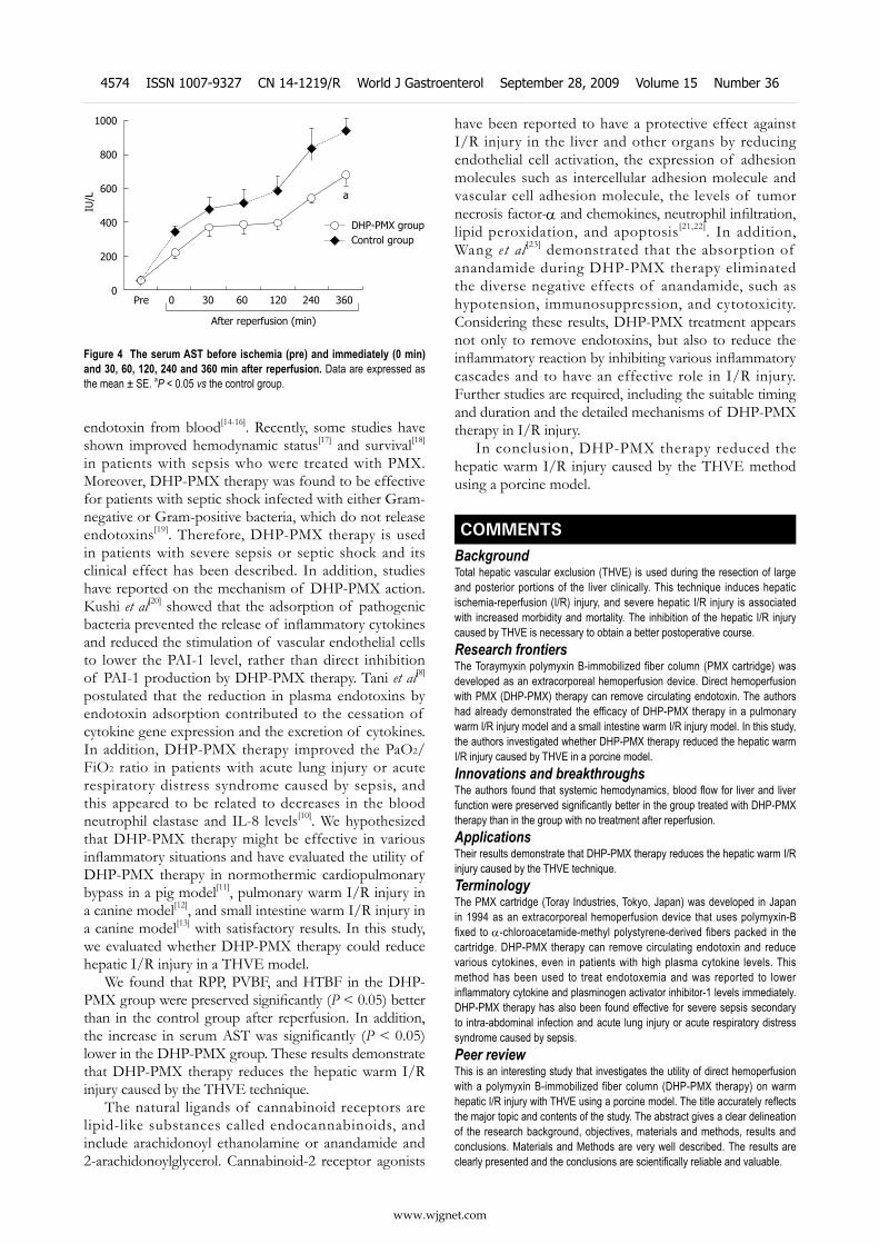
www.wjgnet.com
endotoxin from blood[14-16]. Recently, some studies have shown improved hemodynamic status[17] and survival[18] in patients with sepsis who were treated with PMX. Moreover, DHP-PMX therapy was found to be effective for patients with septic shock infected with either Gram-negative or Gram-positive bacteria, which do not release endotoxins[19]. Therefore, DHP-PMX therapy is used in patients with severe sepsis or septic shock and its clinical effect has been described. In addition, studies have reported on the mechanism of DHP-PMX action. Kushi et al[20] showed that the adsorption of pathogenic bacteria prevented the release of inflammatory cytokines and reduced the stimulation of vascular endothelial cells to lower the PAI-1 level, rather than direct inhibition of PAI-1 production by DHP-PMX therapy. Tani et al[8] postulated that the reduction in plasma endotoxins by endotoxin adsorption contributed to the cessation of cytokine gene expression and the excretion of cytokines. In addition, DHP-PMX therapy improved the PaO2/FiO2 ratio in patients with acute lung injury or acute respiratory distress syndrome caused by sepsis, and this appeared to be related to decreases in the blood neutrophil elastase and IL-8 levels[10]. We hypothesized that DHP-PMX therapy might be effective in various inflammatory situations and have evaluated the utility of DHP-PMX therapy in normothermic cardiopulmonary bypass in a pig model[11], pulmonary warm I/R injury in a canine model[12], and small intestine warm I/R injury in a canine model[13] with satisfactory results. In this study, we evaluated whether DHP-PMX therapy could reduce hepatic I/R injury in a THVE model.
We found that RPP, PVBF, and HTBF in the DHP-PMX group were preserved significantly (P < 0.05) better than in the control group after reperfusion. In addition, the increase in serum AST was significantly (P < 0.05) lower in the DHP-PMX group. These results demonstrate that DHP-PMX therapy reduces the hepatic warm I/R injury caused by the THVE technique.
The natural ligands of cannabinoid receptors are lipid-like substances called endocannabinoids, and include arachidonoyl ethanolamine or anandamide and 2-arachidonoylglycerol. Cannabinoid-2 receptor agonists
have been reported to have a protective effect against I/R injury in the liver and other organs by reducing endothelial cell activation, the expression of adhesion molecules such as intercellular adhesion molecule and vascular cell adhesion molecule, the levels of tumor necrosis factor-α and chemokines, neutrophil infiltration, lipid peroxidation, and apoptosis [21,22]. In addition, Wang et al [23] demonstrated that the absorption of anandamide during DHP-PMX therapy eliminated the diverse negative effects of anandamide, such as hypotension, immunosuppression, and cytotoxicity. Considering these results, DHP-PMX treatment appears not only to remove endotoxins, but also to reduce the inflammatory reaction by inhibiting various inflammatory cascades and to have an effective role in I/R injury. Further studies are required, including the suitable timing and duration and the detailed mechanisms of DHP-PMX therapy in I/R injury.
In conclusion, DHP-PMX therapy reduced the hepatic warm I/R injury caused by the THVE method using a porcine model.
COMMENTSBackgroundTotal hepatic vascular exclusion (THVE) is used during the resection of large and posterior portions of the liver clinically. This technique induces hepatic ischemia-reperfusion (I/R) injury, and severe hepatic I/R injury is associated with increased morbidity and mortality. The inhibition of the hepatic I/R injury caused by THVE is necessary to obtain a better postoperative course.Research frontiersThe Toraymyxin polymyxin B-immobilized fiber column (PMX cartridge) was developed as an extracorporeal hemoperfusion device. Direct hemoperfusion with PMX (DHP-PMX) therapy can remove circulating endotoxin. The authors had already demonstrated the efficacy of DHP-PMX therapy in a pulmonary warm I/R injury model and a small intestine warm I/R injury model. In this study, the authors investigated whether DHP-PMX therapy reduced the hepatic warm I/R injury caused by THVE in a porcine model.Innovations and breakthroughsThe authors found that systemic hemodynamics, blood flow for liver and liver function were preserved significantly better in the group treated with DHP-PMX therapy than in the group with no treatment after reperfusion.ApplicationsTheir results demonstrate that DHP-PMX therapy reduces the hepatic warm I/R injury caused by the THVE technique.TerminologyThe PMX cartridge (Toray Industries, Tokyo, Japan) was developed in Japan in 1994 as an extracorporeal hemoperfusion device that uses polymyxin-B fixed to α-chloroacetamide-methyl polystyrene-derived fibers packed in the cartridge. DHP-PMX therapy can remove circulating endotoxin and reduce various cytokines, even in patients with high plasma cytokine levels. This method has been used to treat endotoxemia and was reported to lower inflammatory cytokine and plasminogen activator inhibitor-1 levels immediately. DHP-PMX therapy has also been found effective for severe sepsis secondary to intra-abdominal infection and acute lung injury or acute respiratory distress syndrome caused by sepsis.Peer reviewThis is an interesting study that investigates the utility of direct hemoperfusion with a polymyxin B-immobilized fiber column (DHP-PMX therapy) on warm hepatic I/R injury with THVE using a porcine model. The title accurately reflects the major topic and contents of the study. The abstract gives a clear delineation of the research background, objectives, materials and methods, results and conclusions. Materials and Methods are very well described. The results are clearly presented and the conclusions are scientifically reliable and valuable.
1000
800
600
400
200
0
IU/L
Pre 0 30 60 120 240 360
After reperfusion (min)
a
DHP-PMX groupControl group
Figure 4 The serum AST before ischemia (pre) and immediately (0 min) and 30, 60, 120, 240 and 360 min after reperfusion. Data are expressed as the mean ± SE. aP < 0.05 vs the control group.
COMMENTS
4574 ISSN 1007-9327 CN 14-1219/R World J Gastroenterol September 28, 2009 Volume 15 Number 36
www.wjgnet.com
REFERENCES1 Fondevila C, Busuttil RW, Kupiec-Weglinski JW. Hepatic
ischemia/reperfusion injury--a fresh look. Exp Mol Pathol 2003; 74: 86-93
2 Laroux FS, Pavlick KP, Hines IN, Kawachi S, Harada H, Bharwani S, Hoffman JM, Grisham MB. Role of nitric oxide in inflammation. Acta Physiol Scand 2001; 173: 113-118
3 Glantzounis GK, Salacinski HJ, Yang W, Davidson BR, Seifalian AM. The contemporary role of antioxidant therapy in attenuating liver ischemia-reperfusion injury: a review. Liver Transpl 2005; 11: 1031-1047
4 Bismuth H, Castaing D, Garden OJ. Major hepatic resection under total vascular exclusion. Ann Surg 1989; 210: 13-19
5 Delva E, Barberousse JP, Nordlinger B, Ollivier JM, Vacher B, Guilmet C, Huguet C. Hemodynamic and biochemical monitoring during major liver resection with use of hepatic vascular exclusion. Surgery 1984; 95: 309-318
6 Tsuzuki H, Tani T, Ueyama H, Kodama M. Lipopolysac-charide: neutralization by polymyxin B shuts down the signaling pathway of nuclear factor kappaB in peripheral blood mononuclear cells, even during activation. J Surg Res 2001; 100: 127-134
7 Shoji H. Extracorporeal endotoxin removal for the treatment of sepsis: endotoxin adsorption cartridge (Toraymyxin). Ther Apher Dial 2003; 7: 108-114
8 Tani T, Hanasawa K, Kodama M, Imaizumi H, Yonekawa M, Saito M, Ikeda T, Yagi Y, Takayama K, Amano I, Shimaoka H, Ohta M, Okahisa T, Koga N, Fujita N, Yamasa H. Correlation between plasma endotoxin, plasma cytokines, and plasminogen activator inhibitor-1 activities in septic patients. World J Surg 2001; 25: 660-668
9 Vincent JL, Laterre PF, Cohen J, Burchardi H, Bruining H, Lerma FA, Wittebole X, De Backer D, Brett S, Marzo D, Nakamura H, John S. A pilot-controlled study of a polymyxin B-immobilized hemoperfusion cartridge in patients with severe sepsis secondary to intra-abdominal infection. Shock 2005; 23: 400-405
10 Kushi H, Miki T, Okamaoto K, Nakahara J, Saito T, Tanjoh K. Early hemoperfusion with an immobilized polymyxin B fiber column eliminates humoral mediators and improves pulmonary oxygenation. Crit Care 2005; 9: R653-R661
11 Ohki S, Oshima K, Takeyoshi I, Matsumoto K, Morishita Y. Endotoxin removal with a polymyxin B-immobilized hemoperfusion cartridge improves cardiopulmonary function after cardiopulmonary bypass. J Surg Res 2008; 145: 74-79
12 Oshima K, Akao T, Kobayashi K, Muraoka M, Matsumoto K, Takeyoshi I. The effect of direct hemoperfusion with a polymyxin B-immobilized fiber column (DHP-PMX
therapy) on pulmonary ischemia-reperfusion injury in a canine model. J Invest Surg 2008; 21: 127-132
13 Sato H, Oshima K, Arakawa K, Kobayashi K, Yamazaki H, Suto Y, Takeyoshi I. Direct hemoperfusion with a polymyxin B-immobilized cartridge in intestinal warm ischemia reperfusion. World J Gastroenterol 2008; 14: 5436-5441
14 King RC, Binns OA, Rodriguez F, Kanithanon RC, Daniel TM, Spotnitz WD, Tribble CG, Kron IL. Reperfusion injury significantly impacts clinical outcome after pulmonary transplantation. Ann Thorac Surg 2000; 69: 1681-1685
15 Cohen J, Aslam M, Pusey CD, Ryan CJ. Protection from endotoxemia: a rat model of plasmapheresis and specific adsorption with polymyxin B. J Infect Dis 1987; 155: 690-695
16 Aoki H, Kodama M, Tani T, Hanasawa K. Treatment of sepsis by extracorporeal elimination of endotoxin using polymyxin B-immobilized fiber. Am J Surg 1994; 167: 412-417
17 Tetta C, Gianotti L, Cavaillon JM, Wratten ML, Fini M, Braga M, Bisagni P, Giavaresi G, Bolzani R, Giardino R. Coupled plasma filtration-adsorption in a rabbit model of endotoxic shock. Crit Care Med 2000; 28: 1526-1533
18 Uriu K, Osajima A, Hiroshige K, Watanabe H, Aibara K, Inada Y, Segawa K, Anai H, Takagi I, Ito A, Kamochi M, Kaizu K. Endotoxin removal by direct hemoperfusion with an adsorbent column using polymyxin B-immobilized fiber ameliorates systemic circulatory disturbance in patients with septic shock. Am J Kidney Dis 2002; 39: 937-947
19 Kawamata T , Imaizumi H, Yoshida M, Kaneko M. Polymyxin B-immobilized fiber improves hyperdynamic state in MRSA septic patients. Intensive Care Med 1997; 23: 130-131
20 Kushi H, Nakahara J, Miki T, Okamoto K, Saito T, Tanjo K. Hemoperfusion with an immobilized polymyxin B fiber column inhibits activation of vascular endothelial cells. Ther Apher Dial 2005; 9: 303-307
21 Bátkai S, Osei-Hyiaman D, Pan H, El-Assal O, Rajesh M, Mukhopadhyay P, Hong F, Harvey-White J, Jafri A, Haskó G, Huffman JW, Gao B, Kunos G, Pacher P. Cannabinoid-2 receptor mediates protection against hepatic ischemia/reperfusion injury. FASEB J 2007; 21: 1788-1800
22 Rajesh M, Pan H, Mukhopadhyay P, Bátkai S, Osei-Hyiaman D, Haskó G, Liaudet L, Gao B, Pacher P. Cannabinoid-2 receptor agonist HU-308 protects against hepatic ischemia/reperfusion injury by attenuating oxidative stress, inflammatory response, and apoptosis. J Leukoc Biol 2007; 82: 1382-1389
23 Wang Y, Liu Y, Sarker KP, Nakashima M, Serizawa T, Kishida A, Akashi M, Nakata M, Kitajima I, Maruyama I. Polymyxin B binds to anandamide and inhibits its cytotoxic effect. FEBS Lett 2000; 470: 151-155
S- Editor Tian L L- Editor Logan S E- Editor Zheng XM
Sato H et al . PMX therapy improves liver function after I/R injury 4575
www.wjgnet.com
BRIEF ARTICLES
Epithelioid angiomyolipoma of the liver: Cross-sectional imaging findings of 10 immunohistochemically-verified cases
Peng-Ju Xu, Yan Shan, Fu-Hua Yan, Yuan Ji, Ying Ding, Mei-Lin Zhou
Online Submissions: wjg.wjgnet.com World J Gastroenterol 2009 September 28; 15(36): [email protected] World Journal of Gastroenterology ISSN 1007-9327doi:10.3748/wjg.15.4576 © 2009 The WJG Press and Baishideng. All rights reserved.
Peng-Ju Xu, Yan Shan, Fu-Hua Yan, Ying Ding, Mei-Lin Zhou, Department of Radiology, Zhongshan Hospital, Fudan University, 180 Fenglin Road, Shanghai 200032, ChinaYuan Ji, Department of Pathology, Zhongshan Hospital, Fudan University, 180 Fenglin Road, Shanghai 200032, ChinaAuthor contributions: Xu PJ and Shan Y contributed equally to this work; Xu PJ, Shan Y, Yan FH designed the research; Xu PJ, Shan Y, Ji Y, Ding Y, Zhou ML performed the research; Xu PJ, Shan Y, Yan FH analyzed the data; Xu PJ, Shan Y, Yan FH wrote the paper.Correspondence to: Fu-Hua Yan, Professor, Department of Radiology Zhongshan Hospital Fudan University, 180 Fenglin Road, Shanghai 200032, China. [email protected]: +86-21-64041990-3682 Fax: +86-21-64439906Received: July 1, 2009 Revised: August 18, 2009Accepted: August 25, 2009Published online: September 28, 2009
AbstractAIM: To retrospectively evaluate the computed tomo-graphy (CT)/magnetic resonance imaging (MRI) imaging features of epithelioid angiomyolipoma of the liver (Epi-HAML), with pathology as a reference.
METHODS: The CT/MRI findings (number, diameter, lobar location, and appearance of lesions) in a series of 10 patients with 12 pathologically proven epithelioid angiomyolipomas of the liver were retrospectively analyzed. The imaging features, including attenuation/signal intensity characteristics, presence of fat, hypervascular, outer rim, and vessels within lesion, were evaluated and compared with that of non-Epi-HAML in 11 patients (13 lesions). The Fisher exact test was used to compare difference in probability of imaging features between the two types.
RESULTS: For 21 patients, CT images of 15 patients and MR images of six patients were available. No patient underwent two examinations. For the 15 patients with a CT scan, all HAML lesions in the two groups (10 Epi-HAML and seven non-Epi-HAML) manifested as hypoattenuation. For the six patients with MRI, all lesions (two Epi-HAML and six non-Epi-HAML) were hypointense on T1WI (fat suppression) and hyperintense on T2WI. There were 10 non-Epi-HAML, but only two Epi-HAML lesions showed the presence of
fat, which significantly different between the two types (P = 0.005). On the dynamic contrast enhancement (DCE) imaging, eight Epi-HAML, and 13 non-Epi lesions manifested as hypervascular. Punctate or curved vessels were displayed in 10 Epi-HAML as well as in nine non-Epi lesions and outer rim enhancement could be found with eight Epi-HAML as well as six non-Epi lesions.
CONCLUSION: Little or no presence of adipose tissue was found to be an imaging feature of Epi-HAML, compared with the non-Epi type. In addition, hypervascularity with opacification of central punctiform or filiform vessels on DCE would be a characteristic enhancement pattern for Epi-HAML.
© 2009 The WJG Press and Baishideng. All rights reserved.
Key words: Epithelioid angiomyolipoma; Liver; Immuno-histochemical staining; Magnetic resonance imaging; Computed X-ray tomography
Peer reviewer: Xiao-Peng Zhang, Department of Radiology, Peking University School of Oncology, Beijing Cancer Hospital & Institute, No.52 Haidian District, Beijing 100142, China
Xu PJ, Shan Y, Yan FH, Ji Y, Ding Y, Zhou ML. Epithelioid angiomyolipoma of the liver: Cross-sectional imaging findings of 10 immunohistochemically-verified cases. World J Gastroenterol 2009; 15(36): 4576-4581 Available from: URL: http://www.wjgnet.com/1007-9327/15/4576.asp DOI: http://dx.doi.org/10.3748/wjg.15.4576
INTRODUCTIONHepatic Angiomyolipoma (HAML) is a rare benign tumor and the etiology is unclear. Some cases have been associated with the tuberous sclerosis complex (TSC). HAML belongs to a family of tumors that have collectively been called ‘‘PEComa’’[1-3]. Histologically, HAML is composed of a heterogeneous mixture of blood vessels, smooth muscle, and adipose cells, of varying proportions and distributions, not only among different tumors, but from area to area within the same tumor. Thus, according to the line of differentiation and the predominance of tissue components, HAML is usually subcategorized into mixed,

Xu PJ et al . Imaging and angiomyolipoma of the liver 4577
www.wjgnet.com
lipomatous (≥ 70% fat), myomatous (≤ 10% fat), and angiomatous types. The smooth muscle cell component is the most specific for the diagnosis. Depending on the dominant cell type, HAML can be subcategorized into epithelioid, spindle, and intermediate forms[1]. Epithelioid AML (Epi-AML) was first described in the liver in 2000[4], diagnosis can be difficult, for example the differentiation of Epi-AML from hepatocellular carcinoma and the metastatic sarcomatoid variant of renal cell carcinoma. Only one case report of Epi-AML has been mentioned or reviewed in the literature[4-7]. In this paper, 10 immunohistochemically verified cases were retrospectively analyzed and the CT/MR imaging findings were summarized and compared with those of non-epi-HAML.
MATERIALS AND METHODSPatientsThe imaging examinations of 21 cases (10 Epi-HAML and 11 non-Epi-HAML) with pathologically proven HAML were included in this study. Proof of diagnosis was based on findings at liver resection and pathological manifestations, including immunohistochemical staining. Cases were collected from one university hospital over a seven-year period and were identified by reviewing pathology databases. In our patients, AML were incidentally detected on cross-sectional imaging performed for various reasons, such as abdominal pain (n = 15), suspected gallbladder stone (n = 3), or urinary (n =3) diseases. Institutional review board approval and patient consent were not required for this retrospective study because patient privacy was maintained and patient care was not impacted.
Imaging protocols and methodsHelical multiphasic CT was performed in 15 patients using a Siemens Sensation 16 (Siemens Medical Solutions) or a PHILIPS Marconi MX8000 4 slice CT unit with 5 to 7 mm contiguous sections. After non-enhanced acquisitions of the liver, patients underwent helical multiphase CT that included both hepatic arterial phase and portal venous phase imaging (30-35 s and 80-85 s, respectively), after i.v. infusion of 90-100 mL nonionic contrast material (iopromide, Ultravist 300, Bayer Schering Pharma). Contrast material was injected at a rate of 3 mL/s with a power injector (Envision CT, MEDRAD).
MRI was performed in six patients using 1.5-T MR units (Magnetom avanto, Siemens Medical Solutions) with the combination of a phased-array body coil and spine array coil for signal reception. Baseline MR images, including a respiratory-navigated T2-weighted turbo spin-echo sequence [TR/TE, 2000/104 ms; slice thickness, 7 mm; flip angle, 150°; matrix, 207 (phase) × 384 (read); FOV, 33-38 cm] and a breath-hold T1-weighted fast low angle shot (FLASH) sequence [TR/TE, 112/4.76 ms; slice thickness, 7 mm flip angle, 70°; matrix, 114 (phase) × 256 (read); FOV, 33-38 cm]. Dynamic imaging, breath-hold T1-weighted FLASH sequence was performed using the following parameters: TR/TE, 230/2.47 ms; flip angle, 70°; matrix, 135 (phase) × 256 (read); effective slice thickness, 7 mm; and FOV, 33-38 cm. Dynamic imaging was
performed before and after administration of gadopentate dimeglumine (Magnevist; Bayer Schering Pharma), consisting of late arterial (delay time 20-25 s), portal (70- 90 s), and equilibrium (180 s) phases. The contrast-enhanced imaging was acquired after a bolus injection of 30-35 mL of contrast with a fixed delay. The contrast material was injected into the antecubital vein at a rate of 2.5 mL/s via a power injector (Spectris, Medrad, Indianola, PA, USA). Three dynamic phases were repeated for 18-21 s during a single breath-hold.
Imaging analysisImaging studies were evaluated on film by two abdominal radiologists (with experience ranging from 5 to 10 years) in consensus. Readers were not blinded to the pathology results.
The following imaging criteria were analyzed: number of lesions; lesion diameter; lesion location according to the hepatic segment numbering system of Couinaud; attenuation at non-enhanced CT, classified as hypoattenuating, isoattenuating, or hyperattenuating to the adjacent liver parenchyma; signal intensity characteristics of the lesions at non-enhanced T1-weighted (including T1WI and T1WI with fat suppression) and T2-weighted MRI; presence of fat tissue, hypoattenuating foci (-20 to -120 Hu) on non-enhanced CT images or hyperintense on T1WI but hypointense on T1WI with fat suppression; enhancement pattern at contrast-enhanced CT or MRI with regard to three-phase dynamic enhancement; presence of the central vessels in lesion at contrast-enhanced imaging; and presence of outer rim enhancement.
PathologyAll tissues were reviewed independently by one patholo-gist. Histopathological diagnosis was made according to the World Health Organization’s classification of tumors of the liver and intrahepatic bile ducts[8]. The most important diagnostic criterion was the presence of HMB-45-positive myoid cells. All tumor tissues had been fixed in neutral buffered formalin and were routinely embedded in paraffin. Hematoxylin-eosin stained sections were evaluated and immunohistochemical studies were performed on representative blocks by the EnVision Plus system (DAKO, Glostrup, Denmark) with a panel of antibodies (HMB-45, SMA, S-100, CD34, A103, MSA, Vimentin, CD68, CD117, HepPar-1, AFP, AE1/AE3, and CK8).
Statistical analysisStatistical analysis was performed by using software (Intercooled Stata, version 9.0 for Windows, 2005; Stata Corp, College Station, TX, USA). We used the Fisher exact test to compare probability of these imaging features for Epi-HAML and non-Epi lesion. A P value less than 0.05 was considered statistically significant.
RESULTSGeneral dataThe data are summarized in Tables 1 and 2. Age at diagnosis of hepatic angiomyolipoma varied between

www.wjgnet.com
17 and 55 years (mean 40.7 years). In the Epi-HAML group, there was history of TSC for one patient, and a combination of left kidney AML for another one. Among 10 patients, eight had solitary lesions and two had two lesions. Lesions had a mean diameter of 4.6 (range, 1.5-10.0). In the non-Epi-HAML group, there was right kidney AML in one patient, 10 had solitary lesions, and one had three lesions. Lesions had a mean diameter of 4.9 (range, 1.0-12.0).
Comparison of CT/MRI findings between Epi-HAML and non-Epi-HAMLThe CT/or MR images of 21 patients (CT images for 15 patients and MR for six) were available for retrospective analysis. The results are show in Tables 1-3.
At non-enhanced CT, 10 Epi-HAML and seven non-Epi HAML were hypoattenuating to the surrounding liver. Two Epi-HAML and six non-epi lesions were hypointense on T1WI (fat suppression) and hyperintense on T2WI. The presence of fat was detected in 10 non-Epi HAML but only two Epi type lesions (Figure 1) and there were significant differences between the two lesion types (P < 0.01, Table 3).
On arterial phase, eight Epi-HAML and 13 non-Epi-HAML showed obvious enhancement (Figures 1-3). Punctate or curved vessels could be seen within 10 Epi-HAML as well as nine non-Epi type lesions (Figures 1-4) on arterial or/and portal phase. Outer rim enhancement could be found in eight Epi-HAML as well as in six non-Epi-HAML on enhanced imaging (Figures 3 and 4).
PathologyImmunohistochemical studies showed that tumor cells were positive for HMB-45 and A103, but negative for cytokeratin (HepPar-1, AFP, AE1/AE3, CK8.) in all cases. Smooth muscle actin (SMA) and desmin staining were weak to moderate in epithelioid cells. MSA staining was similar to desmin but was more weak when positive. The endothelial cells lining the blood vessels were positive for CD34. CD117 and S-100 were negative in all cases.
Follow-upImaging and clinical follow-up was available in 10 patients with Epi-HAML and ranged from one to six years.
Table 2 Synopsis of demographics and imaging findings in 11 patients with non-Epi-HAML
No. Age (yr)/sex Size (cm), segment Unenhanced CT/MR Fat Vascularity Outer rim Vessels in lesion
1 45/F 6.0, Ⅵ/Ⅶ Hypo Yes Hypervascular No No2 34/F 4.5, Ⅱ/Ⅲ Hypo No Hypervascular No Yes3 37/F 5.5, Ⅷ/Ⅳ T1 hypo, T2 hyper Yes Hypervascular Yes Yes4 40/F 12.0, Ⅶ/Ⅷ/Ⅳ T1 hypo, T2 hyper Yes Hypervascular Yes Yes
2.0, 1.0, Ⅱ/Ⅲ5 50/F 6.5, Ⅳ T1 hypo, T2 hyper Yes Hypervascular Yes Yes6 43/M 2.0, Ⅵ Hypo Yes Hypervascular No No7 47/M 2.0, Ⅳ Hypo Yes Hypervascular No No8 46/M 3.0, Ⅷ Hypo Yes Hypervascular No Yes9 44/F 4.0, Ⅱ Hypo No Hypervascular No No10 50/F 3.0, Ⅵ Hypo Yes Hypervascular No Yes11 21/F 12.0, Ⅵ/Ⅵ T1 hypo, T2 hyper No Hypervascular Yes Yes
Table 1 Synopsis of demographics and imaging findings in 10 patients with Epi-HAML
No. Age (yr)/sex Size (cm), segment Unenhanced CT/MR Fat Vascularity Outer rim Vessels in lesion
1 51/F 6.5, 4.0 Ⅵ/Ⅶ, Ⅳ Hypo No Hypovascular Yes Yes2 42/F 4.2, Ⅰ Hypo No Hypovascular No No3 35/F 7.5, Ⅵ Hypo No Hypervascular No Yes4 36/F 1.5, Ⅳ Hypo No Hypervascular Yes Yes5 17/F 10.0, Ⅴ/Ⅶ/Ⅷ Hypo No Hypervascular Yes Yes6 55/F 5.0, Ⅱ/Ⅲ Hypo No Hypervascular Yes Yes71 33/F 6.0, 1.0, Ⅱ/Ⅲ/Ⅳ, Ⅶ Hypo Yes Hypervascular No Yes8 36/F 3.0, Ⅳ Hypo No Hypervascular Yes Yes9 46/F 4.0, Ⅱ/Ⅲ T1 hypo, T2 hyper No Hypervascular Yes Yes10 47/F 2.5, Ⅵ T1 hypo, T2 hyper No Hypervascular Yes Yes
1The demonstration of hypervascularity and vessels with lesion were only for one lesion (large) of No. 7 patient. Hyper: Hyperattenuation; Hypo: Hypoattenuation; T1 hypo: Hypointense on T1WI (fat suppression); T2 hyper: Hyperintense on T2WI.
Table 3 Comparison of imaging features between Epi-HAML and non-Epi-HAML1
Epi-HAML, n = 12 (10)
Non-Epi-HAML, n = 13 (11)
P value
Fat 2 (1) 10 (8) 0.005Hypervascular 8 (8) 13 (11) 0.082Vessels in lesions 10 (9) 9 (7) 0.645Outer rim 8 (7) 6 (4) 0.428
1Data without parentheses are the number of lesions and data in parentheses are the number of patients. The Fisher exact test was used to analyze difference in per-lesion probability of imaging features between two types.
4578 ISSN 1007-9327 CN 14-1219/R World J Gastroenterol September 28, 2009 Volume 15 Number 36

www.wjgnet.com
Recurrent hepatic lesions were found in one patient (Figure 1), pubic bone destruction and metastatic nodule in body soft tissue were proved with biopsy in another.
DISCUSSIONPrevious literatures showed variable imaging appearances for HAML[9-12]. The imaging characteristics of HAML are correlated with its histological components, and demonstration of blood vessels and mature adipose tissue are the most important radiographic features. Color Doppler sonography shows a punctiform or filiform vascular distribution pattern. Contrast-enhanced CT shows marked enhancement of the soft tissue components in the arterial phase. MRI is the most
specific imaging entity for the detection of lipomatous components; however, because hepatic AML can have such variation in the amount of adipose tissue present, detection varies based on the percentage of the lesion that is composed of adipose tissue. MRI findings include hypointensity or hyperintensity on T1WI, slight hyperintensity on T2WI, dense enhancement in the arterial phase, and hypointensity in the delayed phase.
In our study, most of the Epi-AML tumors (10/12) were completely devoid of adipose tissue, which is the characteristic finding compared with that of non-Epi type. The result was consistent with a previous report[1]. After i.v. contrast administration, there was obvious enhancement on the arterial phase for most of these lesions, suggesting that Epi-AML was a hypervascular tumor. Most lesions (seven) manifested as hypoattenuation or hypointensity on portal/equilibrium phase. By adjusting the window
Figure 2 55-year-old woman with epithelioid angiomyolipoma in left lobe of liver (patient 6). A: Non-enhanced CT scan shows hypoattenuating lesion in segment Ⅱ; B: contrast-enhanced CT scan shows obviously enhanced lesion on arterial phase; C: the punctate enhanced vessels (arrowhead) within the lesion is show on an arterial phase image by adjusting the window width and level.
C
B
AA
B
C
Figure 1 33-year-old woman with epithelioid angiomyolipoma in left lobe of liver (patient 7). A: Non-enhanced CT scan shows hypoattenuating lesion with fat components (arrow, CT value mean-35 Hu) in segments Ⅱ/Ⅲ/Ⅳ; B: Contrast-enhanced CT scan shows inhomogeneous enhancement lesion with punctiform or filiform enhanced vessels (arrowheads, the window width and level was adjusted) on arterial phase; C: Contrast-enhanced CT scan at 1-year follow-up shows enhanced recurrent nodule (arrow) on arterial phase image after the left lobe surgery.
Xu PJ et al . Imaging and angiomyolipoma of the liver 4579

www.wjgnet.com
width and level, punctate or curved vessels could be seen within 10 lesions on enhanced scanning. In addition, intact or discrete outer rim enhancement within 8 lesions was observed. According to the pathology specimens and literature report, it is not true capsular but a pseudocapsule, which is composed of the compressed liver parenchyma and sparse fibrosis tissues with small vessels, resulting in delayed enhancement on late phase[13]. A similar manifestation can be found in other hypervascular lesion, such as hepatocellular carcinoma(HCC) and focal nodular hyperplasia (FNH). The distinction between these lesions can be difficult. However, the enhancement patterns were somewhat different among Epi-AML, HCC, and FNH. Most of the HCC enhanced markedly in the arterial phase
and decreased rapidly on the portal venous/equilibrium phase. Additionally, capsule could be found in most of the HCC, so the margins of HCC were more clear than those of AML in the portal venous phase, a suggestive finding for correct diagnosis[14]. The enhancement pattern of FNH was similar to AML, in both of them prolonged enhancement could be shown in the portal venous phase. However, most of FNH enhanced homogeneously on the arterial phase except the central scar, which was characteristic of FNH and could be enhanced on the portal venous phase/delayed phase[15].
Five cases of hepatic malignant angiomyolipoma have been reported[5,16-19]. Strict histology criteria for defining hepatic angiomyolipoma as malignant have not been put
BA
Figure 4 46-year-old woman with epithelioid angiomyolipoma in left lobe of liver (patient 9). A: Non-enhanced MRI T2WI shows hyperintense lesion in segment Ⅱ/Ⅲ; B: Contrast-enhanced MRI shows obvious enhanced lesions with outer rim enhancement (arrow) and opacification of punctate or curved vessels (arrowhead) on arterial phase.
C
BA
D
Figure 3 17-year-old girl with epithelioid angiomyolipoma in right lobe of liver (the patient had a history of tuberous sclerosis complex, patient 5). A: Non-enhanced CT scan shows homogeneous hypoattenuating lesion in segment Ⅴ/Ⅶ/Ⅷ; B: Contrast-enhanced CT scan shows inhomogeneous enhanced lesion with opacification of central vessels (arrow) on the arterial phase; C: The slight enhanced outer rim (arrow) around the inhomogeneous enhancement lesions is shown on portal phase contrast-enhanced CT; D: Immunohistochemical staining for HMB-45 shows diffusely positive staining in tumor cells within the cytoplasm (EnVision, × 100).
4580 ISSN 1007-9327 CN 14-1219/R World J Gastroenterol September 28, 2009 Volume 15 Number 36

www.wjgnet.com
forward, but it should be suspected, especially in tumors with many mitoses. Those with necrosis and significant cellular pleomorphism might show aggressive behavior. There is clinical evidence of aggressive behavior such as recurrence and metastases beyond the liver, for two cases in these 10 Epi-AML cases. However, there are no characteristic findings for malignant HAML on cross-sectional imaging. Therefore, although most HAMLs are biologically benign, this tumor should be considered to have malignant potential, especially for Epi-AML. So resection and careful follow-up are recommended.
It is important to recognize the limitations of our study. Firstly, the study is retrospective; secondly, the numbers reported are limited and no one patient had both CT and MRI. A further limitation is that not every patient underwent regular imaging follow-up and documentation was incomplete.
In summary, little or no adipose tissue was found to be an imaging feature of Epi-HAML, vs non-Epi-HAML. In addition, hypervascularity with opacification of central punctiform or filiform vessels on DCE would be a characteristic enhancement pattern for Epi-HAML.
COMMENTSBackgroundHepatic Angiomyolipoma (HAML) is a rare benign tumor and belongs to a family of tumors that have collectively been called ‘‘PEComa’’. The immense variability of the morphological appearance is due to the different heterogeneous mixtures of vessels, epithelioid cells, and lipocytes. As a specific form, the epithelioid HAML (Epi-HAML) has characteristics in pathology and diagnosis that can be difficult, e.g. differentiation of Epi-AML from hepatocellular carcinoma and the metastatic sarcomatoid variant of renal cell carcinoma.Research frontiersPrevious studies showed variable imaging appearances for HAML. The imaging characteristics of HAML are correlated with its histological components. Both CT and MRI, for the most part, do not allow the definitive differentiation of HAML from hepatocellular carcinoma, adenoma, liposarcoma, lipoma, hamartoma, and sometimes even from focal nodular hyperplasia, especially if the fat content is low. The Epi-HAML has morphological characteristics of tumor cells. In addition, the tumors are devoid of fat or only scattered fat cells distribution in pathology. However, the imaging features of Epi-HAML have not been unequivocally described and evaluated. In this study, the authors summarized CT/MRI imaging features of Epi-HAML and compared with those of non-Epi-HAML.Innovations and breakthroughsPrevious reports have showed demonstration of mature adipose tissue is the most important radiographic features and highlighted the importance in the diagnosis of HAML, in particular for mixed, lipomatous types of HAML. This is the first study to report that blood vessels are also an imaging feature in Epi-HAML. Furthermore, the study would suggest that little or no adipose tissue might be imaging characteristics of Epi-HAML, versus non-Epi-HAML.ApplicationsBy understanding the imaging characteristics, this study could help radiologists familiarize themselves with the appearance of Epi-HAML, and improve the confidence in the diagnosis of incidental liver tumor, especially in cases where the fat content is low and the interpretation of histological findings is difficult. TerminologyImmunohistochemistry is a method of analyzing and identifying cell types based on the binding of antibodies to specific components of the cell. The most important diagnostic criterion for HAML is the presence of HMB-45-positive myoid cells.Peer reviewThe manuscript reported the imaging characteristics of Epi-HAML, only a few reports had been published about the lesion before. It will be helpful for radiologists to obtain knowledge of the lesion.
REFERENCES1 Tsui WM, Colombari R, Portmann BC, Bonetti F, Thung
SN, Ferrell LD, Nakanuma Y, Snover DC, Bioulac-Sage P, Dhillon AP. Hepatic angiomyolipoma: a clinicopathologic study of 30 cases and delineation of unusual morphologic variants. Am J Surg Pathol 1999; 23: 34-48
2 Xu AM , Zhang SH, Zheng JM, Zheng WQ, Wu MC. Pathological and molecular analysis of sporadic hepatic angiomyolipoma. Hum Pathol 2006; 37: 735-741
3 Jiang TA, Zhao QY, Chen MY, Wang LJ, Ao JY. Diagnostic analysis of hepatic angiomyolipoma. Hepatobiliary Pancreat Dis Int 2005; 4: 152-155
4 Yamasaki S, Tanaka S, Fujii H, Matsumoto T, Okuda C, Watanabe G, Suda K. Monotypic epithelioid angiomyolipoma of the liver. Histopathology 2000; 36: 451-456
5 Dalle I, Sciot R, de Vos R, Aerts R, van Damme B, Desmet V, Roskams T. Malignant angiomyolipoma of the liver: a hitherto unreported variant. Histopathology 2000; 36: 443-450
6 Tryggvason G, Blöndal S, Goldin RD, Albrechtsen J, Björnsson J, Jónasson JG. Epithelioid angiomyolipoma of the liver: case report and review of the literature. APMIS 2004; 112: 612-616
7 Garcia TR, Mestre de Juan MJ. Angiomyolipoma of the liver and lung: a case explained by the presence of perivascular epithelioid cells. Pathol Res Pract 2002; 198: 363-367
8 Hirohashi S, Blum HE, Ishak KG. Tumours of the liver and intrahepatic bile ducts. In: Hamilton SR, Aaltonen LA, editors. Pathology and genetics. Tumours of the digestive system. World Health Organization classification of tumours. Lyon: IARC Press, 2000: 157-202
9 Prasad SR, Wang H, Rosas H, Menias CO, Narra VR, Middleton WD, Heiken JP. Fat-containing lesions of the liver: radiologic-pathologic correlation. Radiographics 2005; 25: 321-331
10 Yoshimura H, Murakami T, Kim T, Nakamura H, Hirabuki N, Sakon M, Wakasa K, Inoue Y. Angiomyolipoma of the liver with least amount of fat component: imaging features of CT, MR, and angiography. Abdom Imaging 2002; 27: 184-187
11 Yan F, Zeng M, Zhou K, Shi W, Zheng W, Da R, Fan J, Ji Y. Hepatic angiomyolipoma: various appearances on two-phase contrast scanning of spiral CT. Eur J Radiol 2002; 41: 12-18
12 Högemann D , Flemming P, Kreipe H, Galanski M. Correlation of MRI and CT findings with histopathology in hepatic angiomyolipoma. Eur Radiol 2001; 11: 1389-1395
13 Chang JC, Lee YW, Kim HJ. Preoperative diagnosis of angiomyolipoma of the liver. Abdom Imaging 1994; 19: 546-548
14 Iannaccone R , Piacentini F, Murakami T, Paradis V, Belghiti J, Hori M, Kim T, Durand F, Wakasa K, Monden M, Nakamura H, Passariello R, Vilgrain V. Hepatocellular carcinoma in patients with nonalcoholic fatty liver disease: helical CT and MR imaging findings with clinical-pathologic comparison. Radiology 2007; 243: 422-430
15 Mortelé KJ, Praet M, Van Vlierberghe H, de Hemptinne B, Zou K, Ros PR. Focal nodular hyperplasia of the liver: detection and characterization with plain and dynamic-enhanced MRI. Abdom Imaging 2002; 27: 700-707
16 McKinney CA, Geiger JD, Castle VP, Ruiz RE, Strouse PJ. Aggressive hepatic angiomyolipoma in a child. Pediatr Hematol Oncol 2005; 22: 17-24
17 Mizuguchi T, Katsuramaki T, Nobuoka T, Nishikage A, Oshima H, Kawasaki H, Kimura S, Satoh M, Hirata K. Growth of hepatic angiomyolipoma indicating malignant potential. J Gastroenterol Hepatol 2004; 19: 1328-1330
18 Croquet V, Pilette C, Aubé C, Bouju B, Oberti F, Cervi C, Arnaud JP, Rousselet MC, Boyer J, Calès P. Late recurrence of a hepatic angiomyolipoma. Eur J Gastroenterol Hepatol 2000; 12: 579-582
19 Nguyen TT, Gorman B, Shields D, Goodman Z. Malignant hepatic angiomyolipoma: report of a case and review of literature. Am J Surg Pathol 2008; 32: 793-798
S- Editor Tian L L- Editor Stewart GJ E- Editor Lin YP
COMMENTS
Xu PJ et al . Imaging and angiomyolipoma of the liver 4581

En-Hua Xiao, Dong Guo, Du-Jun Bian, Department of Radiology, the Second Xiangya Hospital, Central South University, Changsha 410011, Hunan Province, China Author contributions: Xiao EH designed the research; Xiao EH, Guo D and Bian DJ performed the research; Guo D and Bian DJ contributed new reagents/analytic tools; Xiao EH, Guo D and Bian DJ analyzed the data; Xiao EH and Guo D wrote the paper. Supported by The National Science and Technology Projects of 11th Five-Year Plan, No. 2007BAI05B06; the National Council of Natural Sciences of China, No. 30070235, 30470508, 30870695, the Provincial Council of Natural Sciences of Hunan, No. 06JJ20081 Correspondence to: Dr. En-Hua Xiao, Department of Radi-ology, the Second Xiangya Hospital, Central South University, Changsha 410011, Hunan Province, China. [email protected]: +86-731-85292116 Fax: +86-731-85533525Received: May 7, 2009 Revised: July 30, 2009Accepted: August 6, 2009Published online: September 28, 2009
AbstractAIM: To evaluate the effects of four types of preopera-tive transcatheter arterial chemoembolization (TACE) on angiogenesis of hepatocellular carcinoma (HCC) cells.
METHODS: A total of 136 patients with HCC under-went liver resection. One to five courses of TACE prior to liver resection were performed in 79 patients (TACE group), in which one to four courses of chemotherapy alone were performed in 11 patients (group A); one to five courses of chemotherapy combined with iodized oil were performed in 33 patients (group B); one to three courses of chemotherapy combined with iodized oil and gelatin sponge were performed in 23 patients (group C); one to three courses of chemotherapy combined with iodized oil, ethanol and gelatin sponge were performed in 12 patients (group D). The other 57 patients only re-ceived liver resection (non-TACE group). The microves-sels were marked by CD31. The expression of CD31 and vascular endothelial growth factor (VEGF) protein were detected by immunohistochemical methods.
RESULTS: The mean microvessel density (MVD) in HCC cells was significantly higher in groups A, B, C and D than in the non-TACE group (P < 0.05). The expres-sion of VEGF protein in HCC cells were significantly higher in groups A, B, C and D than in the non-TACE group (P < 0.05). MVD and the expression of VEGF
protein were positively correlated. Mean MVD and the expression of VEGF protein were closely related to the number of courses of TACE and the interval of TACE.
CONCLUSION: Four different types of preoperative TACE regimens enhanced angiogenesis in HCC cells by up-regulating the expression of VEGF protein. It is necessary to repress angiogenesis of liver cancer after TACE.
© 2009 The WJG Press and Baishideng. All rights reserved.
Key words: Angiogenesis; Hepatocellular carcinoma;
Immunohistochemistry; Transcatheter arterial chemo-embolization; Vascular endothelial growth factor
Peer reviewer: Dr. Paolo Del Poggio, Hepatology Unit, Department of Internal Medicine, Treviglio Hospital, Piazza Ospedale 1, Treviglio Bg 24047, Italy
Xiao EH, Guo D, Bian DJ. Effect of preoperative trans-catheter arterial chemoembolization on angiogenesis of he-patocellular carcinoma cells. World J Gastroenterol 2009; 15(36): 4582-4586 Available from: URL: http://www.wjgnet.com/1007-9327/15/4582.asp DOI: http://dx.doi.org/10.3748/wjg.15.4582
INTRODUCTIONHepatocellular carcinoma (HCC) is one of the most common malignancies in Asian countries. It is responsible for more than 250 000 deaths worldwide each year, 40% of which occur in China ranking HCC second after gastric carcinoma[1-3]. Surgical resection is recognized as the most effective treatment method for patients with HCC[4]. Although recent advances in treatment have helped prolong the survival of patients with HCC, they consequently increase the risk of intrahepatic recurrence and extrahepatic metastasis. Only a minority of patients currently diagnosed with HCC may benefit from this radical option[5].
Transcatheter arterial chemoembolization (TACE) has become one of the most popular and effective palliative methods for patients with HCC[6-11]. Various mixtures of anticancer drugs, lipiodol and gelatin sponge have been used as TACE agents. There have been few reports comparing the efficacy of different TACE regimens in patients with HCC[12-14].
Online Submissions: wjg.wjgnet.com World J Gastroenterol 2009 September 28; 15(36): [email protected] World Journal of Gastroenterology ISSN 1007-9327doi:10.3748/wjg.15.4582 © 2009 The WJG Press and Baishideng. All rights reserved.
Effect of preoperative transcatheter arterial chemoembolization on angiogenesis of hepatocellular carcinoma cells
En-Hua Xiao, Dong Guo, Du-Jun Bian
BRIEF ARTICLES
www.wjgnet.com

There is ample evidence that tumor angiogenesis is the pathological basis and a necessary condition for solid tumor growth and metastasis[15]. Vascular endothelial growth factor (VEGF) is a strong angiogenesis factor in HCC[16], and plays an important role in the development and prognosis of liver cancer. In the present study, we examined the effects of the four main types of TACE used clinically (pure intra-arterial chemotherapy, chemo-therapy plus lipiodol, chemotherapy plus lipiodol plus gelatin sponge, and chemotherapy plus lipiodol plus alcohol plus gelatin sponge) on angiogenesis of HCC cells in vivo.
MATERIALS AND METHODSPatientsFrom February 1992 to February 2001, a total of 136 patients with HCC were referred to our hospital for surgery, of which, 122 were men and 14 were women with a mean age of 45 years (range: 20 to 70 years). A diagnosis of HCC was obtained for all patients by preoperative ultrasound (US) and/or computed tomography (CT) and/or magnetic resonance imaging (MRI) and/or plasma AFP levels, and was then confirmed by biopsy.
Surgical procedureThe patients were randomly divided into two groups. In the TACE group, 79 patients underwent 1-5 courses of chemoembolization prior to liver resection, in which one to four courses of chemotherapy alone were performed in 11 patients (group A), one to five courses of chemotherapy combined with iodized oil were performed in 33 patients (group B), one to three courses of chemotherapy combined with iodized oil and gelatin sponge were performed in 23 patients (group C), and one to three courses of chemotherapy combined with iodized oil, ethanol and gelatin sponge were performed in 12 patients (group D). Fifty patients underwent one course of TACE, 19 patients underwent two courses of TACE and 10 patients underwent three or more courses of TACE during an interval of 52.8 ± 12.2 d (mean ± SD). Twenty-five patients had an interval of one month or less, 29 patients had an interval of two months or less, 16 patients had an interval of three months or less and 9 patients had an interval of more than three months. In the non-TACE group, 57 patients received initial liver resection without preoperative TACE. The extent of liver resection carried out was based on the location of the tumor, the severity of concomitant liver cirrhosis and preoperative liver reserve function.
Immunohistochemical methodsThe formalin-fixed, paraffin-embedded specimens were examined immunohistochemically using anti-CD31 antibody and anti-VEGF antibody (LSAB Kit, Dako). Breast cancers were used as positive controls. Negative controls were generated by substituting the primary antibody with phosphate-buffered saline (PBS). CD31 and VEGF immunostained cells showed a brownish-yellow color in the cytoplasm (Figure 1A and B). Microvessel
density (MVD) counting followed the method of Weidner et al[17], i.e. first, the regions with the highest density of CD31-positive cells were chosen and counted under a low-power microscope (× 40). Then microvessel numbers were counted under a high-power microscope (× 400). Each isolated brown vascular endothelial cell or cluster of endothelial cells was counted as a vascularization. VEGF counting followed the method of Park et al[18], i.e. negative (-) was < 5% of positively stained cells, weakly positive (+) was 5% to 15% of positively stained cells, moderately positive (++) was 15% to 50% of positively stained cells and strongly positive (+++) was > 50% of positively stained cells. All slides were reviewed and scored in a blind test by two observers without knowledge of the corresponding clinical data. A few cases with discrepant scoring were jointly re-evaluated until agreement was reached.
Statistical analysisMVD was expressed as mean ± SD and analyzed using the two-sample t-test for the two groups, and by analysis of variance for multiple comparisons. VEGF was analyzed using table χ2 test. The correlation between MVD and VEGF was analyzed using Pearson correlation analysis. P-value < 0.05 was considered statistically significant.
RESULTSCorrelation between methods of TACE and expression of CD31 and VEGF protein The mean MVD was (47.71 ± 23.33), (56.05 ± 22.45),
Xiao EH et al . TACE on HCC 4583
www.wjgnet.com
Figure 1 Expression of CD31 (A) and VEGF (B) protein detected by immunohistochemical methods after chemotherapy combined with ethanol, iodized oil and gelatin sponge (Dako Envision, peroxidase method × 400).
A
B

(54.36 ± 24.46), (51.90 ± 19.41) and (44.36 ± 17.67) in groups A, B, C, D and the non-TACE group, respective-ly. The mean MVD was significantly higher in groups A, B, C and D than in the non-TACE group (P < 0.05). The expression of VEGF protein was significantly higher in groups B, C and D than in group A or the non-TACE group (χ2 = 12.63, P < 0.05) (Table 1). When the expres-sion of VEGF protein was increased, MVD increased significantly, and both were positively correlated (Pearson correlation, r = 0.445, P < 0.05) (Table 2).
Correlation between courses of TACE and expression of CD31 and VEGF proteinThe mean MVD was (44.36 ± 17.67), (54.01 ± 23.83), (53.38 ± 22.64) and (51.94 ± 22.64) in the non-TACE group, the one-course TACE group, the two-course TACE group and the three-, four- and five-course TACE group, respectively. The mean MVD was signifi-cantly higher in the one-course TACE group than in the non-TACE group (P < 0.05). The expression of VEGF protein was higher in the TACE groups than in the non-TACE group (χ2 = 16.786, P > 0.05) and decreased as the courses of TACE increased (Pearson correlation, r = 0.331) (Table 3).
Correlation between interval of TACE and expression of CD31 and VEGF proteinThe mean MVD was (44.36 ± 17.67), (49.20 ± 19.84), (55.30 ± 23.31), (61.48 ± 26.63) and (44.25 ± 17.52) in the non-TACE group, the 1 mo-interval TACE group, the 1-2 mo interval TACE group, the 2-3 mo interval TACE group, and the > 3 mo interval TACE group, respectively. A comparison between the groups showed that the mean MVD was higher in the 1 mo-interval TACE group, the 1-2 mo interval TACE group and the 2-3 mo interval TACE group than in the non-TACE group (P < 0.05). The expression of VEGF protein was higher in the TACE interval groups than in the non-
TACE group (χ2 = 12.488, P = 0.407) and was highest in the 1-2 mo interval TACE group (Table 4).
DISCUSSIONTACE is one of the most common and effective palliative treatments. The prognosis of patients treated with TACE depends not only on the use of an effective TACE regimen but also on tumor factors[12]. According to the literature, very limited data are currently available regarding the molecular mechanism of TACE treatment in patients with HCC[13-15]. We believe that the current study is the first to detail the correlations between the expression of CD31 and VEGF protein and different TACE regimens, courses of TACE and interval of TACE.
There is ample evidence that tumor angiogenesis is the pathological basis and a necessary condition for the growth and metastasis of solid tumors[15]. VEGF is a strong factor in the angiogenesis of HCC[16], and plays an important role in the development and prognosis of liver cancer. It also has a biological effect by combining its specific VEGF receptor (vascular endothelial growth factor receptor, VEGFR), of which VEGFR-1 and VEGFR-2 are mainly distributed in vascular endothelial cells. Binding of VEGFR-1 and VEGF allows vascular endothelial cell migration, maintains tubular structure and regulates vascular permeabil i ty. Binding of VEGFR-2 and VEGF also promotes vascular endothelial cell proliferation and maturation[19]. This study showed that MVD of HCC was positively correlated with the expression of VEGF protein.
Whether angiogenesis of liver cancer after TACE is enhanced is still controversial. Some reports have suggested that the mean MVD of HCC specimens before and after TACE were not statistically significantly
www.wjgnet.com
Table 1 Expression of VEGF protein in groups A, B, C, D, and the non-TACE group
Group Cases Expression of VEGF (%)
- + ++ +++
A 11 2 (18.2) 4 (36.4) 3 (27.3) 2 (18.2)B 33 5 (15.2) 6 (18.2) 9 (27.3) 13 (39.4)C 23 2 (8.7) 6 (26.1) 4 (17.4) 11 (47.8)D 12 1 (8.3) 2 (16.7) 4 (33.3) 5 (41.7)Non-TACE group 57 17 (29.8) 16 (28.1) 13 (22.8) 11 (19.3)
Table 2 Correlation between expression of CD31 and VEGF protein
Group Cases MVD
VEGF (-) 27 35.47 ± 17.35VEGF (+) 34 44.12 ± 15.84VEGF (++) 33 52.56 ± 17.29VEGF (+++) 42 60.72 ± 23.46
Table 3 Correlation between courses of TACE and expression of VEGF protein
Group Cases Expression of VEGF (%)
- + ++ +++
One-course 50 5 (10.0) 14 (28.0) 12 (24.0) 19 (38.0)Two-course 19 3 (15.8) 4 (21.1) 3 (15.8) 9 (47.4)Three-, four- 10 2 (20.0) 0 (0.0) 5 (50.0) 3 (30.0)or five-courseNon-TACE group 57 17 (29.8) 16 (28.1) 13 (22.8) 11 (19.3)
Table 4 Correlation between interval of TACE and expression of VEGF protein
Group Cases Expression of VEGF (%)
- + ++ +++
1 mo interval 27 4 (14.8) 7 (25.9) 7 (25.9) 9 (33.3)1-2 mo interval 28 2 (7.1) 7 (25.0) 6 (21.4) 13 (46.4)2-3 mo interval 16 3 (18.8) 2 (12.5) 5 (31.3) 6 (37.5)> 3 mo interval 8 1 (12.5) 2 (25.0) 2 (25.0) 3 (37.5)Non-TACE group 57 17 (29.8) 16 (28.1) 13 (22.8) 11 (19.3)
4584 ISSN 1007-9327 CN 14-1219/R World J Gastroenterol September 28, 2009 Volume 15 Number 36

different[20-23]. However, there are other reports which show that the mean MVD was significantly higher in the TACE group than in the non-TACE group[24-26]. The current study showed that the mean MVD of HCC was significantly higher in the TACE groups than in the non-TACE group. The expression of VEGF protein was significantly higher in the TACE groups than in the non-TACE group. After chemoembolization, the expression of VEGF protein increased, and the change in expression and MVD showed a significant positive correlation. It has also been reported that serum VEGF in patients with liver cancers was significantly increased after TACE[27,28]. Tumor tissue ischemia and hypoxia after TACE are important in promoting increased VEGF expression[29]. Therefore, it is necessary to repress angiogenesis in liver cancer after TACE.
The study on the effects of the interval between surgical resection of the tumor and the end of embo-lization on MVD, revealed that MVD was not significantly different in the group with an interval of less than 30 d or in the group with an interval of more than 90 d, but was significantly increased in the group with an interval of 31-90 d compared with the control group. This showed that after chemoembolization, tumor hypoxia-ischemia results in a series of biochemical changes which cause increased angiogenesis and a gradual increase in MVD, reaching a peak in 1-3 mo. More than three months later, the blood supply to the residual tumor improves, the formation of tumor angiogenesis slows, the residual cancer cells grow, infiltration occurs and damage to generated tumor angiogenesis occurs.
The mean MVD and expression of VEGF protein in liver cancer has a tendency to decrease as the time from TACE therapy increases, although this difference was not significant. It was reported that survival after a number of pre-operative TACE treatments was significantly better than that after a single TACE treatment[30]. Therefore, multiple pre-operative TACEs should be carried out in suitable patients.
In conclusion, the present study demonstrated that angiogenesis of residual HCC cells following treatment with four types of TACE is significantly increased and is positively correlated with the expression of VEGF protein. The effect of TACE on angiogenesis of HCC cells has a close correlation with the number of courses of TACE and the interval of TACE.
COMMENTSBackgroundTranscatheter arterial chemoembolization (TACE) has become one of the most popular and effective palliative methods for patients with hepatocellular carcinoma (HCC). Various mixtures of anticancer drugs, lipiodol and gelatin sponge have been used as TACE agents. However, there have been few reports comparing the effects of different TACE regimens on angiogenesis of HCC cells.Applications According to the results of this study, it is necessary to repress angiogenesis of liver cancer after TACE.TerminologyTACE indicates transcatheter arterial chemoembolization; MVD indicates
microvessel density.Peer reviewTACE stimulates angiogenesis. It would be very useful from a clinical standpoint to identify which type of TACE stimulates more angiogenesis and give appropriate “prophylactic” anti-angiogenic therapy. The authors have the merit of confirming that TACE is an angiogenesis stimulating procedure and also give new information on this process -- e.g. angiogenesis peaks about 1 mo after TACE and then seems to wane off. The number of observations and the histological specimen (surgical) are adequate.
REFERENCES1 McGlynn KA, London WT. Epidemiology and natural
history of hepatocellular carcinoma. Best Pract Res Clin Gastroenterol 2005; 19: 3-23
2 Ramsey WH, Wu GY. Hepatocellular carcinoma: update on diagnosis and treatment. Dig Dis 1995; 13: 81-91
3 Tang ZY, Yu YQ, Zhou XD, Ma ZC, Yang BH, Lin ZY, Lu JZ, Liu KD, Fan Z, Zeng ZC. Treatment of unresectable primary liver cancer: with reference to cytoreduction and sequential resection. World J Surg 1995; 19: 47-52
4 Cai J, Hu J, Che X, Zhao J, Bi X, Shao Y. Prognosis of primary liver carcinoma treated with local resection. Chin Med J (Engl) 2003; 116: 187-190
5 Ezaki T, Koyanagi N, Yamagata M, Kajiyama K, Maeda T, Sugimachi K. Postoperative recurrence of solitary small hepatocellular carcinoma. J Surg Oncol 1996; 62: 115-122
6 Xie WF, Cai HP. Research progress of primary liver cancer. Zhongguo Shiyong Neike Zazhi 2006; 26: 1931-1933
7 Llovet JM, Bruix J. Systematic review of randomized trials for unresectable hepatocellular carcinoma: Chemoem-bolization improves survival. Hepatology 2003; 37: 429-442
8 Lo CM, Ngan H, Tso WK, Liu CL, Lam CM, Poon RT, Fan ST, Wong J. Randomized controlled trial of transarterial lipiodol chemoembolization for unresectable hepatocellular carcinoma. Hepatology 2002; 35: 1164-1171
9 Camma C, Schepis F, Orlando A, Albanese M, Shahied L, Trevisani F, Andreone P, Craxi A, Cottone M. Transarterial chemoembolization for unresectable hepatocellular carcinoma: meta-analysis of randomized controlled trials. Radiology 2002; 224: 47-54
10 Xiao E, Li D, Shen S, Zhou S, Tan L, Wang Y, Luo J, Wu Y, Tan C, Liu H, Zhu H. Effect of preoperative transcatheter arterial chemoembolization on apoptosis of hepatocellular carcinoma cells. Chin Med J (Engl) 2003; 116: 203-207
11 Xiao EH , Hu GD, Li JQ, Huang JF . Transcatheter arterial chemoembolization (TACE) in the treatment of hepatocellular carcinoma. Zhonghua Zhongliu Zazhi 2005; 27: 478-482
12 Ueno K, Miyazono N, Inoue H, Nishida H, Kanetsuki I, Nakajo M. Transcatheter arterial chemoembolization therapy using iodized oil for patients with unresectable hepatocellular carcinoma: evaluation of three kinds of regimens and analysis of prognostic factors. Cancer 2000; 88: 1574-1581
13 Xiao EH , L i JQ, Huang JF . Ef fect of preoperat ive transcatheter arterial chemoembolization on proliferation of hepatocellular carcinoma cells. World J Gastroenterol 2007; 13: 4509-4513
14 Xiao EH , Hu GD, Li JQ. The effect of preoperative transcatheter arterial chemoembolization on prognosis of patients with hepatocellular carcinoma in different sizes. Zhonghua Fangshexue Zazhi 2001: 35: 598
15 Folkman J. Angiogenesis in cancer, vascular, rheumatoid and other disease. Nat Med 1995; 1: 27-31
16 Li XM, Tang ZY, Zhou G, Lui YK, Ye SL. Significance of vascular endothelial growth factor mRNA expression in invasion and metastasis of hepatocellular carcinoma. J Exp Clin Cancer Res 1998; 17: 13-17
17 Weidner N. Current pathologic methods for measuring intratumoral microvessel density within breast carcinoma and other solid tumors. Breast Cancer Res Treat 1995; 36:
www.wjgnet.com
COMMENTS
Xiao EH et al . TACE on HCC 4585

169-18018 Park YN, Kim YB, Yang KM, Park C. Increased expression
of vascular endothelial growth factor and angiogenesis in the early stage of multistep hepatocarcinogenesis. Arch Pathol Lab Med 2000; 124: 1061-1065
19 Barleon B, Siemeister G, Martiny-Baron G, Weindel K, Herzog C, Marme D. Vascular endothelial growth factor up-regulates its receptor fms-like tyrosine kinase 1 (FLT-1) and a soluble variant of FLT-1 in human vascular endothelial cells. Cancer Res 1997; 57: 5421-5425
20 Xu H, Wang B, Gao ZQ, Yu DX, Cao GW, Sun YG. Effects of hepatic arterial chemo-embolization of hepatocellular carcinoma on tumor angiogenesis. Shiyong Fangshexue Zazhi 2004; 20: 620-622
21 Wang B, Xu H, Cao GW, Sun YG, Yu DX, Ni HF. Effects of hepatic arterial chemo-embolization of hepatocellular carcinoma on tumor angiogenesis and vascular endothelial growth factor expression. Zhonghua Fangshexue Zazhi 2005; 39: 204-206
22 Shao GL, Wang JH, Zhou KR. Study of embolization microvessel density and vascular endothelial growth factor expression of HCC residual tumor after chemoembolization. Zhonghua Ganzangbing Zazhi 2002; 10 : 170-173
23 Li X, Feng GS, Zheng CS. Experimental study of effect of TACE on tumor angiogenesis. Zhonghua Fangshexue Zazhi 2002; 36: 689-693
24 Liao XF, Yi JL, Yang ZF. Changes of tumor microvessel densi ty of hepatocel lular carcinoma after chemo-embolization. Huazhong Keji Daxue Xuebao (Yixueban) 2002;
31: 544-54625 Xiang XT, Liu ZW, Shi XJ. Expressions of microvascular
density and vascular endothelial growth factor of liver cancer after transcatheter hepatic artery chemo-embolization. Jiefangjun Yixue Zazhi 2005; 30: 45
26 Liao XF, Yi JL, Zhang WJ. Changes of tumor angiogenesis of rabbit liver cancer after the hepatic artery embolization. Zhonghua Shiyan Waike Zazhi 2004; 21: 1173-1175
27 Guo WJ, Li J, Chen Z, Zhuang JY, Gu WH, Zhang L, Pang J, Lu CH, Zhang WZ, Cheng YF. Transient increased expression of VEGF and MMP-1 in a rat liver tumor model after hepatic arterial occlusion. Hepatogastroenterology 2004; 51: 381-386
28 Xiong ZP, Yang SR, Liang ZY, Xiao EH, Yu XP, Zhou SK, Zhang ZS. Association between vascular endothelial growth factor and metastasis after transcatheter arterial chemoembolization in patients with hepatocellular carcinoma. Hepatobiliary Pancreat Dis Int 2004; 3: 386-390
29 Makino Y, Uenishi R, Okamoto K, Isoe T, Hosono O, Tanaka H, Kanopka A, Poellinger L, Haneda M, Morimoto C. Transcriptional up-regulation of inhibitory PAS domain protein gene expression by hypoxia-inducible factor 1 (HIF-1): a negative feedback regulatory circuit in HIF-1-mediated signaling in hypoxic cells. J Biol Chem 2007; 282: 14073-14082
30 Zhang ZJ, Wu MC, Liu Q. The effect of preoperative transcatheter hepatic arterial chemoembolization on disease-free survival after hepatectomy for hepatocellular carcinoma. Zhonghua Zhongliu Zazhi 1999; 21: 214-216
S- Editor Tian L L- Editor Webster JR E- Editor Ma WH
www.wjgnet.com
4586 ISSN 1007-9327 CN 14-1219/R World J Gastroenterol September 28, 2009 Volume 15 Number 36

Toshihide Okada, Hiroshi Mibayashi, Kenkei Hasatani, Yoshiaki Hayashi, Shigetsugu Tsuji, Yoshibumi Kaneko, Masashi Yoshimitsu, Masakazu Yamagishi, Department of Internal Medicine, Kanazawa University Graduate School of Medical Science, 13-1,Takaramachi, Kanazawa 920-8641, Japan Takashi Tani, Department of Gastroenterologic Surgery, Kanazawa University Hospital, 13-1, Takaramachi, Kanazawa 920-8641, JapanYoh Zen, Department of Human Pathology, Kanazawa University Hospital, Kanazawa 920-8641, JapanAuthor contributions: Okada T and Mibayashi H contributed equally to this work; Hasatani K, Hayashi Y, Tsuji S, Kaneko Y and Yoshimitsu M perfomed clinical examinations; Tani T was in charge of surgery; Zen Y was in charge of pathological diagnosis; Okada T, Mibayashi H and Yamagishi M wrote the paper.Correspondence to: Toshihide Okada, MD, Department of Internal Medicine, Kanazawa University Graduate School of Medical Science, 13-1,Takaramachi, Kanazawa 920-8641, Japan. [email protected] Telephone: +81-76-2652000 Fax: +81-76-2344251Received: April 10, 2009 Revised: August 14, 2009 Accepted: August 21, 2009Published online: September 28, 2009
AbstractWe report a case of two pseudolymphomas of the liver in a 63-year-old Japanese woman with primary biliary cirrhosis. One of the lesions was found incidentally during a medical examination, presenting as a 10 mm hypodense nodule that revealed hyperdensity in the early phase and hypodensity in the late phase in computed tomography (CT) after injection of contrast medium. Retrospectively, the 10 mm nodule had first been discovered as a 4 mm nodule during CT 4 years previously. Superparamagnetic iron oxide-enhanced MRI revealed another 4 mm hyperintense nodule in segment 6 in addition to the 10 mm hyperintense nodule in segment 7. CT during arterial portography revealed two hypointense nodules. Findings with other imaging modalities such as ultrasonography, magnetic resonance imaging, and hepatic angiography were consistent with hepatocellular carcinoma. A right posterior segmentectomy was performed, and the lesions were microscopically diagnosed as pseudolymphoma. To the best of our knowledge, only 31 other cases of this disease have ever been reported, with a highly asymmetrical male:female ratio of 1:9.7. Although we could find only one case of transformation of hepatic
pseudolymphoma into lymphoma in the liver, the exact nature of development from benign pseudolymphoma to malignant lymphoma is still not fully understood and cases of hepatic lymphoma need to be followed carefully.
© 2009 The WJG Press and Baishideng. All rights reserved.
Key words: Liver diseases; Pseudolymphoma; Primary biliary cirrhosis
Peer reviewer: Hong-Xiang Liu, PhD, Department of Pathology, Division of Molecular Histopathology, University of Cambridge, Box 231, Level 3, Lab Block, Addenbrooke’s Hospital, Hills Road, Cambridge CB2 2QQ, United Kingdom
Okada T, Mibayashi H, Hasatani K, Hayashi Y, Tsuji S, Kaneko Y, Yoshimitsu M, Tani T, Zen Y, Yamagishi M. Pseudolymphoma of the liver associated with primary biliary cirrhosis: A case report and review of literature. World J Gastroenterol 2009; 15(36): 4587-4592 Available from: URL: http://www.wjgnet.com/1007-9327/15/4587.asp DOI: http://dx.doi.org/10.3748/wjg.15.4587
INTRODUCTIONPseudolymphoma in the liver is an extremely rare disease. Although the exact etiology remains unknown, it is speculated that the disorder is a reactive immunological response to a chronic infection or inflammation[1]; hepatic pseudolymphoma can develop in patients with autoimmune diseases[2,3], malignancy[4], or hepatitis[5] or who are administered interferon therapy[6]. It has been reported that diagnosis of pseudolymphoma is difficult without histopathological examination, since image findings are quite similar to hepatocellular carcinoma (HCC)[2]. Although pseudolymphoma is generally thought to be benign, the risk of malignant transformation into lymphoma remains controversial.
We report a case of hepatic pseudolymphoma in a female Japanese patient with primary biliary cirrhosis (PBC) and discuss the literature.
CASE REPORTA 63-year-old female with a history of PBC and resection of the left adrenal gland for primary aldosteronism, was admitted to our hospital for further evaluation of
Online Submissions: wjg.wjgnet.com World J Gastroenterol 2009 September 28; 15(36): [email protected] World Journal of Gastroenterology ISSN 1007-9327doi:10.3748/wjg.15.4587 © 2009 The WJG Press and Baishideng. All rights reserved.
Pseudolymphoma of the liver associated with primary biliary cirrhosis: A case report and review of literature
Toshihide Okada, Hiroshi Mibayashi, Kenkei Hasatani, Yoshiaki Hayashi, Shigetsugu Tsuji, Yoshibumi Kaneko, Masashi Yoshimitsu, Takashi Tani, Yoh Zen, Masakazu Yamagishi
CASE REPORT
www.wjgnet.com

a hepatic lesion incidentally discovered in abdominal computed tomography (CT). The patient had taken only ursodeoxycholic acid 300 mg/d for PBC for 10 years and had not taken immunosuppressive agents. The patient was asymptomatic on admission and her condition was generally good. She had a surgical scar on her abdomen. We noted no hepatospenomegaly, lymphadenopathy, or peripheral edema. Laboratory tests showed a prothrombin time of 11.8 s (normal, 10.8-13.3), a total bilirubin level of 0.6 mg/dL (normal, 0.3-1.2), and an albumin level of 4.0 g/dL (normal, 4.0-5.0). Her serum alkaline phosphatase level was 350 IU/L (normal, 115-359), γ-glutamyltransferase 48 IU/L (normal, 10-47), aspartate aminotransferase 24 IU/L (normal, 13-33), and alanine aminotransferase 16 IU/L (normal, 6-27). Serology examinations for hepatitis B and C viruses were negative. Rheumatoid factor antibodies, Sjögren syndrome-A antibodies, and Sjögren syndrome-B antibodies were negative. Antinuclear antibodies and antimitochondrial
an t ibod ie s were pos i t ive . Immunog lobu l in G, immunoglobulin M, and immunoglobulin A were within normal ranges. Alpha-fetoprotein, protein induced by vitamin K absence, and carcinoembryonic antigen were within normal ranges. A urea breath test was negative. Abdominal ultrasonography showed a hypoechoic lesion, 10 mm in diameter in segment 7 (data not shown). CT demonstrated a 10 mm hypodense nodule that revealed hyperdensity in the early phase and hypodensity in the late phase after injection of contrast medium (Figure 1). Retrospectively, this nodule had previously shown up as a 4 mm nodule in a CT performed for examination of an adrenal tumor 4 years previously (data not shown). Magnetic resonance imaging (MRI) showed a hypointense nodule on T1-weighted images and a hyperintense nodule on T2-weighted images in segment 7. Following injection of contrast MRI, a hyperintense nodule in the arterial phase and a hypointense nodule in the portal phase were revealed (Figure 2). Superparamagnetic iron oxide-
www.wjgnet.com
Figure 1 Computed tomography (CT) showing a 10 mm nodule in segment 7. A: A hypodense nodule in plane phase; B: A hyperdense nodule in the early phase after injection of contrast medium; C: A hypodense nodule in the late phase after injection of contrast medium.
A B
C
←
←
←
4588 ISSN 1007-9327 CN 14-1219/R World J Gastroenterol September 28, 2009 Volume 15 Number 36
Figure 2 Magnetic resonance imaging (MRI) showing a 10 mm nodule in segment 7. A: A hypointense nodule on T1-weighted images; B: A hyperintense nodule on T2-weighted images; C: A hyperintense nodule in the early phase after injection of contrast medium; D: A hypointense nodule in the late phase after injection of contrast medium.
A B
C← ←
D

enhanced MRI revealed another 4 mm hyperintense nodule in segment 6 in addition to the 10 mm hyperintense nodule in segment 7 (Figure 3). Although angiography via the common hepatic artery did not demonstrate tumor staining, CT during arterial portography revealed two hypointense nodules (Figure 4). Imaging findings suggested HCC, although no other hypervascular tumor could be excluded. The patient received an explanation of the results, including the possibility of a benign tumor, and expressed a desire for surgical extraction of the nodules. A right posterior segmentectomy was
performed. Macroscopically, the lesion in segment 7 was white and hard with clear margins (Figure 5A) and the lesion in segment 6 was not detected. Microscopically, two lesions showed similar histological features. The lesions exhibited a nodular infiltration of mature small lymphoid cells with many lymph follicles. No obvious atypical cells were identified in both lesions. Bile ducts were identified at the periphery of the lesions, although characteristic lymphoepithelial lesions could not be identified. No necrosis or granulomatous inflammation was identified (Figure 5B). Pseudolymphoma and extranodal marginal
www.wjgnet.com
Figure 3 Superparamagnetic iron oxide-enhanced MRI showing hyperintense nodules. A: 10 mm nodule in segment 7; B: 4 mm nodule in segment 6.
A B
←
←
Figure 4 CT during arterial portography showing hypointense nodules. A: 10 mm nodule in segment 7; B: 4 mm nodule in segment 6.
A B
←
←
Figure 5 The pathological findings of the lesions. A: Macroscopically, the lesion was white and hard with a clear margin (a lesion in segment 7); B: Microscopically, the lesion consisted of a nodular lymphoid infiltrate with germinal centers (a lesion in segment 7), HE stain (× 40); Lymphocytes in the lesions consisted of CD3-positive T-cells (C) and CD20-positive B-cells (D), Both × 100; E: Stained for κ light chains with in situ hybridization (× 200); F: Stained for λ light chains with in situ hybridization (× 200).
A B C
FED
Okada T et al . Pseudolymphoma of the liver 4589
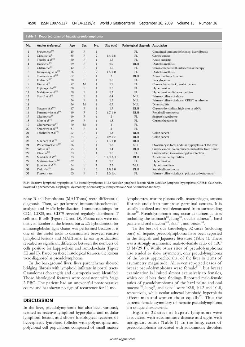
zone B-cell lymphoma (MALToma) were differential diagnosis. Then, we performed immunohistochemical analysis and in situ hybridization. Immunostainings for CD3, CD20, and CD79 revealed regularly distributed T cells and B cells (Figure 5C and D). Plasma cells were not many in number in the lesions, but in situ hybridization for immunoglobulin light chains was performed because it is one of the useful tools to discriminate between reactive lymphoid lesions and MALToma. In situ hybridization revealed no significant difference between the numbers of cells positive for kappa-chain and lambda-chain (Figure 5E and F). Based on these histological features, the lesions were diagnosed as pseudolymphoma.
In the background liver, liver parenchyma showed bridging fibrosis with lymphoid infiltrate in portal tracts. Granulomas cholangitis and ductopenia were identified. Those histological features were consistent with Stage 2 PBC. The patient had an uneventful postoperative course and has shown no sign of recurrence for 11 mo.
DISCUSSIONIn the liver, pseudolymphoma has also been variously termed as reactive lymphoid hyperplasia and nodular lymphoid lesion, and shows histological features of hyperplastic lymphoid follicles with polymorphic and polyclonal cell populations composed of small mature
lymphocytes, mature plasma cells, macrophages, stroma fibrosis and often numerous germinal centers. It is usually localized and well demarcated from surrounding tissue[1]. Pseudolymphoma may occur at numerous sites including the stomach[7], lung[8], ocular adnexa[9], hard palate and oral mucosa[10], skin[11], and breast[12].
To the best of our knowledge, 32 cases (including ours) of hepatic pseudolymphoma have been reported in the English and Japanese literature (Table 1). There was a strongly asymmetric male-to-female ratio of 1:9.7 (3 M/29 F). While other sites of pseudolymphoma also tended to show asymmetry, only pseudolymphoma of the breast approached that of the liver in terms of asymmetry magnitude. All seven reported cases of breast pseudolymphoma were female[12], but breast examination is limited almost exclusively to females, which could bias these findings. Reported male-female ratios of pseudolymphoma of the hard palate and oral mucosa[10], lung[8], and skin[11] were 1:2.8, 1:1.2 and 1:1.8, respectively, while ocular adnexal lymphoid hyperplasia affects men and women about equally[9]. Thus the extreme female asymmetry of hepatic pseudolymphoma is a unique characteristic.
Eight of 32 cases of hepatic lymphoma were associated with autoimmune disease and eight with malignant tumor (Table 1). In the lung, cases of pseudolymphoma associated with autoimmune disorders
www.wjgnet.com
Table 1 Reported cases of hepatic pseudolymphoma
No. Author (reference) Age Sex No. Size (cm) Pathological diagnosis Association
1 Snover et al[32] 15 F 1 PL Combined immunodeficiency, liver fibrosis 2 Grouls et al[1] 85 F 2 1.4, 0.8 PL Gastric cancer 3 Tanabe et al[33] 30 F 1 1.5 PL Acute enteritis 4 Isobe et al[34] 59 F 1 0.9 RLH Diabetes mellitus 5 Ohtsu et al[6] 42 F 1 1.5 PL Chronic hepatitis B, interferon-α therapy 6 Katayanagi et al[35] 66 F 2 1.5, 1.0 PL Diabetes mellitus 7 Tanizawa et al[36] 67 F 1 2 RLH Abnormal liver function 8 Endo et al[37] 38 F 1 1.8 PL Pancytopenia 9 Kim et al[5] 72 M 1 1.7 PL Chronic hepatitis C, gastric cancer10 Fujinaga et al[38] 58 F 1 1.5 PL Hypertension11 Nishijima et al[39] 58 F 1 1.2 PL Hypertension, diabetes mellitus12 Sharifi et al[3] 52 F 1 0.4 NLL Primary biliary cirrhosis13 56 F 1 1.5 NLL Primary biliary cirrhosis, CREST syndrome14 56 M 1 0.7 NLL Diverticulitis15 Nagano et al[40] 47 F 1 1.7 RLH Chronic thyroiditis, high titer of ANA16 Pantanowitz et al[41] 69 F 2 1.7, 1.0 RLH Renal cell carcinoma17 Okubo et al[2] 49 F 1 2 PL Sjögren's syndrome18 Mori et al[42] 49 F 1 1.8 PL Chronic hepatitis B19 Okuhama et al[43] 70 M 1 4 PL20 Shiozawa et al[44] 51 F 1 2 PL21 Takahashi et al[45] 77 F 1 1.5 RLH Colon cancer22 64 F 2 0.9, 0.7 RLH Colon cancer23 Maehara et al[46] 72 F 2 1.3, 1.0 RLH24 Willenbrock et al[47] 36 F 1 1.8 NLL Ovarian cyst, focal nodular hyperplasia of the liver25 Sato et al[4] 75 F 1 1.4 RLH Gastric cancer, colon cancers, metastatic liver tumor27 Ota et al[20] 63 F 1 1.6 PL Gastric ulcer, Helicobacter pylori infection28 Machida et al[48] 53 F 3 1.5, 1.2, 1.0 RLH Autoimmune thyroiditis29 Matsumoto et al[49] 67 F 1 1.5 PL Hypertension30 Jiménez et al[50] 34 F 1 2.3 NLH Hypothyroidism31 Park et al[51] 46 F 2 1.0, 1.0 RLH Renal cell carcinoma32 Present case 63 F 2 1.3, 0,4 PL Primary biliary cirrhosis, primary aldosteronism
RLH: Reactive lymphoid hyperplasia; PL: Pseudolymphoma; NLL: Nodular lymphoid lesion; NLH: Nodular lymphoid hyperplasia; CREST: Calcinosis, Raynaud’s phenomenon, esophageal dysmotility, sclerodactyly, telangiectasia; ANA: Antinuclear antibody.
4590 ISSN 1007-9327 CN 14-1219/R World J Gastroenterol September 28, 2009 Volume 15 Number 36

such as Sjögren’s disease were reported[13], which is similar to the liver. In other organs, several factors are thought to be associated with pseudolymphoma, i.e. Epstein-Barr virus (hard palate and oral mucosa[14]), mechanical stimulation (ear[15]), anticonvulsant drugs (skin[16]) and Helicobacter pylori (H pylori) (stomach[17,18]). Regarding the association of H pylori with gastric pseudolymphoma, successful treatment of the pseudolymphoma by eradication of H pylori has been reported[19]. Ota et al[20] reported a case of hepatic pseudolymphoma in which the diameter decreased after H pylori eradication. In the case reported here, the patient had no H pylori infection.
Transformation of pseudolymphoma to lymphoma has been discussed. Malignant transformation of pseudolymphomas in the lung[21] and stomach[22] have been reported. These reports, however, either predated or did not include the use of immunofluorescent techniques. It is likely that these cases were in fact the early stage of primary lymphoma misinterpreted as benign[8]. On the other hand, evidence of progression from histologically benign, immunohistochemically polyclonal lymphoid infiltrates to malignant lymphoma in cutaneous pseudolymphoma is well delineated in the literature[23]. A potential association between lymphoma and PBC is suggested on the basis of individual reports in the literature[24-29]. In a retrospective study by Panjala et al[30] based on an estimated 2,912 patients evaluated at their institution during a 22-year period, only 13 (an estimated 0.6%) patients were evaluated in referral visits for evidence of lymphoma. Although we could find one case of transformation of hepatic pseudolymphoma into lymphoma[31], we were unable to deduce the long term natural course of hepatic pseudolymphoma since most reported cases of hepatic lymphoma underwent surgical resection.
In conclusion, we have reported a case of hepatic pseudolymphoma associated with PBC. Hepatic pseudo-lymphoma appears unique in its female preponderance and associated diseases. If hypervascular nodules in the liver of female patients with autoimmune disease are found, the possibility of pseudolymphoma should be considered. Cases of hepatic pseudolymphoma should be followed carefully as the exact nature of this disorder is still not fully understood.
REFERENCES1 Grouls V. Pseudolymphoma (inflammatory pseudotumor)
of the liver. Zentralbl Allg Pathol 1987; 133: 565-5682 Okubo H, Maekawa H, Ogawa K, Wada R, Sekigawa I, Iida
N, Maekawa T, Hashimoto H, Sato N. Pseudolymphoma of the liver associated with Sjogren's syndrome. Scand J Rheumatol 2001; 30: 117-119
3 Sharifi S, Murphy M, Loda M, Pinkus GS, Khettry U. Nodular lymphoid lesion of the liver: an immune-mediated disorder mimicking low-grade malignant lymphoma. Am J Surg Pathol 1999; 23: 302-308
4 Sato K, Ueda Y, Yokoi M, Hayashi K, Kosaka T, Katsuda S. Reactive lymphoid hyperplasia of the liver in a patient with multiple carcinomas: a case report and brief review. J Clin Pathol 2006; 59: 990-992
5 Kim SR, Hayashi Y, Kang KB, Soe CG, Kim JH, Yang MK, Itoh H. A case of pseudolymphoma of the liver with chronic
hepatitis C. J Hepatol 1997; 26: 209-2146 Ohtsu T, Sasaki Y, Tanizaki H, Kawano N, Ryu M, Satake
M, Hasebe T, Mukai K, Fujikura M, Tamai M. Development of pseudolymphoma of liver following interferon-alpha therapy for chronic hepatitis B. Intern Med 1994; 33: 18-22
7 Tokunaga O, Watanabe T, Morimatsu M. Pseudolymphoma of the stomach. A clinicopathologic study of 15 cases. Cancer 1987; 59: 1320-1327
8 Holland EA , Ghahremani GG, Fry WA, Victor TA. Evolution of pulmonary pseudolymphomas: clinical and radiologic manifestations. J Thorac Imaging 1991; 6: 74-80
9 Knowles DM, Jakobiec FA, McNally L, Burke JS. Lymphoid hyperplasia and malignant lymphoma occurring in the ocular adnexa (orbit, conjunctiva, and eyelids): a prospective multiparametric analysis of 108 cases during 1977 to 1987. Hum Pathol 1990; 21: 959-973
10 Menasce LP, Shanks JH, Banerjee SS, Harris M. Follicular lymphoid hyperplasia of the hard palate and oral mucosa: report of three cases and a review of the literature. Histopathology 2001; 39: 353-358
11 Baldassano MF, Bailey EM, Ferry JA, Harris NL, Duncan LM. Cutaneous lymphoid hyperplasia and cutaneous marginal zone lymphoma: comparison of morphologic and immunophenotypic features. Am J Surg Pathol 1999; 23: 88-96
12 Maldonado ME, Sierra RD. Pseudolymphoma of the breast: case report and literature review. Mil Med 1994; 159: 469-471
13 Song MK, Seol YM, Park YE, Kim YS, Lee MK, Lee CH, Jeong YJ. Pulmonary nodular lymphoid hyperplasia associated with Sjogren's syndrome. Korean J Intern Med 2007; 22: 192-196
14 Samoszuk M, Ramzi E, Ravel J. Disseminated persistent lymphoid hyperplasia containing Epstein-Barr virus and clonal rearrangements of DNA. Diagn Mol Pathol 1993; 2: 57-60
15 Zilinsky I, Tsur H, Trau H, Orenstein A. Pseudolymphoma of the earlobes due to ear piercing. J Dermatol Surg Oncol 1989; 15: 666-668
16 Harris DW, Ostlere L, Buckley C, Whittaker S, Sweny P, Rustin MH. Phenytoin-induced pseudolymphoma. A report of a case and review of the literature. Br J Dermatol 1992; 127: 403-406
17 Lee EY, Brady L, Yousefzadeh DK, Benya EC. Lymphoid hyperplasia of the stomach caused by Helicobacter pylori: upper gastrointestinal findings. AJR Am J Roentgenol 1999; 173: 362-363
18 Chen XY, Liu WZ, Shi Y, Zhang DZ, Xiao SD, Tytgat GN. Helicobacter pylori associated gastric diseases and lymphoid tissue hyperplasia in gastric antral mucosa. J Clin Pathol 2002; 55: 133-137
19 Weston AP, Campbell DR, McGregor DH, Cherian R. Endoscopic and histologic resolution of gastric pseudo-lymphoma (reactive lymphoid hyperplasia) following treatment with bismuth and oral antibiotics. Dig Dis Sci 1994; 39: 2567-2574
20 Ota H, Isoda N, Sunada F, Kita H, Higashisawa T, Ono K, Sato S, Ido K, Sugano K. A case of hepatic pseudolymphoma observed without surgical intervention. Hepatol Res 2006; 35: 296-301
21 Koss MN , Hochholzer L, Nichols PW, Wehunt WD, Lazarus AA. Primary non-Hodgkin's lymphoma and pseudolymphoma of lung: a study of 161 patients. Hum Pathol 1983; 14: 1024-1038
22 Brooks JJ, Enterline HT. Gastric pseudolymphoma. Its three subtypes and relation to lymphoma. Cancer 1983; 51: 476-486
23 Kulow BF, Cualing H, Steele P, VanHorn J, Breneman JC, Mutasim DF, Breneman DL. Progression of cutaneous B-cell pseudolymphoma to cutaneous B-cell lymphoma. J Cutan Med Surg 2002; 6: 519-528
24 Prabhu RM, Medeiros LJ, Kumar D, Drachenberg CI, Papadimitriou JC, Appelman HD, Johnson LB, Laurin J, Heyman M, Abruzzo LV. Primary hepatic low-grade B-cell
www.wjgnet.com
Okada T et al . Pseudolymphoma of the liver 4591

lymphoma of mucosa-associated lymphoid tissue (MALT) associated with primary biliary cirrhosis. Mod Pathol 1998; 11: 404-410
25 Ye MQ, Suriawinata A, Black C, Min AD, Strauchen J, Thung SN. Primary hepatic marginal zone B-cell lymphoma of mucosa-associated lymphoid tissue type in a patient with primary biliary cirrhosis. Arch Pathol Lab Med 2000; 124: 604-608
26 Ijichi S, Iwata S, Une F, Tara M, Igata A. [B cell malignancy associated with Sjogren syndrome and primary biliary cirrhosis: a case report and review] Rinsho Ketsueki 1987; 28: 911-916
27 de Figueiredo M, Lima M, Macedo G, Ribeiro P. Association of splenic lymphoma with villous lymphocytes and primary biliary cirrhosis in a man. Sangre (Barc) 1996; 41: 262-263
28 Goldin R, Sayer J, Wilkins M, Price P, Thomas H. Primary liver lymphoma associated with primary biliary cirrhosis. Histopathology 1993; 22: 184-185
29 Lizarralde E, Martinez P, Ibanez T, Gutierrez A. Primary hepatic lymphoma and primary biliary cirrhosis. Am J Gastroenterol 2000; 95: 562-563
30 Panjala C, Talwalkar JA, Lindor KD. Risk of lymphoma in primary biliary cirrhosis. Clin Gastroenterol Hepatol 2007; 5: 761-764
31 Sato S, Masuda T, Oikawa H, Satoh T, Suzuki Y, Takikawa Y, Yamazaki K, Suzuki K, Sato S. Primary hepatic lymphoma associated with primary biliary cirrhosis. Am J Gastroenterol 1999; 94: 1669-1673
32 Snover DC , F i l ipovich AH, Dehner LP, Krivi t W. 'Pseudolymphoma'. A case associated with primary immunodeficiency disease and polyglandular failure syndrome. Arch Pathol Lab Med 1981; 105: 46-49
33 Tanabe Y, Yano K, Yoshida Y, Sato T, Sakai K, Koyanagi N. A resectable case of pseudlymphoma of the liver (in Japanese). Jpa J Gastroenterol 1991; 16: 240
34 Isobe H, Sakamoto S, Sakai H, Masumoto A, Sonoda T, Adachi E, Nawata H. Reactive lymphoid hyperplasia of the liver. J Clin Gastroenterol 1993; 16: 240-244
35 Katayanagi K, Terada T, Nakanuma Y, Ueno T. A case of pseudolymphoma of the liver. Pathol Int 1994; 44: 704-711
36 Tanizawa T, Eishi Y, Kamiyama R, Nakahara M, Abo Y, Sumita T, Kawano N. Reactive lymphoid hyperplasia of the liver characterized by an angiofollicular pattern mimicking Castleman's disease. Pathol Int 1996; 46: 782-786
37 Nishijima K, Shimizu Y, Oonishi K, Hasebe K, Tani S, Hashimoto T, Yagi M, Miwa K, Nonomura A. A case of pseudolymphoma of the liver (in Japanese). Acta Hepatol Jpa 1998; 39: 23-27
38 Fujinaga Y, Matsui O, Shimizu K. Reactive lymphoid hyperplasia of the liver (in Japanese). Jpn J Diagn Imaging 1997; 17: 586
39 Nichijima K, Shimizu Y, Oonishi K, Hasebe K, Tani S, Hashimoto T. A case of pseudolymphoma of the liver (in Japanese). Acta Hepatol Jpa 1998; 39: 23-27
40 Nagano K, Fukuda Y, Nakano I, Katano Y, Toyoda H, Nonami T, Nagasaka T, Hayakawa T. Reactive lymphoid hyperplasia of liver coexisting with chronic thyroiditis: radiographical characteristics of the disorder. J Gastroenterol Hepatol 1999; 14: 163-167
41 Pantanowitz L, Saldinger PF, Kadin ME. Pathologic quiz case: Hepatic mass in a patient with renal cell carcinoma. Arch Pathol Lab Med 2001; 125: 577-578
42 Mori M, Koga Y, Dairaku K, Kishikawa M. A case of pseudolymphoma of the liver (in Japanese). Acta Hepatol Jpa 2002; 43: 376-380
43 Okuhama Y, Kenjyo T, Oomine M, Kaneshiro T, Shikiya M. A case of pseudolymphoma of the liver (in Japanese). Shoukakigeka 2003; 26; 1557-1562
44 Shiozawa K, Kinoshita H, Tsuruta H, Nakamura K, Naito S, Koga M, Takeshima F, Omagari K, Mizuta Y, Murata I, Kohno S. [A case of pseudolymphoma of the liver diagnosed before operation] Nippon Shokakibyo Gakkai Zasshi 2004; 101: 772-778
45 Takahashi H, Sawai H, Matsuo Y, Funahashi H, Satoh M, Okada Y, Inagaki H, Takeyama H, Manabe T. Reactive lymphoid hyperplasia of the liver in a patient with colon cancer: report of two cases. BMC Gastroenterol 2006; 6: 25
46 Maehara N, Chijiiwa K, Makino I, Ohuchida J, Kai M, Kondo K, Moriguchi S, Marutsuka K, Asada Y. Segmentectomy for reactive lymphoid hyperplasia of the liver: Report of a case. Surg Today 2006; 36: 1019-1023
47 Willenbrock K, Kriener S, Oeschger S, Hansmann ML. Nodular lymphoid lesion of the liver with simultaneous focal nodular hyperplasia and hemangioma: discrimination from primary hepat ic MALT-type non-Hodgkin’s lymphoma. Virchows Arch 2006; 448: 223-227
48 Machida T, Takahashi T, Itoh T, Hirayama M, Morita T, Horita S. Reactive lymphoid hyperplasia of the liver: a case report and review of literature. World J Gastroenterol 2007; 13: 5403-5407
49 Matsumoto N, Ogawa M, Kawabata M, Tohne R, Hiroi Y, Furuta T, Yamamoto T, Gotoh I, Ishiwata H, Ono Y, Arakawa Y, Kinukawa N. Pseudolymphoma of the liver: Sonographic findings and review of the literature. J Clin Ultrasound 2007; 35: 284-288
50 Jiménez R, Beguiristain A, Ruiz-Montesinos I, Villar F, Medrano MA, Garnateo F, Vaquero J, Elizondo ME. Nodular lymphoid hyperplasia of the liver. Pseudolymphoma. Rev Esp Enferm Dig 2007; 99: 299-306
51 Park HS, Jang KY, Kim YK, Cho BH, Moon WS. Histiocyte-rich reactive lymphoid hyperplasia of the liver: unusual morphologic features. J Korean Med Sci 2008; 23: 156-160
S- Editor Li LF L- Editor Cant MR E- Editor Ma WH
www.wjgnet.com
4592 ISSN 1007-9327 CN 14-1219/R World J Gastroenterol September 28, 2009 Volume 15 Number 36

Seong Hun Kim, In Hee Kim, Sang Wook Kim, Seung-Ok Lee, Division of Gastroenterology and Hepatology, Department of Internal Medicine, Chonbuk National University Medical School, Jeonju 561-712, South KoreaAuthor contributions: Kim SH and Lee SO designed this work; Kim SH, Kim IH, Kim SW and Lee SO analyzed references; Kim SH wrote the paper.Correspondence to: Seung-Ok Lee, MD, Associate Professor, Division of Gastroenterology and Hepatology, Department of Internal Medicine, Chonbuk National University Medical School and Hospital, 634-18, Geum-am dong, Deokjin-gu, Jeonju, Jeonbuk 561-712, South Korea. [email protected] Telephone: +82-63-2501289 Fax: +82-63-2541609Received: June 30, 2009 Revised: July 20, 2009Accepted: July 27, 2009Published online: September 28, 2009
AbstractThis study reports a case of unresectable intrahepatic mass-forming cholangiocarcinoma which showed a dra-matic response to gemcitabine that led to curative resec-tion and a long-term survival of more than five years. Six and five cycles of gemcitabine monotherapy were admin-istered separately over a three-year period and a radical excision was performed at 4.5 years after diagnosis. This case indicates the role of gemcitabine as a neoadjuvant chemotherapeutic agent for cholangiocarcinoma and guarantees a randomized controlled prospective study.
© 2009 The WJG Press and Baishideng. All rights reserved.
Key words: Cholangiocarcinoma; Gemcitabine; Neoadjuvant chemotherapy
Peer reviewer: Wei Tang, MD, EngD, Assistant Professor, H-B-P Surgery Division, Artificial Organ and Transplantation Division, Department of surgery, Graduate School of Medicine, The University of Tokyo, Tokyo 113-8655, Japan
Kim SH, Kim IH, Kim SW, Lee SO. Repetitive response to gemcitabine that led to curative resection in cholangiocarcinoma. World J Gastroenterol 2009; 15(36): 4593-4595 Available from: URL: http://www.wjgnet.com/1007-9327/15/4593.asp DOI: http://dx.doi.org/10.3748/wjg.15.4593
INTRODUCTIONCholangiocarcinoma is a devastating malignancy with a
poor prognosis. Early diagnosis and extensive surgery offers the only chance for cure although other treatment modalit ies have been applied. Response rates to chemotherapy are relatively low although several kinds of chemotherapeutic agents such as gemcitabine are known to be effective. Furthermore, the role of neoadjuvant chemotherapy for unresectable cholangiocarcinoma has not been established either. Here we report the case of a patient with huge intrahepatic mass-forming cholangiocarcinoma who showed repetitive responses to a gemcitabine single regimen, and eventually underwent curative resection that led to long-term survival of more than five years.
CASE REPORTA 54-year-old female patient was admitted with mild epigastric discomfort and a liver mass was detected by ultrasonography in January 2004. She had a history of cholecystectomy due to gallstone disease 20 years ago. She denied any recent history of medication or herbal treat-ments. She had never consumed raw fresh-water fish. An abdominal computed tomography (CT) scan showed a 10 cm sized irregular hypovascular mass occupying the left hepatic lobe and growing inferiorly to compress the gastric antrum and the pancreas (Figure 1A). Invasion of the middle hepatic vein and left portal vein were strongly suggested. Multiple lymph node enlargements were noted at the liver hilum and lesser omentum. Laboratory data showed a white blood cell count of 9560/mm3 (normal 4800-10 800 mm3), hemoglobin 15.8 g/dL (normal 13-18 g/dL), platelet count of 203 000 /mm3, AST 33 IU/L (normal 12-33 IU/L), ALT 20 IU/L (normal 5-35 IU/L), total bilirubin 0.9 mg/dL (normal 0.2-1.2 mg/dL), and albumin 4.6 g/dL (normal 3.5-5.3 g/dL). A tumor marker study revealed AFP 2.3 ng/mL (normal 0-8 ng/mL), CEA 2.2 ng/mL (normal 0-5 ng/mL), and CA19-9 17.6 U/mL (normal 0-36 U/mL). A serologic study showed HBs Ag (-), ANA (-), HCV Ab (+) and HCV RNA PCR (-). Esophagogastroduodenoscopy and colonoscopy were nonspecific except for mild chronic gastritis and a small hyperplasic rectal polyp. An abdominal magnetic reso-nance imaging (MRI) reported T1 low and T2 slightly high lobulated contour solid mass showing a similar invasion pattern with the CT scan. Biliary dilatation at the upper portion of the mass was seen (image not shown). Percu-taneous needle biopsy from the mass reported well differ-entiated cholangiocarcinoma (image not shown). Histo-
Online Submissions: wjg.wjgnet.com World J Gastroenterol 2009 September 28; 15(36): [email protected] World Journal of Gastroenterology ISSN 1007-9327doi:10.3748/wjg.15.4593 © 2009 The WJG Press and Baishideng. All rights reserved.
Repetitive response to gemcitabine that led to curative resection in cholangiocarcinoma
Seong Hun Kim, In Hee Kim, Sang Wook Kim, Seung-Ok Lee
CASE REPORT
www.wjgnet.com

chemical and immunohistochemical study showed tumor cells were positive for CK-7, and negative for CK-19, and CK-20. An extended surgery was recommended to the patient after explaining the degree of tumor invasion and low possibility of curative resection. However, the patient declined any kind of treatment including surgery and chemoradiotherapy. After 3 mo from diagnosis, the patient was persuaded to undertake chemotherapy. Gem-citabine monotherapy was provided as intravenous 2 h infusions of 1000 mg/m2 body surface area weekly every three out of 4 wk. A total of six cycles of chemotherapy were treated tolerably.
The CT scan taken on October 2004 (10 mo after diagnosis) showed that the tumor mass dramatically decreased in size and was smaller (half of the diameter) than that of the previous CT (Figure 1B). Surgery was again recommended to the patient. However, the patient
declined surgery and further chemotherapy. After ending chemotherapy treatment the tumor mass did not show any significant change on a regular CT scan over 3 years and 3 mo except for a little sclerotic change (Figure 1C).
Unfortunately, an abdominal CT scan in January 2008 showed a slight increase of the mass with a symp-tomatic complaint of fatigue (Figure 1D). Gemcitabine treatment was restarted under the same protocol as the previous one. The patient barely finished five cycles of treatment due to various symptoms such as weakness or edema. On follow up CT scan in May 2008, a partial response was repeated with a slight decrease in the mass size (Figure 1E). Radical surgery was recommended and the patient agreed to it. A curative resection of atrophied left liver including the mid-hepatic vein and extrahe-patic bile duct was performed. A hepatico-enterostomy was followed after the resection. Histological findings
www.wjgnet.com
Figure 1 Abdominal CT scan images at two different levels are shown serially. A: At left portal vein level (black arrowhead), a 10 cm sized irregular hypovascular mass (asterisk) occupying left hepatic lobe is shown; B: At lower, main portal vein level (black arrow), the inferiorly grown mass is shown. Invasion of middle hepatic vein and left portal vein were strongly suggested with multiple lymph node enlargements at the liver hilum and lesser omentum; C and D: On CT scan taken after the end of the first 6 cycles (18 times) of gemcitabine chemotherapy, the tumor mass decreased in size dramatically, smaller than half of the previous diameter; E and F: On CT scan on 3 years and 3 mo from diagnosis, mass does not show any significant change except for a little sclerotic change; G and H: On CT scan on 4 years from diagnosis, tumor mass shows re-increment of the size than previous study; I and J: On follow up CT scan after second 5 cycles of gemcitabine chemotherapy, partial response is shown again with slight decrease of the mass size; K and L: On CT scan taken 11 months after R0 resection, there is no any recurrence or new lesion growing.
A C E
B D F
G I K
H J L
↑
*
*
4594 ISSN 1007-9327 CN 14-1219/R World J Gastroenterol September 28, 2009 Volume 15 Number 36

of the resected specimen showed well differentiated cholangiocarcinoma composed of a glandular structure lined by cuboidal mucin-producing epithelium resem-bling cholangiocytes and a dense fibrous stroma with desmotic reaction (Figure 2). All resection margins were negative for malignancy microscopically. One cycle of adjuvant gemcitabine chemotherapy was administered due to concern about remnant tumor cells. On a CT scan taken 11 mo after R0 resection, there was no recur-rence or new growing lesion (Figure 1F). In July 2009, 5 years and 6 mo from diagnosis (1 year from the surgical resection) the patient is living well without any symp-toms.
DISCUSSIONThe establishment of an optimal chemotherapeutic regimen for cholangiocarcinoma is necessary because most of the patients are diagnosed in an advanced stage and curative resection would not be viable. Gemcitabine is known as one of the most effective agents for cholan-giocarcinoma and response rates were reported as 20%-35%[1] for a single regimen[2] or in combination
with other agents such as 5-FU (or capecitabine)[3], mitomycin-C[4], or a platinum analog[5]. This case study showed repetitive responses to a gemcitabine single agent regimen given separately over a long-term interval. Tumors were observed to be strongly repressed for more than three years after the first cycle of treatment.
Studies in neoadjuvant settings for unresectable cholangiocarcinoma are very few although there was a remarkable success of a neoadjuvant protocol with the use of radiotherapy and capecitabine prior to liver transplantation[6]. There was no report about gemcitabine based chemotherapy as a neoadjuvant approach combined with surgical tumor excision. In our case, a very large unresectable intrahepatic mass-forming cholangio-carcinoma showed a definite response to gemcitabine alone that eventually led the patient to a curative resection and long-term survival. This result suggests that gemcitabine based chemotherapy should be tried more actively for unresectable cholangiocarcinoma.
REFERENCES1 Verslype C , Prenen H, Van Cutsem E. The role of
chemotherapy in biliary tract carcinoma. HPB (Oxford) 2008; 10: 164-167
2 Kubicka S, Rudolph KL, Tietze MK, Lorenz M, Manns M. Phase II study of systemic gemcitabine chemotherapy for advanced unresectable hepatobiliary carcinomas. Hepatogastroenterology 2001; 48: 783-789
3 Riechelmann RP, Townsley CA, Chin SN, Pond GR, Knox JJ. Expanded phase II trial of gemcitabine and capecitabine for advanced biliary cancer. Cancer 2007; 110: 1307-1312
4 Kornek GV, Schuell B, Laengle F, Gruenberger T, Penz M, Karall K, Depisch D, Lang F, Scheithauer W. Mitomycin C in combination with capecitabine or biweekly high-dose gemcitabine in patients with advanced biliary tract cancer: a randomised phase II trial. Ann Oncol 2004; 15: 478-483
5 Kim ST, Park JO, Lee J, Lee KT, Lee JK, Choi SH, Heo JS, Park YS, Kang WK, Park K. A Phase II study of gemcitabine and cisplatin in advanced biliary tract cancer. Cancer 2006; 106: 1339-1346
6 Rea DJ, Heimbach JK, Rosen CB, Haddock MG, Alberts SR, Kremers WK, Gores GJ , Nagorney DM. Liver transplantation with neoadjuvant chemoradiation is more effective than resection for hilar cholangiocarcinoma. Ann Surg 2005; 242: 451-458; discussion 458-461
S- Editor Tian L L- Editor O'Neill M E- Editor Ma WH
www.wjgnet.com
Figure 2 Histological finding of resected specimen is shown. Well differentiated adenocarcinoma is seen with glandular structure and a dense fibrous stroma. Scattered lymphocytes are also present (40 × magnification). In higher magnification, tumor is composed of glands lined by cuboidal mucin-producing epithelium resemble biliary epithelium ( Inset, 400 × magnification).
Kim SH et al . Gemcitabine neoadjuvant chemotherapy for cholangiocarcinoma 4595

I-Ming Kuo, Frank Wang, Keng-Hao Liu, Yi-Yin Jan, Department of Surgery, Chang Gung Memorial Hospital, Chang Gung University, Linkou, Taiwan, ChinaAuthor contributions: Kuo IM contributed to manuscript preparation, manuscript editing and was the primary writer of the manuscript; Wang F revised the manuscript; Liu KH contributed to data collection and manuscript review; Jan YY reviewed the manuscript.Correspondence to: Dr. Keng-Hao Liu, Department of Surgery, Chang Gung Memorial Hospital, 5, Fu-Shin Street, Kweishan, Taoyuan, Taiwan, China. [email protected]: +886-3-3281200 Fax: +886-3-3285818Received: July 11, 2009 Revised: August 6, 2009 Accepted: August 13, 2009Published online: September 28, 2009
AbstractGastrectomy is commonly performed for both be-nign and malignant lesions. Although the incidence of post-gastrectomy acute pancreatitis (PGAP) is low compared to other well-recognized post-operative complications, it has been reported to be associated with a high mortality rate. In this article, we describe a 70-year-old man with asymptomatic pancreatic divisum who underwent palliative subtotal gastrectomy for an advanced gastric cancer with liver metastasis. His post-operative course was complicated by acute pancreatitis and intra-abdominal sepsis. The patient eventually succumbed to multiple organ failure despite surgical debridement and drainage, together with aggressive antibiotic therapy and nutritional support. For patients with pancreas divisum or dominant duct of Santorini who fail to follow the normal post-operative course after gastrectomy, clinicians should be alert to the possibility of PGAP as one of the potential diagnoses. Early detection and aggressive treatment of PGAP might improve the prognosis.
© 2009 The WJG Press and Baishideng. All rights reserved.
Key words: Acute Pancreatitis; Gastrectomy; Pancreas divisum; Duct of Santorini; Laparoscopy
Peer reviewers: Jose Sahel, Professor, Hepato-gastroen-terology, Hospital sainti Marevenite, I270 Boolevard AE Sainti Margrenise, Marseille 13009, France; Chikashi Shibata, Department of Surgery, Tohoku University, 1-1 Seiryo-machi, Aoba-ku, Japan
Kuo IM, Wang F, Liu KH, Jan YY. Post-gastrectomy acute pan-creatitis in a patient with gastric carcinoma and pancreas divi-sum. World J Gastroenterol 2009; 15(36): 4596-4600 Available from: URL: http://www.wjgnet.com/1007-9327/15/4596.asp DOI: http://dx.doi.org/10.3748/wjg.15.4596
INTRODUCTIONAlthough gastrectomies for lesions of the stomach and duodenum have been performed safely for more than one hundred years, major complications such as anastomosis leakage, duodenal stump leakage and post-operative bleeding cannot be completely avoided even by experienced surgeons. In the past, most gastrecto-mies were performed for benign gastric or duodenal ulcer. One of the complications associated with these procedures was post-gastrectomy acute pancreati-tis (PGAP). The pathogenesis of this condition is hypothesized to be related to pancreatic parenchymal in-jury secondary to severe adhesion of peripancreatic tis-sue, compromised pancreatic micro-circulation, hyper-pressure of the duodenum, and edema or spasm of the major papilla[1,2]. The incidence of PGAP has decreased in recent years, coinciding with the dramatic reduction in the need for surgical intervention of complicated peptic ulcer disease in the advent of improved medical and endoscopic management. Gastrectomies nowadays are performed mainly for patients with gastric malig-nancy. The “pancreatitis-like” presentation is mostly seen in patients with afferent loop obstruction after gas-trectomies, mostly following Billroth Ⅱ reconstruction. The true PGAP might be related to the extended lymph node dissection and resection of adjacent organs (such as splenectomy and distal pancreatectomy) in radical gastrectomies for gastric cancer[3-5]. However, the cor-relation between pancreatic anomalies and PGAP has not been well documented in the literature. This article, therefore, reports a patient with concurrent advanced gastric cancer and pancreas divisum (PD) who had PGAP and died from multiple organ failure post-opera-tively.
CASE REPORTA 70-year-old gentleman with a medical background of hypertension and asthma, presented with a 3 mo history
Online Submissions: wjg.wjgnet.com World J Gastroenterol 2009 September 28; 15(36): [email protected] World Journal of Gastroenterology ISSN 1007-9327doi:10.3748/wjg.15.4596 © 2009 The WJG Press and Baishideng. All rights reserved.
Post-gastrectomy acute pancreatitis in a patient with gastric carcinoma and pancreas divisum
I-Ming Kuo, Frank Wang, Keng-Hao Liu, Yi-Yin Jan
CASE REPORT
www.wjgnet.com

of poor appetite, 10 kg weight loss, postprandial fullness and nausea. There was, however, no history of tarry stool, hematemesis, fever or abdominal pain. The physical ex-amination did not reveal any palpable abdominal mass, lymphadenopathy, anemia, or jaundice. All laboratory data, including biochemistry examination and hemogram, were within normal ranges except for a low hemoglobin level (10.3 g/dL). Esophagogastroduodenoscopy (EGD) was performed and one ulcerative mass over the posterior wall of antrum was identified (Figure 1). The histopathological examination of the biopsy of the gastric lesion confirmed that it was an adenocarcinoma. Dynamic CT was arranged for pre-operative staging. It revealed several cystic lesions of the liver and possible deformity of the duodenal bulb without definite evidence of metastatic disease (Figure 2). In addition, the gallbladder was contracted with suspicious wall thickening and heterogeneous content. At laparotomy, one unsuspected nodule was seen in segment 3 of the liver. The lesion was confirmed to be a metastatic liver nodule by frozen section. At the time of the operation, severe adhesion between the head of the pancreas and the peripyloric tissue was noted. As a result, a palliative subtotal gastrectomy with Billroth Ⅱ gastrojejunostomy was performed due to partial outlet obstruction.
Post operatively, the patient had low-grade fever and persistent epigastric pain without jaundice, tarry stools, or active upper gastrointestinal hemorrhage. The laboratory tests on the 3rd postoperative day demonstrated a raised white cell count with left shift (11 500/μL), a platelet count of 176 000/μL and a hemoglobin of 9.5 g/dL. Serum biochemistry revealed a normal liver function and renal function but a raised C-reactive protein (CRP) of 193.27 mg/L. Slightly elevated amylase (185 U/L) and normal lipase (39 U/L) level were also noted. Chest X ray and urinary analysis were obtained and disclosed no pneu-monia or urinary tract infection. Although the drainage fluid was clear, there was some yellowish and cloudy discharge from the abdominal wound. We collected the fluid for microbial analysis only and culture, which grew a yeast-like organism. Fever persisted despite the administration of intravenous antibiotics. Abdominal CT arranged on the 8th postoperative day demonstrated ir-regular pancreatic contours with diffuse enlargement and fluid accumulation in the lesser sac with retroperitoneal extension (Figure 3), suggestive of either an acute pan-creatitis or duodenal stump leakage. The laboratory tests on the 10th postoperative day demonstrated a white cell count with marked left shift (8400/μL, 86% in segment form and 10% in band form), a decreased platelet count of 58 000/μL, a raised CRP of 247.95 mg/L, and normal serum level of pancreatic enzymes (amylase level was 79 U/L and lipase level was 26 U/L). In view of continuing clinical deterioration associated with peritonitis and intra-abdominal sepsis from a possible anastomosis leak rather than acute pancreatitis (according to the normal amylase and lipase levels), the patient underwent an exploratory laparotomy instead of percutaneous drainage of the fluid 10 d after the first operation. At laparotomy, colorless turbid fluid accumulation was found at the same regions as reported on the abdominal CT scan. There was no
evidence of bile leak, and the duodenal stump and anasto-mosis were intact. The necrotic tissues around the cavity of the loculated fluid collection had a similar appearance to fat necrosis in acute pancreatitis. Limited debridement was performed and a sump drain tube was placed for post-operative irrigation and drainage.
Bile-stained fluid was noted in the sump drain 2 d after the second operation, and the biochemistry study of the drainage fluid disclosed lower amylase and lipase levels (14 U/L and 9 U/L). Blood culture grew Bacteroides fragilis and hemogram showed progressive pancytopenia. A repeat CT scan suggested possible duodenal stump leakage with abscess formation (Figure 4). Despite total parenteral nutrition, hemodialysis for acute renal failure, and sump drain irrigation-drainage with intravenous antibiotics for septicemia and intra-abdominal infection, the patient eventually died of profound septic shock and multiple organ failure on day 30 after the first operation. A retrospective review of the patient’s abdominal CT demonstrated a previously undiagnosed, asymptomatic PD (Figure 5).
DISCUSSIONGastrectomy is a commonly practiced procedure for both benign and malignant lesions of the stomach. Major complications such as postoperative bleeding, anastomotic leak and delayed gastric emptying are well documented[6]. In the past, most gastrectomies were performed for benign gastric or duodenal ulcer. The incidence of PGAP might be as high as 40.8% and the mortality rates in this group of patients ranged from 12.6% to 62.5%[1,2]. Recently, gastrectomies have mostly been performed for gastric malignancies, and the com-plication and mortality rates are much less because of improved surgical technique and postoperative care. The incidence of PGAP nowadays, although difficult to ac-curately estimate, has been reported to be less than 5%[7]. The mortality rate, however, can be up to 33.3%-50%[8,9] and is higher than acute pancreatitis of other etiolo-gies[10]. Chen et al[3] reported a higher incidence of PGAP in patients having total gastrectomy compared with other types of gastrectomies (7.4% vs 0.8%), with a 33.3% mortality rate. Another study reported higher
Kuo IM et al . Post-gastrectomy acute pancreatitis 4597
www.wjgnet.com
Figure 1 The esophagogastroduodenoscopy (EGD) showed an ulcerated, annular lesion over the gastric antrum.

PGAP rate in patients undergoing extended lymph node dissection[4]. Despite the various advantages of laparoscopically assisted gastrectomy (LAG), the rates of acute pancreatitis after LAG have also been shown to range from 0.7% to 2.3%[8,9,11]. The poorer prognosis of PGAP might be related to postoperative immuno-suppression, increased pancreatitis-related hemorrhage because of extensive soft tissue/lymph node dissection, or anastomotic leak secondary to necrotizing pancreatitis
and intra-abdominal sepsis. PGAP may be a result of several factors. The stom-
ach/duodenum might adhere to the underlying pancreas because of severe peptic ulceration or desmoplastic change from malignancy. Sometimes, the pancreas might actually be the base of the ulcer. Injury to the pancreatic parenchyma by electrocauterization or traction, microcir-culation compromise, and direct injury to the pancreatic duct might occur during gastrectomy[7,11]. Other causes
www.wjgnet.com
Figure 2 Preoperative abdominal CT images in coronary and transverse sections. The white solid arrows indicate diffuse wall thickening at the gastric antrum. The white dotted arrow indicates a contracted gallbladder with eccentric wall thickening. The black solid arrows point to the apparent deformity of the duodenal bulb with adhesion to the head of the pancreas. There is no evidence of intra-abdominal metastasis in this study.
←
←
←
←←
←
←←
Figure 3 Abdominal CT 8 d after the first operation demonstrates massive fluid accumulation in the peripancreatic area and the lesser sac. The homogenous fluid extends to the retroperitoneal space. The status of the duodenal stump (black arrow) cannot be clearly assessed. The pancreas is well enhanced and enlarged, and the head shows an uneven and infiltrative margin (white arrow).
←
←
Figure 4 Follow-up abdominal CT after the second operation. Severe fat stranding is seen. An edematous duodenal stump is observed without a clear fat plane surrounding it. This is highly suspicious of a stump leak (black arrow). The loculated f lu id w i th he terogeneous changes indicates the possibility of an abscess formation (white arrow).
4598 ISSN 1007-9327 CN 14-1219/R World J Gastroenterol September 28, 2009 Volume 15 Number 36

of PGAP such as duodenal hyperpressure[1] and post-operative spasm of the major papilla[2] have been hy-pothesized, but there is a general lack of evidence in the literature to substantiate these theories. Although some studies suggested insertion of a nasogastric tube or a duodenostomy tube after gastrectomy for decom-pression and releasing duodenal hyperpressure, Hsu et al[12] reported no difference in PGAP in spite of NG tube insertion. On the contrary, the “pancreatitis-like” presentation (such as hyperamylasemia and epigastric pain) caused by afferent loop obstruction can be cured under careful management of operations or decompres-sion procedures[7,13]. The condition should be taken into consideration because the prognosis, timing of interven-tion and treatment options are far different from PGAP without A-loop obstruction.
The diagnosis of acute pancreatitis is mainly de-pendent on suggestive clinical features and laboratory studies. Physical examination may be variable and non-specific in postoperative patients because of pain and symptoms related to the operation. Elevated lipase levels 3-fold or more above the normal range appearing within 48 h is the most reliable test[14]. However, normal serum pancreatic enzyme levels cannot exclude acute pancre-atitis absolutely[14]. Abdominal CT scan is particularly helpful in making a definitive diagnosis and excluding other differential diagnoses such as anastomotic leak, intra-abdominal abscess and hemorrhage[15]. However, the anatomical distortion post-operatively often makes the interpretation of CT images difficult[16]. This is ex-emplified by the present case report whereby a definitive diagnosis of PGAP was delayed because of equivocal pancreatic enzymes and non-specific CT findings of intra-abdominal fluid collection.
PD is a common congenital anomaly of the pancreas, with an incidence rate of 4% to 10%[17-19]. Anatomically, the dorsal duct (duct of Santorini) becomes the domi-nant duct draining the majority of the pancreatic juice through the minor papilla. The ventral duct (duct of Wirsung) does not fuse with the dorsal duct and only drains a small portion of the pancreatic head through the major papilla. The diagnosis of PD is made with
endoscopic retrograde pancreatography (ERP) or mag-netic resonance pancreatography (MRP). However, with the advent of multi-detector CT scans, high sensitivity and specificity for diagnosis of PD can be achieved by CT images in some particular conditions[20-22]. The CT features consistent with PD include the presence of the dominant dorsal duct sign (the dorsal duct being larger than the ventral duct, or a missing ventral duct) and the miss-communication between the two pancreatic ducts[20]. Both of these features were observed on the patient’s CT images retrospectively (Figure 5). About 5% to 45.5% of patients with PD present with pancre-atitis or chronic abdominal pain[23,24]. The pathogenesis of this is attributable to the “relative stenosis” of the minor papilla as it drains the majority of the pancreatic juice through a small opening[24], resulting in an increase in the intra-ductal pressure of the dorsal duct system (20-28 mmHg vs 8-14 mmHg in normal pressure of major papilla)[25]. In addition, the unusual anatomical ar-rangement of the sphincter of the minor papilla could also contribute to the development of intra-ductal hypertension leading to pancreatitis[26]. Kamisawa et al[27] reported the high incidence of pancreatitis or pancreat-ic-type abdominal pain in patients with dominant duct of Santorini but without pancreas divisum.
According to these findings, it is reasonable to suppose that patients with PD might be more sensitive to minor injury of the pancreas and disturbance of its microcircu-lation. Once developed, it might progress precipitously to a more severe form and patients are slower to gain full recovery. Prophylactic administration of octreotide in the post-operative setting has been suggested in an attempt to prevent the development of PGAP[7]. The roles of preop-eratively prophylactic cannulation of minor papilla, stent placement, and sphincterotomy have not been reported yet. The potential benefits of these invasive procedures in patients with PD or dominant duct of Santorini undergoing gastrectomy warrant further investigation.
In conclusion, PGAP is a less frequent post-gastrec-tomy complication in the current era. However, diagno-sis and intervention should be made as early as possible because of the relatively high mortality rate associated with PGAP. Theoretically, patients with pancreas divisum or dominant duct of Santorini might connect to PGAP but the absolute relationship should be explored. Although serum pancreatic enzymes play an important role in the diagnosis of acute pancreatitis, imaging study such as an abdominal CT scan is particularly helpful in patients at high risk for PGAP. The roles and benefits of prophylactic octreotide, preoperative cannulation/stenting of the minor papilla, and sphincterotomy of minor papilla have not been well documented and would therefore warrant further investigation.
REFERENCES1 Bacchini I, Martino G, Falaschi CF, Viti M, Sammartano
C, Mantovani R. [Postoperative acute pancreatitis (PAP). Direct personal experience] Minerva Chir 1980; 35: 421-427
2 Lubianskiĭ VG, Nasonov SV. [Acute pancreatitis after resection of stomach for low duodenal ulcer] Khirurgiia
www.wjgnet.com
Figure 5 The preoperative CT scan. The dominant dorsal duct (duct of Santorini) drains into the minor papilla (white arrow). The CBD is seen in the section (black arrow) but the ventral duct (duct of Wirsung) can not be traced. These features are consistent with pancreas divisum.
←←
Kuo IM et al . Post-gastrectomy acute pancreatitis 4599

(Mosk) 2001; 8-113 Chen MM, Zhu ZG, Yan M, Chen J, Xiang M, Li C, Zhang
J, Yao XX, Yang QM. Etiology and management of early postoperative severe acute pancreatitis following radical gastrectomy. Shanghai Jiaotong Daxue Xuebao (Yixueban) 2007; 27: 566-568
4 Li FN, Chen D, Wang HY. Acute pancreatitis following radical gastrectomy for carcinoma of the stomach: Its features, prevention, and treatment. Qingdao Daxue Yixueyuan Xuebao 2003; 39: 259-267
5 Doglietto GB, Pacelli F, Caprino P, Bossola M, Di Stasi C. Pancreas-preserving total gastrectomy for gastric cancer. Arch Surg 2000; 135: 89-94
6 Siewert JR, Bumm R. Distal gastrectomy with Billroth I, Billroth II or Roux-Y reconstruction. In: Fischer JE, Bland KI, editors. Mastery of Surgery. 5th ed. Philadelphia: Lippincott Williams and Wilkins; 2007: 849-860
7 Soybel DI, Zinner MJ. Complications following gastric operations. In: Zinner MJ, Schwartz SI, editors. Maingot’s Abdominal Operations. 10th ed. Stamford, CT: Appleton and Lange; 1997: 1029-1056
8 Park JM, Jin SH, Lee SR, Kim H, Jung IH, Cho YK, Han SU. Complications with laparoscopically assisted gastrectomy: multivariate analysis of 300 consecutive cases. Surg Endosc 2008; 22: 2133-2139
9 Ibanez Aguirre FJ, Azagra JS, Erro Azcarate ML, Goergen M, Rico Selas P, Moreno Elola-Olaso A, Clemares de Lama M, de Simone P, Echenique Elizondo MM. Laparoscopic gastrectomy for gastric adenocarcinoma. Long-term results. Rev Esp Enferm Dig 2006; 98: 491-500
10 Tonsi AF, Bacchion M, Crippa S, Malleo G, Bassi C. Acute pancreatitis at the beginning of the 21st century: the state of the art. World J Gastroenterol 2009; 15: 2945-2959
11 Bo T, Zhihong P, Peiwu Y, Feng Q, Ziqiang W, Yan S, Yongliang Z, Huaxin L. General complications following laparoscopic-assisted gastrectomy and analysis of techniques to manage them. Surg Endosc 2009; 23: 1860-1865
12 Hsu SD, Yu JC, Chen TW, Chou SJ, Hsieh HF, Chan DC. Role of Nasogastric Tube Insertion after Gastrectomy. Chir Gastroenterol 2007; 23: 303-306
13 Kim HJ, Kim JW, Kim KH, Jo KW, Hong JH, Baik SK, Kim HS. [A case of afferent loop syndrome treated by endoscopic drainage procedure using nasogastric tube] Korean J Gastroenterol 2007; 49: 173-176
14 Cartier T, Sogni P, Perruche F, Meyniard O, Claessens YE,
Dhainaut JF, Der Sahakian G. Normal lipase serum level in acute pancreatitis: a case report. Emerg Med J 2006; 23: 701-702
15 Steer ML. Exocrine pancreas. In: Townsend CM, Beauchamp RD, editors. Sabiston Textbook of Surgery. 17th ed. Philadelphia: Elsevier Saunders; 2004: 1643-1678
16 Kim KW, Choi BI, Han JK, Kim TK, Kim AY, Lee HJ, Kim YH, Choi JI, Do KH, Kim HC, Lee MW. Postoperative anatomic and pathologic f indings at CT following gastrectomy. Radiographics 2002; 22: 323-336
17 Agha FP, Williams KD. Pancreas divisum: incidence, detection, and clinical significance. Am J Gastroenterol 1987; 82: 315-320
18 Delhaye M, Engelholm L, Cremer M. Pancreas divisum: congenital anatomic variant or anomaly? Contribution of endoscopic retrograde dorsal pancreatography. Gastroenterology 1985; 89: 951-958
19 Millbourn E. On the excretory ducts of the pancreas in man, with special reference to their relations to each other, to the common bile duct and to the duodenum. Acta Anat (Basel) 1950; 9: 1-34
20 Soto JA, Lucey BC, Stuhlfaut JW. Pancreas divisum: depiction with multi-detector row CT. Radiology 2005; 235: 503-508
21 Anderson SW, Soto JA. Pancreatic duct evaluation: accuracy of portal venous phase 64 MDCT. Abdom Imaging 2009; 34: 55-63
22 Itoh S, Takada A, Satake H, Ota T, Ishigaki T. Diagnostic value of multislice computed tomography for pancreas divisum: assessment with oblique coronal reconstruction images. J Comput Assist Tomogr 2005; 29: 452-460
23 Varshney S, Johnson CD. Pancreas divisum. Int J Pancreatol 1999; 25: 135-141
24 Gregg JA. Pancreas divisum: its association with pancreatitis. Am J Surg 1977; 134: 539-543
25 Staritz M, Meyer zum Buschenfelde KH. Elevated pressure in the dorsal part of pancreas divisum: the cause of chronic pancreatitis? Pancreas 1988; 3: 108-110
26 Valverde Barbato de Prates NE , Smanio T, De Maio Domingos M, Ferraz de Carvalho CA. "Sphincter" of the minor papilla of the human duodenum. Clin Anat 1996; 9: 34-40
27 Kamisawa T, Egawa N, Nakajima H, Okamoto A. Clinical and radiological findings in dominance of Santorini's duct. Digestion 2004; 70: 146-151
S- Editor Li LF L- Editor O'Neill M E- Editor Ma WH
www.wjgnet.com
4600 ISSN 1007-9327 CN 14-1219/R World J Gastroenterol September 28, 2009 Volume 15 Number 36

www.wjgnet.com
CASE REPORT
Hemorrhagic hepatic cysts mimicking biliary cystadenoma
You-Lei Zhang, Lei Yuan, Feng Shen, Yi Wang
Online Submissions: wjg.wjgnet.com World J Gastroenterol 2009 September 28; 15(36): [email protected] World Journal of Gastroenterology ISSN 1007-9327doi:10.3748/wjg.15.4601 © 2009 The WJG Press and Baishideng. All rights reserved.
You-Lei Zhang, Lei Yuan, Feng Shen, Yi Wang, Eastern Hepatobiliary Surgery Hospital, Second Military Medical University, Shanghai 200438, ChinaAuthor contributions: Zhang YL and Yuan L contributed equally to this work; Yuan L and Shen F analyzed the data; Zhang YL and Wang Y wrote the paper.Correspondence to: Yi Wang, MD, Eastern Hepatobiliary Surgery Hospital, The Second Military Medical University, Shanghai 200438, China. [email protected] Telephone: +86-21-81875512 Fax: +86-21-81875511Received: July 27, 2009 Revised: August 17, 2009Accepted: August 24, 2009Published online: September 28, 2009
AbstractThe hemorrhagic simple hepatic cyst is extremely rare and can sometimes be confused with biliary cystadenoma or cystadenocarcinoma. Here we present two cases of huge hemorrhagic simple hepatic cysts. Case 1 was a 43-year-old man with a cystic lesion measuring 13 cm × 12 cm in the right hepatic lobe. Ultrasound and computed tomography showed several mural nodules on the irregularly thickened wall and high-density straps inside the cyst. Case 2 was a 60-year-old woman with a huge cyst measuring 15 cm × 14 cm in the central liver. Ultrasound and magnetic resonance imaging showed the cystic wall was unevenly thickened and there were some flame-like prominences on the wall. The iconographic representations of the two cases mimicked biliary cystadenoma. Cystectomy and left hepatectomy were performed for the two patients, respectively. Both patients recovered quickly after their operations and showed no recurrence.
© 2009 The WJG Press and Baishideng. All rights reserved.
Key words: Simple hepatic cyst; Intracystic hemorrhage; Biliary cystadenoma; Diagnosis; Treatment
Peer reviewer: Hiroshi Yoshida, MD, First Department of Surgery, Nippon Medical School, 1-1-5 Sendagi, Bunkyo-ku, Tokyo 113-8603, Japan
Zhang YL, Yuan L, Shen F, Wang Y. Hemorrhagic hepatic cysts mimicking biliary cystadenoma. World J Gastroenterol 2009; 15(36): 4601-4603 Available from: URL: http://www.wjgnet.com/1007-9327/15/4601.asp DOI: http://dx.doi.org/10.3748/wjg.15.4601
INTRODUCTIONIntrahepatic cysts are generally classified as congenital cyst, traumatic cyst, infectious cyst, parasitic cyst, or neoplastic cyst. Congenital hepatic cysts include simple hepatic cysts and adult polycystic liver disease. A few simple hepatic cysts can reach large sizes and occasional complications, such as rupture, infection, hemorrhage, obstructive jaundice, or portal hypertension may occur.
Intracyst ic hemor rhage is an extremely rare complication of simple hepatic cysts and few cases have been reported worldwide. The hemorrhage usually occurs in solitary huge hepatic cysts in older patients. Massive bleeding into the cysts can significantly enlarge the cysts in a short time, which might partly contribute to the huge size of the cysts. The clinical manifestations of the hemorrhagic hepatic cysts are usually lack of specificity and the iconographic representations often mimic biliary cystadenoma or cystadenocarcinoma. Therefore, it is sometimes very difficult to make a precise diagnosis and select the appropriate treatment for this disease in clinical practice.
Here we present two cases of huge hemorrhagic simple hepatic cysts mimicking biliary cystadenoma, and discuss the diagnosis and the treatment of the disease.
CASE REPORTCase 1 was a 43-year-old man admitted to hospital complaining of right upper quadrant abdominal pain for seven days. No positive clinical signs were found upon physical examination. The patient’s serum carbohydrate antigen 19-9 (CA 19-9) concentration was four times higher than normal. Abdominal ultrasound examination showed a liquid anechoic area measuring 13 cm × 12 cm in right hepatic lobe. The cyst had an irregularly thickened wall with several mamelons on it. There were flocculation echoes inside the cyst. A computed tomography (CT) scan showed a low density cystic lesion with high density straps inside. The thickened cyst wall and mural nodules were not enhanced on contrast CT (Figure 1).
Case 2 was a 60-year-old woman admitted to hospital with a huge cystic liver mass found by regular ultrasound examination without any symptoms. The mass could be partly palpated below the right costal margin. The patient’s serum concentration of CA 19-9 was normal. Abdominal ultrasound examination showed

www.wjgnet.com
a huge echoless cyst measuring 15 cm × 14 cm in the central liver. The margin of the cyst was clear but its wall was unevenly thickened. There were some flame-like prominences on the wall. MRI showed the cyst content was liquid; the flame-like prominences were seen but not enhanced with contrast (Figure 2).
It was difficult to make a diagnosis in either case preoperatively. Biliary cystadenoma was highly suspected. Operations were performed for both cases. Case 1 received a cystectomy and case 2 received a left hepatectomy. The pathological diagnosis was hepatic simple cysts accompanied by intracystic hemorrhage. The mural nodules and prominences on the cyst wall were blood clots. Both patients recovered quickly after their operations and have been alive for six years and two years, respectively.
DISCUSSIONThe clinical manifestation of this disease is lack of specificity. Generally the patients might have acute or chronic upper abdomen pain, but some patients have no symptoms[1-4]. Extra attention should be paid to patients whose symptoms are relative to the severe complications caused by the intracystic hemorrhage. For example, Iguchi et al[5] reported a 76-year-old man with severe edema of both lower extremities and a CT scan revealed an enlarged hemorrhagic hepatic cyst compressing inferior vena cava (IVC) in which massive thromboses formed. Buyse et al[6] reported a patient with simple hepatic cyst developed acute dyspnoea. It was found that the intracystic bleeding led to the enlargement of the cyst and compression of the IVC, followed by pulmonary embolism and dyspnoea. The clinical signs of hemorrhagic simple hepatic cysts depend on the size, location, and complications of the cysts, in which abdominal mass is more common.
The iconographic representations of hemorrhagic hepatic cyst sometimes mimic biliary cystadenoma or cystadenocarcinoma. The common manifestation on ultrasound includes hepatic cystic mass, hyperechoic intracystic straps, and irregularly thickened cyst wall with flame-like prominences or convex palliates. The mural nodules can sometimes be seen to be mildly enhanced
on contrast enhanced CT and MRI[2], which increases the difficulty in differential diagnosis with cystic neoplasms. In fact, as proved by surgery and histological examination, the mural nodules are usually blood clots or organized hematomas[1,2]. In our cases, although the mural nodules and prominences on the cyst wall were not enhanced on contrast CT and MRI, the suspicion of biliary cystadenoma could not be precluded.
The distinction between hemorrhagic simple hepatic cysts and biliary cystic neoplasms can prove difficult to determine by clinical and iconographic features alone. For differential diagnosis, percutaneous transhepatic aspiration might be helpful if serosanguineous fluid is obtained[4]. However, the puncture should be forbidden if malignancy is highly suspected. Naganuma et al [7] reported that contrast-enhanced ultrasound clearly showed microbubbles oozing from the cyst wall into the cyst cavity in a case of hepatic cyst with intracystic bleeding, suggesting contrast-enhanced sonography might be a useful diagnostic tool for the disease. Akiyama et al[8] also reported that Levovist ultrasonography imaging could play an important role in the correct diagnosis of a simple hemorrhagic cyst, by demonstrating the avascularity of the visualized intracystic structures. Horsmans et al[9] found in a four-patient small group that both serum and cystic fluid CA 19-9 levels were elevated in the two patients with cystadenoma or cystadenocarcinoma, but remained normal in the other two patients with hemorrhagic simple cyst. This suggested that the determination of serum and cyst fluid CA 19-9 levels might be of help in distinguishing between hemorrhagic simple cyst and cystadenoma or cystadenocarcinoma. However, other studies revealed that the CA 19-9 levels in neither serum nor cyst fluid had a relationship to the benign or malignant property of hepatic cystic lesions[10-12]. In the present two cases, the serum CA 19-9 level was high in one case but normal in the other case, which seems to agree with the latter standpoint. Except for biliary cystadenoma and cystadenocarcinoma, other hepatic lesions such as hemangioma[13], abscess, and parasitic cyst should also be differentially diagnosed.
Treatment for the hemorrhagic simple hepatic cysts should be active because intracystic bleeding can enlarge
Figure 2 MRI (T2 weighted sequence) showing a huge cyst in the central liver with liquid content and flame-like prominences inside.
Figure 1 CT scan showing a huge low density cystic lesion in the right liver with high density straps (arrowhead) inside and mural nodules (arrow) on the cyst wall.
4602 ISSN 1007-9327 CN 14-1219/R World J Gastroenterol September 28, 2009 Volume 15 Number 36

www.wjgnet.com
the cysts significantly and cause severe complications, such as rupture, infection, and compression of the IVC leading to venous return obstruction or thrombosis. For poorly conditioned patients, transcatheter arterial embolization, transhepatic cyst drainage, intracystic ethanol injection or noninvasive methods are indicated. These methods can obtain therapeutic effect in some patients, though the bleeding may recur afterwards[3-5,14,15]. Surgery should be performed for most well conditioned patients, especially those whose diagnosis does not preclude malignancy. The operations can include partial hepatectomy, cystectomy, and fenestration. Partial hepatectomy and cystectomy can remove the whole cyst and reach curative effect[1-4,8,9,11,13,16]. Occasionally, a fenestration operation is performed to simplify the procedure, which can also obtain a good therapeutic effect[6].
REFERENCES1 Kitajima Y, Okayama Y, Hirai M, Hayashi K, Imai H,
Okamoto T, Aoki S, Akita S, Gotoh K, Ohara H, Nomura T, Joh T, Yokoyama Y, Itoh M. Intracystic hemorrhage of a simple liver cyst mimicking a biliary cystadenocarcinoma. J Gastroenterol 2003; 38: 190-193
2 Hagiwara A, Inoue Y, Shutoh T, Kinoshita H, Wakasa K. Haemorrhagic hepatic cyst: a differential diagnosis of cystic tumour. Br J Radiol 2001; 74: 270-272
3 Ishikawa H, Uchida S, Yokokura Y, Iwasaki Y, Horiuchi H, Hiraki M, Kinoshita H, Shirouzu K. Nonparasitic solitary huge liver cysts causing intracystic hemorrhage or obstructive jaundice. J Hepatobiliary Pancreat Surg 2002; 9: 764-768
4 Yoshida H, Onda M, Tajiri T, Mamada Y, Taniai N, Uchida E, Arima Y, Akimaru K, Yamashita K. Intracystic hemorrhage of a simple hepatic cyst. Hepatogastroenterology 2002; 49: 1095-1097
5 Iguchi S, Kasai A, Kishimoto H, Suzuki K, Ito S, Ogawa Y, Nishi S, Gejyo F, Ohno Y. Thrombosis in inferior vena cava (IVC) due to intra-cystic hemorrhage into a hepatic local cyst with autosomal dominant polycystic kidney disease (ADPKD). Intern Med 2004; 43: 209-212
6 Buyse S, Asselah T, Vilgrain V, Paradis V, Sauvanet A, Consigny Y, Dufour V, Fantin B, Valla D, Marcellin P. Acute pulmonary embolism: a rare complication of a large non-parasitic hepatic cyst. Eur J Gastroenterol Hepatol 2004; 16: 1241-1244
7 Naganuma H, Funaoka M, Fujimori S, Ishida H, Komatsuda T, Yamada M, Furukawa K. Hepatic cyst with intracystic bleeding: contrast-enhanced sonographic findings. J Med Ultrasonics 2006; 33: 105-107
8 Akiyama T, Inamori M, Saito S, Takahashi H, Yoneda M, Fujita K, Fujisawa T, Abe Y, Kirikoshi H, Kubota K, Ueda M, Tanaka K, Togo S, Ueno N, Shimada H, Nakajima A. Levovist ultrasonography imaging in intracystic hemorrhage of simple liver cyst. World J Gastroenterol 2008; 14: 805-807
9 Horsmans Y, Laka A, Gigot JF, Geubel AP. Serum and cystic fluid CA 19-9 determinations as a diagnostic help in liver cysts of uncertain nature. Liver 1996; 16: 255-257
10 Iwase K, Takenaka H, Oshima S, Yagura A, Nishimura Y, Yoshidome K, Tanaka T. Determination of tumor marker levels in cystic fluid of benign liver cysts. Dig Dis Sci 1992; 37: 1648-1654
11 Yamaguchi M, Kuzume M, Matsumoto T, Matsumiya A, Nakano H, Kumada K. Spontaneous rupture of a nonparasitic liver cyst complicated by intracystic hemorrhage. J Gastroenterol 1999; 34: 645-648
12 Park KH, Kim JS, Lee JH, Kim HJ, Kim JY, Yeon JE, Park JJ, Byun KS, Bak YT, Lee CH. [Significances of serum level and immunohistochemical stain of CA19-9 in simple hepatic cysts and intrahepatic biliary cystic neoplasms] Korean J Gastroenterol 2006; 47: 52-58
13 Uchiyama T, Akahane T, Watanabe M, Kitayama T, Ise H. [Case of giant liver cyst with angiogenesis mimicking hemangioma that was difficult to differentiate from cystadenocarcinoma of the liver] Nippon Shokakibyo Gakkai Zasshi 2008; 105: 1634-1639
14 Kanazawa A, Yoshioka Y, Inoi O, Kubo S, Kinoshita H. Intracystic hemorrhage with spontaneous rupture of liver cyst complicated by infection: a case report. Osaka City Med J 2003; 49: 57-60
15 Zanen AL, van Tilburg AJ. Bleeding into a liver cyst can be treated conservatively. Eur J Gastroenterol Hepatol 1995; 7: 91-93
16 Takahashi G, Yoshida H, Mamada Y, Taniai N, Bando K, Tajiri T. Intracystic hemorrhage of a large simple hepatic cyst. J Nippon Med Sch 2008; 75: 302-305
S- Editor Li LF L- Editor Stewart GJ E- Editor Lin YP
Zhang YL et al . Hemorrhagic hepatic cysts 4603
ACKNOWLEDGMENTS
Acknowledgments to reviewers of World Journal of Gastroenterology
Many reviewers have contributed their expertise and time to the peer review, a critical process to ensure the quality of World Journal of Gastroenterology. The editors and authors of the articles submitted to the journal are grateful to the following reviewers for evaluating the articles (including those published in this issue and those rejected for this issue) during the last editing time period.
Dr. Sk Md Fazle Akbar, Assistant ProfessorThird Department of Internal Medicine, Ehime University School of Medicine, Shigenobu-Cho, Ehime 791-0295, Japan
Mark Bloomston, MD, FACS, Assistant Professor of SurgeryDivision of Surgical Oncology, N924 Doan Hall, 410W.10th
Avenue, Columbus, Ohio 43082, United States
Roberto Berni Canani, MD, PhD, ProfessorDepartment of Pediatrics, University of Naples Federico II, via S.Pansini 5 80131, Naples, Italy
Dr. Yogesh K Chawla, ProfessorDepartment of Hepatology, Postgraduate Institute of Medical Education and Research, Chandigarh 160012, India
Dr. Olivier DetryDepartment of Abdominal Surgery and Transplantation, University of Liège, CHU Sart Tilman B35, B-4000 Liège, Belgium
William DickeyAltnagelvin Hospital, Londonderry, BT47 6SB, Northern Ireland, United Kingdom
Isabel Fabregat, PhD, Associate ProfessorLaboratori d’Oncologia Molecular, Institut d’Investigación Bio-mèdica de Bellvitge , Gran Via, Km 2, 7, L’Hospitalet, 08907 Bar-celona, Spain
Kazuma Fujimoto, ProfessorDepartment of Internal Medicine, Saga Medical School, Nabeshi-ma, Saga, Saga 849-8501, Japan
Guiu Boris Guiu, MDDepartment of Radiology, CHU le Bocage, Bd Maréchal de Lattre de Tassigny, 21000 Dijon, France
Keiji Hirata, MDSurgery 1, University of Occupational and Environmental Health, 1-1 Iseigaoka, Yahatanishi-ku, Kitakyushu 807-8555, Japan
Toru Ishikawa, MDDepartment of Gastroenterology, Saiseikai Niigata Second Hospi-tal, Teraji 280-7, Niigata, Niigata 950-1104, Japan
Ezio Laconi, MD, PhD, Professor of General PathologyDepartment of Sciences and Biomedical Technologies, Unit of Experimental Pathology, University of Cagliari, Via Porcell, 4-IV Piano, 09125-Cagliari, Italy
James Neuberger, ProfessorLiver Unit, Queen Elizabeth Hospital, Birmingham B15 2TH, United Kingdom
Dr. Jean RosenbaumInserm E362, Universite Victor Segalen Bordeaux 2, Bordeaux 33076, France
Alain L Servin, PhDFaculty of Pharmacy, French National Institute of Health and Medical Research, Unit 756, Rue J.-B. Clément, F-922296 Châtenay-Malabry, France
Mitsuo Shimada, ProfessorDepartment of Digestive and Pediatric Surgery, Tokushima University, Kuramoto 3-18-15, Tokushima 770-8503, Japan
Ami D Sperber, MD, MSPH, Professor of MedicineDepartment of Gastroenterology, Soroka Medical Center, Beer-Sheva 84101, Israel
Yasuhiko Sugawara, MDArtificial Organ and Transplantation Division, Department of Surgery, Graduate School of Medicine University of Tokyo, Tokyo, Japan
Kiichi Tamada, MDDepartment of Gastroenterology, Jichi Medical Shool, 3311-1 Yakushiji, Minamikawachi, Kawachigun, Tochigi 329-0498, Japan
Albert Frederik Pull ter Gunne, MDDepartement of General Surgery, St. Elisabeth Hospital, Hilvarenbeekseweg 60, 5022 GC, Tilburg, The Netherlands
Takato Ueno, ProfessorResearch Center for Innovative Cancer Therapy, Kurume University, 67 Asahi-machi, Kurume 830-0011, Japan
David Ian Watson, Professor, HeadFlinders University Department of Surgery, Room 3D211, Flinders Medical Center, Bedford Park, South Australia 5042, Australia
George Y Wu, ProfessorDepartment of Medicine, Division of Gastroenterology-Hepato-logy, University of Connecticut Health Center, 263 Farmington Ave, Farmington, CT 06030, United States
Satoshi Yamagiwa, MD, PhDDivision of Gastroenterology and Hepatology, Niigata University Graduate School of Medical and Dental Sciences, 757 Asahimachi-dori, Chuo-ku, Niigata 951-8510, Japan
Online Submissions: wjg.wjgnet.com World J Gastroenterol 2009 September 28; 15(36): [email protected] World Journal of Gastroenterology ISSN 1007-9327www.wjgnet.com © 2009 The WJG Press and Baishideng. All rights reserved.

Online Submissions: wjg.wjgnet.com World J Gastroenterol 2009 September 28; 15(36): [email protected] World Journal of Gastroenterology ISSN 1007-9327www.wjgnet.com © 2009 The WJG Press and Baishideng. All rights reserved.
Meetings
Events Calendar 2009
January 12-15, 2009Hyatt Regency San Francisco, San Francisco, CAMouse Models of Cancer
January 21-24, 2009 Westin San Diego Hotel, San Diego, CAAdvances in Prostate Cancer Research
February 3-6, 2009Carefree Resort and Villas, Carefree, AZ (Greater Phoenix Area)Second AACR ConferenceThe Sc ience of Cancer Heal th Disparities in Racial/Ethnic Minorities and the Medically Underserved
February 7-10, 2009Hyatt Regency Boston, Boston, MATranslation of the Cancer Genome
February 8-11, 2009Westin New Orleans Canal Place, New Orleans, LAChemistry in Cancer Research: A Vital Partnership in Cancer Drug Discovery and Development
February 13-16, 2009H o n g K o n g C o n v e n t i o n a n d Exhibition Centre, Hong Kong, China19th Conference of the APASLhttp://www.apasl2009hongkong.org/en/home.aspx
February 27-28, 2009Orlando, FloridaAGAI/AASLD/ASGE/ACG Training Directors' Workshop
February 27-Mar 1, 2009Vienna, AustriaEASL/AASLD Monothemat ic : Nuclear Receptors and Liver Diseasewww.easl.ch/vienna2009
March 13-14, 2009Phoenix, ArizonaAGAI/AASLD Academic Skills Workshop
March 20-24, 2009Marriott Wardman Park HotelWashington, DC13th International Symposium on Viral Hepatitis and Liver Disease
Global Collaboration for GastroenterologyFor the first time in the history of gastroenterology, an international conference will take place which joins together the forces of four pre-eminent organisations: Gastro 2009, UEGW/WCOG London. The United European Gastroenterology Federation (UEGF) and the World Gastroenterology Organisation (WGO), together with the World O r g a n i s a t i o n o f D i g e s t i v e E n d o s c o p y ( O M E D ) a n d t h e British Society of Gastroenterology (BSG), are jointly organising a landmark meet ing in London from November 21-25, 2009. This collaboration will ensure the perfect balance of basic science and clinical practice, will cover all disciplines in gastroenterology (endoscopy, digest ive oncology, nutr i t ion, digestive surgery, hepatology, gastroenterology) and ensure a truly global context; all presented in the exciting setting of the city of London. Attendance is expected to reach record heights as participants are provided with a compact “all-in-one” programme merging the best of several GI meetings. Faculty and participants from all corners of the earth will merge to provide a truly global environment conducive to the exchange of ideas and the forming of friendships and collaborations.
March 23-26, 2009Glasgow, Scotland British Society of Gastroenterology (BSG) Annual Meeting Email: [email protected]
April 8-9, 2009Silver Spring, Maryland2009 Hepatotoxicity Special Interest Group Meeting
April 18-22, 2009Colorado Convent ion Center , Denver, COAACR 100th Annual Meeting 2009
April 22-26, 2009Copenhagen, Denmarkthe 44th Annual Meeting of the European Association for the Study of the Liver (EASL)http://www.easl.ch/
May 17-20, 2009Denver, Colorado, USADigestive Disease Week 2009
May 29-June 2, 2009Orange County Convention CenterOrlando, Florida45th ASCO Annual Meetingwww.asco.org/annualmeeting
May 30, 2009Chicago, IllinoisEndpoints Workshop: NASH
May 30-June 4, 2009McCormick Place, Chicago, ILDDW 2009http://www.ddw.org
June 17-19, 2009North Bethesda, MDAccelerating Anticancer Agent Development
June 20-26, 2009Flims, SwitzerlandMethods in Clinical Cancer Research (Europe)
June 24-27 2009Barcelona, SpainESMO Conference: 11th World Congress on Gastrointestinal Cancerwww.worldgicancer.com
June 25-28, 2009Beijing International Convention Center (BICC), Beijing, ChinaWorld Conference on Interventional Oncologyhttp://www.chinamed.com.cn/wcio2009/
July 5-12, 2009Snowmass, CO, United StatesPathobiology of Cancer: The Edward A. Smuckler Memorial Workshop
July 17-24, 2009Aspen, CO, United StatesMolecular Biology in Cl inical Oncology
August 1-7, 2009Vail Marriott Mountain Resort, Vail, CO, United StatesMethods in Clinical Cancer Research
August 14-16, 2009Bell Harbor Conference Center, Seattle, Washington, United StatesPractical Solutions for Successful Managementh t t p : / / w w w . a s g e . o r g / i n d e x .aspx?id=5040
September 23-26, 2009Beijing International Convention Center (BICC), Beijing, China1 9 t h W o r l d C o n g r e s s o f t h e I n t e r n a t i o n a l A s s o c i a t i o n o f Surgeons, Gastroenterologists and Oncologists(IASGO)http://iasgo2009.org/en/index.shtml
September 27-30, 2009Taipei, ChinaAsian Pacific Digestive Weekh t t p : / / w w w . a p d w c o n g r e s s .org/2009/index.shtml
October 7-11, 2009Boston Park Plaza Hotel and Towers, Boston, MA, United StatesFrontiers in Basic Cancer Research
October 13-16, 2009Hyatt Regency Mission Bay Spa and Marina, San Diego, CA, United StatesAdvances in Breast Cancer Research: Genetics, Biology, and Clinical Applications
October 20-24, 2009Versailles, FranceFifth International Conference on Tumor Microenvironment : P r o g r e s s i o n , T h e r a p y , a n d Prevention
October 30-November 3, 2009Boston, MA, United StatesThe Liver Meeting
November 15-19, 2009John B. Hynes Veterans Memorial Convention Center, Boston, MA, United StatesAACR-NCI-EORTC Molecular Targets and Cancer Therapeutics
November 21-25, 2009London, UKGastro 2009 UEGW/World Congress of Gastroenterologywww.gastro2009.org

Online Submissions: wjg.wjgnet.com World J Gastroenterol 2009 September 28; 15(36): [email protected] World Journal of Gastroenterology ISSN 1007-9327www.wjgnet.com © 2009 The WJG Press and Baishideng. All rights reserved.
Instructions to authors
GENERAL INFORMATIONWorld Journal of Gastroenterology (World J Gastroenterol ISSN 1007-9327, DOI: 10.3748) is a weekly, peer-reviewed, online, open-access (OA) journal supported by an editorial board of 1126 experts in gastroenterology and hepatology from 60 countries.
The biggest advantage of the OA model is that it provides free, full-text articles in PDF and other formats for experts and the public without registration, which eliminates the obstacle that traditional journals possess and usually delays the speed of the propagation and communication of scientific research results. The open access model has been proven to be a true approach that may achieve the ultimate goal of the journals, i.e. the maximization of the value to the readers, authors and society.
Maximization of the value of the readers can be comprehended in two ways. First, the journal publishes articles that can be directly read or downloaded free of charge at any time, which attracts more readers. Second, the readers can apply the knowledge in clinical practice without delay after reading and understanding the information in their fields. In addition, the readers are encouraged to propose new ideas based on those of the authors, or to provide viewpoints that are different from those of the authors. Such discussions or debates among different schools of thought will definitely boost advancements and developments in the fields. Maximization of the value of the authors refers to the fact that these journals provide a platform that promotes the speed of propagation and communication to a maximum extent. This is also what the authors really need. Maximization of the value of the society refers to the maximal extent of the social influences and impacts produced by the high quality original articles published in the journal. This is also the main purpose of many journals around the world.
The major task of WJG is to report rapidly the most recent results in basic and clinical research on esophageal, gastrointestinal, liver, pancreas and biliary tract diseases, Helicobacter pylori, endoscopy and gastrointestinal surgery, including: gastroesophageal reflux disease, gastrointestinal bleeding, infection and tumors; gastric and duodenal disorders; intestinal inflammation, microflora and immunity; celiac disease, dyspepsia and nutrition; viral hepatitis, portal hypertension, liver fibrosis, liver cirrhosis, liver transplantation, and metabolic liver disease; molecular and cell biology; geriatric and pediatric gastroenterology; diagnosis and screening, imaging and advanced technology.
The columns in WJG will include the following. (1) Editorial: to introduce and comment on major advances in rapidly developing areas and their importance. (2) Frontier: to review recent developments and comment on current research status in important fields, and propose directions for future research. (3) Topic Highlight: this column consists of three formats, including: (a) 10 invited review articles on a hot topic; (b) a commentary on common issues associated with this hot topic; and (c) a commentary on the 10 individual articles. (4) Observation: to update the development of old and new questions, highlight unsolved problems, and provide strategies for their resolution. (5) Guidelines for Basic Research: as suggested by the title. (6) Guidelines for Clinical Practice: to provide guidelines for clinical diagnosis and treatment. (7) Review: to review systemically the most representative progress and unsolved problems, comment on current research status, and make suggestions for future work. (8) Original Article: to report original and innovative findings. (9) Brief Articles: to report briefly on novel and innovative findings. (10) Case Report: To report a rare or typical case. (11) Letters to the Editor: to discuss and reply to contributions published in WJG, or to introduce and comment on a controversial issue of general interest. (12) Book Reviews: to introduce and comment on quality monographs. (13) Guidelines: To introduce consensuses and guidelines reached by international and national academic authorities on basic research and clinical practice
Indexed and abstracted inCurrent Contents®/Clinical Medicine, Science Citation Index Expanded (also known as SciSearch®), Journal Citation Reports/Science Edition, Index Medicus, MEDLINE, PubMed, Chemical Abstracts, EMBASE/Excerpta Medica, Abstracts Journals, PubMed Central, Digital Object Identifier, CAB Abstracts, and Global Health. ISI, Thomson Reuters, 2008 Impact Factor: 2.081 (32/55 Gastroenterology and Hepatology).
Published byThe WJG Press and Baishideng
SUBMISSION OF MANUSCRIPTSManuscripts should be typed in 1.5 line spacing and 12 pt. Book Antiqua with ample margins. Number all pages consecutively, and start each of the following sections on a new page: Title Page, Abstract, Introduction, Materials and Methods, Results, Discussion, Acknowledgments,
References, Tables, Figures, and Figure Legends. Neither the editors nor the publisher are responsible for the opinions expressed by contributors. Manuscripts formally accepted for publication become the permanent property of The WJG Press and Baishideng, and may not be reproduced by any means, in whole or in part, without the written permission of both the authors and the publisher. We reserve the right to copy-edit and put onto our website accepted manuscripts. Authors should follow the relevant guidelines for the care and use of laboratory animals of their institution or national animal welfare committee. For the sake of transparency in regard to the performance and reporting of clinical trials, we endorse the policy of the International Committee of Medical Journal Editors to refuse to publish papers on clinical trial results if the trial was not recorded in a publicly-accessible registry at its outset. The only register now available, to our knowledge, is http://www. clinicaltrials.gov sponsored by the United States National Library of Medicine, and we encourage all potential contributors to register with it. However, in the event that other registers become available, you will be duly notified. A letter of recommendation from each author’s organization should be provided with the contributed article to ensure the privacy and secrecy of research is protected.
Authors should retain one copy of the text, tables, photographs and illustrations because rejected manuscripts will not be returned to the corresponding author(s) and the editors will not be responsible for loss or damage to photographs and illustrations sustained during mailing.
Online submissionsManuscripts should be submitted through the Online Submission System at: http://wjg.wjgnet.com/wjg. Authors are highly recommended to consult the ONLINE INSTRUCTIONS TO AUTHORS (http://www.wjgnet.com/wjg/help/instructions.jsp) before attempting to submit online. For assistance, authors encountering problems with the Online Submission System may send an email describing the problem to [email protected], or by telephone: +86-10-85381892. If you submit your manuscript online, do not make a postal contribution. Repeated online submission for the same manuscript is strictly prohibited.
MANUSCRIPT PREPARATIONAll contributions should be written in English. All articles must be submitted using word-processing software. All submissions must be typed in 1.5 line spacing and 12 pt. Book Antiqua with ample margins. Style should conform to our house format. Required information for each of the manuscript sections is as follows:
Title pageTitle: Title should be less than 12 words.
Running title: A short running title of less than 6 words should be provided.
Authorship: Authorship credit should be in accordance with the standard proposed by International Committee of Medical Journal Editors, based on (1) substantial contributions to conception and design, acquisition of data, or analysis and interpretation of data; (2) drafting the article or revising it critically for important intellectual content; and (3) final approval of the version to be published. Authors should meet conditions 1, 2, and 3.
Institution: Author names should be given first, then the complete name of institution, city, province and postcode. For example, Xu-Chen Zhang, Li-Xin Mei, Department of Pathology, Chengde Medical College, Chengde 067000, Hebei Province, China. One author may be represented from two institutions, for example, George Sgourakis, Department of General, Visceral, and Transplantation Surgery, Essen 45122, Germany; George Sgourakis, 2nd Surgical Department, Korgialenio-Benakio Red Cross Hospital, Athens 15451, Greece
Author contributions: The format of this section should be: Author contributions: Wang CL and Liang L contributed equally to this work; Wang CL, Liang L, Fu JF, Zou CC, Hong F and Wu XM designed the research; Wang CL, Zou CC, Hong F and Wu XM performed the research; Xue JZ and Lu JR contributed new reagents/analytic tools; Wang CL, Liang L and Fu JF analyzed the data; and Wang CL, Liang L and Fu JF wrote the paper.
Supportive foundations: The complete name and number of supportive foundations should be provided, e.g., Supported by National Natural Science Foundation of China, No. 30224801
Correspondence to: Only one corresponding address should be

Instructions to authors 4607
provided. Author names should be given first, then author title, affiliation, the complete name of institution, city, postcode, province, country, and email. All the letters in the email should be in lower case. A space interval should be inserted between country name and email address. For example, Montgomery Bissell, MD, Professor of Medicine, Chief, Liver Center, Gastroenterology Division, University of California, Box 0538, San Francisco, CA 94143, United States. [email protected]
Telephone and fax: Telephone and fax should consist of +, country number, district number and telephone or fax number, e.g., Telephone: +86-10-59080039, Fax: +86-10-85381893
Peer reviewers: All articles received are subject to peer review. Normally, three experts are invited for each article. Decision for acceptance is made only when at least two experts recommend an article for publication. Reviewers for accepted manuscripts are acknowledged in each manuscript, and reviewers of articles which were not accepted will be acknowledged at the end of each issue. To ensure the quality of the articles published in WJG, reviewers of accepted manuscripts will be announced by publishing the name, title/position and institution of the reviewer in the footnote accompanying the printed article. For example, reviewers: Professor Jing-Yuan Fang, Shanghai Institute of Digestive Disease, Shanghai, Affiliated Renji Hospital, Medical Faculty, Shanghai Jiaotong University, Shanghai, China; Professor Xin-Wei Han, Department of Radiology, The First Affiliated Hospital, Zhengzhou University, Zhengzhou, Henan Province, China; and Professor Anren Kuang, Department of Nuclear Medicine, Huaxi Hospital, Sichuan University, Chengdu, Sichuan Province, China.
AbstractThere are unstructured abstracts (no more than 256 words) and structured abstracts (no more than 480). The specific requirements for structured abstracts are as follows:
An informative, structured abstracts of no more than 480 words should accompany each manuscript. Abstracts for original contributions should be structured into the following sections. AIM (no more than 20 words): Only the purpose should be included. Please write the aim as the form of “To investigate/study/…; MATERIALS AND METHODS (no more than 140 words); RESULTS (no more than 294 words): You should present P values where appropriate and must provide relevant data to illustrate how they were obtained, e.g. 6.92 ± 3.86 vs 3.61 ± 1.67, P < 0.001; CONCLUSION (no more than 26 words). Available from:http://www.wjgnet.com/wjg/help/8.doc
Key wordsPlease list 5-10 key words, selected mainly from Index Medicus, which reflect the content of the study.
TextFor articles of these sections, original articles, rapid communication and case reports, the main text should be structured into the following sections: INTRODUCTION, MATERIALS AND METHODS, RESULTS and DISCUSSION, and should include appropriate Figures and Tables. Data should be presented in the main text or in Figures and Tables, but not in both. The main text format of these sections, editorial, topic highlight, case report, letters to the editors, can be found at: http://www.wjgnet.com/wjg/help/instructions.jsp.
IllustrationsFigures should be numbered as 1, 2, 3, etc., and mentioned clearly in the main text. Provide a brief title for each figure on a separate page. Detailed legends should not be provided under the figures. This part should be added into the text where the figures are applicable. Figures should be either Photoshop or Illustrator files (in tiff, eps, jpeg formats) at high-resolution. Examples can be found at : http ://www.wjgnet .com/1007-9327/13/4520.pdf; http://www.wjgnet.com/1007-9327/13/4554.pdf; http://www.w jgne t . com/1007 -9327/13/4891 .pd f ; h t tp : //www.wjgnet .com/1007-9327/13/4986.pdf ; h t tp ://www.wjgnet .com/1007-9327/13/4498.pdf. Keeping all elements compiled is necessary in line-art image. Scale bars should be used rather than magnification factors, with the length of the bar defined in the legend rather than on the bar itself. File names should identify the figure and panel. Avoid layering type directly over shaded or textured areas. Please use uniform legends for the same subjects. For example: Figure 1 Pathological changes in atrophic gastritis after treatment. A: ...; B: ...; C: ...; D: ...; E: ...; F: ...; G: …etc. It is our principle to publish high resolution-figures for the printed and E-versions.
TablesThree-line tables should be numbered 1, 2, 3, etc., and mentioned clearly in the main text. Provide a brief title for each table. Detailed legends should not be included under tables, but rather added into the text where applicable. The information should complement, but not duplicate the text. Use one horizontal line under the title, a second under column heads, and a third below the Table, above any footnotes. Vertical and italic lines should be omitted.
Notes in tables and illustrationsData that are not statistically significant should not be noted. aP < 0.05, bP < 0.01 should be noted (P > 0.05 should not be noted). If there are other series of P values, cP < 0.05 and dP < 0.01 are used. A third series of P values can be expressed as eP < 0.05 and fP < 0.01. Other notes in tables or under illustrations should be expressed as 1F, 2F, 3F; or sometimes as other symbols with a superscript (Arabic numerals) in the upper left corner. In a multi-curve illustration, each curve should be labeled with ●, ○, ■, □, ▲, △, etc., in a certain sequence.
AcknowledgmentsBrief acknowledgments of persons who have made genuine contributions to the manuscript and who endorse the data and conclusions should be included. Authors are responsible for obtaining written permission to use any copyrighted text and/or illustrations.
REFERENCESCoding systemThe author should number the references in Arabic numerals according to the citation order in the text. Put reference numbers in square brackets in superscript at the end of citation content or after the cited author’s name. For citation content which is part of the narration, the coding number and square brackets should be typeset normally. For example, “Crohn’s disease (CD) is associated with increased intestinal permeability[1,2]”. If references are cited directly in the text, they should be put together within the text, for example, “From references[19,22-24], we know that...”
When the authors write the references, please ensure that the order in text is the same as in the references section, and also ensure the spelling accuracy of the first author’s name. Do not list the same citation twice.
PMID and DOIPleased provide PubMed citation numbers to the reference list, e.g. PMID and DOI, which can be found at http://www.ncbi.nlm.nih.gov/sites/entrez?db=pubmed and http://www.crossref.org/SimpleTextQuery/, respectively. The numbers will be used in E-version of this journal.
Style for journal referencesAuthors: the name of the first author should be typed in bold-faced letters. The family name of all authors should be typed with the initial letter capitalized, followed by their abbreviated first and middle initials. (For example, Lian-Sheng Ma is abbreviated as Ma LS, Bo-Rong Pan as Pan BR). The title of the cited article and italicized journal title (journal title should be in its abbreviated form as shown in PubMed), publication date, volume number (in black), start page, and end page [PMID: 11819634 DOI: 10.3748/wjg.13.5396].
Style for book referencesAuthors: the name of the first author should be typed in bold-faced letters. The surname of all authors should be typed with the initial letter capitalized, followed by their abbreviated middle and first initials. (For example, Lian-Sheng Ma is abbreviated as Ma LS, Bo-Rong Pan as Pan BR) Book title. Publication number. Publication place: Publication press, Year: start page and end page.
FormatJournals English journal article (list all authors and include the PMID where applicable)1 Jung EM, Clevert DA, Schreyer AG, Schmitt S, Rennert J, Kubale
R, Feuerbach S, Jung F. Evaluation of quantitative contrast harmonic imaging to assess malignancy of liver tumors: A prospective controlled two-center study. World J Gastroenterol 2007; 13: 6356-6364 [PMID: 18081224 DOI: 10.3748/wjg.13.6356]
Chinese journal article (list all authors and include the PMID where applicable)2 Lin GZ, Wang XZ, Wang P, Lin J, Yang FD. Immunologic effect
of Jianpi Yishen decoction in treatment of Pixu-diarrhoea. Shijie Huaren Xiaohua Zazhi 1999; 7: 285-287
In press3 Tian D, Araki H, Stahl E, Bergelson J, Kreitman M. Signature of

AbbreviationsStandard abbreviations should be defined in the abstract and on first mention in the text. In general, terms should not be abbreviated unless they are used repeatedly and the abbreviation is helpful to the reader. Permissible abbreviations are listed in Units, Symbols and Abbreviations: A Guide for Biological and Medical Editors and Authors (Ed. Baron DN, 1988) published by The Royal Society of Medicine, London. Certain commonly used abbreviations, such as DNA, RNA, HIV, LD50, PCR, HBV, ECG, WBC, RBC, CT, ESR, CSF, IgG, ELISA, PBS, ATP, EDTA, mAb, can be used directly without further explanation.
ItalicsQuantities: t time or temperature, c concentration, A area, l length, m mass, V volume.Genotypes: gyrA, arg 1, c myc, c fos, etc.Restriction enzymes: EcoRI, HindI, BamHI, Kbo I, Kpn I, etc.Biology: H. pylori, E coli, etc.
SUBMISSION OF THE REVISED MANUSCRIPTS AFTER ACCEPTEDPlease revise your article according to the revision policies of WJG. The revised version including manuscript and high-resolution image figures (if any) should be copied on a floppy or compact disk. The author should send the revised manuscript, along with printed high-resolution color or black and white photos, copyright transfer letter, and responses to the reviewers by courier (such as EMS/DHL).
Editorial Office World Journal of GastroenterologyEditorial Department: Room 903, Building D, Ocean International Center, No.62 Dongsihuan Zhonglu, Chaoyang District, Beijing 100025, ChinaTelephone: +86-10-59080039Fax: +86-10-85381893E-mail: [email protected]://www.wjgnet.com
Language evaluation The language of a manuscript will be graded before it is sent for revision. (1) Grade A: priority publishing; (2) Grade B: minor language polishing; (3) Grade C: a great deal of language polishing needed; (4) Grade D: rejected. Revised articles should reach Grade A or B.
Copyright assignment formPlease download a Copyright assignment form from http://www.wjgnet.com/wjg/help/10.doc.
Responses to reviewersPlease revise your article according to the comments/suggestions provided by the reviewers. The format for responses to the reviewers’ comments can be found at: http://www.wjgnet.com/wjg/help/9.doc.
Proof of financial supportFor paper supported by a foundation, authors should provide a copy of the document and serial number of the foundation.
Links to documents related to the manuscript WJG will be initiating a platform to promote dynamic interactions between the editors, peer reviewers, readers and authors. After a manuscript is published online, links to the PDF version of the submitted manuscript, the peer-reviewers’ report and the revised manuscript will be put on-line. Readers can make comments on the peer reviewer’s report, authors’ responses to peer reviewers, and the revised manuscript. We hope that authors will benefit from this feedback and be able to revise the manuscript accordingly in a timely manner.
Science news releasesAuthors of accepted manuscripts are suggested to write a science news item to promote their articles. The news will be released rapidly at EurekAlert/AAAS (http://www.eurekalert.org). The title for news items should be less than 90 characters; the summary should be less than 75 words; and main body less than 500 words. Science news items should be lawful, ethical, and strictly based on your original content with an attractive title and interesting pictures.
Publication feeAuthors of accepted articles must pay a publication fee.EDITORIAL, TOPIC HIGHLIGHTS, BOOK REVIEWS and LETTERS TO THE EDITOR are published free of charge.
4608 ISSN 1007-9327 CN 14-1219/R World J Gastroenterol September 28, 2009 Volume 15 Number 36
balancing selection in Arabidopsis. Proc Natl Acad Sci USA 2006; In press
Organization as author4 Diabetes Prevention Program Research Group. Hypertension,
insulin, and proinsulin in participants with impaired glucose tolerance. Hypertension 2002; 40: 679-686 [PMID: 12411462 PMCID:2516377 DOI:10.1161/01.HYP.0000035706.28494.09]
Both personal authors and an organization as author 5 Vallancien G, Emberton M, Harving N, van Moorselaar RJ; Alf-One
Study Group. Sexual dysfunction in 1274 European men suffering from lower urinary tract symptoms. J Urol 2003; 169: 2257-2261 [PMID: 12771764 DOI:10.1097/01.ju.0000067940.76090.73]
No author given6 21st century heart solution may have a sting in the tail. BMJ 2002;
325: 184 [PMID: 12142303 DOI:10.1136/bmj.325.7357.184]Volume with supplement7 Geraud G, Spierings EL, Keywood C. Tolerability and safety
of frovatriptan with short- and long-term use for treatment of migraine and in comparison with sumatriptan. Headache 2002; 42 Suppl 2: S93-99 [PMID: 12028325 DOI:10.1046/j.1526-4610.42.s2.7.x]
Issue with no volume8 Banit DM, Kaufer H, Hartford JM. Intraoperative frozen section
analysis in revision total joint arthroplasty. Clin Orthop Relat Res 2002; (401): 230-238 [PMID: 12151900 DOI:10.1097/00003086-200208000-00026]
No volume or issue9 Outreach: Bringing HIV-positive individuals into care. HRSA
Careaction 2002; 1-6 [PMID: 12154804]
BooksPersonal author(s)10 Sherlock S, Dooley J. Diseases of the liver and billiary system. 9th
ed. Oxford: Blackwell Sci Pub, 1993: 258-296Chapter in a book (list all authors)11 Lam SK. Academic investigator’s perspectives of medical treatment
for peptic ulcer. In: Swabb EA, Azabo S. Ulcer disease: investigation and basis for therapy. New York: Marcel Dekker, 1991: 431-450
Author(s) and editor(s)12 Breedlove GK, Schorfheide AM. Adolescent pregnancy. 2nd
ed. Wieczorek RR, editor. White Plains (NY): March of Dimes Education Services, 2001: 20-34
Conference proceedings13 Harnden P, Joffe JK, Jones WG, editors. Germ cell tumours V.
Proceedings of the 5th Germ cell tumours Conference; 2001 Sep 13-15; Leeds, UK. New York: Springer, 2002: 30-56
Conference paper14 Christensen S, Oppacher F. An analysis of Koza's computational
effort statistic for genetic programming. In: Foster JA, Lutton E, Miller J, Ryan C, Tettamanzi AG, editors. Genetic programming. EuroGP 2002: Proceedings of the 5th European Conference on Genetic Programming; 2002 Apr 3-5; Kinsdale, Ireland. Berlin: Springer, 2002: 182-191
Electronic journal (list all authors)15 Morse SS. Factors in the emergence of infectious diseases. Emerg
Infect Dis serial online, 1995-01-03, cited 1996-06-05; 1(1): 24 screens. Available from: URL: http//www.cdc.gov/ncidod/EID/eid.htm
Patent (list all authors)16 Pagedas AC, inventor; Ancel Surgical R&D Inc., assignee. Flexible
endoscopic grasping and cutting device and positioning tool assembly. United States patent US 20020103498. 2002 Aug 1
Statistical dataWrite as mean ± SD or mean ± SE.
Statistical expressionExpress t test as t (in italics), F test as F (in italics), chi square test as χ2 (in Greek), related coefficient as r (in italics), degree of freedom as υ (in Greek), sample number as n (in italics), and probability as P (in italics).
UnitsUse SI units. For example: body mass, m (B) = 78 kg; blood pressure, p (B) = 16.2/12.3 kPa; incubation time, t (incubation) = 96 h, blood glucose concentration, c (glucose) 6.4 ± 2.1 mmol/L; blood CEA mass concentration, p (CEA) = 8.6 24.5 mg/L; CO2 volume fraction, 50 mL/L CO2, not 5% CO2; likewise for 40 g/L formaldehyde, not 10% formalin; and mass fraction, 8 ng/g, etc. Arabic numerals such as 23, 243, 641 should be read 23 243 641.
The format for how to accurately write common units and quantums can be found at: http://www.wjgnet.com/wjg/help/14.doc.