Embed Size (px)
Citation preview
Breakthrough Technologies
Multiplex Fluorescent, Activity-Based Protein ProfilingIdentifies Active a-Glycosidases and Other Hydrolasesin Plants1[OPEN]
Amjad M. Husaini,a,f Kyoko Morimoto,a Balakumaran Chandrasekar ,a Steven Kelly,a Farnusch Kaschani,b
Daniel Palmero,c Jianbing Jiang,d Markus Kaiser,b Oussama Ahrazem,e Hermen S. Overkleeft,d andRenier A. L. van der Hoorna,2
aDepartment of Plant Sciences, University of Oxford, Oxford OX1 3RB, United KingdombChemische Biologie, Zentrum für Medizinische Biotechnologie, Fakultät für Biologie, Universität Duisburg-Essen, 45117 Essen, GermanycUniversidad Politécnica de Madrid, Escuela Técnica Superior de Ingenieria Agronómica, Alimentaria cv deBiosistemas, Ciudad Universitaria s/n, 28040 Madrid, SpaindGorlaeus Laboratories, Leiden Institute of Chemistry and Netherlands Center for Proteomics, 2333 CC Leiden,The NetherlandseInstituto Botánico, Facultad de Farmacia, Universidad de Castilla-La Mancha, Campus Universitario s/n,Albacete 02071, SpainfSher-e-Kashmir University of Agricultural Sciences and Technology of Kashmir, Shalimar- 190025, Jammu andKashmir, India
ORCID IDs: 0000-0003-1293-3118 (A.M.H.); 0000-0001-8583-5362 (S.K.); 0000-0002-1442-1274 (D.P.); 0000-0003-4792-8140 (J.J.);0000-0001-9183-9319 (O.A.); 0000-0002-3692-7487 (R.A.L.v.d.H.).
With nearly 140 a-glycosidases in 14 different families, plants are well equipped with enzymes that can break the a-glucosidicbonds in a large diversity of molecules. Here, we introduce activity-based protein profiling (ABPP) of a-glycosidases in plantsusing a-configured cyclophellitol aziridine probes carrying various fluorophores or biotin. In Arabidopsis (Arabidopsis thaliana),these probes label members of the GH31 family of glycosyl hydrolases, including endoplasmic reticulum-residenta-glucosidase-II Radial Swelling3/Priority for Sweet Life5 (RSW3/PSL5) and Golgi-resident a-mannosidase-II HybridGlycosylation1 (HGL1), both of which trim N-glycans on glycoproteins. We detected the active state of extracellulara-glycosidases such as a-xylosidase XYL1, which acts on xyloglucans in the cell wall to promote cell expansion, anda-glucosidase AGLU1, which acts in starch hydrolysis and can suppress fungal invasion. Labeling of a-glycosidasesgenerates pH-dependent signals that can be suppressed by a-glycosidase inhibitors in a broad range of plant species. Todemonstrate its use on a nonmodel plant species, we applied ABPP on saffron crocus (Crocus sativus), a cash crop for theproduction of saffron spice. Using a combination of biotinylated glycosidase probes, we identified and quantified 67 activeglycosidases in saffron crocus stigma, of which 10 are differentially active. We also uncovered massive changes in hydrolaseactivities in the corms upon infection with Fusarium oxysporum using multiplex fluorescence labeling in combination with probesfor serine hydrolases and cysteine proteases. These experiments demonstrate the ease with which active a-glycosidases andother hydrolases can be analyzed through ABPP in model and nonmodel plants.
Carbohydrates in the form of glycoproteins, poly-saccharides, and glycolipids play significant roles in cellphysiology and the development of plants, animals,and microbes. The enzymes that cleave and build theglycosidic bonds of glycoconjugates, oligosaccharides,and polysaccharides act on some of the most structur-ally diverse substrates in nature. Based on amino acidsequence similarity, these carbohydrate-active enzymesare classified into over 200 families of glycosidases,glycosyltransferases, carbohydrate esterases, and pol-ysaccharide lyases in the Carbohydrate-Active En-zymes database (Coutinho and Henrissat, 1999).
Enzymes that catalyze the synthesis and break-down of glycosidic bonds account for 1% to 3% of theproteins encoded by the genomes of most organisms.
For instance, the genome of Arabidopsis (Arabidopsisthaliana) encodes for 400 glycosidases. These glycosi-dases play critical roles ranging from the biosynthesisof glycoproteins to the digestion and decomposition ofpolysaccharides. The a-glycosidases are an importantsubset of these enzymes. The Carbohydrate-ActiveEnzymes database counts 138 a-glycosidases for Ara-bidopsis, subdivided into 14 families based on sequencehomology (Lombard et al., 2014). These include a-galactosidases, a-amylases, a-xylosidases, a-rhamnosi-dases, a-fucosidases, a-threhalases, a-mannosidases,a-arabinofuranosidases, and a-glucosidases.
The Glycosyl Hydrolase31 (GH31) family containsimportant a-glycosidases that act across the plantkingdom in protein N-glycosylation (Radial Swelling3
24 Plant Physiology�, May 2018, Vol. 177, pp. 24–37, www.plantphysiol.org � 2018 American Society of Plant Biologists. All Rights Reserved.
Dow
nloaded from https://academ
ic.oup.com/plphys/article/177/1/24/6117040 by guest on 08 July 2022
[RSW3] and Hybrid Glycosylation1 [HGL1]), cell wallmodulation (XYL1), and starch degradation (AGLU1).All GH31 family members retain a-glycosidases thatretain the stereocenter of the C1 atom of the substratewith relatively large molecular mass values (;100 kD).Other a-glycosidases can be found in 13 additional GHfamilies. Most of these glycosidases are inverting en-zymes that invert the stereocenter of the C1 atom in thesubstrate. GH63, for example, contains the invertinga-glucosidase-I (GCS1; KNOPF), which resides in theendoplasmic reticulum, and removes the first Glc fromthe N-glycan of glycoproteins (Boisson et al., 2001).Plant a-glycosidases can have interesting properties.For instance, some a-glycosidases mediate the releaseof monoterpenoid aroma in apricot (Prunus armeniaca;Krammer at al., 2002). Furthermore, the a-glycosidaseof the hyperthermophilic archaebacterium Pyrococcusfuriosus remains active at 105°C to 115°C (Costantinoet al., 1990).Many a-glycosidases can be blocked by selective in-
hibitors. Compounds likemiglitol and acarbose arewell-described a-glucosidase inhibitors used as antidiabeticdrugs (Hillman et al., 1989; Carrascosa et al., 2001). Thesecompounds have occasionally been used to conduct re-search in plants; for instance, a-amylase was inhibited tostudy barley (Hordeum vulgare) seed germination(Frandsen et al., 2000; Stanley et al., 2011). Iminosugarsalso have been used to study plant cell wall metabolismand starch remobilization (Andriotis et al., 2016).The large number of a-glycosidases, their complex
regulation, and their varied subcellular localization callfor new tools to detect when and where these enzymesare active. Activity-based protein profiling (ABPP) is asimple and widely applicable technique that can dis-play active enzymes in crude extracts and living tissues(Cravatt et al., 2008). ABPP is based on the use of smallchemical probes that mimic a substrate but lock theenzymatic mechanism in the covalent intermedi-ate state, labeling only active enzymes. Fluorescentprobes facilitate the detection of the labeled proteomeon protein gels by fluorescence scanning, whereas
biotinylated probes facilitate the purification andidentification of labeled proteins by mass spectrometry(MS).
ABPP technology was initially developed in medicalbiology (Verhelst and Bogyo, 2005; Cravatt et al., 2008;Willems et al., 2014) and is used increasingly in mi-crobiology (Sadler and Wright, 2015) and plant biology(Morimoto and van der Hoorn, 2016). For instance, wehave used activity-based probes to study the activitiesof the proteasome, Ser hydrolases, Cys proteases, glu-tathione transferases, aldehyde hydrogenases, metal-loproteases, and ATP-binding proteins in plants(Kaschani et al., 2009; Gu et al., 2010, 2013; Kolodziejeket al., 2011; Lenger et al., 2012; Richau et al., 2012;Misas-Villamil et al., 2013, 2017; Stiti et al., 2016). These probesalso have been used by other plant research laboratoriesto investigatemaize (Zea mays) smut effectors, herbicideactivation, wheat (Triticum aestivum) leaf senescence,and apical dominance in potato (Solanum tuberosum)tubers (Gershater et al., 2007; Martínez et al., 2007;Mueller et al., 2013; Teper-Bamnolker et al., 2017).
We previously validated activity-based probes forretaining b-glycosidases in plants (Chandrasekar et al.,2014). Using b-configured cyclophellitol aziridine probes,we were able to monitor the activity of dozens ofb-glycosidases, including b-myrosinases, b-glucosi-dases,b-xylosidases, andb-galactosidases (Chandrasekaret al., 2014). Here, we validate the use of a-configuredcyclophellitol aziridine probes for retaining a-glycosi-dases in plants. These probes were recently developedand tested on mammalian proteomes (Jiang et al., 2016).We characterize the labeling profile, identify labeled pro-teins, and characterize the inhibitor sensitivity of labeling.We also show their broad applicability for studying gly-cosidases in nonmodel plant species, exemplified by saf-fron crocus (Crocus sativus), and demonstrate their use incombination with other fluorescent probes for profilingmultiple classes of hydrolase activities simultaneously.
RESULTS
Distinct Activity-Based Probes for a- and b-GHs
We studied the labeling of three different activity-based probes that carry a cyclophellitol aziridine reac-tive group with glucopyranose in the a-configuration(Fig. 1A). This reactive group has been designed totarget retaining a-glucoside hydrolases (aGHs; Jianget al., 2016). The three probes used differ only in thereporter tag: JJB382 carries a Bodipy(FL) fluorophore,JJB383 carries a Cy5 fluorophore, and JJB384 carries abiotin affinity tag (Supplemental Fig. S1). These probesare distinct from the probes introduced earlier, whichcontained b-configured cyclophellitol aziridine reactivegroups, aimed at labeling b-glucoside hydrolases(bGHs; Kallemeijn et al., 2012; Chandrasekar et al.,2014). The b-configured probes carry different reportertags: KY371 carries an alkyne minitag, JJB70 carries aBodipy(FL) fluorophore, JJB75 carries a Bodipy(TMR)fluorophore, and JJB111 carries a biotin affinity tag
1 This work was supported by Department of Biotechnology of theGovernment of India (A.M.H.), ERC Consolidator grant616449 GreenProteases (R.A.L.v.d.H.), John Fell Fund (K.M.), ERCstarting grant (M.K., grant no. 258413), the Deutsche Forschungsge-meinschaft (M.K., grant no. INST 20876/127-1 FUGG), and the ChinaScholarship Council (J.J., grant no. 2011618045).
2 Address correspondence to [email protected] author responsible for distribution of materials integral to the
findings presented in this article in accordance with the policy de-scribed in the Instructions for Authors (www.plantphysiol.org) is:Renier A.L. van der Hoorn ([email protected]).
A.M.H., K.M., B.C., and R.A.L.v.d.H. designed the research; A.M.H.,K.M., and B.C. performed research; D.P. and O.A. provided Fox-in-fected corms; F.K. and M.K. performed MS analysis; J.J. and H.S.O.contributed new analytic tools; S.K. generated the saffron proteomedatabase; A.M.H., K.M., B.C., and F.K. analyzed data; R.A.L.v.d.H.wrote the article with input from all authors.
[OPEN] Articles can be viewed without a subscription.www.plantphysiol.org/cgi/doi/10.1104/pp.18.00250
Plant Physiol. Vol. 177, 2018 25
a-Glycosidase Activity Profiling
Dow
nloaded from https://academ
ic.oup.com/plphys/article/177/1/24/6117040 by guest on 08 July 2022
(Fig. 1A). When we previously tested these b-configuredprobes in plants,we found that they labeled a broad rangeof bGHs of the GH1, GH3, GH5, GH30, GH35, GH51,
GH52, and GH79 families but not aGHs from GH31(Chandrasekar et al., 2014).
The a-Glucosidase Probes Label a Distinct Subproteome
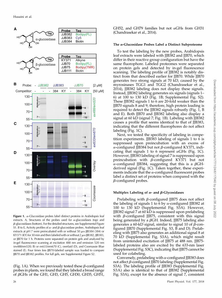
To test the labeling by the new probes, Arabidopsisleaf extracts were labeled with JJB382 and JJB70, whichdiffer in their reactive group configuration but have thesame fluorophore. Labeled proteomes were separatedon protein gels and detected by in-gel fluorescencescanning. The labeling profile of JJB382 is notably dis-tinct from that described earlier for JJB70. While JJB70generates two strong signals at 70 kD, caused by themyrosinases TGG1 and TGG2 (Chandrasekar et al.,2014), JJB382 labeling does not display these signals.Instead, JJB382 labeling generates six signals (signals 1–6) at 100 to 130 kD (Fig. 1B; Supplemental Fig. S2).These JJB382 signals 1 to 6 are 20-fold weaker than theJJB70 signals 8 and 9; therefore, high protein loading isrequired to detect the JJB382 signals robustly (Fig. 1, Band E). Both JJB70 and JJB382 labeling also display asignal at 60 kD (signal 7; Fig. 1B). Labeling with JJB382causes a profile that seems identical to that of JJB383,indicating that the different fluorophores do not affectlabeling (Fig. 1C).
Next, we tested the specificity of labeling in compe-tition experiments. JJB383 labeling of signals 1 to 6 issuppressed upon preincubation with an excess ofa-configured JJB384 but not b-configured KY371, indi-cating that signals 1 to 6 represent aGHs (Fig. 1C).However, JJB383 labeling of signal 7 is suppressed uponpreincubation with b-configured KY371 but nota-configured JJB384, suggesting that this is a bGH-derived signal (Fig. 1C). Taken together, these experi-ments indicate that the a-configured fluorescent probeslabel a distinct set of proteins when compared with theb-configured probes.
Multiplex Labeling of a- and b-Glycosidases
Prelabeling with b-configured JJB75 does not affectthe labeling of signals 1 to 6 by a-configured JJB382 at100 to 130 kD (Supplemental Fig. S3A). However,JJB382 signal 7 at 60 kD is suppressed upon prelabelingwith b-configured JJB75, consistent with this signalbeing generated by a bGH. Indeed, JJB75 labeling alsogenerates a 60-kD signal, similar to signal 10 of b-con-figured JJB70 (Supplemental Fig. S3, B and D). Prelab-eling with JJB75 also generates an additional signal 8 at70 kD (Supplemental Fig. S3A), which might resultfrom unintended excitation of JJB75 at 488 nm. JJB75-labeled proteins also are excited by the 633-nm laser(Supplemental Fig. S3C), indicating that JJB75 cannot beused for colabeling.
Conversely, prelabelingwith a-configured JJB383 doesnot affect b-configured JJB70 labeling (Supplemental Fig.S3A). The labeling profile of JJB383 (Supplemental Fig.S3A) also is identical to that of JJB382 (SupplementalFig. S3A), except for the absence of signal 7, consistent
Figure 1. a-Glucosidase probes label distinct proteins in Arabidopsis leafextracts. A, Structures of the probes used for a-glucosidases (top) andb-glucosidases (bottom). For the detailed structures, see Supplemental FigureS1. B to E, Activity profiles of a- and b-glucosidase probes. Arabidopsis leafextracts at pH 7 were preincubated with or without 50 mM JJB384 (384) orKY371 (KY) for 30minand then labeledwith orwithout 2mM JJB382, JJB383,or JJB70 for 1 h. Proteins were separated on protein gels and analyzed byin-gel fluorescence scanning at excitation 488 nm and emission 520 nm(ex488/em520; B) or ex633/em670 (C), overlaid (D), and Coomassie Bluestained (E). Four times less JJB70-labeled sample was loaded to compareJJB70 and JJB382 profiles. For full gels, see Supplemental Figure S2.
26 Plant Physiol. Vol. 177, 2018
Husaini et al.
Dow
nloaded from https://academ
ic.oup.com/plphys/article/177/1/24/6117040 by guest on 08 July 2022
with it being a bGH labeled by JJB70. Taken together,these data demonstrate that the a-configured probescause distinct signals at 100 to 130 kD and that probeshaving different fluorophores can be used for colabeling.Also, all fluorophores except for the Bodipy(TMR) ofJJB75 are detected selectively using distinctive excitationand emission wavelengths.
The a-Glycosidases Have Distinct Labeling Characteristics
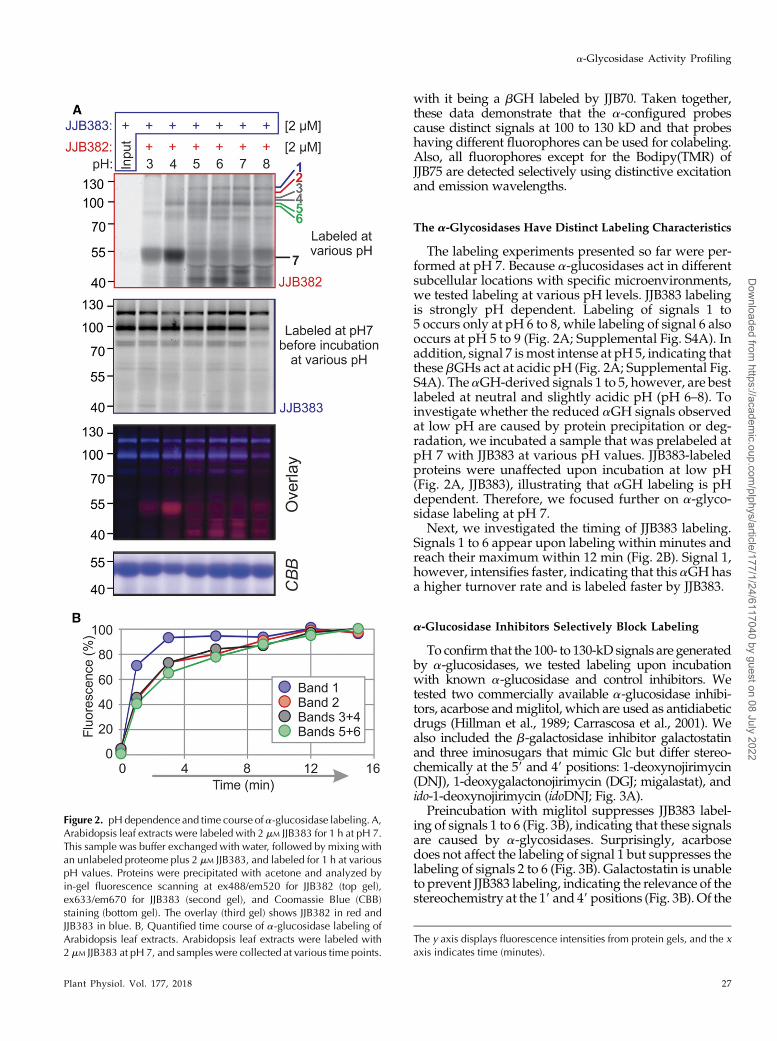
The labeling experiments presented so far were per-formed at pH 7. Because a-glucosidases act in differentsubcellular locations with specific microenvironments,we tested labeling at various pH levels. JJB383 labelingis strongly pH dependent. Labeling of signals 1 to5 occurs only at pH 6 to 8, while labeling of signal 6 alsooccurs at pH 5 to 9 (Fig. 2A; Supplemental Fig. S4A). Inaddition, signal 7 ismost intense at pH 5, indicating thatthese bGHs act at acidic pH (Fig. 2A; Supplemental Fig.S4A). The aGH-derived signals 1 to 5, however, are bestlabeled at neutral and slightly acidic pH (pH 6–8). Toinvestigate whether the reduced aGH signals observedat low pH are caused by protein precipitation or deg-radation, we incubated a sample that was prelabeled atpH 7 with JJB383 at various pH values. JJB383-labeledproteins were unaffected upon incubation at low pH(Fig. 2A, JJB383), illustrating that aGH labeling is pHdependent. Therefore, we focused further on a-glyco-sidase labeling at pH 7.
Next, we investigated the timing of JJB383 labeling.Signals 1 to 6 appear upon labeling within minutes andreach their maximum within 12 min (Fig. 2B). Signal 1,however, intensifies faster, indicating that this aGH hasa higher turnover rate and is labeled faster by JJB383.
a-Glucosidase Inhibitors Selectively Block Labeling
To confirm that the 100- to 130-kD signals are generatedby a-glucosidases, we tested labeling upon incubationwith known a-glucosidase and control inhibitors. Wetested two commercially available a-glucosidase inhibi-tors, acarbose andmiglitol, which are used as antidiabeticdrugs (Hillman et al., 1989; Carrascosa et al., 2001). Wealso included the b-galactosidase inhibitor galactostatinand three iminosugars that mimic Glc but differ stereo-chemically at the 59 and 49 positions: 1-deoxynojirimycin(DNJ), 1-deoxygalactonojirimycin (DGJ; migalastat), andido-1-deoxynojirimycin (idoDNJ; Fig. 3A).
Preincubation with miglitol suppresses JJB383 label-ing of signals 1 to 6 (Fig. 3B), indicating that these signalsare caused by a-glycosidases. Surprisingly, acarbosedoes not affect the labeling of signal 1 but suppresses thelabeling of signals 2 to 6 (Fig. 3B). Galactostatin is unableto prevent JJB383 labeling, indicating the relevance of thestereochemistry at the 19 and 49 positions (Fig. 3B). Of the
Figure 2. pHdependence and time course of a-glucosidase labeling. A,Arabidopsis leaf extracts were labeled with 2 mM JJB383 for 1 h at pH 7.This sample was buffer exchanged with water, followed by mixing withan unlabeled proteome plus 2 mM JJB383, and labeled for 1 h at variouspH values. Proteins were precipitated with acetone and analyzed byin-gel fluorescence scanning at ex488/em520 for JJB382 (top gel),ex633/em670 for JJB383 (second gel), and Coomassie Blue (CBB)staining (bottom gel). The overlay (third gel) shows JJB382 in red andJJB383 in blue. B, Quantified time course of a-glucosidase labeling ofArabidopsis leaf extracts. Arabidopsis leaf extracts were labeled with2 mM JJB383 at pH 7, and samples were collected at various time points.
The y axis displays fluorescence intensities from protein gels, and the xaxis indicates time (minutes).
Plant Physiol. Vol. 177, 2018 27
a-Glycosidase Activity Profiling
Dow
nloaded from https://academ
ic.oup.com/plphys/article/177/1/24/6117040 by guest on 08 July 2022
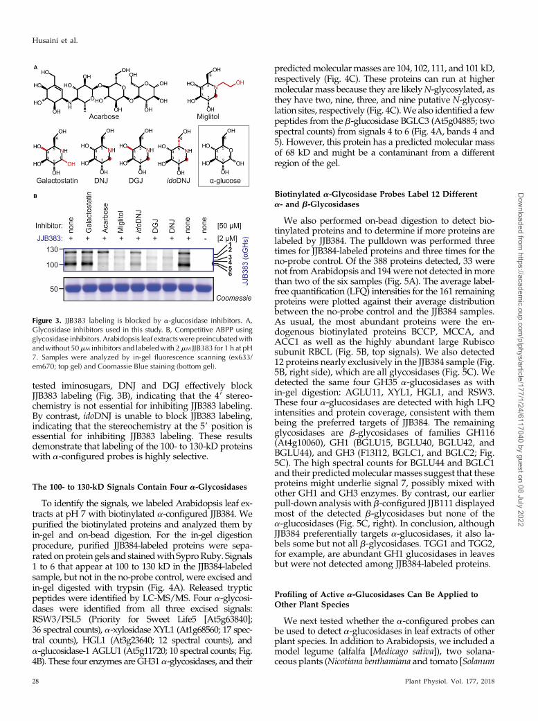
tested iminosugars, DNJ and DGJ effectively blockJJB383 labeling (Fig. 3B), indicating that the 49 stereo-chemistry is not essential for inhibiting JJB383 labeling.By contrast, idoDNJ is unable to block JJB383 labeling,indicating that the stereochemistry at the 59 position isessential for inhibiting JJB383 labeling. These resultsdemonstrate that labeling of the 100- to 130-kD proteinswith a-configured probes is highly selective.
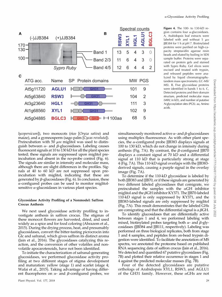
The 100- to 130-kD Signals Contain Four a-Glycosidases
To identify the signals, we labeled Arabidopsis leaf ex-tracts at pH 7 with biotinylated a-configured JJB384. Wepurified the biotinylated proteins and analyzed them byin-gel and on-bead digestion. For the in-gel digestionprocedure, purified JJB384-labeled proteins were sepa-rated onprotein gels and stainedwith SyproRuby. Signals1 to 6 that appear at 100 to 130 kD in the JJB384-labeledsample, but not in the no-probe control, were excised andin-gel digested with trypsin (Fig. 4A). Released trypticpeptides were identified by LC-MS/MS. Four a-glycosi-dases were identified from all three excised signals:RSW3/PSL5 (Priority for Sweet Life5 [At5g63840];36 spectral counts), a-xylosidase XYL1 (At1g68560; 17 spec-tral counts), HGL1 (At3g23640; 12 spectral counts), anda-glucosidase-1 AGLU1 (At5g11720; 10 spectral counts; Fig.4B). These four enzymes are GH31 a-glycosidases, and their
predictedmolecular masses are 104, 102, 111, and 101 kD,respectively (Fig. 4C). These proteins can run at highermolecularmass because they are likelyN-glycosylated, asthey have two, nine, three, and nine putative N-glycosy-lation sites, respectively (Fig. 4C).We also identified a fewpeptides from the b-glucosidase BGLC3 (At5g04885; twospectral counts) from signals 4 to 6 (Fig. 4A, bands 4 and5). However, this protein has a predicted molecular massof 68 kD and might be a contaminant from a differentregion of the gel.
Biotinylated a-Glycosidase Probes Label 12 Differenta- and b-Glycosidases
We also performed on-bead digestion to detect bio-tinylated proteins and to determine if more proteins arelabeled by JJB384. The pulldown was performed threetimes for JJB384-labeled proteins and three times for theno-probe control. Of the 388 proteins detected, 33 werenot fromArabidopsis and 194were not detected inmorethan two of the six samples (Fig. 5A). The average label-free quantification (LFQ) intensities for the 161 remainingproteins were plotted against their average distributionbetween the no-probe control and the JJB384 samples.As usual, the most abundant proteins were the en-dogenous biotinylated proteins BCCP, MCCA, andACC1 as well as the highly abundant large Rubiscosubunit RBCL (Fig. 5B, top signals). We also detected12 proteins nearly exclusively in the JJB384 sample (Fig.5B, right side), which are all glycosidases (Fig. 5C). Wedetected the same four GH35 a-glucosidases as within-gel digestion: AGLU11, XYL1, HGL1, and RSW3.These four a-glucosidases are detected with high LFQintensities and protein coverage, consistent with thembeing the preferred targets of JJB384. The remainingglycosidases are b-glycosidases of families GH116(At4g10060), GH1 (BGLU15, BGLU40, BGLU42, andBGLU44), and GH3 (F13I12, BGLC1, and BGLC2; Fig.5C). The high spectral counts for BGLU44 and BGLC1and their predictedmolecular masses suggest that theseproteins might underlie signal 7, possibly mixed withother GH1 and GH3 enzymes. By contrast, our earlierpull-down analysis with b-configured JJB111 displayedmost of the detected b-glycosidases but none of thea-glucosidases (Fig. 5C, right). In conclusion, althoughJJB384 preferentially targets a-glucosidases, it also la-bels some but not all b-glycosidases. TGG1 and TGG2,for example, are abundant GH1 glucosidases in leavesbut were not detected among JJB384-labeled proteins.
Profiling of Active a-Glucosidases Can Be Applied toOther Plant Species
We next tested whether the a-configured probes canbe used to detect a-glucosidases in leaf extracts of otherplant species. In addition to Arabidopsis, we included amodel legume (alfalfa [Medicago sativa]), two solana-ceous plants (Nicotiana benthamiana and tomato [Solanum
Figure 3. JJB383 labeling is blocked by a-glucosidase inhibitors. A,Glycosidase inhibitors used in this study. B, Competitive ABPP usingglycosidase inhibitors. Arabidopsis leaf extractswere preincubatedwithandwithout 50mM inhibitors and labeledwith 2 mM JJB383 for 1 h at pH7. Samples were analyzed by in-gel fluorescence scanning (ex633/em670; top gel) and Coomassie Blue staining (bottom gel).
28 Plant Physiol. Vol. 177, 2018
Husaini et al.
Dow
nloaded from https://academ
ic.oup.com/plphys/article/177/1/24/6117040 by guest on 08 July 2022
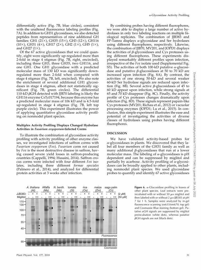
lycopersicum]), two monocots (rice [Oryza sativa) andmaize), and a gymnosperm (sago palm [Cycas revoluta]).Preincubation with 50 mM miglitol was used to distin-guish between a- and b-glucosidases. Labeling causesfluorescent signals at 10 to 130 kD for all the plant speciestested; these signals are suppressed upon miglitol pre-incubation and absent in the no-probe control (Fig. 6).The signals are similar in intensity and molecular mass,although there are slight differences in the profiles. Sig-nals at 40 to 60 kD are not suppressed upon pre-incubation with miglitol, indicating that these aregenerated by b-glucosidases (Fig. 6). In conclusion, thesea-configured probes can be used to monitor miglitol-sensitive a-glucosidases in various plant species.
Glycosidase Activity Profiling of a Nonmodel: SaffronCrocus Anthesis
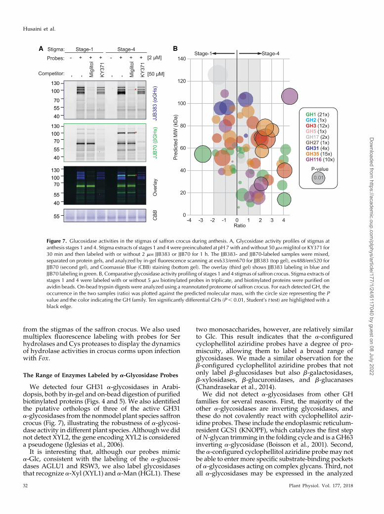
We next used glycosidase activity profiling to in-vestigate anthesis in saffron crocus. The stigmas ofthese monocot flowers are harvested, dried, and usedwidely as a spice and for food coloring (Ahrazem et al.,2015). During the drying process, heat, and presumablyglucosidases, convert the bitter-tasting picrocrocin intoGlc and safranal, which gives saffron its distinct aroma(Jain et al., 2016). The glycosidases catalyzing this re-action, and the conversion of other volatiles and non-volatile apocarotenoids, have not been identified.To initiate the characterization of safranal-generating
glucosidases, we performed glucosidase activity pro-filing at two different stages of stigma developmentand maturation: yellow (stage 1) and scarlet (stage 4;Wafai et al., 2015). Taking advantage of having differ-ent fluorophores on a- and b-configured probes, we
simultaneously monitored active a- and b-glucosidasesusing multiplex fluorescence. As with other plant spe-cies, the a-configured probe JJB383 displays signals at100 to 130 kD, which do not change in intensity duringanthesis (Fig. 7A). By contrast, the b-configured JJB70displays a constant signal at 70 kD and a differentialsignal at 110 kD that is particularly strong at stage4 (Fig. 7A). This 110-kD signal overlaps with the JJB383-derived signals, causing a purple signal in the overlayimage (Fig. 7A).
To determine if the 110-kD glycosidase is labeled byboth JJB383 and JJB70, or if these signals are generated bytwo different labeled glycosidases that comigrate, wepreincubated the samples with the aGH inhibitormiglitol and the bGH inhibitor KY371. The JJB70-labeled110-kD signal is only suppressed by KY371, and theJJB383-labeled signals are only suppressed by miglitol(Fig. 7A). This result demonstrates that the labeled GHsare comigrating and that the differential signal is a bGH.
To identify glucosidases that are differentially activebetween stages 1 and 4, we performed labeling withmixed, biotinylated probes targeting both a- and b-gly-cosidases (JJB384 and JJB111, respectively). Labeling wasperformed on three biological replicates, both from stage1 and 4 samples, and peptides from on-bead trypsin di-gestion were identified. To facilitate the annotation of MSspectra, we annotated the proteome based on publishedRNA sequencing data of saffron crocus (Jain et al., 2016).We detected and quantified 67 putative glycosidases (Fig.7B) and plotted their relative occurrence in stages 1 and4 against the predicted molecular masses (Fig. 7B).
Four of the detected glycosidases are putativeorthologs of Arabidopsis XYL1, RSW3, and AGLU1of the GH31 family. However, these aGHs are not
Figure 4. The 100- to 130-kD re-gion contains four a-glucosidases.A, Arabidopsis leaf extracts werelabeled with and without 5 mM
JJB384 for 1 h at pH 7. Biotinylatedproteins were purified on high-ca-pacity streptavidin agarose resinbeads and eluted by boiling in SDSsample buffer. Proteins were sepa-rated on protein gels and stainedwith Sypro Ruby. Gel slices wereexcised and treated with trypsin,and released peptides were ana-lyzed by liquid chromatography-tandemmass spectrometry (LC-MS/MS). B, Five glycosidase proteinswere identified in bands 1 to 6. C,Detected proteins and their domainstructure, predicted molecular mass(MW; in kD), and number of putativeN-glycosylation sites (PGS). aa, Aminoacids.
Plant Physiol. Vol. 177, 2018 29
a-Glycosidase Activity Profiling
Dow
nloaded from https://academ
ic.oup.com/plphys/article/177/1/24/6117040 by guest on 08 July 2022
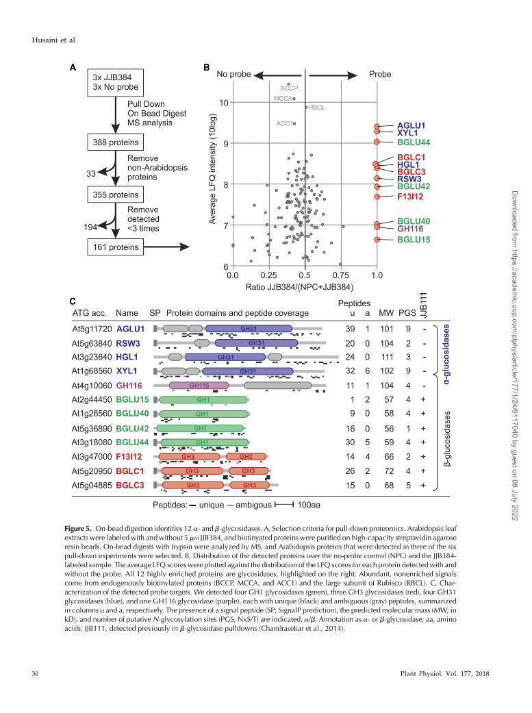
Figure 5. On-bead digestion identifies 12 a- and b-glycosidases. A, Selection criteria for pull-down proteomics. Arabidopsis leafextracts were labeledwith andwithout 5 mM JJB384, and biotinyated proteins were purified on high-capacity streptavidin agaroseresin beads. On-bead digests with trypsin were analyzed by MS, and Arabidopsis proteins that were detected in three of the sixpull-down experiments were selected. B, Distribution of the detected proteins over the no-probe control (NPC) and the JJB384-labeled sample. The average LFQ scoreswere plotted against the distribution of the LFQ scores for each protein detectedwith andwithout the probe. All 12 highly enriched proteins are glycosidases, highlighted on the right. Abundant, nonenriched signalscome from endogenously biotinylated proteins (BCCP, MCCA, and ACC1) and the large subunit of Rubisco (RBCL). C, Char-acterization of the detected probe targets. We detected four GH1 glycosidases (green), three GH3 glycosidases (red), four GH31glycosidases (blue), and one GH116 glycosidase (purple), each with unique (black) and ambiguous (gray) peptides, summarizedin columns u and a, respectively. The presence of a signal peptide (SP; SignalP prediction), the predicted molecular mass (MW; inkD), and number of putativeN-glycosylation sites (PGS; NxS/T) are indicated. a/b, Annotation as a- or b-glycosidase; aa, aminoacids; JJB111, detected previously in b-glycosidase pulldowns (Chandrasekar et al., 2014).
30 Plant Physiol. Vol. 177, 2018
Husaini et al.
Dow
nloaded from https://academ
ic.oup.com/plphys/article/177/1/24/6117040 by guest on 08 July 2022
differentially active (Fig. 7B, blue circles), consistentwith the unaltered fluorescence labeling profiles (Fig.7A). In addition to GH31 glycosidases, we also detectedpeptides from representatives of nine additional GHfamilies: GH1 (213), GH35 (153), GH3 (123), GH116(103), GH31 (43), GH17 (23), GH2 (13), GH5 (13),and GH27 (13).Of the 67 active glycosidases that we could quan-
tify, eight were significantly up-regulated more than2-fold in stage 4 stigmas (Fig. 7B, right, encircled),including three GH3, three GH35, two GH116, andone GH1. One GH1 glycosidase with a predictedmolecular mass of 130 kD was significantly down-regulated more than 2-fold when compared withstage 4 stigmas (Fig. 7B, left, encircled). We also notethe enrichment of several additional GH1 glycosi-dases in stage 4 stigmas, albeit not statistically sig-nificant (Fig. 7B, green circles). The differential110-kD bGH detected with JJB70 labeling is likely theGH116 enzyme CsTc017194, because this enzyme hasa predicted molecular mass of 106 kD and is 4.5-foldup-regulated in stage 4 stigmas (Fig. 7B, left toppurple circle). This experiment illustrates the powerof applying quantitative glycosidase activity profil-ing on nonmodel plant species.
Multiplex Activity Profiling Displays Changed HydrolaseActivities in Fusarium oxysporum-Infected Corms
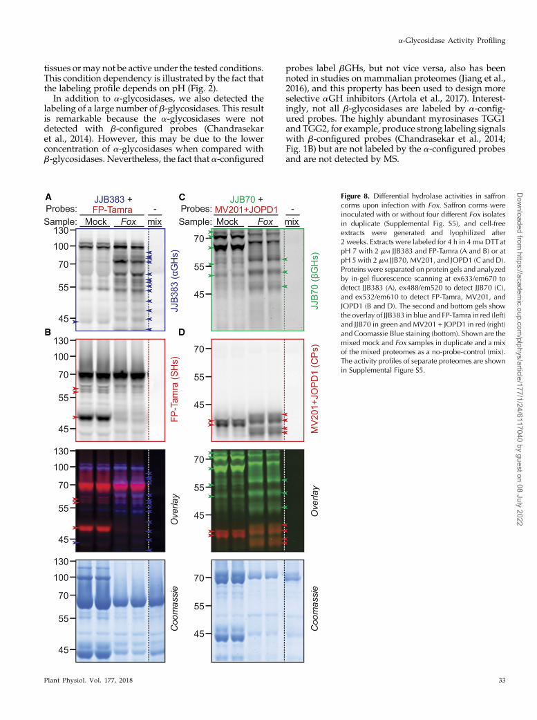
To illustrate the combination of glycosidase activityprofiling with activity profiling of other enzyme clas-ses, we investigated infections of saffron corms withFusarium oxysporum (Fox). Fusarium corm rot causedby Fox is the most destructive disease in saffron, hav-ing caused severe yield losses in saffron-producingcountries (Cappelli, 1994; Husaini, 2014). Saffron cro-cus corms were infected with four different Fox iso-lates, including three different formae speciales(Palmero et al., 2014), and analyzed for differentialprotein activities at 3 weeks after infection.
By combining probes having different fluorophores,we were able to display a large number of active hy-drolases in only two labeling reactions on multiple bi-ological replicates. The combination of JJB383 andFP-Tamra displays a-glycosidase and Ser hydrolasesusing different fluorophores, respectively. Likewise,the combination of JJB70, MV201, and JOPD1 displaysthe activities of b-glycosidases and Cys proteases us-ing different fluorophores. These experiments dis-played remarkably different profiles upon infection,irrespective of the Fox isolate used (Supplemental Fig.S5). The activities of both 100-kD putative a-glucosi-dase and putative b-glucosidases at 50 to 70 kD areincreased upon infection (Fig. 8A). By contrast, theactivities of one strong 50-kD and several weaker60-kD Ser hydrolase signals are reduced upon infec-tion (Fig. 8B). Several active b-glucosidases of 45 to60 kD appear upon infection, while strong signals at65 and 70 kD disappear (Fig. 8C). Finally, the activityprofile of Cys proteases changes dramatically uponinfection (Fig. 8D). These signals represent papain-likeCys proteases (MV201; Richau et al., 2012) or vacuolarprocessing enzymes (JOPD1; Lu et al., 2015). In con-clusion, this simple experiment illustrates the ease andpotential of investigating the activities of diverseclasses of hydrolases using probes having differentfluorophores.
DISCUSSION
We have validated activity-based probes fora-glycosidases in plants. We discovered that they la-bel all four members of the GH31 family as well asmany additional b-glycosidases that run at a lowermolecular mass. The labeling of a-glycosidases is pHdependent and can be suppressed by miglitol andpartially by acarbose. Activity profiling of a-glycosi-dases can be broadly applied to other plants, includ-ing nonmodel plant species. We used glycosidaseprobes to quantify and identify 67 active glycosidases
Figure 6. a-Glucosidase profiling in leaves ofother plant species. Leaf extracts were pre-incubated with or without 50 mM miglitol andthen labeled with or without 2 mM JJB383 at pH7 for 1 h. Samples were analyzed by in-gelfluorescence scanning (ex633/em670; top gel)and Coomassie Blue staining (bottom gel). Pu-tative aGH signals are suppressed by miglitolpreincubation (white dots), whereas putativebGH signals are not (black dots).
Plant Physiol. Vol. 177, 2018 31
a-Glycosidase Activity Profiling
Dow
nloaded from https://academ
ic.oup.com/plphys/article/177/1/24/6117040 by guest on 08 July 2022
from the stigmas of the saffron crocus. We also usedmultiplex fluorescence labeling with probes for Serhydrolases and Cys proteases to display the dynamicsof hydrolase activities in crocus corms upon infectionwith Fox.
The Range of Enzymes Labeled by a-Glycosidase Probes
We detected four GH31 a-glycosidases in Arabi-dopsis, both by in-gel and on-bead digestion of purifiedbiotinylated proteins (Figs. 4 and 5). We also identifiedthe putative orthologs of three of the active GH31a-glycosidases from the nonmodel plant species saffroncrocus (Fig. 7), illustrating the robustness of a-glycosi-dase activity in different plant species. Althoughwe didnot detect XYL2, the gene encoding XYL2 is considereda pseudogene (Iglesias et al., 2006).
It is interesting that, although our probes mimica-Glc, consistent with the labeling of the a-glucosi-dases AGLU1 and RSW3, we also label glycosidasesthat recognize a-Xyl (XYL1) and a-Man (HGL1). These
two monosaccharides, however, are relatively similarto Glc. This result indicates that the a-configuredcyclophellitol aziridine probes have a degree of pro-miscuity, allowing them to label a broad range ofglycosidases. We made a similar observation for theb-configured cyclophellitol aziridine probes that notonly label b-glucosidases but also b-galactosidases,b-xylosidases, b-glucuronidases, and b-glucanases(Chandrasekar et al., 2014).
We did not detect a-glycosidases from other GHfamilies for several reasons. First, the majority of theother a-glycosidases are inverting glycosidases, andthese do not covalently react with cyclophellitol azir-idine probes. These include the endoplasmic reticulum-resident GCS1 (KNOPF), which catalyzes the first stepofN-glycan trimming in the folding cycle and is a GH63inverting a-glycosidase (Boisson et al., 2001). Second,the a-configured cyclophellitol aziridine probe may notbe able to enter more specific substrate-binding pocketsof a-glycosidases acting on complex glycans. Third, notall a-glycosidases may be expressed in the analyzed
Figure 7. Glucosidase activities in the stigmas of saffron crocus during anthesis. A, Glycosidase activity profiles of stigmas atanthesis stages 1 and 4. Stigma extracts of stages 1 and 4were preincubated at pH 7with andwithout 50mMmiglitol or KY371 for30 min and then labeled with or without 2 mM JJB383 or JJB70 for 1 h. The JJB383- and JJB70-labeled samples were mixed,separated on protein gels, and analyzed by in-gel fluorescence scanning at ex633/em670 for JJB383 (top gel), ex488/em520 forJJB70 (second gel), and Coomassie Blue (CBB) staining (bottom gel). The overlay (third gel) shows JJB383 labeling in blue andJJB70 labeling in green. B, Comparative glycosidase activity profiling of stages 1 and 4 stigmas of saffron crocus. Stigma extracts ofstages 1 and 4 were labeled with or without 5 mM biotinylated probes in triplicate, and biotinylated proteins were purified onavidin beads. On-bead trypsin digests were analyzed using a reannotated proteome of saffron crocus. For each detected GH, theoccurrence in the two samples (ratio) was plotted against the predicted molecular mass, with the circle size representing the Pvalue and the color indicating the GH family. Ten significantly differential GHs (P, 0.01, Student’s t test) are highlighted with ablack edge.
32 Plant Physiol. Vol. 177, 2018
Husaini et al.
Dow
nloaded from https://academ
ic.oup.com/plphys/article/177/1/24/6117040 by guest on 08 July 2022
tissues ormay not be active under the tested conditions.This condition dependency is illustrated by the fact thatthe labeling profile depends on pH (Fig. 2).In addition to a-glycosidases, we also detected the
labeling of a large number of b-glycosidases. This resultis remarkable because the a-glycosidases were notdetected with b-configured probes (Chandrasekaret al., 2014). However, this may be due to the lowerconcentration of a-glycosidases when compared withb-glycosidases. Nevertheless, the fact that a-configured
probes label bGHs, but not vice versa, also has beennoted in studies on mammalian proteomes (Jiang et al.,2016), and this property has been used to design moreselective aGH inhibitors (Artola et al., 2017). Interest-ingly, not all b-glycosidases are labeled by a-config-ured probes. The highly abundant myrosinases TGG1and TGG2, for example, produce strong labeling signalswith b-configured probes (Chandrasekar et al., 2014;Fig. 1B) but are not labeled by the a-configured probesand are not detected by MS.
Figure 8. Differential hydrolase activities in saffroncorms upon infection with Fox. Saffron corms wereinoculated with or without four different Fox isolatesin duplicate (Supplemental Fig. S5), and cell-freeextracts were generated and lyophilized after2 weeks. Extracts were labeled for 4 h in 4 mM DTTatpH 7 with 2 mM JJB383 and FP-Tamra (A and B) or atpH 5 with 2 mM JJB70, MV201, and JOPD1 (C and D).Proteins were separated on protein gels and analyzedby in-gel fluorescence scanning at ex633/em670 todetect JJB383 (A), ex488/em520 to detect JJB70 (C),and ex532/em610 to detect FP-Tamra, MV201, andJOPD1 (B and D). The second and bottom gels showthe overlay of JJB383 in blue and FP-Tamra in red (left)and JJB70 in green and MV201 + JOPD1 in red (right)and Coomassie Blue staining (bottom). Shown are themixed mock and Fox samples in duplicate and a mixof the mixed proteomes as a no-probe-control (mix).The activity profiles of separate proteomes are shownin Supplemental Figure S5.
Plant Physiol. Vol. 177, 2018 33
a-Glycosidase Activity Profiling
Dow
nloaded from https://academ
ic.oup.com/plphys/article/177/1/24/6117040 by guest on 08 July 2022
The Identity of the 100- to 130-kD Signals in thea-Glycosidase Activity Profiles
The a-glycosidases cause distinct signals in the 100- to130-kD region, consistent with their predicted molecularmasses (Fig. 4). Unfortunately, the signals are too close toseparate and identify the proteins corresponding to eachsignal. However, we can predict that the bottom signalsrepresent AGLU1 and XYL1 for several reasons. First,this signal also appears upon labeling at pH 4 to5 (Fig. 2A), consistent with their function in the apoplast.Second, this signal is sensitive to acarbose inhibition (Fig.3), and AGLU1 is known to be inhibited by acarbose(Frandsen et al., 2000). Third, AGLU1 and XYL1 havethe lowest predicted molecular masses (Fig. 5C). Con-versely, our data suggest thatHGL1may generate one ofthe top signals, as it is expected to have the higher mo-lecular mass and to be active in the Golgi (pH 6.5). Thisresult implies that HGL1 may be insensitive to acarbosebut not to miglitol inhibition.
a-Glycosidase Activity Profiling Is Broadly Applicable inPlant Science
Our work demonstrates that a-glycosidase activityprofiling is broadly applicable. We were able to displaymiglitol-sensitive labeling of 100- to 130-kDproteins fromleaves of monocot and dicot plants and the leaves of agymnosperm (Fig. 6). These signals very likely representthe orthologs of the Arabidopsis a-glycosidases RSW3,HGL1, AGLU1, and XYL1. Three of these enzymes alsowere detected upon labeling of saffron crocus anthers(Fig. 5C).We displayed activity profiles ofa-glycosidasesin leaves, stigmas, and corms (Figs. 6–8), further sup-porting the broad applicability of this technique. Sincethe probes are uncharged, and b-configured probes havebeen used for in vivo labeling (Chandrasekar et al., 2014),we believe that the probes also can be used for the in vivolabeling of a-glycosidases.
We also applied a-glycosidase activity profiling tostigmas and corms of the saffron crocus to study stigmadevelopment (Fig. 7) and Fox infection (Fig. 8). These ex-periments illustrate the ease with which a-glycosidaseactivity profiling can be applied to cash-crop plants togenerate data for follow-up studies. For instance, our datasuggest candidate glycosidases responsible for the con-version of picrocrocin into safranal in harvested stigmas.Likewise, we detect the suppressed activity of an a-gly-cosidase upon infection with Fox, consistent with the no-tion that Fox is likely to suppress AGLU1 in the apoplastduring infection to overcome the antifungal activity of thisenzyme (Xiao et al., 1994; Monroe et al., 1999).
Multiplex Fluorescence Activity Profiling DisplaysDynamic Changes in Hydrolase Activities
The use of probes having different fluorophores greatlyexpands the ease with which we can profile multipleclasses of hydrolases simultaneously. Multiplex labeling
will drastically accelerate research and miniaturize theexperiments. By labeling of saffron stigmas and infectedcorms (Figs. 7A and 8), we have illustrated the ease withwhichmultiplexfluorescence simultaneously displays theactivity profiles of different enzyme classes.
The changes in hydrolase activities upon infection ofcorms by Fox are interesting and robust (Fig. 8;Supplemental Fig. S5). The signals that appear ininfected corms may be generated by plant-producedhydrolases aimed to suppress the disease or maycome from Fox itself, to macerate the host tissue(Mohamed et al., 2017). The reduced activity of a 50-kDplant Ser hydrolase upon Fox infection may be causedby a depletion of this 50-kD protein but also may becaused by the suppression of the activity of this proteinby Fox. We have detected a similar suppression of hosthydrolase activities upon bacterial infection (Hong andvan der Hoorn, 2014; Chandrasekar et al., 2017). Thesuppression of host enzymes can be studied furtherusing convolution ABPP, where samples from infectedand noninfected samples are mixed before and afterlabeling (Chandrasekar et al., 2017).
In conclusion, we have validated activity profilingfor a-glycosidases, showed its broad applicability byprofiling other tissues, plant species, and biologicalphenomena, and introduced multiplex labeling to speedup and miniaturize activity profiling in plant science.
MATERIALS AND METHODS
Probes and Inhibitors
The activity-basedprobes JJB382, JJB347, JJB383, and JJB384 (Jiang et al., 2016)and JJB70, JJB75, JJB111, and KY371 (Kallemeijn et al., 2012; Chandrasekar et al.,2014) have been described previously. Immunosugars DNJ (Wennekes et al.,2007), DGJ, and idoDNJ (Wennekes et al., 2010) have been described previously.Acarbose, miglitol, and galactostatin were purchased from Sigma-Aldrich,Tocris, and Santa Cruz, respectively.
Plant Material, Growth Conditions, and Fox Infections
Arabidopsis (Arabidopsis thaliana) ecotype Columbia, alfalfa (Medicago sat-iva), Nicotiana benthamiana, tomato (Solanum lycopersicum), rice (Oryza sativa),maize (Zea mays), and sago palm (Cycas revoluta) were grown on soil understandard greenhouse conditions. Healthy corms of saffron crocus (Crocus sat-ivus), previously disinfected with 5% [v/v] sodium hypochlorite for 15 minfollowed by three washes with sterile water, were planted in sterile substrateand held for 3 weeks with controlled temperature and light (12/12 h of light/dark and 25°C/21°C). The inoculum consisted of a suspension of conidiaobtained after 1 week in potato (Solanum tuberosum)-Glc medium, stirred at150 rpm. To remove the mycelium, the suspension was filtered with a doublelayer of cheesecloth. The conidia suspension was adjusted to 105 conidia mL21
and used for the inoculation of plant material. The plant roots were immersedfor 24 h in the suspension of conidia (200 mL of suspension), then transplantedback and kept under the same conditions of temperature and light for 3 weeks.
Protein Extraction
Two leaf discs (0.9 cm diameter) were taken from the leaves of various plantspecies and homogenized with 300 mL of a buffer with suitable pH (50 mM
sodium acetate buffer for pH 3 and 4, 50 mM MES buffer for pH 5 and 6, 50 mM
MOPS buffer for pH 7 and 8, and 50 mM Tris buffer for pH 9 and 10). After thetissues had been ground in a 1.5-mL tube, the samples were centrifuged at10,000g (4°C for 10 min) followed by 11,000g (4°C for 5 min) to remove celldebris, and the supernatant containing soluble proteins was used for labeling.
34 Plant Physiol. Vol. 177, 2018
Husaini et al.
Dow
nloaded from https://academ
ic.oup.com/plphys/article/177/1/24/6117040 by guest on 08 July 2022
Labeling Plant Extracts
All probes were prepared as 0.1 to 10 mM stock solutions in dimethyl sulf-oxide. Equal volumes of dimethyl sulfoxide were used as a no-probe control.Labeling was performed as described previously (Chandrasekar et al., 2014).For fluorescence gel imaging, the extracts were incubated with 2 mM probes for1 h at room temperature in the dark at a 50-mL total reaction volume. For thecompetition experiments, the extracts were preincubated with the corre-sponding inhibitors at 50 mM for 30 min prior to labeling with the probe.
The labeling reactionswere quenchedbyadding43gel loadingbuffer (200mM
Tris-HCl [pH 6.8], 400 mM DTT, 8% [w/v] SDS, 0.04% [w/v] bromophenol blue,and 40% [v/v] glycerol) at 13 final concentration and heating at 95°C for 5 min.The labeled proteins were separated on 10% protein gels and detected on theprotein gelswith the TyphoonFLA9000 scanner at ex488/em520or ex633/em670(GE Healthcare Life Sciences). Subsequent to fluorescence imaging, the gels werestained with Coomassie Brilliant Blue R-250. The fluorescence of the labeledproteins was quantified using ImageJ. For pull-down experiments, the extractsfrom three biological replicates were incubatedwith 5mM probes for 1.5 h at roomtemperature in the dark at a 1-mL total reaction volume. The labeling reactionswere quenched by precipitating the total proteins via the chloroform/methanolprecipitation method (Wessel and Flugge, 1984).
Pull-Down and On-Bead, In-Gel Trypsin Digestion
Pull-down experiments and on-bead and in-gel trypsin digestions wereperformed as described with minor modifications (Chandrasekar et al., 2014).For Arabidopsis, healthy leaf discs (1.5 g) were collected from middle/topleaves of 5-week-old Arabidopsis plants growing in different pots at the samepoint of time. Leaf discsweremixed, and three separate extracts were generatedin 50 mM MOPS at pH 7. For saffron crocus sigmas, four stigmas, each from asingle flower, were harvested for each stage from different plants of the sameage but at different time points. These four stigmas were pooled for the pull-down experiment, and three of these pooled samples were used as biologicalreplicates. The trypsin-digested peptides were purified using Sep-Pak C18columns (Waters; WAT020515). The columns were equilibrated with 2% (v/v)acetonitrile (ACN) and 0.1% (v/v) formic acid (FA) before peptide loading.Peptides were washed with 10 mL of 2% (v/v) ACN and 0.1% (v/v) FA andeluted with 2 3 1 mL of 65% (v/v) ACN and 0.1% (v/v) FA. Purified peptideswere dried in a vacuum centrifuge and subjected to MS analysis. After elutionfrom the Sep-Pak, samples were dried using a vacuum concentrator (Eppen-dorf), and the peptides were resuspended in 0.1% [v/v] FA solution (15 mL).
LC-MS/MS
Experiments were performed on an Orbitrap Elite instrument (Thermo;Michalski et al., 2012) that was coupled to an EASY-nLC 1000 liquid chro-matograph (Thermo). The liquid chromatograph was operated in the one-column mode. The analytical column was a fused silica capillary (75 mm 320 cm or 75 mm 3 40 cm) with an integrated PicoFrit emitter (New Objective)packed in house with Reprosil-Pur 120 C18-AQ 1.9-mm resin (Dr. Maisch). Theanalytical column was encased by a column oven (Sonation) and attached to ananospray flex ion source (Thermo). The column oven temperature was ad-justed to 45°C during data acquisition and in all other modes at 30°C. The liquidchromatograph was equipped with two mobile phases: solvent A (0.1% [v/v]FA in water) and solvent B (0.1% [v/v] FA in ACN). All solvents were of ultra-performance liquid chromatography grade (Sigma-Aldrich). Peptides wereloaded directly onto the analytical column with a maximum flow rate thatwould not exceed the set pressure limit of 980 bar (usually around 0.6–1 mLmin21). Peptides were subsequently separated on the analytical column byrunning a 70- or 140-min gradient of solvents A and B (70-min gradient: startwith 7% B; gradient 7% to 35% B for 60 min; gradient 35% to 100% B for 5 min;and 100% B for 5 min; 140-min gradient: start with 7% B; gradient 7% to 35% Bfor 120 min; gradient 35% to 100% B for 10 min; and 100% B for 10 min) at aflow rate of 300 nL min21. The mass spectrometer was operated using Xca-libur software (version 2.2 SP1.48) and was set in positive ion mode. Pre-cursor ion scanning was performed in the Orbitrap analyzer (Fouriertransform mass spectrometry) in the scan range of m/z 300 to 1,800 and at aresolution of 60,000 with the internal lock mass option turned on (lock masswas 445.120025 m/z, polysiloxane; Olsen et al., 2005). Product ion spectrawere recorded in a data-dependent fashion in the ion trap in a variable scanrange and at a rapid scan rate. The ionization potential (spray voltage) was setto 1.8 kV. Peptides were analyzed using a repeating cycle consisting of a full
precursor ion scan (1 3 106 ions or 50 ms) followed by 12 or 15 product ionscans (1 3 104 ions or 80–100 ms) where peptides are isolated based on theirintensity in the full survey scan (threshold of 500 counts) for tandem massspectrum (MS2) generation that permits peptide sequencing and identifica-tion. Collision-induced dissociation energy was set to 35% for the generationof MS2. During MS2 data acquisition, dynamic ion exclusion was set to 60 to120 s with a maximum list of excluded ions consisting of 500 members and arepeat count of one. Ion injection time prediction, preview mode for Fouriertransform mass spectrometry, monoisotopic precursor selection, and chargestate screening were enabled. Only charge states of greater than 1 wereconsidered for fragmentation.
Peptide and Protein Identification Using MaxQuant
Spectra (RAW files) were submitted to an Andromeda (Cox et al., 2011)search inMaxQuant (version 1.5.0.25 or 1.5.3.30) using the default settings (Coxand Mann, 2008). Label-free quantification and match-between-runs were ac-tivated (Cox et al., 2014). For Arabidopsis samples, the MS2 data were searchedagainst an Arabidopsis (taxonomy identifier 3702) database downloaded fromthe TAIR repository (TAIR10_pep_20110103.fasta; 27,416 entries). For saffroncrocus (taxonomy identifier 82528) samples, we generated a dedicated proteindatabase by translating the publicly available RNA sequencing data from Jainet al. (2016) and filtering for open reading frames that encode proteins of morethan 50 amino acids (Supplemental File S1; Saffron.PROTEIN_50.fasta; 83,422entries). All searches included a contaminants database search (as implementedin MaxQuant; 245 sequences). The contaminants database contains known MScontaminants and was included to estimate the level of contamination. An-dromeda searches allowed oxidation of Met residues (16 D) and acetylation ofthe protein N terminus (42 D) as dynamic modifications and the static modi-fication of Cys (57 D, alkylation with iodoacetamide). Enzyme specificity wasset to trypsin/P with two missed cleavages allowed. The instrument type inAndromeda searches was set to Orbitrap, and the precursor mass tolerance wasset to 620 ppm (first search) and 64.5 ppm (main search). The MS/MS matchtolerance was set to 60.5 D. The peptide spectrum match false discovery rateand the protein false discovery rate were set to 0.01 (based on the target-decoyapproach). The minimum peptide length was seven amino acids. For proteinquantification, unique and razor peptides were allowed. Modified peptideswere allowed for quantification. Theminimum score for modified peptides was40. Label-free protein quantification was switched on, and unique and razorpeptides were considered for quantification with a minimum ratio count of 2.Retention times were recalibrated based on the built-in nonlinear time-rescaling algorithm. MS/MS identifications were transferred betweenLC-MS/MS runs with the match between runs option, in which the maximalmatch time window was set to 0.7 min and the alignment time window wasset to 20 min. The quantification was based on the value at maximum of theextracted ion current. At least two quantification events were required foreach protein. Further analysis and filtering of the results were done in Perseusversion 1.5.5.3 (Tyanova et al., 2016). Briefly, only protein groups with at leasttwo identified unique peptides over all runs were considered for furtheranalysis. Comparison of protein group quantities (relative quantification)between different MS runs was based solely on the LFQs, as calculated byMaxQuant (MaxLFQ algorithm; Cox et al., 2014). Imputed values weregenerated over the whole matrix, and the fold change and P values werecalculated over the three biological replicates.
Open Reading Frame Detection and Domain Annotation
The complete set of de novo assembled transcripts was subject to openreading frame detection using three different prediction algorithms: Gene-MarkS-T (Tang et al., 2015), TransDecoder (Haas et al., 2013), and Prodigal(Hyatt et al., 2010). Prodigal was run in both intronless eukaryotic and pro-karyotic modes; thus, up to four open reading frame predictions were gener-ated for each transcript. To select the single best open reading frame for eachtranscript, the following process was applied. If multiple methods predictedoverlapping open reading frames, then the longest was chosen. Where multiplemethods disagreed on the correct open reading frame, then the following de-cision process was followed. If all methods disagreed (either in frame or loca-tion), then the priority for assignment was taken as GeneMarkS-T,TransDecoder, Prodigal (eukaryotic settings), and Prodigal (prokaryotic set-tings). If some methods agreed, then the open reading frame that was detectedby the largest number of methods was chosen. Only open reading frames of50 or more amino acids were selected for further analysis. To annotate
Plant Physiol. Vol. 177, 2018 35
a-Glycosidase Activity Profiling
Dow
nloaded from https://academ
ic.oup.com/plphys/article/177/1/24/6117040 by guest on 08 July 2022
conserved domains in the predicted open reading frames, the full PFAM-Adatabase (Finn et al., 2016) was searched against the complete set of openreading frames with an e-value cutoff of 1e-5.
Data Availability
The MS proteomics data have been deposited to the ProteomeXchangeConsortium via the PRIDE (Vizcaíno et al., 2016) partner repository with thedata set identifier PXD009014. The samples are named as follows: in-gel digestsArabidopsis leaves [ACE0111: bands 1-4 (AH7-9)]; on-bead digest Arabidopsisleaves [ACE0149: JJB384 (AH2,5,8) and no-probe control (AH1,4,7)]; andon-bead digest saffron stigma [ACE0158: Stage-1 (AS13-15), Stage-4 (AS3-6),and no-probe control (AS1-3)].
Accession Numbers
Accession numbers are as follows: At5g11720 (AGLU1), At5g63840 (RSW3/PSL5), At3g23640 (HGL1), At1g68560 (XYL1), At4g10060 (GH116), At2g44450(BGLU15), At1g26560 (BGLU40), At5g36890 (BGLU42), At3g18080 (BHLU44),At3g47000 (F13I12), At5g20950 (BGLC1), and At5g04885 (BGLC3).
Supplemental Data
The following supplemental materials are available.
Supplemental Figure S1. Structures of the probes used.
Supplemental Figure S2. Activity profiles of a- and b-glucosidase probes.
Supplemental Figure S3. Subsequent labeling of a- and b-glucosidases.
Supplemental Figure S4. Duplicate of the pH course.
Supplemental Figure S5. Replicates of Fox infection of saffron corms.
Supplemental File S1. The saffron protein database.
ACKNOWLEDGMENTS
We thank Ursula Pyzio for excellent plant care.
Received March 2, 2018; accepted March 6, 2018; published March 19, 2018.
LITERATURE CITED
Ahrazem O, Rubio-Moraga A, Nebauer SG, Molina RV, Gómez-Gómez L(2015) Saffron: its phytochemistry, developmental processes, and bio-technological prospects. J Agric Food Chem 63: 8751–8764
Andriotis VME, Rejzek M, Barclay E, Rugen MD, Field RA, Smith AM(2016) Cell wall degradation is required for normal starch mobilisationin barley endosperm. Sci Rep 6: 33215
Artola M, Wu L, Ferraz MJ, Kuo CL, Raich L, Breen IZ, Offen WA, Codée JDC,van derMarel GA, Rovira C, et al (2017) 1,6-Cyclophellitol cyclosulfates: a newclass of irreversible glycosidase inhibitor. ACS Cent Sci 3: 784–793
Boisson M, Gomord V, Audran C, Berger N, Dubreucq B, Granier F,Lerouge P, Faye L, Caboche M, Lepiniec L (2001) Arabidopsis gluco-sidase I mutants reveal a critical role of N-glycan trimming in seeddevelopment. EMBO J 20: 1010–1019
Cappelli C (1994) Occurrence of Fusarium oxysporum f. sp. gladioli on saf-fron in Italy. Phytopathol Mediterr 33: 93–94
Carrascosa JM, Molero JC, Fermín Y, Martínez C, Andrés A, Satrústegui J(2001) Effects of chronic treatment with acarbose on glucose and lipid me-tabolism in obese diabetic Wistar rats. Diabetes Obes Metab 3: 240–248
Chandrasekar B, Colby T, Emran Khan Emon A, Jiang J, Hong TN, Vil-lamor JG, Harzen A, Overkleeft HS, van der Hoorn RAL (2014) Broad-range glycosidase activity profiling. Mol Cell Proteomics 13: 2787–2800
Chandrasekar B, Hong TN, van der Hoorn RAL (2017) Inhibitor discoveryby convolution ABPP. Methods Mol Biol 1491: 47–56
Costantino HR, Brown SH, Kelly RM (1990) Purification and characteri-zation of an a-glucosidase from a hyperthermophilic archaebacterium,Pyrococcus furiosus, exhibiting a temperature optimum of 105 to 115°C. JBacteriol 172: 3654–3660
Coutinho PM, Henrissat B (1999) Carbohydrate-active enzymes: an inte-grated approach. In HJ Gilbert, GJ Davies, B Svensson, B Henrissat, eds,Recent Advances in Carbohydrate Engineering. Royal Society ofChemistry, Cambridge, UK, pp 3–12
Cox J, Hein MY, Luber CA, Paron I, Nagaraj N, Mann M (2014) Accurateproteome-wide label-free quantification by delayed normalization andmaximal peptide ratio extraction, termed MaxLFQ. Mol Cell Proteomics13: 2513–2526
Cox J, Mann M (2008) MaxQuant enables high peptide identification rates,individualized p.p.b.-range mass accuracies and proteome-wide proteinquantification. Nat Biotechnol 26: 1367–1372
Cox J, Neuhauser N, Michalski A, Scheltema RA, Olsen JV, Mann M(2011) Andromeda: a peptide search engine integrated into the Max-Quant environment. J Proteome Res 10: 1794–1805
Cravatt BF, Wright AT, Kozarich JW (2008) Activity-based protein pro-filing: from enzyme chemistry to proteomic chemistry. Annu Rev Bio-chem 77: 383–414
Finn RD, Coggill P, Eberhardt RY, Eddy SR, Mistry J, Mitchell AL, PotterSC, Punta M, Qureshi M, Sangrador-Vegas A, et al (2016) The Pfamprotein families database: towards a more sustainable future. NucleicAcids Res 44: D279–D285
Frandsen TP, Lok F, Mirgorodskaya E, Roepstorff P, Svensson B (2000) Pu-rification, enzymatic characterization, and nucleotide sequence of a high-isoelectric-point a-glucosidase from barley malt. Plant Physiol 123: 275–286
Gershater MC, Cummins I, Edwards R (2007) Role of a carboxylesterase inherbicide bioactivation in Arabidopsis thaliana. J Biol Chem 282: 21460–21466
Gu C, Kolodziejek I, Misas-Villamil J, Shindo T, Colby T, Verdoes M,Richau KH, Schmidt J, Overkleeft HS, van der Hoorn RAL (2010)Proteasome activity profiling: a simple, robust and versatile methodrevealing subunit-selective inhibitors and cytoplasmic, defense-inducedproteasome activities. Plant J 62: 160–170
Gu C, Shannon DA, Colby T, Wang Z, Shabab M, Kumari S, VillamorJG, McLaughlin CJ, Weerapana E, Kaiser M, et al (2013) Chemicalproteomics with sulfonyl fluoride probes reveals selective labeling offunctional tyrosines in glutathione transferases. Chem Biol 20: 541–548
Haas BJ, Papanicolaou A, Yassour M, Grabherr M, Blood PD, Bowden J,Couger MB, Eccles D, Li B, Lieber M, et al (2013) De novo transcriptsequence reconstruction from RNA-seq using the Trinity platform forreference generation and analysis. Nat Protoc 8: 1494–1512
Hillman RJ, Scott M, Gray RS (1989) Effect of alpha-glucosidase inhibition onglucose profiles in insulin dependent diabetes. Diabetes Res 10: 81–84
Hong TN, van der Hoorn RAL (2014) DIGE-ABPP by click chemistry:pairwise comparison of serine hydrolase activities from the apoplast ofinfected plants. Methods Mol Biol 1127: 183–194
Husaini AM (2014) Challenges of climate change: omics-based biology ofsaffron plants and organic agricultural biotechnology for sustainablesaffron production. GM Crops Food 5: 97–105
Hyatt D, Chen GL, Locascio PF, Land ML, Larimer FW, Hauser LJ (2010)Prodigal: prokaryotic gene recognition and translation initiation siteidentification. BMC Bioinformatics 11: 119
Iglesias N, Abelenda JA, Rodiño M, Sampedro J, Revilla G, Zarra I (2006)Apoplastic glycosidases active against xyloglucan oligosaccharides ofArabidopsis thaliana. Plant Cell Physiol 47: 55–63
Jain M, Srivastava PL, Verma M, Ghangal R, Garg R (2016) De novo tran-scriptome assembly and comprehensive expression profiling in Crocus sativusto gain insights into apocarotenoid biosynthesis. Sci Rep 6: 22456
Jiang J, Kuo CL, Wu L, Franke C, Kallemeijn WW, Florea BI, van Meel E,van der Marel GA, Codée JDC, Boot RG, et al (2016) Detection of activemammalian GH31 a-glucosidases in health and disease using in-class,broad-spectrum activity-based probes. ACS Cent Sci 2: 351–358
Kallemeijn WW, Li KY, Witte MD, Marques ARA, Aten J, Scheij S, Jiang J,Willems LI, Voorn-Brouwer TM, van Roomen CPAA, et al (2012) Novelactivity-based probes for broad-spectrum profiling of retaining b-exogluco-sidases in situ and in vivo. Angew Chem Int Ed Engl 51: 12529–12533
Kaschani F, Gu C, Niessen S, Hoover H, Cravatt BF, van der Hoorn RAL(2009) Diversity of serine hydrolase activities of unchallenged andBotrytis-infected Arabidopsis thaliana. Mol Cell Proteomics 8: 1082–1093
Kolodziejek I, Misas-Villamil JC, Kaschani F, Clerc J, Gu C, Krahn D,Niessen S, Verdoes M, Willems LI, Overkleeft HS, et al (2011) Pro-teasome activity imaging and profiling characterizes bacterial effectorsyringolin A. Plant Physiol 155: 477–489
36 Plant Physiol. Vol. 177, 2018
Husaini et al.
Dow
nloaded from https://academ
ic.oup.com/plphys/article/177/1/24/6117040 by guest on 08 July 2022
Krammer G, Winterhalter P, Schwab M, Shreier P (2002) Glycosidicallybound aroma compounds in the fruits of Prunus species: apricot (P. ar-meniaca L.), peach (P. persica L.), yellow plum (P. domestica L. ssp. sy-riaca). Postharvest Biol Technol 39: 778–781
Lenger J, Kaschani F, Lenz T, Dalhoff C, Villamor JG, Köster H, SewaldN, van der Hoorn RAL (2012) Labeling and enrichment of Arabidopsisthaliana matrix metalloproteases using an active-site directed,marimastat-based photoreactive probe. Bioorg Med Chem 20: 592–596
Lombard V, Golaconda Ramulu H, Drula E, Coutinho PM, Henrissat B(2014) The Carbohydrate-Active Enzymes database (CAZy) in 2013.Nucleic Acids Res 42: D490–D495
Lu H, Chandrasekar B, Oeljeklaus J, Misas-Villamil JC, Wang Z, ShindoT, Bogyo M, Kaiser M, van der Hoorn RAL (2015) Subfamily-specificfluorescent probes for cysteine proteases display dynamic protease ac-tivities during seed germination. Plant Physiol 168: 1462–1475
Martínez DE, Bartoli CG, Grbic V, Guiamet JJ (2007) Vacuolar cysteineproteases of wheat (Triticum aestivum L.) are common to leaf senescenceinduced by different factors. J Exp Bot 58: 1099–1107
Michalski A, Damoc E, Lange O, Denisov E, Nolting D, Müller M, VinerR, Schwartz J, Remes P, Belford M, et al (2012) Ultra high resolutionlinear ion trap Orbitrap mass spectrometer (Orbitrap Elite) facilitates topdown LC MS/MS and versatile peptide fragmentation modes. Mol CellProteomics 11: O111.013698
Misas-Villamil JC, Toenges G, Kolodziejek I, Sadaghiani AM, KaschaniF, Colby T, Bogyo M, van der Hoorn RAL (2013) Activity profiling ofvacuolar processing enzymes reveals a role for VPE during oomyceteinfection. Plant J 73: 689–700
Misas-Villamil JC, van der Burgh AM, Grosse-Holz F, Bach-Pages M, KovácsJ, Kaschani F, Schilasky S, Emon AE, Ruben M, Kaiser M, et al (2017)Subunit-selective proteasome activity profiling uncovers uncoupled protea-some subunit activities during bacterial infections. Plant J 90: 418–430
Mohamed MSM, Saleh AM, Abdel-Farid IB, El-Naggar SA (2017)Growth, hydrolases and ultrastructure of Fusarium oxysporum as affectedby phenolic rich extracts from several xerophytic plants. Pestic BiochemPhysiol 141: 57–64
Monroe JD, Gough CM, Chandler LE, Loch CM, Ferrante JE, Wright PW(1999) Structure, properties, and tissue localization of apoplastica-glucosidase in crucifers. Plant Physiol 119: 385–397
Morimoto K, van der Hoorn RAL (2016) The increasing impact of activity-based protein profiling in plant science. Plant Cell Physiol 57: 446–461
Mueller AN, Ziemann S, Treitschke S, Aßmann D, Doehlemann G (2013)Compatibility in the Ustilago maydis-maize interaction requires inhibi-tion of host cysteine proteases by the fungal effector Pit2. PLoS Pathog 9:e1003177
Olsen JV, de Godoy LM, Li G, Macek B, Mortensen P, Pesch R, MakarovA, Lange O, Horning S, Mann M (2005) Parts per million mass accuracyon an Orbitrap mass spectrometer via lock mass injection into a C-trap.Mol Cell Proteomics 4: 2010–2021
Palmero D, Rubio-Moraga A, Galvez-Paron L, Nogueras J, Abato C,Gomez-Gomez L, Ahrazem O (2014) Pathogenicity and genetic
diversity of Fusarium oxysporum isolates from corms of Crocus sativus.Ind Crops Prod 61: 186–192
Richau KH, Kaschani F, Verdoes M, Pansuriya TC, Niessen S, Stüber K,Colby T, Overkleeft HS, Bogyo M, van der Hoorn RA (2012) Subclassifi-cation and biochemical analysis of plant papain-like cysteine proteases dis-plays subfamily-specific characteristics. Plant Physiol 158: 1583–1599
Sadler NC, Wright AT (2015) Activity-based protein profiling of microbes.Curr Opin Chem Biol 24: 139–144
Stanley D, Rejzek M, Naested H, Smedley M, Otero S, Fahy B, Thorpe F,Nash RJ, Harwood W, Svensson B, et al (2011) The role of a-glucosidasein germinating barley grains. Plant Physiol 155: 932–943
Stiti N, Chandrasekar B, Strubl L, Mohammed S, Bartels D, van derHoorn RAL (2016) Nicotinamide cofactors suppress active-site labelingof aldehyde dehydrogenases. ACS Chem Biol 11: 1578–1586
Tang S, Lomsadze A, Borodovsky M (2015) Identification of protein cod-ing regions in RNA transcripts. Nucleic Acids Res 43: e78
Teper-Bamnolker P, Buskila Y, Belausov E, Wolf D, Doron-Faigenboim A,Ben-Dor S, van der Hoorn RAL, Lers A, Eshel D (2017) Vacuolar pro-cessing enzyme activates programmed cell death in the apical meristeminducing loss of apical dominance. Plant Cell Environ 40: 2381–2392
Tyanova S, Temu T, Sinitcyn P, Carlson A, Hein MY, Geiger T, Mann M,Cox J (2016) The Perseus computational platform for comprehensiveanalysis of (prote)omics data. Nat Methods 13: 731–740
Verhelst SH, Bogyo M (2005) Chemical proteomics applied to targetidentification and drug discovery. Biotechniques 38: 175–177
Vizcaíno JA, Csordas A, del-Toro N, Dianes JA, Griss J, Lavidas I, Mayer G,Perez-Riverol Y, Reisinger F, Ternent T, et al (2016) 2016 update of thePRIDE database and its related tools. Nucleic Acids Res 44: D447–D456
Wafai AH, Bukhari S, Mokhdomi TA, Amin A, Wani Z, Hussaini A, MirJI, Qadri RA (2015) Comparative expression analysis of senescence geneCsNAP and B-class floral development gene CsAP3 during differentstages of flower development in saffron (Crocus sativus L.). Physiol MolBiol Plants 21: 459–463
Wennekes T, Meijer AJ, Groen AK, Boot RG, Groener JE, van Eijk M,Ottenhoff R, Bijl N, Ghauharali K, Song H, et al (2010) Dual-actionlipophilic iminosugar improves glycemic control in obese rodents byreduction of visceral glycosphingolipids and buffering of carbohydrateassimilation. J Med Chem 53: 689–698
Wennekes T, van den Berg RJ, Donker W, van der Marel GA, Strijland A,Aerts JM, Overkleeft HS (2007) Development of adamantan-1-yl-methoxy-functionalized 1-deoxynojirimycin derivatives as selective inhibitors of glu-cosylceramide metabolism in man. J Org Chem 72: 1088–1097
Wessel D, Flügge UI (1984) A method for the quantitative recovery ofprotein in dilute solution in the presence of detergents and lipids. AnalBiochem 138: 141–143
Willems LI, Overkleeft HS, van Kasteren SI (2014) Current developments inactivity-based protein profiling. Bioconjug Chem 25: 1181–1191
Xiao J, Ohshima A, Kamakura T, Ishiyama T, Yamaguchi I (1994) Extra-cellular glycoprotein(s) associated with cellular differentiation in Mag-naporta grisea. Mol Plant Microbe Interact 7: 639–644
Plant Physiol. Vol. 177, 2018 37
a-Glycosidase Activity Profiling
Dow
nloaded from https://academ
ic.oup.com/plphys/article/177/1/24/6117040 by guest on 08 July 2022