Embed Size (px)
Citation preview
PNAS Nexus, 2022, 1, 1–14
https://doi.org/10.1093/pnasnexus/pgac056Advance access publication date: 9 June 2022
Research Report
Aldehyde dehydrogenase 3A1 deficiency leads tomitochondrial dysfunction and impacts salivary glandstem cell phenotype
Vignesh Viswanathan a, Hongbin Caoa, Julie Saikia, Dadi Jiangb, Aaron Mattinglyc, Dhanya Nambiara, Joshua Bloomstein a,
Yang Lia, Sizun Jiang d, Manish Chamoli e, Davud Sirjanif, Michael Kaplanf, F Christopher Holsingerf, Rachel Lianga,
Rie Von Eybena, Haowen Jianga, Li Guana, Edward Lagorya, Zhiping Fengg, Garry Noland, Jiangbin Ye a, Nicholas Denkoh,
Sarah Knox c, Daria-Mochly Rosen g and Quynh-Thu Le a,*
aDepartment of Radiation Oncology, Stanford School of Medicine, Stanford, CA 94305, USAbDepartment of Radiation Oncology, The University of Texas MD Anderson Cancer Center, Houston, TX 77030, USAcDepartment of Cell and Tissue Biology, University of California San Francisco, San Francisco, CA 94143, USAdDepartment of Microbiology and Immunology, Stanford University School of Medicine, Stanford, CA 94305, USAeBuck Institute for Research on Aging, 8001 Redwood Blvd., Novato, CA 94945, USAfDepartment of Otolaryngology–Head and Neck Surgery, Stanford University School of Medicine, Stanford, CA 94305, USAgDepartment of Chemical and Systems Biology, Stanford University School of Medicine, Stanford, CA 94305, USAhThe Ohio State University Wexner Medical Center and OSU Comprehensive Cancer Center, Columbus, OH 43210, USA∗To whom correspondence should be addressed: Email: [email protected] By: Patrick Stover
Abstract
Adult salivary stem/progenitor cells (SSPC) have an intrinsic property to self-renew in order to maintain tissue architecture and home-ostasis. Adult salivary glands have been documented to harbor SSPC, which have been shown to play a vital role in the regenerationof the glandular structures postradiation damage. We have previously demonstrated that activation of aldehyde dehydrogenase 3A1(ALDH3A1) after radiation reduced aldehyde accumulation in SSPC, leading to less apoptosis and improved salivary function. We sub-sequently found that sustained pharmacological ALDH3A1 activation is critical to enhance regeneration of murine submandibulargland after radiation damage. Further investigation shows that ALDH3A1 function is crucial for SSPC self-renewal and survival evenin the absence of radiation stress. Salivary glands from Aldh3a1–/– mice have fewer acinar structures than wildtype mice. ALDH3A1deletion or pharmacological inhibition in SSPC leads to a decrease in mitochondrial DNA copy number, lower expression of mito-chondrial specific genes and proteins, structural abnormalities, lower membrane potential, and reduced cellular respiration. Loss orinhibition of ALDH3A1 also elevates ROS levels, depletes glutathione pool, and accumulates ALDH3A1 substrate 4-hydroxynonenal (4-HNE, a lipid peroxidation product), leading to decreased survival of murine SSPC that can be rescued by treatment with 4-HNE specificcarbonyl scavengers. Our data indicate that ALDH3A1 activity protects mitochondrial function and is important for the regenerationactivity of SSPC. This knowledge will help to guide our translational strategy of applying ALDH3A1 activators in the clinic to preventradiation-related hyposalivation in head and neck cancer patients.
Significance Statement:
Radiation therapy in head and neck cancer patients are associated with severe side effects such as xerostomia/dry mouth. It occursdue to the radiation induced salivary gland atrophy and dysfunction. Our goal is to identify ways to protect salivary gland fromradiation damage. We previously reported a role of a specific enzyme Aldehyde dehydrogenase 3A1 (ALDH3A1) activator Alda-341in improving salivary gland function after radiation in mice by decreasing radiation associate aldehyde load. Here, we report thatALDH3A1 activity is also important for mitochondrial function to influence survival of salivary gland stem cells. This study bolstersthe translational potential of the activator molecule to improve salivary gland function in patients.
IntroductionSalivary glands function to produce and secrete saliva that aids inthe digestion of food and maintenance of oral health. In humans,there are three pairs of major salivary gland namely parotid, sub-mandibular, and sublingual that provide 90% of resting saliva vol-umes (1). Loss of function due to pathological atrophy of salivary
glands (xerostomia) is a common complication associated withradiation therapy (RT) in head and neck cancer (HNC) patients.Xerostomia leads to difficulty in chewing, swallowing food, den-tal decay, weight loss, and overall poor quality of life (2). Becauseof their proximity to the draining lymph nodes, the submandibu-lar glands (SMGs) are most likely to be damaged by radiation
Competing Interest: The authors declare no competing interest.Received: November 8, 2021. Accepted: May 10, 2022C© The Author(s) 2022. Published by Oxford University Press on behalf of the National Academy of Sciences. This is an Open Access articledistributed under the terms of the Creative Commons Attribution License (https://creativecommons.org/licenses/by/4.0/), which permitsunrestricted reuse, distribution, and reproduction in any medium, provided the original work is properly cited.
Dow
nloaded from https://academ
ic.oup.com/pnasnexus/article/1/2/pgac056/6604846 by guest on 12 August 2022

2 | PNAS Nexus, 2022, Vol. 1, No. 2
, including intensity modulated radiotherapy (IMRT) (3, 4). Theonly approved drug to prevent RT-related xerostomia is Amifos-tine, which is rarely used because of its significant side effects.Other interventions, such as pilocarpine for symptom relief, areminimally effective and require use for many years (5). This ther-apeutic gap has led us to focus on understanding salivary glandbiology in order to protect them from radiation-induced damage(6–9).
Morphologically, SMGs are primarily composed of ductal andacinar cells. Acinar cells can be mucous or serous types thatproduce saliva, which is then moved through an organized duc-tal system into the oral cavity via the Wharton duct. Inde-pendent studies including lineage tracings or label retainingexperiments have identified ductal stem cell markers, such as C-kit, Ascl3, Ck5, and Ck14 (10–15). Recently, the existence of self-renewing cells in the acinar compartment that could undergoself-duplication to regenerate and maintain homeostasis has alsobeen described (16). Multiple reports investigating the responseof salivary stem/progenitor cells (SSPC) to stressors using mousesalivary gland embryogenesis model, lineage tracing as well asadult murine salivary gland irradiation models have illustratedcomplex signaling mechanisms driving self-renewal, differentia-tion, and regeneration (17).
We previously identified a novel role of aldehyde dehydroge-nase 3A1 (ALDH3A1) in facilitating recovery of salivary glandfunction in irradiated murine SMG models (9). ALDH3A1 is one ofthe 19 isoforms of the ALDH family that is primarily known for itsdetoxification function in corneal epithelial cells (18). We demon-strated that increasing ALDH3A1 activity using its specific activa-tor, Alda-341 (d-limonene), protected murine SMG SSPC from RT-induced toxic aldehydes, leading to less SSPC cell deaths and im-provement of salivary gland function. Intriguingly, we noted thatsustained ALDH3A1 activation with Alda-341 long after clearanceof RT-induced toxic aldehydes is critical to enhance regenerationof murine SMG after RT damage. Since Aldh3a1 is one of the topenriched genes found in SSPC compared to non-SSPC from SMGs(6, 9), we hypothesize that it is also important for stem cell func-tion and salivary gland development regardless of any stressor.Unexpectedly, we found that loss of Aldh3a1 leads to mitochon-drial dysfunction that can negatively affect self-renewal, differ-entiation, and survival of murine SSPC.
ResultsEnhanced ALDH3A1 activity after irradiation isessential for SSPC survival and regenerationWe had previously demonstrated that irradiation of murine SMGsreduced saliva function substantially, which could be partiallyrescued by treatment of ALDH3A1 activator, Alda-341, through areduction of the aldehyde load (9). If reduced aldehyde load fromradiation is the main driver to mitigate RT damage and restorefunction, then short-term treatment with the activator shouldsuffice in sustaining improved saliva function. To address this hy-pothesis, we designed an experiment where Alda-341 treatmentwas started immediately after RT (30 Gy in five fractions to mimicfractionated radiation in the clinic), continued for 8 weeks, theneither stopped and observed or continued for a total of 20 weeks(Fig. 1A). We had previously reported that this fractionated radi-ation regimen to the SMGs decreased salivary output by ∼60% inirradiated mice compared to unirradiated control mice, and con-tinuous treatment of Alda-341 for 20 weeks improved saliva pro-duction over the nondrug-treated irradiated group (9). Interest-
ingly, early termination of Alda-341 at 8 weeks post-RT resultedin a significant decline in saliva function at week 20, down to thelevel of the nondrug treated irradiated group and much lower thanthat of the continuously treated group (Fig. 1B). These results sug-gest that ALDH3A1 activation up to 8 weeks is not sufficient tolead to sustained improvement of salivary function. Of note to de-termine whether sustained treatment would result in long-termbenefit, we treated another group of mice for 18 weeks and ob-served them for 4 weeks. An 18-week treatment led to sustainedimprovement that did not decline to the nondrug-treated irradi-ated level with drug withdrawal (Figure S1A, Supplementary Ma-terial). Thus, we hypothesize that apart from reducing RT-inducedaldehyde load, ALDH3A1 may play another role in the regener-ation process, and sustained enzyme activation is a requisite tosee maximum benefits. These findings, in addition to our previousobservation of elevated ALDH3A1 expression in SSPC, lead to thehypothesis that ALDH3A1 may play a role in SSPC function andsurvival.
Aldh3a1 deficiency alters murine SMG tissuemorphology in adults and branchingmorphogenesis of embryonic explantsWe assessed differences in tissue morphology of the three majorsalivary glands: parotid, submandibular, and sublingual in naïveAldh3a1–/– mice as compared to WT mice. In both parotid andSMGs, we initially noted more ductal and less acinar compart-ments in the Aldh3a1–/– as compared to the WT mice on H&Estaining (Figure S1B, Supplementary Material). Using Ck7 as duc-tal and Aqp5 as acinar marker, we confirmed that Aldh3a1–/– SMGshad significantly fewer Aqp5+ acinar and more Ck7+ ductal cellscompared to WT SMGs (Fig. 1C).
To investigate the role of Aldh3a1 in development, we ana-lyzed the expression pattern of Aldh3a1 mRNA in murine SMGsat E14.5, 15.5, 16.5, P0, and P1 using RNAscope. Murine Aldh3a1expression was ubiquitous and diffuse in early stage (E14.5 to15.5) and became limited and concentrated in the stem cell com-partment marked by c-kit expression during later stages of de-velopment of murine SMGs (P0 and P1) as shown in Fig. 2(A).Quantification of RNAscope data shows a significant decrease inpercentage of cells coexpressing c-kit and Aldh3a1 at postnatalSMG as compared to embryonic stages (Fig. 2B). We also analyzeddata from a published single cell RNA sequencing study done onmurine embryonic and adult salivary glands to identify the cellpopulations that express Aldh3a1 during development, postna-tal, and adult murine SMG (19). UMAP plots show ubiquitous ex-pression of Aldh3a1 across different cell types at E12; the expres-sion becomes restricted to specific cell populations at E14 and E16(“Krt19+” and “Basal duct+ cell cluster”; Figure S2A, Supplemen-tary Material). In the postnatal and adult glands, Aldh3a1 mRNAis found to be enriched primarily in cell clusters of ductal origin(“krt19+ duct,” “Basal duct,” and “Gstt1+ ducts”; Figure S2B, Sup-plementary Material). In humans, ALDH3A1 was found to be ex-pressed predominantly in the ductal compartment as representedby immunostaining images from three different patient-derivedSMGs (Figure S3A, Supplementary Material). These results bol-ster our findings that Aldh3a1 expression is predominantly limitedto ductal cells during development, postnatal, and adult murineSMGs. Based on the morphological differences between Aldh3a1–/–
and WT adult SMG, we hypothesized that ALDH3A1 may influ-ence murine embryonic SMG differentiation. SMGs isolated fromE13.5 Aldh3a1–/– and WT mice were carefully dissected to removethe epithelium from the mesenchyme; the epithelial rudiments
Dow
nloaded from https://academ
ic.oup.com/pnasnexus/article/1/2/pgac056/6604846 by guest on 12 August 2022

Viswanathan et al. | 3
Fig. 1. Sustained ALDH3A1 activity improves salivary function post-RT in mice and its genetic deletion alters SMG tissue morphology. (A) SMGs of micewere irradiated (30 Gy, 6 Gy x 5) followed by Alda-341 drug treatment for various time points up to 20 weeks. (B) Salivary index (saliva volumes/gm ofbody weight) of mice from different treatment groups at week 20 post 30 Gy (6 Gy/day) radiation to the salivary glands (30 Gy + Alda-341 continuousfor 20 weeks, 30Gy + with Alda-341 withdrawn at week 8, 30 Gy + no drug, and N = 7 to 8 per group. (C) Immunofluorescent staining of acinar (Aqp5)and ductal (Ck7, Sox9) markers in SMGs derived from WT and Aldh3a1–/– mice imaged at 200x total magnification and is quantified and represented asmean fluorescence intensity (MFI) in the lower panel (10 images per staining, n = 3 mice/group). Scale bar: 100 μM. One-way ANOVA with multiplecomparison was used to calculate the p value for panel (B). Student’s t test was used to determine P-value in panel (C). Error bars represent SD(∗represents P-value < 0.05 and ∗∗∗< 0.001).
branch to form end buds when grown for 24 hours as describedpreviously (11). We observed that Aldh3a1–/– murine embryonicglands had fewer end buds with associated smaller total epithelialarea as compared to WT glands (Fig. 2C and D). We then assessedthe effect of activating ALDH3A1 with an activator Alda-341 (d-limonene) on branching morphogenesis of E13.5 murine SMGsand observed a significant dose-dependent increase in branching
with more end buds in the Alda-341 treated group as comparedto controls (Fig. 2E). In the Alda-341-treated group, stem cell c-kitexpression was found to be limited in the end buds as comparedto the control (Figure S3B, Supplementary Material). QuantitativePCR analyses revealed that the phenotypic difference after Alda-341 treatment appears to be orchestrated by increased expressionof genes regulating differentiation (Aqp5, Etv5, and Mist1) and a
Dow
nloaded from https://academ
ic.oup.com/pnasnexus/article/1/2/pgac056/6604846 by guest on 12 August 2022

4 | PNAS Nexus, 2022, Vol. 1, No. 2
Fig. 2. ALDH3A1 is expressed during development of embryonic salivary glands and is crucial for branching morphogenesis. (A) Representative imagesof RNAscope hybridization analyses of ALDH3A1 (red) and c-kit (green) in embryonic salivary glands isolated at various stages of development. Arrowpoints at enrichment of ALDH3A1 and c-kit expression in salivary glands of postnatal (p0 and p1) mice (5 to 6 epithelia/stage of development). Scalebar: 50 μM. (B) Quantification of percentage of cells showing overlap of c-kit and Aldh3a1 expression from RNAscope hybridization assay in differentstages of development (eight independent field of view across multiple epithelia was used for analyses using QuPath). (C) Represents embryonic SMGepithelia (E13.5) from WT (left) and Aldh3a1–/– (right) mouse embryos cultured for 24 hours and imaged at 10x magnification. (D) Represents budnumber counted from 17 WT and 16 Aldh3a1–/– epithelia (left panel) and average epithelial area quantified from 15 WT and 14 Aldh3a1–/– epitheliausing Image J (NIH) and normalized to WT (right panel). (E) Representative images of E13.5 SMG epithelia from murine embryos that were treated withvehicle control, 100 μM, and 200 μM Alda-341 (left to right), cultured for 24 hours and imaged at 100x total magnification (N = 6 to 7 epithelia pergroup). Lower left panel: bud number was counted for each epithelium and averaged per group. Lower right panel: epithelial area was quantified usingImage J (NIH) and normalized to vehicle control. (F) Reverse transcription quantitative PCR of RNA extracted from four epithelia per group. RNAexpression of epithelia treated with 200 μM Alda-341 represented as a log2 fold change over RNA expression of epithelia treated with vehicle control.Error bars represent SD. One-way ANOVA with multiple comparisons was used to calculate P-value for panels (B) and (E). Student’s t test was used tocalculate the p value for panel (D) (∗represents P-value < 0.05, ∗∗∗< 0.001, and ∗∗∗∗< 0.0001).
Dow
nloaded from https://academ
ic.oup.com/pnasnexus/article/1/2/pgac056/6604846 by guest on 12 August 2022
Viswanathan et al. | 5
decreased expression of stemness-related genes (Sox2 and Ck5;Fig. 2F).
ALDH3A1 activity is essential for self-renewal ofmurine and human SSPCTo determine if ALDH3A1 function is crucial for the growth prop-erties of murine SSPC, we investigated the effect of ALDH3A1 de-ficiency in self-renewal in vitro. Primary dissociated cells frommurine salivary gland derived from age and gender matchedWT and Aldh3a1–/– mice were FACS sorted for cells with Ep-CAM/CD24 high expression, which has previously been shownto be enriched for SSPC (7). These cells were then grown on alayer of growth factor reduced (GFR) matrigel supplemented withstem cell growth media (Figure S3C, Supplementary Material). Byday 7, Aldh3a1–/– SSPC gave rise to significantly fewer spheres ascompared to WT, which was consistent across multiple passages(Fig. 3A). To test the reverse, we treated murine SSPC, either WT orAldh3a1–/–, with the ALDH3A1 activator Alda-341. Treatment withAlda-341 significantly increased the number of spheres only in theWT but not in Aldh3a1–/– group compared to vehicle treated con-trols (Fig. 3B). These data indicated that ALDH3A1 activation in-creased sphere formation and the effect of Alda-341 is specific toits activation of ALDH3A1 enzyme. Increased self-renewal of SSPCby Alda-341 was also extended to human samples; treatment ofSSPC from five patient-derived SMG cultures with two concentra-tions of Alda-341 similarly resulted in increased sphere formationcompared to vehicle control (Fig. 3C).
Gene expression analyses identifieddysregulated mitochondrial gene expression inAldh3a1–/– SSPCTo identify molecular pathways impacted by Aldh3a1 deficiency,we performed RNA-seq analyses on freshly isolated WT andAldh3a1–/– murine SSPC (n = 3/group). In total, 175 genes showeda significant log fold change with an FDR < 0.1 (Figure S3D, Sup-plementary Material). Gene Ontology analysis of these 175 genesusing MetaCORE identified mitochondrial pathways to be signifi-cantly different in the Aldh3a1–/– SSPC as compared to WT (FigureS3E, Supplementary Material). A total of nine mitochondrial en-coded genes were downregulated in the Aldh3a1–/– cells as com-pared to WT cells (Figure S4A, Supplementary Material). Q-PCR ofmitochondrial genes in freshly isolated murine WT and Aldh3a1–/–
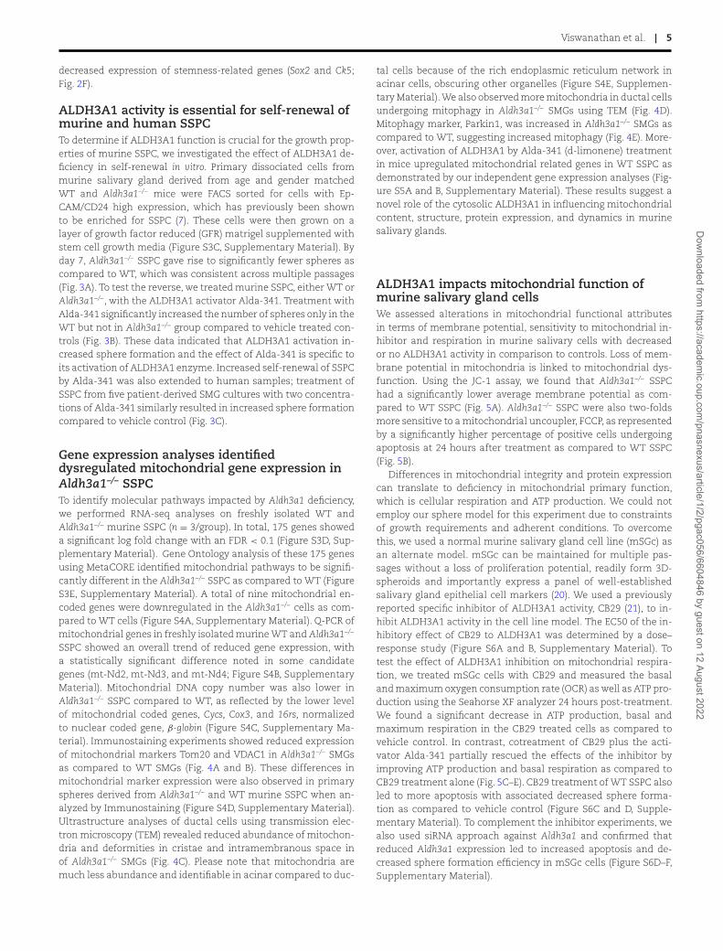
SSPC showed an overall trend of reduced gene expression, witha statistically significant difference noted in some candidategenes (mt-Nd2, mt-Nd3, and mt-Nd4; Figure S4B, SupplementaryMaterial). Mitochondrial DNA copy number was also lower inAldh3a1–/– SSPC compared to WT, as reflected by the lower levelof mitochondrial coded genes, Cycs, Cox3, and 16rs, normalizedto nuclear coded gene, β-globin (Figure S4C, Supplementary Ma-terial). Immunostaining experiments showed reduced expressionof mitochondrial markers Tom20 and VDAC1 in Aldh3a1–/– SMGsas compared to WT SMGs (Fig. 4A and B). These differences inmitochondrial marker expression were also observed in primaryspheres derived from Aldh3a1–/– and WT murine SSPC when an-alyzed by Immunostaining (Figure S4D, Supplementary Material).Ultrastructure analyses of ductal cells using transmission elec-tron microscopy (TEM) revealed reduced abundance of mitochon-dria and deformities in cristae and intramembranous space inof Aldh3a1–/– SMGs (Fig. 4C). Please note that mitochondria aremuch less abundance and identifiable in acinar compared to duc-
tal cells because of the rich endoplasmic reticulum network inacinar cells, obscuring other organelles (Figure S4E, Supplemen-tary Material). We also observed more mitochondria in ductal cellsundergoing mitophagy in Aldh3a1–/– SMGs using TEM (Fig. 4D).Mitophagy marker, Parkin1, was increased in Aldh3a1–/– SMGs ascompared to WT, suggesting increased mitophagy (Fig. 4E). More-over, activation of ALDH3A1 by Alda-341 (d-limonene) treatmentin mice upregulated mitochondrial related genes in WT SSPC asdemonstrated by our independent gene expression analyses (Fig-ure S5A and B, Supplementary Material). These results suggest anovel role of the cytosolic ALDH3A1 in influencing mitochondrialcontent, structure, protein expression, and dynamics in murinesalivary glands.
ALDH3A1 impacts mitochondrial function ofmurine salivary gland cellsWe assessed alterations in mitochondrial functional attributesin terms of membrane potential, sensitivity to mitochondrial in-hibitor and respiration in murine salivary cells with decreasedor no ALDH3A1 activity in comparison to controls. Loss of mem-brane potential in mitochondria is linked to mitochondrial dys-function. Using the JC-1 assay, we found that Aldh3a1–/– SSPChad a significantly lower average membrane potential as com-pared to WT SSPC (Fig. 5A). Aldh3a1–/– SSPC were also two-foldsmore sensitive to a mitochondrial uncoupler, FCCP, as representedby a significantly higher percentage of positive cells undergoingapoptosis at 24 hours after treatment as compared to WT SSPC(Fig. 5B).
Differences in mitochondrial integrity and protein expressioncan translate to deficiency in mitochondrial primary function,which is cellular respiration and ATP production. We could notemploy our sphere model for this experiment due to constraintsof growth requirements and adherent conditions. To overcomethis, we used a normal murine salivary gland cell line (mSGc) asan alternate model. mSGc can be maintained for multiple pas-sages without a loss of proliferation potential, readily form 3D-spheroids and importantly express a panel of well-establishedsalivary gland epithelial cell markers (20). We used a previouslyreported specific inhibitor of ALDH3A1 activity, CB29 (21), to in-hibit ALDH3A1 activity in the cell line model. The EC50 of the in-hibitory effect of CB29 to ALDH3A1 was determined by a dose–response study (Figure S6A and B, Supplementary Material). Totest the effect of ALDH3A1 inhibition on mitochondrial respira-tion, we treated mSGc cells with CB29 and measured the basaland maximum oxygen consumption rate (OCR) as well as ATP pro-duction using the Seahorse XF analyzer 24 hours post-treatment.We found a significant decrease in ATP production, basal andmaximum respiration in the CB29 treated cells as compared tovehicle control. In contrast, cotreatment of CB29 plus the acti-vator Alda-341 partially rescued the effects of the inhibitor byimproving ATP production and basal respiration as compared toCB29 treatment alone (Fig. 5C–E). CB29 treatment of WT SSPC alsoled to more apoptosis with associated decreased sphere forma-tion as compared to vehicle control (Figure S6C and D, Supple-mentary Material). To complement the inhibitor experiments, wealso used siRNA approach against Aldh3a1 and confirmed thatreduced Aldh3a1 expression led to increased apoptosis and de-creased sphere formation efficiency in mSGc cells (Figure S6D–F,Supplementary Material).
Dow
nloaded from https://academ
ic.oup.com/pnasnexus/article/1/2/pgac056/6604846 by guest on 12 August 2022

6 | PNAS Nexus, 2022, Vol. 1, No. 2
Fig. 3. ALDH3A1 deficiency impacts self-renewal of murine and human patient derived SSPC. (A) Left panel shows the graph of the average number ofspheres per well at day 7 for each passage by imaging each well and quantifying by Image J (NIH). Cells were passaged every 7 d for three passages (sixreplicates per group). Middle and right panel reflect the size of the spheres by quantifying the average frequency for the different sphere areas in WTand Aldh3a1–/- SSPC. Representative images of spheres from WT and Aldh3a1–/– SSPC are also shown. (B) Number of spheres from murine WT andAldh3a1–/– SSPC at day 7 post-treatment with 200 μM Alda-341 or vehicle control. Sphere number per well was quantified by Image J (NIH), (sixreplicates per group). (C) Average number of human salivary cells (dissociated from embedded human salivary spheres in matrigel) at day 7 in thepresence of vehicle control, 100 or 200 μM Alda-341 (n = 5 patients, 3 technical replicates). Representative image of human salivary gland spheres isalso shown. Error bars represent SD. Student’s t test was used to calculate the P-value for panels (B) and (C). Two-way ANOVA with multiplecomparisons was used to estimate P-value for panel (D) (∗represents P-value < 0.05, ∗∗< 0.01, ∗∗∗< 0.001, and ∗∗∗∗< 0.0001).
Levels of ROS, glutathione, and ALDH3A1substrate 4-HNE influences survival of Aldh3a1–/–
SSPCIncreased ROS levels have been associated with mitochondrialdysfunction. In the murine SSPC model, ROS levels were sig-nificantly higher in the Aldh3a1–/– SSPC as compared to WTSSPC (Fig. 6A). We have previously found that Aldh3a1–/– SSPChave higher basal apoptosis as compared to WT cells (9).To demonstrate that ROS accumulation impacts survival, wetested the effect of antioxidant treatment on survival of murineAldh3a1–/– SSPC. Treatment of Aldh3a1–/– cells for 24 hours withmito-TEMPO, a mitochondria-specific ROS scavenging molecule,significantly improved Aldh3a1–/– SSPC survival as compared tovehicle control (Fig. 6B). Glutathione plays antioxidative roleagainst elevated ROS. ALDH3A1 helps to convert oxidized glu-tathione (GSSG) to its reduced form (GSH) (22). As expected, in the
absence of ALDH3A1, we observed an increased GSSG–GSH ratioin murine Aldh3a1–/– SSPC by LC/MS analyses (Fig. 6C), which cancontribute to oxidative stress. To test if GSH depletion has an in-dependent effect on poor survival of Aldh3a1–/– SSPC, we supple-mented WT and Aldh3a1–/– SSPC with N-Acetyl-L-cysteine (NAC, acysteine precursor that can boost GSH levels) and assessed sphereformation at day 7. NAC treatment led to a significant increase insphere formation for Aldh3a1–/– SSPC compared to vehicle controlbut no difference for WT SSPC (Figure S7A, Supplementary Mate-rial).
One of the main substrates of ALDH3A1 is 4-hydoxynonenal (4-HNE) which is a product of lipid peroxidation (LPO).We observedincreased accumulation of 4-HNE in Aldh3a1–/– SMG as comparedto WT SMG via immunostaining and western blotting (Fig. 6Dand E). Hydrazine derivatives have been reported to rescue theeffects of 4-HNE accumulation in smooth muscle cells (23). Asexpected, hydralazine treatment on Aldh3a1–/– SSPC for 24 hours
Dow
nloaded from https://academ
ic.oup.com/pnasnexus/article/1/2/pgac056/6604846 by guest on 12 August 2022
Viswanathan et al. | 7
Fig. 4. Aldh3a1 deficient SMG have lower protein expression, lower mitochondrial abundance, altered morphology, and increased mitophagy. (A)Representative Immunostaining images of mitochondrial (Tom20 and Vdac1), ductal (Sox9), and acinar (Aqp5) markers in WT and Aldh3a1–/– SMGs.Scale bar: 50 μM (10 images per staining, n = 3/group). Right panel shows quantification of Tom20 and Vdac1 staining in WT and Aldh3a1–/– SMG. Atotal of 10 random field of view used for quantification per using ImageJ and represented as the Mean of Fluorescence intensity (MFI). (B) Highmagnification immunostaining image of WT and Aldh3a1–/– SMGs showing mitochondrial marker Tom20 and Acinar marker Aqp5. (C) TEM image ofductal cells in SMGs derived from WT and Aldh3a1–/– showing more mitochondrial abundance and better ultrastructure in WT cells (n = 3/group, scalebar = 2 μM left panel, and 1 μM right panel). (D) Representative TEM images of Aldh3a1–/– ductal cells in SMG showing mitophagy (arrow). (E)Immunostaining analyses of mitophagy marker, Parkin, in WT and Aldh3a1–/– SMGs imaged at 200x total magnification and is quantified andrepresented in the right panel (n = 3/group and scale bar: 130 uM). Error bars represent SD. Student’s t test was used to calculate the P-value(∗represents P-value < 0.05, ∗∗< 0.01, and ∗∗∗< 0.001).
significantly decreased the proportion of cells undergoing apop-tosis and improved survival (Fig. 6F). To confirm if the hydrazineacts like a scavenger for 4-HNE, we exposed WT SSPC for 24 hourswith 4-HNE alone, hydralazine alone, or the two together and as-
sessed apoptosis. There was an induction of cell death with 4-HNE treatment alone, which was rescued upon cotreatment ofhydralazine (Fig. 6G). Overall, our data suggests ALDH3A1 de-ficiency leads to increased accumulation of ROS, its byproduct
Dow
nloaded from https://academ
ic.oup.com/pnasnexus/article/1/2/pgac056/6604846 by guest on 12 August 2022

8 | PNAS Nexus, 2022, Vol. 1, No. 2
Fig. 5. Aldh3a1 deficient SSPC display attributes of dysregulated mitochondrial function as compared to WT. (A) JC-1 assay analysis of WT andAldh3a1–/– murine SSPC showing proportions of PE-high/FITC-high cells with mitochondria that have intact membrane potential. This is quantifiedand represented in the graph in right panel (n = 3 and 2 technical replicates). (B) Quadrant plots showing Annexin-V/PI staining of WT and Aldh3a1–/–
murine SSPC 24 hours post-treatment with mitochondrial inhibitor FCCP; this is quantified and represented in Graph in right panel (n = 3 withtechnical duplicates). The data is normalized to % apoptosis in the WT group. (C) Average OCR over time of mSGc cells treated with vehicle, 10 uMALDH3A1 inhibitor CB29, 100 uM Alda-341, and combination of both. (D) Average ATP production in various treatment groups. (n = 2 with 4 technicalreplicates). (E) Average basal and maximum respiration in various treatment groups. Error bars represent SD. Student’s t test was used to calculate theP-value for panel (B) and one-way ANOVA with multiple comparisons was used for panels (D) and (E) (∗represents P-value < 0.05, ∗∗< 0.01, ∗∗∗< 0.001,and ∗∗∗∗< 0.0001).
4-HNE, and decreased GSH that contributes to poor survival ofSSPC.
DiscussionGene expression profiling of murine SSPC had helped us to iden-tify and validate GDNF and ALDH3A1 as crucial determinants ofsalivary gland function and repair upon radiation stress (7, 8).
ALDH3A1-deficient mice display poorer saliva function after RTas compared to WT. Treatment with the ALDH3A1 activator, Alda-341 (d-limonene), improved function of SMGs by reducing theacute RT-induced aldehyde load and promoting SSPC survival (9).However, our data showing that long-term activation of ALDH3A1(upto 18 to 20 weeks) with Alda341 (d-limonene) is needed forlong-term maintenance of improved saliva function suggest thatALDH3A1 plays a more sustained role in SSPC renewal rather than
Dow
nloaded from https://academ
ic.oup.com/pnasnexus/article/1/2/pgac056/6604846 by guest on 12 August 2022

Viswanathan et al. | 9
Fig. 6. Accumulation of ROS and substrate 4-HNE in Aldh3a1 deficient SSPC drives poor survival. (A) Histogram plots of ROS levels determined by FACSanalyses in WT and Aldh3a1–/– murine SSPC; this is quantified and represented in the right panel (n = 3 and 2 technical replicates). (B) Bar graphshowing % apoptosis of Aldh3a1–/– murine SSPC 24 hours normalized to vehicle control post-treatment with100 nM mito-TEMPO. (n = 2 and 3 technicalreplicates). (C) Carbon labeling experiments demonstrate differences in glutathione turnover in WT and Aldh3a1–/– SSPC represented as the ratio ofabsolute abundance of GSSG and GSH. (D) Immunostaining and (E) Western blot analyses of 4-HNE in WT and Aldh3a1–/– SMGs (n = 3/group for IHC,n = 2 for western blotting). Scale bar: 100 μM. (E) Average % apoptosis in Aldh3a1–/– SSPC 24 hours post-treatment with 10 uM of hydralazinenormalized to control. (n = 3 and 3 technical replicates). (F) Average % apoptosis in WT SSPC 24 hours post-treatment with 10 uM 4-HNE, 10 uM ofhydralazine or the combination with 4-HNE and hydralazine normalized to control (n = 2 and 3 technical replicates). Error bars represent SD. Student’st test was used to calculate the P-value for panels (A) to (D) and (F). One-way ANOVA with multiple comparisons was used for panel G (∗representsP-value < 0.05, ∗∗< 0.01, and ∗∗∗< 0.001).
just acute clearance of radiation-induced aldehyde. This led us toexplore the effect of ALDH3A1 in SSPC phenotype using both em-bryonic and salisphere models.
We found that ALDH3A1 is important for self-renewal, survival,and differentiation of murine SSPC beyond acute radiation stress.Genetic deletion of Aldh3a1 results in morphological differences inmurine SMG that can be attributed to abnormal development pat-terns. Branching morphogenesis of murine embryonic SMGs is an
excellent model to study the effect of cell specific ablation of fac-tors important for differentiation or development (12). Reducedbranching and number of end buds in Aldh3a1 deficient explantssuggests its role in acinar cell development. These findings res-onate with other studies; for example, SOX2 was identified as amarker of SMG progenitor cells that are capable of giving rise toacinar cells but not ductal cells (24). Indeed, differences observedin embryonic morphogenesis of fetal explants can explain why
Dow
nloaded from https://academ
ic.oup.com/pnasnexus/article/1/2/pgac056/6604846 by guest on 12 August 2022

10 | PNAS Nexus, 2022, Vol. 1, No. 2
we see reduced acinar compartment in adult SMGs of KO miceas compared to WT. However, the potential role of ALDH3A1 inthe context of adult salivary stem cell differentiation is yet to beexplored.
ALDH3A1 activity also promoted self-renewal of murine andhuman SSPC. Self-renewal is a characteristic attribute of adultstem cells, which upon division gives rise to a differentiateddaughter cell and a stem cell. Sphere formation assay fromisolated murine SSPC is an in vitro technique to assess changes inself-renewal upon treatment or intervention (25). As sphere for-mation is a balance of proliferation and survival, we predict thatALDH3A1 deficiency affects SSPC survival, thus shifting the over-all balance and reducing sphere formation.
Our in-depth gene expression analyses identifies a strong linkbetween Aldh3a1 deficiency and reduced mitochondrial gene ex-pression and function in SSPC. A survey of the literature showedonly two studies that reported a relationship between ALDH3A1and mitochondrial function. In a yeast model, a homolog ofALDH3A1 plays a critical role in the synthesis of a precursormolecule of coenzyme Q, an important carrier of the mitochon-drial electron transport chain (ETC) (26). In gastric cancer, inhibit-ing ALDH3A1 impairs mitochondrial activity due to reduced betaoxidation of lipids and acetyl co-A flux for TCA cycle (27), whichwas also demonstrated by our LC/MS analyses (Figure S7B, Sup-plementary Material). These cumulative findings strongly demon-strate that activation of the cytosolic ALDH3A1 is needed to sup-port mitochondrial function in SSPCs under basal, nonstressedconditions. Although we primarily focus on ductal cells due to theenrichment of Aldh3a1 expression (single cell RNAseq) and mito-chondria number (TEM) in the ductal compartment, we cannotexclude the possibility that the same process may also be occur-ring in acinar cells, especially self-renewing ones. As shown in themagnified view, increased 4-HNE level is seen in both ductal andacinar cells with Aldh3a1 deletion despite the low expression ofthis gene in the acinar compartment. Future work is needed todetermine the role of ALDH3A1 in acinar cells.
Mechanistically, our data suggest a vicious cycle between lossof ALDH3A1 activity and mitochondrial function: increased ROSlevels in Aldh3a1–/– SSPC leads to higher oxidative stress, reducedglutathione turnover, increased 4-HNE accumulation and mito-chondrial damage, and reduced survival (Figure S8A, Supplemen-tary Material). This is consistent with prior data showing that irra-diation to the saliva glands leads to an increase in mitochondrialROS and loss of membrane potential, resulting in caspase 3 activa-tion and subsequent apoptosis, that can be improved by the treat-ment of mitochondria specific scavenger mito-TEMPO (28,29). Mi-tochondria are the primary source of ROS, which is partially con-verted to toxic products such as 4-Hydroxynonenal (4-HNE) viaLPO (30, 31). ALDH3A1 inactivates 4-HNE, as well as replenishesreduced glutathione pool to maintain homeostasis and viability(18, 26). GSH depletion has been attributed to the activation ofapoptosis via post-translational modifications, thus providing aparallel mechanism that contribute to poor survival of SSPC defi-cient of ALDH3A1 (32). The loss or decrease activity of ALDH3A1leads to accumulation of 4-HNE, which in turn causes proteinadduct formation and inactivation, mitochondrial dysfunction,increased ROS accumulation, and increased apoptosis in cells (33,34). 4-HNE accumulation can induce cell death via extrinsic or in-trinsic apoptotic pathways by causing DNA damage in cells (35,36). In our model, we found higher gammaH2AX foci as well asDNA damage response gene expression (p21 and Bax) in Aldh3a1–/-
SMG and SSPC, respectively as compared to WT (Figure S8B, Sup-plementary Material). These findings is consistent with the previ-
ously identified role of ALDH3A1 to protect against DNA damagein human corneal and bronchial epithelial cells (37, 38).
Physiologically, adult stem cells are found in hypoxic niche;thus, they normally experience low ROS levels (39). In such hy-poxic environment, adult stem cells prefer glycolysis and fattyacid oxidation as metabolic drivers. Based on previously identifiedrole (27), we can postulate that ALDH3A1 function is crucial for ef-ficient FAO in these cells. Adult stem cell niche are also sensitive toROS produced by radiation that can prime them for differentiationand apoptosis (40, 41). Increased ALDH3A1 activity together withreplenished GSH pool operate as oxidative stress responses to pro-tect adult stem cells from ROS damage. Stress such as radiation,increases ROS levels and impairs their ability to self-renew andproliferate long term, preventing regeneration of salivary glandeven with termination of the radiation insult (42, 43). Our datashowed that sustained activation of ALDH3A1 can protect thecells from oxidative damage over time, leading to long-term sur-vival of SSPC and sustained improvement of salivary function. Fu-ture work will explore how ALDH3A1 activity can influence othereffects such as chronic inflammation postradiation that poses agreater risk for tissue regeneration (44). Overall, our data helpedto guide the duration of d-limonene treatment in a phase I trialthat has just been activated in HNC patients (NCT04392622). Inthis trial, d-limonene is administered during and up to 20 weeksafter definitive chemoradiotherapy for salivary gland protectionfrom radiation damage.
In summary, our data indicate that ALDH3A1 activity is re-quired to support embryonic differentiation and self-renewalfunction of adult salivary stem cells, at least in part, by main-taining mitochondrial number and functions. It explains the dif-ferences in tissue architecture when this enzyme is deleted andsupports the continuing use of ALDH3A1 activators after the endof radiation to assist stem cell renewal and possibly differentia-tion in that setting.
MethodsAnimals usedC57BL/6 mice (7 to 10 weeks) were purchased from Jackson Labo-ratories (Bar Harbor, ME). C57BL/6J Aldh3a1–/– mice were obtainedfrom the laboratory of Vasilis Vasiliou at Yale School of PublicHealth, New Haven, CT (45). Experiments were done with 8 to 12weeks old female mice. All the protocols were approved by The Ad-ministrative Panel on Laboratory Animal Care (APLAC) at StanfordUniversity, Stanford, CA. All the experiments involving animalswere done in adherence to the NIH Guide for the Care of and Useof Laboratory Animals. For our analyses, we used female mice be-cause murine SMGs display sexual dimorphism and female SMGsshow closer resemblance to humans (46).
Salivary gland isolation and culture from mouseand human SMGsMurine salivary glands were dissected and isolated from eutha-nized mice as described previously (8). Please see Supplementalmethods for details.
RNA sequencingRNA sequencing experiment was done as previously described (9).Please see Supplemental methods for details.
Dow
nloaded from https://academ
ic.oup.com/pnasnexus/article/1/2/pgac056/6604846 by guest on 12 August 2022

Viswanathan et al. | 11
Single-cell RNA-Seq data analysesReady to use SEURAT objects for Embryonic and Postnatal SMGintegrated datasets were retrieved from figshare as indicated inthe original study (19). The code used for the analyses is providedin Data S1 (Supplementary Material).
RT-PCRRNA was isolated from whole tissue or SSPC using RNAqueousMicro Kit (Ambion, Austin, TX). Total RNA samples were DNase-treated (Ambion) before complementary DNA (cDNA) synthesisusing SuperScript reagents according to manufacturer’s protocol(Invitrogen, Waltham, MA). SYBRgreen RT-qPCR was performedusing cDNA and primers used before (47) or designed usingPrimer3 and Beacon Designer software or found using PrimerBank(http://pga.mgh.harvard.edu/primerbank/). Gene expression wasnormalized to the housekeeping gene S29 (Rps29) or actin.
Embryo salivary gland dissection and branchingmorphogenesis assaySalivary glands were dissected out during various stages ofmurine embryos development as described previously (48). E13.5salivary glands were used for the branching morphogenesis assay.Please refer to Supplemental methods for more details.
EM imagingSalivary glands from WT and Aldh3a1–/– mice were dissected andused for TEM. Please refer to Supplemental methods for details.
Mitochondria respirationA total of 10,000 cells were seeded in Agilent (Santa Clara, CA) Sea-horse XF96 cell culture microplates. Next day, cells were treatedwith vehicle control, CB29 (20 μM), Alda-341 (100 μM), and combi-nation of the two drugs. After 24 hours of incubation, media wasreplaced, and cells were maintained at 37◦C. Extracellular flux as-say to measure mitochondrial respiration and ATP production wascarried out as recommended by manufacturer’s protocol (Agilent).SRB assay (49) was used to quantify cell concentration in samplesfor normalization of data.
Mitochondrial isolation and copy numberanalysesMitochondrial DNA was isolated from a modified version of a pro-tocol (50). Cells were scraped using RIPA lysis buffer and incubatedwith 5 μl of Proteinase K (Invitrogen) for 55◦C for 3 hours. Sam-ples were sonicated briefly to shear the DNA. The debris was re-moved from spinning down the samples at 8,000 g for 15 min. Tothe supernatant, equal volume of phenol/chloroform/Isoamyl al-cohol mixture (25:4:1) was added. The samples were mixed well byvigorous shaking followed by centrifugation at 8,000 g for 15 min.The clear upper layer was collected, added to equal volume ofchloroform/isoamyl alcohol and mixed well. The samples werespun down and the upper layer was transferred to a fresh tube, towhich 40 μl of 3M sodium acetate and 440 μl of Isopropanol wereadded. Samples were incubated at −20◦C for 10 min to facilitateDNA precipitation. Samples were spun again to pellet the precipi-tated DNA. Supernatant was discarded and the pellet was washedwith 70% alcohol. The supernatant was discarded, and the driedpellet was resuspended in molecular grade water (Invitrogen). Forcopy number analyses, 1 ng of DNA was diluted in 100 μl of water.MtDNA and genomic DNA isolated was subjected to qPCR (stan-dard conditions) using primers against mitochondrial genes cycs,
cox3, and 16rs and nuclear encoded gene beta-globin. Primer se-quences and run conditions for mitochondrial genes were as pre-viously described (50). The final data was represented as relativeamount of mitochondrial gene amplification normalized to nu-clear gene amplification.
Mitochondria membrane potential assayJC-1 assay (Adipogen Life Sciences, San Diego, CA) was used toassess mitochondrial membrane potential. SSPC were incubatedwith 0.5 μM of JC-1 reagent in 3% Bovine Serum Albumin (BSA) inPBS for 10 min at 37◦C. Cells were washed once with excess PBSand were resuspended in 200 μl of BSA solution. Samples weresubjected to FACS analyses where dot plots with axes FITC-A vs.PE-A with positive gates set according to the unstained sample.Using FlowJo software (BD biosciences, San Jose, CA) for analy-ses, healthy mitochondria were identified as cell populations thatwere FITChigh PEhigh and unhealthy mitochondria (due to loss ofmembrane potential) were identified as FITChigh PElow population.
Estimation of ROS levelsCellular ROS was estimated using the CM-H2DCFDA reagent(ThermoFisher). Cells were incubated with 5 μM of the reagent in3%BSA in PBS at 37◦C for 30 min. Cells were washed once with ice-cold PBS and then subjected to FACS analyses. Histogram plots ofFITC-Area from 10,000 cells were recorded and mean fluorescenceintensity (MFI) were estimated for analyses using FlowJo software.
RNAscope RNA hybridizationParaffin embedded tissue sections were used for RNAscope. Probesagainst mouse Aldh3a1 and ckit were used (ACD bio, Newark,CA). The slides were baked at 70◦C for an hour. Slides were de-paraffinized with fresh xylene treatment followed by alcohol andfinally rehydrated in distilled water. Antigen retrieval step wasperformed by incubating the slides in 1X RNAscope antigen re-trieval buffer at 97◦C for 30 min. After cooling down, diluted pro-tease reagent was added on to the sections and incubated at 40◦Cfor 10 min. Sides were washed with distilled water two times for2 min each. Slides were incubated in the probes (1:50) at 40◦C for16 hours (overnight step). Next day, slides were washed twice in1X RNAscope wash buffer 2 min each. Three step amplificationwas carried for 15 to 30 min at 40◦C with intermittent wash-ing steps. After the final wash, slides were incubated in washingbuffer containing DAPI (1:10,000) followed by mounting cover slipscarefully with ProlongGold on glass slides. A total of five to six ep-ithelia images were acquired using a total magnification of 630xusing the Leica DMi8 fluorescence inverted microscope. Quan-tification was done using Qupath software following instructionsrecommended by the manufacturer.
ALDH activity assayHuman recombinant ALDH1A1, ALDH1A2, ALDH1A3, ALDH1B1,ALDH2, ALDH3A1, ALDH3A2, and ALDH4A1 were expressed andpurified using nickel column chromatography as previously de-scribed (51). The assay buffer was 100 mM sodium phosphate(pH 8.0) with 1 mM MgCl2. Reaction was conducted in the whiteopaque 96-well assay plates (Corning Costar, flat-bottom, andnontreated) with a total reaction volume of 100 μl for eachwell. Each reaction mixture consisted of 100 nM ALDH enzyme,0.1% DMSO, 1 mM β-mercaptoethanol, 1 mM NAD+, 1 mMacetaldehyde (lastly added), and CB29 of indicated concentra-tion in assay buffer. Enzymatic activity was measured basedon NADH-mediated fluorescence (Ex340/Em460 nm). The activity
Dow
nloaded from https://academ
ic.oup.com/pnasnexus/article/1/2/pgac056/6604846 by guest on 12 August 2022

12 | PNAS Nexus, 2022, Vol. 1, No. 2
was recorded for 10 min on a SpectraMax M2e microplate readeroperated by the SoftMax Pro software. ALDH enzymatic activitycaused by CB29 was normalized to the DMSO control. The single-dose inhibition data and dose–response inhibition curves wereprocessed with the Prism software.
LC/MS analyses of metabolitesFreshly isolated cells from murine WT and Aldh3a1–/– SMGs wereused from LC/MS analyses. Please refer to Supplemental methodsfor details.
siRNA transfectionMSGc (120,000/well) were plated in a 6-well format. A total of 24hours later, cells were transfected with 30 pmoles of siAldh3a1(Ambion, AM16708) and scramble siRNA using Lipofectamine3000 following Manufacturers recommended protocol. A total of24 hours later, cells were trypsinized, counted, and used for down-stream analyses.
Irradiation of salivary glands and salivarycollectionA total radiation dose of 30 Gy was delivered in five fractions(6 Gy/fraction/day) to the SMG with the rest of the body leadshielded. Mice were treated with 10% Alda-341 mixed in dailychow or no treatment. Stimulated saliva was measured as pre-viously described (6). Mice were anesthetized with a ketamine(80 mg/kg) and xylazine (16 mg/kg) mixture delivered by intraperi-toneal injection and subcutaneously injected with 2 mg/kg pilo-carpine. Saliva was collected for 15 min. Saliva volume was calcu-lated by assuming the density as 1, was normalized to the mousebody weight by dividing the total collected saliva volume by themass of the mouse (kg).
Study approvalHuman salivary glands were procured from HNC patients in ac-cordance with the guidelines approved by the Stanford Univer-sity’s Institutional Review Board. Written informed consent wasreceived from participants prior to inclusion in our study. The hu-man tissue was rinsed twice with diluted betadine solution andwashed with excess sterile phosphate buffered saline (PBS) threetimes before dissociation.
StatisticsAll data are represented as averages with standard deviation (SD).Statistical ANOVA and Student’s t tests were used to compare thedata. All tests performed were two-sided with an alpha level of0.05. P ≤ 0.05 was considered to be significant. All data was ana-lyzed using GraphPad Prism 8.4.3 (GraphPad Software Inc, La Jolla,CA).
AcknowledgementsWe would also like to thank Nan Xiao, Daria Mochly-Rosen’sgroup, and Sarah Knox’s group for valuable input and suppliesfor this research work. We would like to sincerely thank Rose-Anne Romano at the University of Buffalo for mSGc cells. Wethank Dr. Vasilis Vasiliou for the Aldh3a1−/− transgenic mice. Wethank Pauline Chu at the Histology core facility, Deana Rae Crys-tal Colburg at the Department of Pathology at Stanford Schoolof Medicine. We thank Dr. John Coller and Vida Shokoohi for theRNA-seq. We thank Sivakumasundari V for her assistance withsingle cell RNA-Seq data analyses. We acknowledge John Perrino
and the EM core facility at the Stanford University for assistancewith EM data.
Supplementary MaterialSupplementary material is available at PNAS Nexus Online.
FundingResearch presented here was supported in part by the TobaccoRelated Disease Research Program Postdoctoral Fellowship Award(27FT-0038) to V.V., by the award number R01 DE029672-01A1 fromNational Institute of Dental and Craniofacial Research to H.C.,D.N., L.G. and Q.T.L., by the ARRA award number 1S10RR026780-01from the National Center for Research Resources (NCRR), by theNational Center For Advancing Translational Sciences of the Na-tional Institutes of Health under award number UL1TR003142, bythe award number 2U10CA180868-06 and P30CA124435 from theNational Cancer Institute to Q.T.L., and by the National Institutesof Health Award AA11147 to D.M-R. The content is solely the re-sponsibility of the authors and does not necessarily represent theofficial views of the National Institutes of Health.
Authors’ ContributionsV.V., J.S., and Q.T.L. conceived the project. V.V., J.S., D.J., A.M., D.N.,S.K., D.M.R., and Q.T.L. designed, performed experiments, and theanalyses. H.C. assisted with the animal experiments. J.B. helpedwith the image analyses and animal experiments. M.C. assistedwith the conceptual study design. S.J., G.N., and L.G. helped withthe RNAscope experiments. Z.F. performed the enzymatic activ-ity assays. Y.L., H.J., and J.Y. performed the LC/MS experimentand analyses. E.L. assisted with the seahorse experiment. N.D. as-sisted with the experiment design and hypothesis. R.V.E. helpedwith the statistical analyses. D.S., F.C.H., and M.K. assisted withproviding human tissue samples. R.L. helped with patient tissueprocurement and IRB protocol approval. V.V. and Q.T.L. wrote themanuscript.
Data AvailabilityData generated in this study is available on public open accessrepository Dyrad (https://doi.org/10.5061/dryad.9w0vt4bgg).
References1. Miletich I. 2010. Introduction to salivary glands: structure, func-
tion and embryonic development. Saliv Glands. 14:1–20.2. Frank RM, Herdly J, Philippe E. 1965. Acquired dental defects and
salivary gland lesions after irradiation for carcinoma. J Am DentAssoc. 70(4):868–883.
3. Ao R, Jb D. 1967. Dental aspects of the problems, care, and treat-ment of the irradiated oral cancer patient. J Am Dent Assoc.74(5):957–966.
4. Valdez IH, Atkinson JC, Ship JA, Fox PC. 1993. Major salivarygland function in patients with radiation-induced xerostomia:Flow rates and sialochemistry. Int J Radiat Oncol Biol Phys.25(1):41–47.
5. Sasportas LS, et al. 2013. Cost-effectiveness landscape analysisof treatments addressing xerostomia in patients receiving headand neck radiation therapy. Oral Surg Oral Med Oral Pathol OralRadiol. 116(1):e37–e51.
Dow
nloaded from https://academ
ic.oup.com/pnasnexus/article/1/2/pgac056/6604846 by guest on 12 August 2022

Viswanathan et al. | 13
6. Banh A, et al. A novel aldehyde dehydrogenase-3 activator leadsto adult salivary stem cell enrichment in vivo. Clin Cancer Res.17:7265–7272. [ updated 2021 Jul 22]. https://clincancerres.aacrjournals.org/content/17/23/7265.
7. Xiao N, et al. 2013. A novel aldehyde dehydrogenase-3 activa-tor (Alda-89) protects submandibular gland function from ir-radiation without accelerating tumor growth. Clin Cancer Res.19(16):4455–4464.
8. Xiao N, et al. 2014. Neurotrophic factor GDNF promotes survivalof salivary stem cells. J Clin Invest. 124(8):3364–3377.
9. Saiki JP, et al. 2018. Aldehyde dehydrogenase 3A1 activation pre-vents radiation-induced xerostomia by protecting salivary stemcells from toxic aldehydes. Proc Natl Acad Sci USA. 115(24):6279–6284.
10. Bullard T, et al. 2008. Ascl3 expression marks a progenitor popu-lation of both acinar and ductal cells in mouse salivary glands.Dev Biol. 320(1):72–78.
11. Hisatomi Y, et al. 2004. Flow cytometric isolation of endodermalprogenitors from mouse salivary gland differentiate into hepaticand pancreatic lineages. Hepatology. 39(3):667–675.
12. Knox SM, et al. 2010. Parasympathetic innervation maintains ep-ithelial progenitor cells during salivary organogenesis. Science.329(5999):1645–1647.
13. Chibly AM, Querin L, Harris Z, Limesand KH. 2014. Label-retaining cells in the adult murine salivary glands possess char-acteristics of adult progenitor cells. PLoS ONE. 9(9):e107893.
14. Kimoto M, Yura Y, Kishino M, Toyosawa S, Ogawa Y. 2008. Label-retaining cells in the rat submandibular gland. J Histochem Cy-tochem. 56(1):15–24.
15. May AJ, et al. 2018. Diverse progenitor cells preserve salivarygland ductal architecture after radiation-induced damage. De-velopment. 145(21):dev166363.
16. Aure MH, Konieczny SF, Ovitt CE. 2015. Salivary gland homeosta-sis is maintained through acinar cell self-duplication. Dev Cell.33(2):231–237.
17. Emmerson E, Knox SM. 2018. Salivary gland stem cells: a reviewof development, regeneration and cancer. Genesis. 56(5):e23211.
18. Estey T, Piatigorsky J, Lassen N, Vasiliou V. 2007. ALDH3A1: acorneal crystallin with diverse functions. Exp Eye Res. 84(1):3–12.
19. Hauser BR, et al. 2020. Generation of a single-cell RNAseq atlasof murine salivary gland development. iScience. 23(12):101838.
20. Min S, et al. 2018. Functional characterization and genomic stud-ies of a novel murine submandibular gland epithelial cell line.PLoS ONE. 13(2):e0192775.
21. Parajuli B, Georgiadis TM, Fishel ML, Hurley TD. 2014. Develop-ment of selective inhibitors for human aldehyde dehydrogenase3A1 (ALDH3A1) for the enhancement of cyclophosphamide cy-totoxicity. Chembiochem. 15(5):701–712.
22. Lassen N, et al. 2006. Antioxidant function of corneal ALDH3A1in cultured stromal fibroblasts. Free Radic Biol Med. 41(9):1459–1469.
23. Galvani S, et al. 2008. Carbonyl scavenger and antiatherogenic ef-fects of hydrazine derivatives. Free Radic Biol Med. 45(10):1457–1467.
24. Emmerson E, et al. 2017. SOX2 regulates acinar cell developmentin the salivary gland. eLife. 6:e26620. Horsley V, editor.
25. Nanduri LSY, et al. 2014. Purification and ex vivo expansionof fully functional salivary gland stem cells. Stem Cell Rep.3(6):957–964.
26. Payet L-A, et al. 2016. Mechanistic details of early steps incoenzyme Q biosynthesis pathway in yeast. Cell Chem Biol.23(10):1241–1250.
27. Lee J-S, et al. 2019. Gastric cancer depends on aldehydedehydrogenase 3A1 for fatty acid oxidation. Sci Rep. 9(1):16313.
28. Liu X, et al. 2017. Radiation inhibits salivary gland func-tion by promoting STIM1 cleavage by caspase-3 and lossof SOCE through a TRPM2-dependent pathway. Sci Signal.10(482):eaal4064.
29. Liu X, Subedi KP, Zheng C, Ambudkar I. 2021. Mitochondria-targeted antioxidant protects against irradiation-induced sali-vary gland hypofunction. Sci Rep. 11(1):7690.
30. Ayala A, Muñoz MF, Argüelles S. 2014. Lipid peroxidation: pro-duction, metabolism, and signaling mechanisms of malon-dialdehyde and 4-hydroxy-2-nonenal. Oxid Med Cell Longev.2014:360438.
31. Forman HJ. 2010. Reactive oxygen species and alpha,beta-unsaturated aldehydes as second messengers in signal trans-duction. Ann N Y Acad Sci. 1203:35–44.
32. Franco R, Cidlowski JA. 2009. Apoptosis and glutathione: beyondan antioxidant. Cell Death Differ. 16(10):1303–1314.
33. Black W, et al. 2012. Molecular mechanisms of ALDH3A1-mediated cellular protection against 4-hydroxy-2-nonenal. FreeRadic Biol Med. 52(9):1937–1944.
34. Dodson M, et al. 2017. Regulation of autophagy, mitochondrialdynamics, and cellular bioenergetics by 4-hydroxynonenal inprimary neurons. Autophagy. 13(11):1828–1840.
35. Dalleau S, Baradat M, Guéraud F, Huc L. 2013. Cell death anddiseases related to oxidative stress:4-hydroxynonenal (HNE) inthe balance. Cell Death Differ. 20(12):1615–1630.
36. Sharma A, et al. 2008. 4-Hydroxynonenal induces p53-mediatedapoptosis in retinal pigment epithelial cells. Arch Biochem Bio-phys. 480(2):85–94.
37. Jang J-H, et al. 2014. Aldehyde dehydrogenase 3A1 protects air-way epithelial cells from cigarette smoke-induced DNA damageand cytotoxicity. Free Radic Biol Med. 68:80–86.
38. Voulgaridou G-P et al. 2020. Aldehyde dehydrogenase 3A1 con-fers oxidative stress resistance accompanied by altered DNAdamage response in human corneal epithelial cells. Free RadicBiol Med. 150:66–74.
39. Mohyeldin A, Garzón-Muvdi T, Quiñones-Hinojosa A. 2010. Oxy-gen in stem cell biology: a critical component of the stem cellniche. Cell Stem Cell. 7(2):150–161.
40. Renault VM, et al. 2009. FoxO3 regulates neural stem cell home-ostasis. Cell Stem Cell. 5(5):527–539.
41. Tothova Z, et al. 2007. FoxOs are critical mediators of hematopoi-etic stem cell resistance to physiologic oxidative stress. Cell.128(2):325–339.
42. Henry E, et al. 2020. Human hematopoietic stem/progenitorcells display reactive oxygen species-dependent long-termhematopoietic defects after exposure to low doses of ionizingradiations. Haematologica. 105(8):2044–2055.
43. Yamaguchi M, Kashiwakura I. 2013. Role of reactive oxygenspecies in the radiation response of human hematopoieticstem/progenitor cells. PLoS ONE. 8:e70503.
44. McBride WH, Schaue D. 2020. Radiation-induced tissue damageand response. J Pathol. 250(5):647–655.
45. Nees DW, Wawrousek EF, Robison WG, Piatigorsky J. 2002.Structurally normal corneas in aldehyde dehydrogenase 3a1-deficient mice. Mol Cell Biol. 22(3):849–855.
46. Jayasinghe NR, Cope GH, Jacob S. 1990. Morphometric studies onthe development and sexual dimorphism of the submandibulargland of the mouse. J Anat. 172:115–127.
47. Manczak M, Jung Y, Park BS, Partovi D, Reddy PH. 2005. Time-course of mitochondrial gene expressions in mice brains: impli-
Dow
nloaded from https://academ
ic.oup.com/pnasnexus/article/1/2/pgac056/6604846 by guest on 12 August 2022

14 | PNAS Nexus, 2022, Vol. 1, No. 2
cations for mitochondrial dysfunction, oxidative damage, andcytochrome c in aging. J Neurochem. 92(3):494–504.
48. Rebustini IT, Hoffman MP. 2009. ECM and FGF-dependent as-say of embryonic submandibular gland epithelial morphogen-esis: investigating growth factor/matrix regulation of gene ex-pression during development. Methods Mol Biol. 522:319–330.
49. Vichai V, Kirtikara K. 2006. Sulforhodamine B colorimetric assayfor cytotoxicity screening. Nat Protoc. 1(3):1112–1116.
50. Guo W, Jiang L, Bhasin S, Khan SM, Swerdlow RH. 2009.DNA extraction procedures meaningfully influence qPCR-based mtDNA copy number determination. Mitochondrion. 9(4):261–265.
51. Chen C-H, Cruz LA, Mochly-Rosen D. 2015. Pharmacological re-cruitment of aldehyde dehydrogenase 3A1 (ALDH3A1) to assistALDH2 in acetaldehyde and ethanol metabolism in vivo. ProcNatl Acad Sci USA. 112(10):3074–3079.
Dow
nloaded from https://academ
ic.oup.com/pnasnexus/article/1/2/pgac056/6604846 by guest on 12 August 2022