Embed Size (px)
Citation preview

1
Submitted: 2 March, 2019; Revised: 25 April, 2019
© Sleep Research Society 2019. Published by Oxford University Press on behalf of the Sleep Research Society. All rights reserved. For permissions, please email: [email protected]
Original article
Simultaneous tonic and phasic REM sleep without atonia best
predicts early phenoconversion to neurodegenerative disease in
idiopathic REM sleep behavior disorderJiri Nepozitek1,*, , Simona Dostalova1, Petr Dusek1, David Kemlink1, , Iva Prihodova1, Veronika Ibarburu Lorenzo y Losada1, Latica Friedrich1,2, Ondrej Bezdicek1, Tomas Nikolai1, Pavla Perinova1, Irene Dall’Antonia1, Pavel Dusek1, Martin Ruml1, Evzen Ruzicka1 and Karel Sonka1
1Department of Neurology and Center of Clinical Neuroscience, First Faculty of Medicine, Charles University and General University Hospital
in Prague, Prague, Czech Republic and 2Department of Neurology, Sveti Duh University Hospital, Zagreb, Croatia
*Corresponding author. Jiri Nepozitek, Department of Neurology and Center of Clinical Neuroscience, First Faculty of Medicine, Charles University and General University Hospital in Prague, Katerinska 30, Prague, 120 00, Czech Republic. Email: [email protected].
Abstract
Study Objectives: Rapid eye movement (REM) sleep without atonia (RWA) is the main polysomnographic feature of idiopathic REM sleep behavior disorder (iRBD)
and is considered to be a promising biomarker predicting conversion to manifested synucleinopathy. Besides conventionally evaluated tonic, phasic and any RWA,
we took into consideration also periods, when phasic and tonic RWA appeared simultaneously and we called this activity “mixed RWA.” The study aimed to evaluate
different types of RWA, to reveal the most relevant biomarker to the conversion.
Methods: A total of 55 patients with confirmed iRBD were recruited with mean follow-up duration 2.3 ± 0.7 years. Scoring of RWA was based on Sleep Innsbruck
Barcelona rules. Positive phenocoversion was ascertained according to standard diagnostic criteria during follow-up. Receiver operator characteristic analysis was
applied to evaluate predictive performance of different RWA types.
Results: A total of nine patients (16%) developed neurodegenerative diseases. Yearly phenoconversion rate was 5.5%. Significantly higher amounts of mixed
(p = 0.009), tonic (p = 0.020), and any RWA (p = 0.049) were found in converters. Optimal cutoffs differentiating the prediction were 16.4% (sensitivity 88.9; specificity
69.6) for tonic, 4.4% (sensitivity 88.9; specificity 60.9) for mixed, and 36.8% (sensitivity 77.8; specificity 65.2) for any RWA. With area under the curve (AUC) 0.778, mixed
RWA has proven to be the best predictive test followed by tonic (AUC 0.749) and any (AUC 0.710).
Conclusions: Mixed, tonic and any RWA may serve as biomarkers predicting the conversion into neurodegenerative disease in iRBD. The best predictive value lies
within mixed RWA, thus it should be considered as standard biomarker.
Key words: REM sleep behavior disorder; movement disorders; sleep and neurodegenerative disorders; REM sleep; parasomnias
Statement of Significance
Idiopathic rapid eye movement sleep behavior disorder is considered to be prodromal stage of neurodegenerative diseases associated with alpha-synuclein ag-
gregation. Amount of muscle activity in rapid eye movement sleep, when muscle atonia is supposed to occur physiologically, is a promising biomarker predicting
early phenoconversion according to current knowledge. Tonic and phasic muscle activity are distinguished conventionally. The present study tests the hypothesis
that simultaneous occurrence of tonic and phasic muscle activity, which could reflect more severe alpha-synuclein involvement, predicts the phenoconversion,
and answers the question which type of muscle activity has the best predictive value.
SLEEPJ, 2019, 1–7
doi: 10.1093/sleep/zsz132Advance Access Publication Date: 13 June 2019Original Article
Dow
nloaded from https://academ
ic.oup.com/sleep/article/42/9/zsz132/5516479 by guest on 07 Septem
ber 2022
2 | SLEEPJ, 2019, Vol. 42, No. 9
Introduction
Rapid eye movement sleep behavior disorder (RBD) is a parasomnia, characterized by abnormal behavior during the rapid eye movement (REM) sleep corresponding to the content of the current dream [1, 2]. The idiopathic form of RBD (iRBD) is known to be a manifestation of prodromal neurodegenerative diseases characterized by abnormal alpha-synuclein aggregation and storage such as Parkinson’s disease, dementia with Lewy bodies, multiple system atrophy, and pure autonomic failure [3–7]. There is a need for reliable biological markers predicting the risk of early phenoconversion into manifest synucleinopathy.
The loss of physiological atonia in REM sleep represents the main polysomnographic (PSG) feature of RBD, manifesting as excessive muscle activity in the electromyographic (EMG) chan-nels and is referred to as REM sleep without atonia (RWA) [8]. To describe the characteristics of muscle activity in RBD, tonic and phasic type of RWA are conventionally distinguished [9]. In a previous study, it was reported that increased tonic EMG muscle activity in iRBD was associated with early phenoconversion to Parkinson’s disease [10] and the observation of tonic RWA as a stable biomarker was confirmed by recent work [11]. Very re-cently it was further presented that higher amounts of RWA at the baseline evaluated in certain combinations of chin and tibi-alis anterior EMG channels (chin tonic RWA, tibialis phasic RWA, combination of chin and tibialis phasic, and any RWA) were ob-served in patients with iRBD, who developed neurodegenerative disorder [12]. Another earlier study shows that excessive tonic and phasic RWA increases over time in subjects with iRBD, but does not comment on the relationship to the conversion [13]. These studies document that the intensity of REM sleep atonia loss and its differentiation may serve as quantifiable biomarker reflecting the severity of neurodegenerative changes in the brainstem [14, 15].
According to current guidelines, when analyzing muscle ac-tivity during REM sleep, tonic and phasic activities are classified as if they occurred separately as independent types. The term “any RWA” was introduced to describe every period containing any EMG activity irrespective of whether it contained tonic, phasic, or a combination of both EMG activities [16].
However, in RWA, phasic activity bursts often appear over a background of tonic activity. This is not taken into account by any scoring classification of RWA. We believe that the simultan-eous tonic and phasic muscle activity in REM sleep could repre-sent more severe involvement of the brainstem and hence the higher risk of early phenoconversion. To quantify this superim-posed EMG activity as a specific entity, we introduced the term “mixed RWA” (Figure 1).
In this study, we aimed to quantify different types of RWA including the mixed RWA and to determine whether amounts of these RWA types are predictive of phenoconversion to neurodegenerative disorders in iRBD.
Methods
Study subjects
The patients were recruited by a stepwise media and internet survey [17]. A total of 55 iRBD patients (5 women and 50 men) with a mean age 65.7 ± standard deviation (SD) 9.1 years were included in the study. All patients were diagnosed with iRBD
according to the latest version of the International Classification of Sleep Disorders, third edition (ICSD-3). The diagnosis of RBD was based on their history of dream-enacting behaviors and nocturnal video-PSG demonstrating prominent EMG activity during REM sleep [8]. The exclusion criteria were as follows: age under 50 years, clinical signs of overt dementia or parkinsonism, RBD associated with narcolepsy, encephalitis, head injury, or focal brain lesion found on magnetic resonance imaging (MRI) indicative of secondary RBD.
Thirteen of the iRBD patients were using antidepressants, however, the patients who used antidepressants referred the be-ginning of RBD symptoms before they started using this medica-tion. Otherwise participants were not treated with drugs with a potential to influence sleep structure, peripheral nerve activity, and muscle activity during the study and prior to the study.
The study was approved by the local Ethics Committee and participants signed informed consents before the study in ac-cordance with the Helsinki Declaration.
Clinical examination
All study participants underwent a comprehensive clinical evaluation including medical history, a neurological examin-ation including the Movement Disorders Society-sponsored Revision of the Unified Parkinson’s Disease Rating Scale (MDS UPDRS), olfactory examination using University of Pennsylvania Smell Identification Test (UPSIT), video-PSG, high-resolution brain MRI (whole brain T2-weighted 0.7 mm3 isotropic scan, TE/TR = 566/3,200 ms), and neuropsychological examination including Montreal Cognitive Assessment (MoCA) testing at the baseline. The MoCA cutoff for the diagnosis of mild cogni-tive impairment (MCI) was set according to Czech normative data study as below 1.5 SD from the normative mean [18]. All participants also completed questionnaires to assess the se-verity of RBD (RBD screening questionnaire—RBD SQ) and autonomic dysfunction (Scales for Outcomes in Parkinson’s Disease-Autonomic—SCOPA-AUT).
All patients had in-person follow-up examinations on a yearly basis that included MDS-UPDRS scoring and MoCA. Hereby in 21 patients (38.2%) the follow-up examination was performed three times, in 27 patients (49.1%) twice and in 7 pa-tients (12.7%) once. Mean follow-up duration was 2.3 ± 0.7 years. Positive phenocoversion was recorded upon the presence of par-kinsonism, dementia, and/or autonomic failure. Parkinsonism was diagnosed according to the MDS criteria [19], which is pres-ence of bradykinesia in association with resting tremor and/or rigidity. Dementia was diagnosed according to the MDS criteria [20]. For diagnosis of dementia with Lewy bodies, the consensual criteria were used [21]. Diagnosis of multiple system atrophy was made according to the consensus criteria [22] and pure autonomic failure was diagnosed in cases with clinical signs compatible with chronic sympathetic deficiency, without asso-ciated parkinsonism, dementia, or cerebellar symptoms [23].
Video-polysomnography
Nocturnal video-PSG was performed using a digital PSG system (RemLogic, version 3.4.1, Embla Systems) and con-sisted of electrooculography (EOG), electroencephalography (F3-M2, C3-M2, O1-M2, F4-M1, C4-M1, O2-M1), surface EMG of
Dow
nloaded from https://academ
ic.oup.com/sleep/article/42/9/zsz132/5516479 by guest on 07 Septem
ber 2022

Nepozitek et al. | 3
the bilateral mentalis muscle, the bilateral flexor digitorum superficialis muscle (FDS), and the bilateral tibialis anterior muscle, electrocardiography, nasal pressure, nasal and oral air flow, thoracic and abdominal respiratory effort, oxygen satur-ation, microphone, and digitally synchronized video monitoring from 10 pm to 6 am according to the American Academy of Sleep Medicine (AASM) recommendation [24].
All features on video-PSG were analyzed visually. Sleep stages, arousals, respiratory events, and limb movements were scored according to AASM Manual for the Scoring of Sleep and Associated Events version 2.2 2015 [24] with an exception for the REM sleep rules allowing to score REM sleep stage despite the prominent EMG activity in the mentalis muscle channel [25]. The beginning and the end of the REM sleep episode were scored according to the Sleep Innsbruck Barcelona (SINBAR) rules [16]: the occurrence of the first rapid eye movements (REMs) in the EOG channel was used to determine the onset of the REM sleep period. The end of the REM sleep period was determined when either no REMs were detected in 3 consecutive minutes or an awakening, K complexes or spindles were observed [16].
The scoring method of RWA was based on the SINBAR mon-tage: REM sleep-related EMG activity was scored in the mentalis muscle and FDS channels. All artifacts and increases in EMG tone due to arousals from respiratory events were excluded from the quantitative scoring before the analysis of EMG activity [16].
For scoring of the EMG activity, each 30-second epoch was divided into ten 3-second miniepochs [26].
Phasic EMG activity was defined as any burst of EMG activity lasting between 0.1 and 5.0 seconds with amplitude exceeding twice the background EMG activity irrespective of its morph-ology. The end of each phasic EMG burst was defined, by an iden-tifiable return to the baseline or by an interburst interval lasting more than 250 milliseconds.
Tonic EMG activity was defined as a sustained increase in EMG activity exceeding 50% of the total 30-second epoch dur-ation with amplitude of at least twice the background EMG muscle tone or more than 10 μV.
Mixed EMG activity, which is a new concept introduced in this paper, was scored when burst of phasic EMG activity had at least twice the amplitude of the background tonic EMG activity within that same 3-second miniepoch. EMG activity with a dur-ation of 5–15 seconds, which could not be classified as neither tonic or phasic and had a character of sustained elevated base-line EMG activity with superimposed phasic activity exceeding at least twice the baseline activity amplitude, was counted into mixed EMG activity as well.
Each 3-second mini-epoch was scored as having or not having “any” EMG activity, irrespective of containing phasic, tonic, or mixed EMG activity [16].
In order to compare the amounts of different types of RWA, 3-second mini-epochs were used in the mentalis muscle chan-nels for quantification of each type. The amounts of each RWA type were expressed as the percentage of REM sleep.
Overall RWA was quantified according to the SINBAR re-commendations: after scoring any EMG activity in the mentalis muscle channels using 3-second mini-epochs, we scored the phasic EMG activity in the FDS channels also in 3-second mini-epochs not having any EMG activity already scored in the mentalis muscle channels [16]. Thus, we obtained a SINBAR score expressing the percentage of REM sleep referring to the amount of REM sleep-related EMG activity.
With regard to the rules for scoring phasic EMG activity in REM sleep, periodic limb movements during REM sleep were identified only when having a periodic pattern [16].
Motor behaviors and/or vocalizations in REM sleep with a complex purposeful component other than comfort moves were identified as REM sleep behavioral events (RBE). Two or more events had to be present to be classified as RBE positive [27].
Statistical analysis
Statistical analyses were performed using the STATISTICA 13 software package (Dell Inc. 2016 software.dell.com). To assess the normality of demographic, clinical, and polysomnographic variables, a Shapiro–Wilks test was performed. Except for age all the other data showed a nonparametric nature. For the com-parison of the between-group differences in qualitative data, Fischer’s exact two-tailed test was used. Mann–Whitney U test was applied to evaluate between-group differences in quantita-tive data. Spearman Rank Correlation Coefficient (rho) was cal-culated to evaluate correlations between variables. We used a cutoff value of rho > 0.4 and/or p < 0.001 for significant signal in the exploratory phase. The Bonferroni correction for multiple comparisons was applied to correct family-wise error (FWE) and the postcorrection cutoff for statistical significance was set at p < 0.05 for quantitative data. Receiver operator characteristic (ROC) analysis was performed for tonic, mixed and any RWA. For each RWA type, ROC curve was plotted and optimal cutoff points with resulting sensitivity and specificity values were obtained. The area under the curve (AUC) was calculated.
ResultsBaseline clinical and PSG results of all subjects as well as the comparison between converter and nonconverter groups are displayed in Table 1.
Yearly phenoconversion rate in our cohort was 5.45%. In 2.3 ± 0.7 years of follow-up examinations, nine patients (16.3%) developed neurodegenerative phenotype; six patients devel-oped Parkinson’s disease, two patients developed probable Lewy body disease, and one patient developed pure autonomic failure.
Comparing converted and nonconverted iRBD patients, sig-nificantly higher MDS UPDRS III and significantly lower UPSIT scores were found among converters. Any, tonic and mixed RWA was significantly higher in converters than in nonconverters while difference in phasic RWA was not significant.
The contribution of mixed to other RWA types needed to be investigated. In order to obtain that, the amounts of any, phasic, and tonic RWA were evaluated separately with subtracted mixed RWA. The differences between converters and nonconverters in either RWA type after the subtraction of mixed RWA turned out to be insignificant.
The optimal cutoff points with resulting sensitivities/specificities for differentiation of converters and nonconverters were 16.4% (sensitivity = 88.9; specificity = 69.6) for tonic RWA, 4.4% (sensitivity = 88.9; specificity = 60.9) for mixed RWA, and 36.8% (sensitivity = 77.8; specificity = 65.2) for any RWA (Figure 2). Results of the ROC analysis are presented in Table 2. Mixed RWA had the highest AUC value (0.778), followed by tonic (0.749) and any (0.710) RWA.
Dow
nloaded from https://academ
ic.oup.com/sleep/article/42/9/zsz132/5516479 by guest on 07 Septem
ber 2022
4 | SLEEPJ, 2019, Vol. 42, No. 9
MCI was detected in 11 patients (20% of the whole group), 2 of them in the converted group and 7 of them in the nonconverted group. Results of the qualitative analysis evaluating the pres-ence of MCI in relation to conversion were nonsignificant.
RBE were captured in 38 patients (69%), 6 in the converted and 32 in the nonconverted group. The comparative ana-lysis in qualitative data showed no association of RBE with conversion.
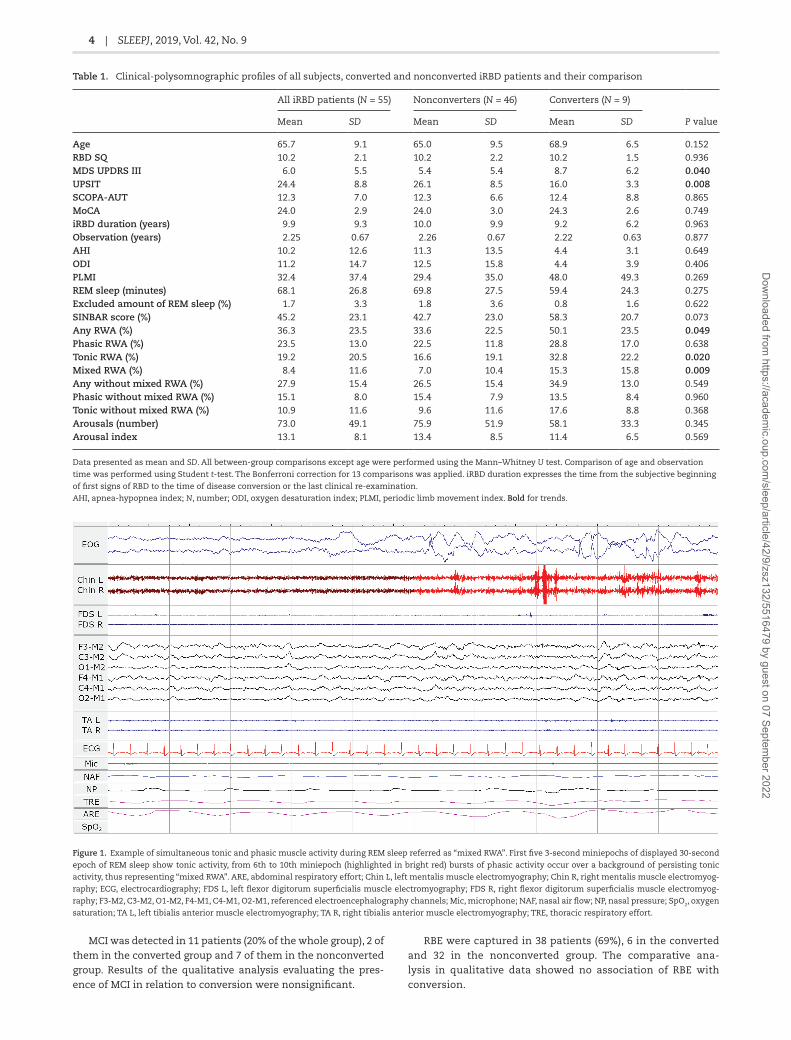
Figure 1. Example of simultaneous tonic and phasic muscle activity during REM sleep referred as “mixed RWA”. First five 3-second miniepochs of displayed 30-second
epoch of REM sleep show tonic activity, from 6th to 10th miniepoch (highlighted in bright red) bursts of phasic activity occur over a background of persisting tonic
activity, thus representing “mixed RWA”. ARE, abdominal respiratory effort; Chin L, left mentalis muscle electromyography; Chin R, right mentalis muscle electromyog-
raphy; ECG, electrocardiography; FDS L, left flexor digitorum superficialis muscle electromyography; FDS R, right flexor digitorum superficialis muscle electromyog-
raphy; F3-M2, C3-M2, O1-M2, F4-M1, C4-M1, O2-M1, referenced electroencephalography channels; Mic, microphone; NAF, nasal air flow; NP, nasal pressure; SpO2, oxygen
saturation; TA L, left tibialis anterior muscle electromyography; TA R, right tibialis anterior muscle electromyography; TRE, thoracic respiratory effort.
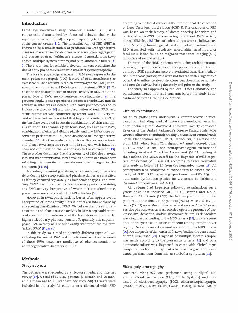
Table 1. Clinical-polysomnographic profiles of all subjects, converted and nonconverted iRBD patients and their comparison
All iRBD patients (N = 55) Nonconverters (N = 46) Converters (N = 9)
P valueMean SD Mean SD Mean SD
Age 65.7 9.1 65.0 9.5 68.9 6.5 0.152RBD SQ 10.2 2.1 10.2 2.2 10.2 1.5 0.936MDS UPDRS III 6.0 5.5 5.4 5.4 8.7 6.2 0.040UPSIT 24.4 8.8 26.1 8.5 16.0 3.3 0.008SCOPA-AUT 12.3 7.0 12.3 6.6 12.4 8.8 0.865MoCA 24.0 2.9 24.0 3.0 24.3 2.6 0.749iRBD duration (years) 9.9 9.3 10.0 9.9 9.2 6.2 0.963Observation (years) 2.25 0.67 2.26 0.67 2.22 0.63 0.877AHI 10.2 12.6 11.3 13.5 4.4 3.1 0.649ODI 11.2 14.7 12.5 15.8 4.4 3.9 0.406PLMI 32.4 37.4 29.4 35.0 48.0 49.3 0.269REM sleep (minutes) 68.1 26.8 69.8 27.5 59.4 24.3 0.275Excluded amount of REM sleep (%) 1.7 3.3 1.8 3.6 0.8 1.6 0.622SINBAR score (%) 45.2 23.1 42.7 23.0 58.3 20.7 0.073Any RWA (%) 36.3 23.5 33.6 22.5 50.1 23.5 0.049Phasic RWA (%) 23.5 13.0 22.5 11.8 28.8 17.0 0.638Tonic RWA (%) 19.2 20.5 16.6 19.1 32.8 22.2 0.020Mixed RWA (%) 8.4 11.6 7.0 10.4 15.3 15.8 0.009Any without mixed RWA (%) 27.9 15.4 26.5 15.4 34.9 13.0 0.549Phasic without mixed RWA (%) 15.1 8.0 15.4 7.9 13.5 8.4 0.960Tonic without mixed RWA (%) 10.9 11.6 9.6 11.6 17.6 8.8 0.368Arousals (number) 73.0 49.1 75.9 51.9 58.1 33.3 0.345Arousal index 13.1 8.1 13.4 8.5 11.4 6.5 0.569
Data presented as mean and SD. All between-group comparisons except age were performed using the Mann–Whitney U test. Comparison of age and observation
time was performed using Student t-test. The Bonferroni correction for 13 comparisons was applied. iRBD duration expresses the time from the subjective beginning
of first signs of RBD to the time of disease conversion or the last clinical re-examination.
AHI, apnea-hypopnea index; N, number; ODI, oxygen desaturation index; PLMI, periodic limb movement index. Bold for trends.
Dow
nloaded from https://academ
ic.oup.com/sleep/article/42/9/zsz132/5516479 by guest on 07 Septem
ber 2022

Nepozitek et al. | 5
The comparative analysis showed that use of antidepres-sants was not associated with an increase of either type of muscle activity in REM sleep (Supplementary Table S1). None of the iRBD patients on antidepressants converted to the neurodegeneration, however, results of the analysis comparing use of antidepressants in the converted and nonconverted group did not reach statistical significance (Supplementary Table S2).
Correlation analysis showed no significant associations of ei-ther RWA type with RBD SQ, MDS UPDRS III, SCOPA-AUT, MoCA, UPSIT scores, subjectively perceived RBD duration, sleep respir-ation parameters, periodic limb movement index, and arousals.
DiscussionThis observational study presents the new concept of mixed RWA referring to simultaneous appearance of phasic and tonic EMG activity.
The results show that tonic, mixed, and any RWA are useful predictive factors of phenoconversion in iRBD patients. No dif-ference of phasic RWA was observed between the converted and nonconverted patients. The observation of tonic but not phasic EMG muscle activity in iRBD associated with early phenoconversion is in accordance with previous reports [10, 11].
It has been suggested that phasic and tonic muscle ac-tivity during REM sleep in RBD have different underlying neural mechanisms [10]. Phasic activity was reported to be generated by cortical and spinal motor neurons [28] and depends upon al-teration of pathways in the ventromedial medulla (VMM) [10, 29, 30]. Tonic activity is considered to be caused by degeneration of sublaterodorsal (SLD) nucleus (or analogous subcoeruleus nucleus in humans) [10, 29, 31]. It has been determined that REM-active neurons of SLD are generally important for gener-ation of REM sleep atonia by projection to inhibitory premotor neurons both in the spinal cord and the VMM [32]. According to this concept, the sole degeneration of SLD would fittingly ex-plain the concurrent occurrence of phasic and tonic activity, represented by mixed RWA. Indeed, it is reasonable to expect the pathophysiological substrate of mixed RWA to be more com-plex. The global degeneration of SLD itself would hardly leave a margin for explanation of isolated tonic RWA. Instead, dif-ferent subpopulations of neurons within SLD could be involved, so mixed EMG activity would be the expression of overlap in-volvement of several functional subsections of the nucleus. It has been further hypothesized that difference between separate types of motor activity may be more quantitative than quali-tative, depending on the excitatory drive breaking through the inhibitory modulation as a continuum [32]. Mixed EMG ac-tivity can also be the expression of overlap neurodegeneration involving multiple brainstem areas and control of REM sleep atonia on multiple different levels [30]. Under this assumption, mixed RWA would be giving a nice picture about the severity of the disease and presumably about its future progression.
Performing ROC analysis for comparison of tonic, mixed, and any RWA, the “mixed RWA” parameter appears to be strong pre-dictive marker with slightly higher AUC compared to those ob-served in tonic and any RWA. Results in any RWA appeared to be
Figure 2. Boxplot representation of the amounts of different types of RWA (tonic, mixed, any) in nonconverted and converted iRBD patients, and corresponding receiver
operating characteristic curves. conv-, nonconverters; conv+, converters.
Table 2. Receiver operator characteristic analysis results of tonic, mixed, and any RWA
AUC 95% CI P value RR 95% CI P value
Tonic RWA 0.749 0.585 to 0.912 0.019 12.0 2.1 to 71.4 0.001Mixed RWA 0.778 0.648 to 0.908 0.009 8.9 1.6 to 53.3 0.006Any RWA 0.710 0.524 to 0.896 0.048 4.9 1.3 to 19.5 0.017
CI, confidence interval; RR, relative risk.
Dow
nloaded from https://academ
ic.oup.com/sleep/article/42/9/zsz132/5516479 by guest on 07 Septem
ber 2022

6 | SLEEPJ, 2019, Vol. 42, No. 9
influenced by the inclusion of phasic activity, which seemed to decrease the statistical significance of the “any RWA” parameter. The results of our study support previous findings showing that simple phasic muscle activity in REM sleep is nonspecific factor with regard to the conversion [10, 11].
In order to reveal the counterpart of the mixed RWA, we have evaluated any, phasic and tonic RWA with subtracted mixed RWA separately. The differences between converters and nonconverters in either RWA type were insignificant after the subtraction. These results suggest that the predictive value of EMG activity lies precisely within the mixed RWA. In other words, mixed RWA is the phenomenon that is worth evaluating in order to estimate the risk of upcoming conversion.
The phenoconversion rate of 16% observed in the pre-sent study is in accordance with previous studies which yielded approximately 15–35% of patients converting to overt neurodegeneration over 2–5 years [33, 34].
Interestingly, the results show no differences in SINBAR score between convertors and nonconvertors. SINBAR score comprise mentalis muscle tone together with FDS tone, which suggests that FDS activity may serve as parameter useful just to improve diagnostic sensitivity, but not as biomarker of short-term conversion.
One of the earlier studies shows that tonic and phasic RWA increases over time in subjects with iRBD [13]. The prediction value of mixed RWA with regard to the conversion would also empirically suggest that the amount of muscle activity increases over time, and thus, it can be a marker of iRBD progression as well. However, such implication cannot be in-ferred from the present results, since only cross-sectional data are available. On the other hand, the study mentioned above does not comment on the relationship of tonic and phasic RWA amounts to the conversion. Further research with a repeated follow-up polysomnographic examinations need to be performed in order to confirm the hypoth-esis that mixed RWA is also a marker of progression of the synucleinopathy.
The scores of RBD SQ did not show any difference between the converters and nonconverters or correlation with quan-titative RWA measures, suggesting that RBD SQ is less likely to be used as a surrogate marker for RBD severity or early phenoconversion risk.
Higher MDS UPDRS III scores found in early converters sup-ports previous observations that clinical examination is sen-sitive to capture discrete motor manifestations present in prodromal stage of synucleinopathies [14, 35].
The results of UPSIT scores confirm the findings from pre-vious studies that olfactory deficit predict the phenoconversion to neurodegenerative disease [35–37].
As to autonomic dysfunction and cognitive decline, no re-lationship to the conversion was found in the present study, although it is reported by earlier studies [35, 38]. One of the reasons could be a relatively short time of follow-up.
In earlier study on RBE in early Parkinson’s disease, it was shown that presence of RBE is a marker of neurodegeneration and it was hypothesized that it may precede iRBD [27]. In line with this idea, it would be expected that presence of RBE at the baseline involves subjects showing later early conversion. Surprisingly, qualitative analysis of RBE showed no association with the conversion. Shorter follow-up and potential bias due to the use of the cover might influence the present result. Further
studies need to be performed in order to confirm the previous observation.
The major limitation of the present study is low number of converters despite the study has replicated the results of pre-vious studies on the relationship of specific RWA types and phenoconversion [10, 11]. Rather broad variability of RWA param-eters reflect this limitation. The evaluation of mixed RWA and validation of the present results in larger cohorts is needed. The inclusion of subjects with only 1 year follow-up is another limi-tation that should be mentioned, as RWA measures of these pa-tients might have biased the results and they could be less prone to convert due to a shorter the follow-up time. To confirm that RWA scores at baseline did not differ between subjects with longer vs. shorter follow-up, subanalysis was performed (Supplementary Table S3). Antidepressant use by a minority of patients must also be considered a limitation. Despite antidepressants are reported to cause increase of muscle activity in REM sleep [39, 40], the medi-cation showed not to be associated with an increase of muscle ac-tivity in REM sleep in the current cohort.
ConclusionsThe severity of mixed, tonic, and any type of RWA at the base-line in iRBD predict the phenoconversion to fully manifested synucleinopathy. Mixed RWA, as the new concept representing simultaneous occurrence of phasic and tonic EMG activity ap-pears to be the best predictive RWA parameter and thus con-sideration should be given to its use as a standard indicator of impending phenoconversion.
Supplementary MaterialSupplementary material is available at SLEEP online.
FundingThis study was supported by The Charles University Grant Agency (grant number GA UK 64216), The Czech Science Foundation (grant number GACR 16-07879S), and The Ministry of Health of the Czech Republic (grant number 16-28914A), all rights reserved.Conflict of interest statement. None declared.
References 1. Schenck CH, et al. Chronic behavioral disorders of
human REM sleep: a new category of parasomnia. Sleep. 1986;9(2):293–308.
2. Arnulf I. REM sleep behavior disorder: motor manifest-ations and pathophysiology. Mov Disord. 2012;27(6):677–689.
3. Schenck CH. Rapid eye movement sleep behavior dis-order: current knowledge and future directions. Sleep Med. 2013;14(8):699–702.
4. Schenck CH, et al. Delayed emergence of a parkinsonian disorder in 38% of 29 older men initially diagnosed with idiopathic rapid eye movement sleep behaviour disorder. Neurology. 1996;46(2):388–393.
5. Iranzo A, et al. Rapid-eye-movement sleep behaviour dis-order as an early marker for a neurodegenerative disorder: a descriptive study. Lancet Neurol. 2006;5(7):572–577.
Dow
nloaded from https://academ
ic.oup.com/sleep/article/42/9/zsz132/5516479 by guest on 07 Septem
ber 2022

Nepozitek et al. | 7
6. Postuma RB, et al. Quantifying the risk of neurodegenerative disease in idiopathic REM sleep behavior disorder. Neurology. 2009;72(15):1296–1300.
7. Claassen DO, et al. REM sleep behavior disorder preceding other aspects of synucleinopathies by up to half a century. Neurology. 2010;75(6):494–499.
8. American Academy of Sleep Medicine. International Classification of Sleep Disorders. 3rd ed. Darien, IL: American Academy of Sleep Medicine; 2014.
9. Lapierre O, et al. Polysomnographic features of REM sleep behavior disorder: development of a scoring method. Neurology. 1992;42(7):1371–1374.
10. Postuma RB, et al. Severity of REM atonia loss in idiopathic REM sleep behavior disorder predicts Parkinson disease. Neurology. 2010;74(3):239–244.
11. Liu Y, et al. Electromyography activity level in rapid eye movement sleep predicts neurodegenerative diseases in idiopathic rapid eye movement sleep behavior disorder: a 5-year longitudinal study. Sleep Med. 2019; 56:128–134.
12. McCarter SJ, et al. Higher Amounts of REM Sleep Without Atonia Predict Phenoconversion to Defined Neurodegenerative Disorders in Idiopathic REM Sleep Behavior Disorder. Presented at: Sleep 2018 Annual Meeting; June 2–6 2018; Baltimore, MD.
13. Iranzo A, et al. Excessive muscle activity increases over time in idiopathic REM sleep behavior disorder. Sleep. 2009;32(9):1149–1153.
14. Högl B, et al. Idiopathic REM sleep behaviour disorder and neurodegeneration - an update. Nat Rev Neurol. 2018;14(1):40–55.
15. McCarter SJ, et al. REM sleep behavior disorder and REM sleep without atonia as an early manifestation of de-generative neurological disease. Curr Neurol Neurosci Rep. 2012;12(2):182–192.
16. Frauscher B, et al.; SINBAR (Sleep Innsbruck Barcelona) Group. Normative EMG values during REM sleep for the diagnosis of REM sleep behavior disorder. Sleep. 2012;35(6):835–847.
17. Bušková J, et al. Screening for REM sleep behavior disorder in the general population. Sleep Med. 2016;24:147.
18. Kopecek M, et al. Montreal cognitive assessment (MoCA): normative data for old and very old Czech adults. Appl Neuropsychol Adult. 2017;24(1):23–29.
19. Postuma RB, et al. MDS clinical diagnostic criteria for Parkinson’s disease. Mov Disord. 2015;30(12):1591–1601.
20. Dubois B, et al. Diagnostic procedures for Parkinson’s dis-ease dementia: recommendations from the movement dis-order society task force. Mov Disord. 2007;22(16):2314–2324.
21. McKeith IG, et al. Diagnosis and management of dementia with Lewy bodies: fourth consensus report of the DLB Consortium. Neurology. 2017;89(1):88–100.
22. Gilman S, et al. Second consensus statement on the diagnosis of multiple system atrophy. Neurology. 2008;71(9):670–676.
23. Garland EM, et al. Pure autonomic failure. Handb Clin Neurol. 2013;117:243–257.
24. Berry RB, et al.; for the American Academy of Sleep Medicine. The AASM Manual for the Scoring of Sleep and Associated Events: Rules, Terminology and Technical Specifications, Version 2.2. Darien, IL: American Academy of Sleep Medicine; 2015.
25. Schenck CH, et al. Rapid eye movement sleep parasomnias. Neurol Clin. 2005;23(4):1107–1126.
26. Frauscher B, et al.; SINBAR (Sleep Innsbruck Barcelona group). Quantification of electromyographic activity during REM sleep in multiple muscles in REM sleep behavior dis-order. Sleep. 2008;31(5):724–731.
27. Sixel-Döring F, et al. Rapid eye movement sleep behav-ioral events: a new marker for neurodegeneration in early Parkinson disease? Sleep. 2014;37(3):431–438.
28. Sunwoo JS, et al. Abnormal activation of motor cortical net-work during phasic REM sleep in idiopathic REM sleep be-havior disorder. Sleep. 2019;42(2). doi:10.1093/sleep/zsy227
29. Valencia Garcia S, et al. Ventromedial medulla inhibitory neuron inactivation induces REM sleep without atonia and REM sleep behavior disorder. Nat Commun. 2018;9(1):504.
30. Boeve BF, et al. Pathophysiology of REM sleep behaviour dis-order and relevance to neurodegenerative disease. Brain. 2007;130(Pt 11):2770–2788.
31. Gjerstad MD, et al. Occurrence and clinical correl-ates of REM sleep behaviour disorder in patients with Parkinson’s disease over time. J Neurol Neurosurg Psychiatry. 2008;79(4):387–391.
32. Ramaligam V, et al. Perspectives on the rapid eye move-ment sleep switch in rapid eye movement sleep behavior disorder. Sleep Med. 2013;14(8):707–713.
33. St Louis EK, et al. REM sleep behavior disorder: diagnosis, clinical implications, and future directions. Mayo Clin Proc. 2017;92(11):1723–1736.
34. Galbiati A, et al. The risk of neurodegeneration in REM sleep behavior disorder: a systematic review and meta-analysis of longitudinal studies. Sleep Med Rev. 2019;43:37–46.
35. Postuma RB, et al. Risk and predictors of dementia and par-kinsonism in idiopathic REM sleep behaviour disorder: a multicentre study. Brain. 2019;142(3):744–759.
36. Mahlknecht P, et al.; Sleep Innsbruck Barcelona Group. Olfactory dysfunction predicts early transition to a Lewy body disease in idiopathic RBD. Neurology. 2015;84(7):654–658.
37. Postuma RB, et al. Olfaction and color vision identify impending neurodegeneration in rapid eye movement sleep behavior disorder. Ann Neurol. 2011;69(5):811–818.
38. Li Y, et al. Predictive markers for early conversion of iRBD to neurodegenerative synucleinopathy diseases. Neurology. 2017;88(16):1493–1500.
39. Lee K, et al. The Prevalence and Characteristics of REM Sleep without Atonia (RSWA) in Patients Taking Antidepressants. J Clin Sleep Med. 2016;12(3):351–355.
40. McCarter SJ, et al. Antidepressants increase REM sleep muscle tone in patients with and without REM sleep be-havior disorder. Sleep. 2015;38(6):907–917.
Dow
nloaded from https://academ
ic.oup.com/sleep/article/42/9/zsz132/5516479 by guest on 07 Septem
ber 2022