Embed Size (px)
Citation preview
...........................................................................................................................
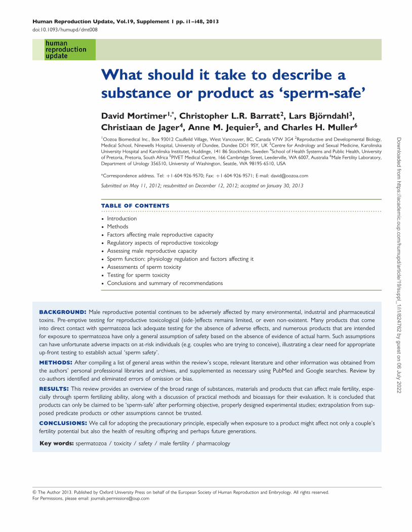
What should it take to describe asubstance or product as ‘sperm-safe’David Mortimer1,*, Christopher L.R. Barratt2, Lars Bjorndahl3,Christiaan de Jager4, Anne M. Jequier5, and Charles H. Muller6
1Oozoa Biomedical Inc., Box 93012 Caulfeild Village, West Vancouver, BC, Canada V7W 3G4 2Reproductive and Developmental Biology,Medical School, Ninewells Hospital, University of Dundee, Dundee DD1 9SY, UK 3Centre for Andrology and Sexual Medicine, KarolinskaUniversity Hospital and Karolinska Institutet, Huddinge, 141 86 Stockholm, Sweden 4School of Health Systems and Public Health, Universityof Pretoria, Pretoria, South Africa 5PIVET Medical Centre, 166 Cambridge Street, Leederville, WA 6007, Australia 6Male Fertility Laboratory,Department of Urology 356510, University of Washington, Seattle, WA 98195-6510, USA
*Correspondence address. Tel: +1-604-926-9570; Fax: +1-604-926-9571; E-mail: [email protected]
Submitted on May 11, 2012; resubmitted on December 12, 2012; accepted on January 30, 2013
table of contents
† Introduction† Methods† Factors affecting male reproductive capacity† Regulatory aspects of reproductive toxicology† Assessing male reproductive capacity† Sperm function: physiology regulation and factors affecting it† Assessments of sperm toxicity† Testing for sperm toxicity† Conclusions and summary of recommendations
background: Male reproductive potential continues to be adversely affected by many environmental, industrial and pharmaceuticaltoxins. Pre-emptive testing for reproductive toxicological (side-)effects remains limited, or even non-existent. Many products that comeinto direct contact with spermatozoa lack adequate testing for the absence of adverse effects, and numerous products that are intendedfor exposure to spermatozoa have only a general assumption of safety based on the absence of evidence of actual harm. Such assumptionscan have unfortunate adverse impacts on at-risk individuals (e.g. couples who are trying to conceive), illustrating a clear need for appropriateup-front testing to establish actual ‘sperm safety’.
methods: After compiling a list of general areas within the review’s scope, relevant literature and other information was obtained fromthe authors’ personal professional libraries and archives, and supplemented as necessary using PubMed and Google searches. Review byco-authors identified and eliminated errors of omission or bias.
results: This review provides an overview of the broad range of substances, materials and products that can affect male fertility, espe-cially through sperm fertilizing ability, along with a discussion of practical methods and bioassays for their evaluation. It is concluded thatproducts can only be claimed to be ‘sperm-safe’ after performing objective, properly designed experimental studies; extrapolation from sup-posed predicate products or other assumptions cannot be trusted.
conclusions: We call for adopting the precautionary principle, especially when exposure to a product might affect not only a couple’sfertility potential but also the health of resulting offspring and perhaps future generations.
Key words: spermatozoa / toxicity / safety / male fertility / pharmacology
& The Author 2013. Published by Oxford University Press on behalf of the European Society of Human Reproduction and Embryology. All rights reserved.For Permissions, please email: [email protected]
Human Reproduction Update, Vol.19, Supplement 1 pp. i1–i48, 2013
doi:10.1093/humupd/dmt008
Dow
nloaded from https://academ
ic.oup.com/hum
upd/article/19/suppl_1/i1/824762 by guest on 06 July 2022
IntroductionIt is well established that male reproductive potential (commonly ‘fer-tility’) has been, and continues to be, adversely affected by many en-vironmental, industrial and pharmaceutical toxins (e.g. Perry, 2008;Jurewicz et al., 2009; European Science Foundation, 2010; Joffe, 2010;Sharpe, 2010; Perry et al., 2011; Sutton et al., 2012). Unfortunately,the reproductive toxicity of the great majority of these problem sub-stances was discovered after-the-fact, and pre-emptive testing for repro-ductive toxicological (side-)effects remains limited, or even non-existent.With the burgeoning markets for nutraceuticals, processed food andfood packaging, cosmetics and personal care products, it thereforeseems apposite that awareness be created regarding such risks tohuman and animal fertility. Beyond the many substances and materialsthat might affect male fertility indirectly, many products that comeinto direct contact with spermatozoa are reaching market without ad-equate testing for absence of adverse effects, even some that receiveCE marking (Conformite Europeenne; a mandatory conformity mark forproducts placed on the market in the European Economic Area) foruse in assisted conception treatment. For example, saying that aproduct is ‘non-spermicidal’ only means that it does not specificallycontain a spermicidal drug (Vargas et al., 2011), and there are numerousproducts that are intended for exposure to spermatozoa where there isnothing documented or known, only a general assumption of ‘safety’based on the absence of any evidence of actual harm. By providing anoverview of the broad range of substances, materials and productsthat can affect male fertility via sperm production or sperm fertilizing po-tential, we hope to raise awareness of the extent of the problem (Suttonet al., 2012) and the increasingly urgent need for action to avoid person-al calamities by individuals (e.g. couples who are trying to conceive) onthe one hand, and severe environmental consequences on the other.
Further information to facilitate understanding of this review bynon-spermatologist/non-andrologist readers can be found in basicandrology reference books, including Hargreave (1994a), Mortimer(1994), Patton and Battaglia (2005); Schill et al. (2006), Bjorndahlet al. (2010), Nieschlag et al. (2010a) and Jequier (2011).
MethodsGiven the recognition that many substances and materials that might affectmale fertility potential either directly or indirectly are reaching marketwithout adequate prior testing for adverse effects on spermatozoa, aninternational group of authors with extensive experience in spermatologyand andrology was formed with D.M. acting as the coordinating author.Following an initial series of telephone conversations and e-mailexchanges, the following general objectives were set for the article:
(a) To compile a comprehensive, critical overview of the physical andchemical factors that can influence human male fertility by affectingsperm production and/or sperm functional potential.
(b) To expand this list to include pharmaceuticals, nutraceuticals, pro-cessed food and food packaging materials, cosmetics and personalcare products, and products used in assisted conception treatment.
(c) To focus primarily on human male fertility, but to augment our under-standing using research from other animal species when necessary.
(d) To make the article understandable by readers from a broad range ofbackgrounds, many of whom would not have specialist knowledge ofsperm physiology and pathophysiology.
Because of the necessarily broad scope of the article, an initial manuscriptoutline was created based on a list of sections that represented thegeneral areas of interest within its scope. The outline for each section wasthen expanded with lists of topics and sub-topics, reviewed and amendedby all authors and then used as the framework for compiling the content.Each author either self-assigned or was asked to contribute to various sec-tions, based on their particular areas of interest, experience and expertise.Relevant literature and other material for each (sub-)topic was obtainedfrom personal professional libraries and archives, and supplemented as ne-cessary using PubMed and Google searches to ensure the breadth of cover-age. While the bibliography of resources employed is extensive, the vastscope of all the pertinent issues precludes its being exhaustive for simplereasons of practicality. Review by co-authors was considered to have ad-equately identified and eliminated any errors of omission or bias. Summar-ization of material was based on the expertise and the contributing authorfor each (sub-)topic, followed by review and acceptance by all co-authors.
At several points during its preparation, the manuscript was circulatedto all co-authors for overall review and comment on both specificcontent and general readability. The coordinating author compiled all revi-sions and suggestions using the principle of unanimity based on implicit ac-ceptance in the absence of contrary comment, combined with consensusestablished via e-mail polling and other exchanges. The final version of themanuscript for submission, as well as the revised version following feed-back from the reviewers, including all summaries and recommendations,was approved by all authors.
Factors affecting malereproductive capacityMale reproductive function is claimed to be decreasing (Carlsen et al.,1992; Sharpe and Skakkebaek, 1993; Sharpe, 2012), specifically a per-ceived decline in sperm counts in the ejaculates of normal men.However, the extent to which measurements of sperm concentrationin complete and incompletely collected semen samples is a reliablemeasure for the fertility status of entire populations has been hotlydebated (Mahmoud et al., 1997; Gottardo and Kliesch, 2011; Isobe,2012). Based on reported decreases in sperm concentrations in thegeneral population, and decreased sperm motility and morphologyin the male partners of couples seeking infertility treatment, it hasbeen hypothesized that environmental chemicals with estrogenicproperties are negative factors for male fertility (Tas et al., 1996;Van Waeleghem et al., 1996; Phillips and Tanphaichitr, 2008;Diamanti-Kandarakis et al., 2009; Giwercman, 2011; Woodruff,2011), even though no apparent and clear decrease in population fer-tility has been noted in epidemiological studies (Akre et al., 1999;Scheike et al., 2008; Joffe, 2010). Nonetheless, although the epi-demiological consequences are unclear, there may be toxicologicaleffects on individuals, since both clinical and laboratory research sug-gests that all the changes in male reproductive health appear inter-related and may have a common origin in fetal life or childhood(Sharpe, 2006; Sharpe and Skakkebaek, 2003; Skakkebaek et al.,2001, 2011; Buck Louis et al., 2008,) and several epidemiologicalstudies implicate exposure to environmental chemicals, mainly endo-crine disruptors, in male reproductive health disorders (Sharpe, 2009,2010; Balabanic et al., 2011).
The mechanisms whereby xenogenic factors exert their adverseeffects on male reproduction through events occurring during fetal
i2 Mortimer et al.
Dow
nloaded from https://academ
ic.oup.com/hum
upd/article/19/suppl_1/i1/824762 by guest on 06 July 2022
testis and germinal cell development, commonly referred to as the‘testicular dysgenesis syndrome’, is clearly outside the scope of thepresent review. However, many effects are mediated either by target-ing pituitary gonadotrophins (Mutoh et al., 2006) or via the geneticregulation of steroidogenesis (Kuhl et al., 2007) at either thegenomic (Liu et al., 2005) or proteomic (Laier et al., 2006; Klinefelteret al., 2012) levels. Gene pathways disrupted included cholesteroltransport and steroidogenesis, as well as pathways involved in intracel-lular lipid and cholesterol homeostasis, insulin signalling, transcriptionalregulation and oxidative stress (Liu et al., 2005). Other gene targetsinclude a-inhibin (which is essential for normal Sertoli cell development)and genes involved with communication between Sertoli cells and gono-cytes (Liu et al., 2005). Environmental toxicants can induce tissue oxida-tive stress and peroxidation (Kabuto et al., 2004) and induce germ cellapoptosis in the human fetal testis (Coutts et al., 2007).
This concept of a ‘critical period of exposure’ applies to both thegonad and the brain, as illustrated by the diethylstilbestrol experience inhumans and experiments in rodents showing that estrogenic compounds,present during a critical pre- or peri-natal period can influence behaviour,accessory glands and reproductive structures (Harris and Waring, 2012).Other compounds present at these times can have effects that are notnecessarily hormonal in nature, and are manifested later in life, particular-ly, epigenetic modifications (Anway et al., 2005, 2006).
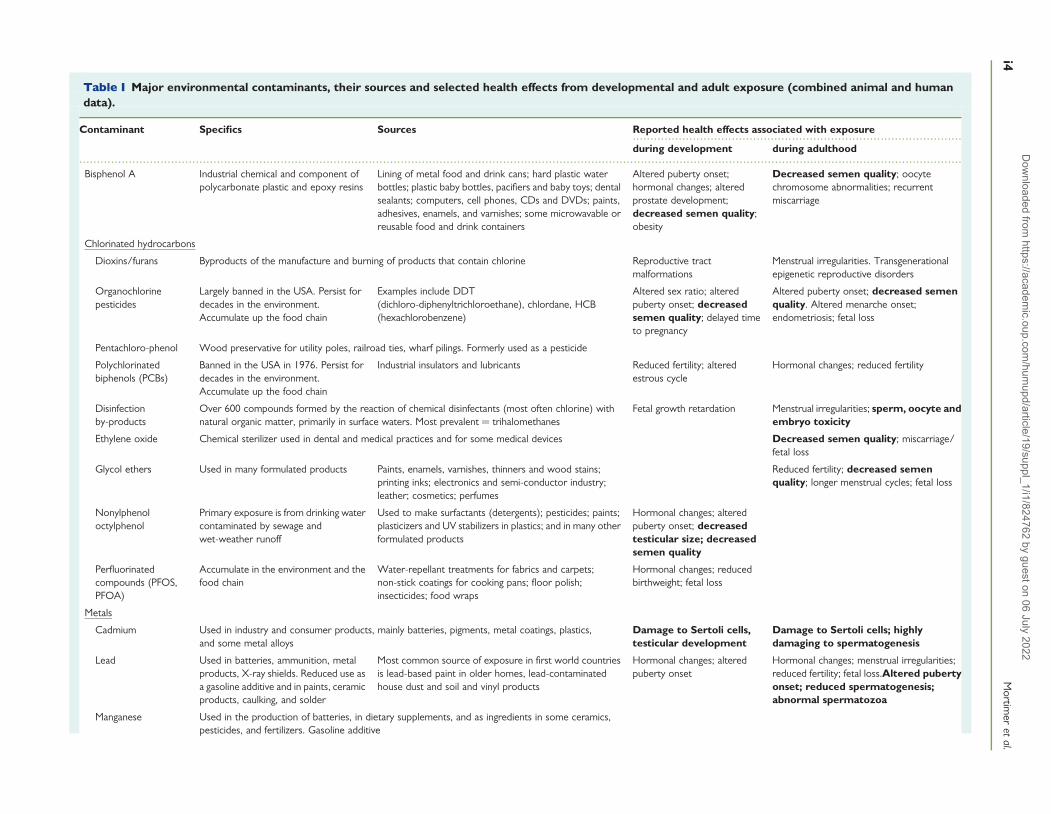
Emerging trends in the male reproductive health of wildlife also raisethe possibility of environmental factors as partial aetiologic contribu-tions to the observed reproductive health decline (Edwards et al.,2006; Hamlin and Guillette, 2010). Experimental results and clinicalreports have suggested that prenatal exposure to exogenous estro-gens may play an aetiologic role in the trends observed in male fertility(Danish Environmental Protection Agency, 1995; Toppari et al., 1996;Skakkebaek et al., 2011; Braw-Tal, 2010). While awareness of the bio-logical risks of chemical toxicity has increased considerably in recentyears (Zhu and Huang, 2011; Rhomberg and Goodman, 2012)(Table I), some of these chemicals have long half-lives and havebeen detected in environmental samples 10–20 years after theywere banned for sale or use (Aitken et al., 2004).
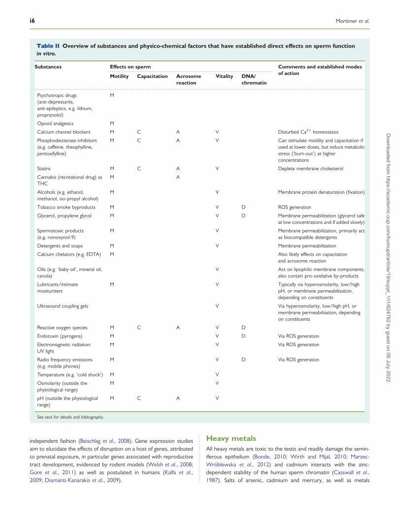
A short list of those substances and physico-chemical factors thathave established direct effects on sperm function in vitro, and henceare of particular interest within the context of the present review,has been provided in Table II.
Environmental endocrine-disruptingchemicalsMankind and wildlife alike are exposed to many general classes of che-micals present in the natural environment, including pesticides, fungi-cides, heavy metals, defoliants and other chemical weapons, as wellas oils and cleaning agents (Colborn et al., 1993, 1997; Sheineret al., 2003; Gore, 2007; Woodruff et al., 2008; see also http://www.ourstolenfuture.org/basics/chemlist.htm). Many of these chemi-cals likely exert their effects through mechanisms involving disruptionof the endocrine system (‘endocrine disruptor chemicals’ or EDCs),which have received a great deal of attention, since the introductionof the term ‘endocrine disruptor’ nearly two decades ago (Colbornet al., 1993).
Endocrine disruption refers to a mechanism of toxicity that hindersthe ability of cells, tissues and organs to communicate hormonally
(Silva et al., 2010), resulting in a wide variety of adverse health out-comes including reduced fertility and fecundity (Giwercman, 2011),spontaneous abortion, skewed sex ratios within the offspring ofexposed communities (Yiee and Baskin, 2010), male and female repro-ductive tract abnormalities (Bornman et al., 2010; Newbold, 2011;Dunbar et al., 2012), precocious puberty (Mouritsen et al., 2010;Deng et al., 2012), polycystic ovary syndrome (Teede et al., 2010), neu-robehavioural disorders, impaired immune function and a wide varietyof cancers (Skakkebaek et al., 2001; Keinan-Boker et al., 2004;Ndebele et al., 2010). Endocrine disruptors represent a wide rangeof chemical classes and include agonists of the estrogen receptor, an-drogen receptor antagonists and aryl hydrocarbon receptor agonists(Beischlag et al., 2008). Some chemicals exhibit more than one mech-anism of action (Phillips and Foster, 2008).
The major groups of environmental chemicals with EDC activityincludes the organochlorine pesticides, other pesticides, herbicidesand fungicides, polychlorinated biphenyls (PCBs), dioxins and furans,alkylphenol polyethoxylates, bisphenol A (BPA), phthalates, phytoes-trogens and other xenoestrogens (Colborn et al., 1993, 1997)(Table I). Many of these chemicals persist in the environment. Someare lipophilic and hence sequestered in adipose tissue and secretedin milk, and others may only be present for short periods of timebut at critical periods of development.
A number of insecticides and herbicides are known to cause in-fertility (Perry, 2008; also http://www.arhp.org/publications-and-resources/clinical-proceedings/RHE/Pesticides, accessed 14 February2013). For example, the nematocide dibromodichloropropane (DBCP)was for many years used to protect the Hawaiian pineapple cropfrom destruction by a weevil that attacks it roots, but transferenceof DBCP into the water supply had devastating impact on human fer-tility by causing azoospermia. Other insecticides, such as dimethyl1-2-dichlorovinyl phosphate, and the PCBs are similarly gonadotoxic.
Exposure to EDCs may occur through environmental routes (air,soil, water, food) or via occupational exposures (Sharpe, 2009;Woodruff, 2011), and individuals may have multiple exposures thatin many cases occur chronically and at low doses. EDCs are verydiverse in structure and potency (Damstra et al., 2002). Humans areexposed to low levels of multiple EDCs, the effects of which can beadditive, and frequently have body burdens comparable to thosethat cause abnormalities in other vertebrates (Kortenkamp, 2007;Kortenkamp et al., 2007; Bornman et al., 2010). There are alsomany spermotoxic and gonadotoxic substances beyond EDCs towhich men are exposed within their normal environment or in theworkplace. Endocrine disruption must be considered in the contextof both individuals and populations, although not every individualwithin a population may be similarly affected, and EDC effects canbe permanent or irreversible (Gore, 2007).
There are a number of mechanisms whereby EDCs can modulateendocrine systems and potentially cause adverse effects (Mnif et al.,2011). The generally accepted paradigm for receptor-mediatedresponses involves a hormone binding to its receptor at the cellsurface, in the cytoplasm or within the nucleus, followed by a complexseries of events within the classical genomic pathway that lead to inter-action of receptors with the DNA by binding to hormone response ele-ments in the target gene promoter area (Gruber et al., 2004; Delbeset al., 2005). Off-target effects as well as cross-talk may occur asmany transcription factors modulate transcription in a DNA-binding
Requirements to describe a substance as sperm-safe i3D
ownloaded from
https://academic.oup.com
/humupd/article/19/suppl_1/i1/824762 by guest on 06 July 2022
.............................................................................................
..........................................................................................................................................................................................................................................................
Table I Major environmental contaminants, their sources and selected health effects from developmental and adult exposure (combined animal and humandata).
Contaminant Specifics Sources Reported health effects associated with exposure
during development during adulthood
Bisphenol A Industrial chemical and component ofpolycarbonate plastic and epoxy resins
Lining of metal food and drink cans; hard plastic waterbottles; plastic baby bottles, pacifiers and baby toys; dentalsealants; computers, cell phones, CDs and DVDs; paints,adhesives, enamels, and varnishes; some microwavable orreusable food and drink containers
Altered puberty onset;hormonal changes; alteredprostate development;decreased semen quality;obesity
Decreased semen quality; oocytechromosome abnormalities; recurrentmiscarriage
Chlorinated hydrocarbons
Dioxins/furans Byproducts of the manufacture and burning of products that contain chlorine Reproductive tractmalformations
Menstrual irregularities. Transgenerationalepigenetic reproductive disorders
Organochlorinepesticides
Largely banned in the USA. Persist fordecades in the environment.Accumulate up the food chain
Examples include DDT(dichloro-diphenyltrichloroethane), chlordane, HCB(hexachlorobenzene)
Altered sex ratio; alteredpuberty onset; decreasedsemen quality; delayed timeto pregnancy
Altered puberty onset; decreased semenquality. Altered menarche onset;endometriosis; fetal loss
Pentachloro-phenol Wood preservative for utility poles, railroad ties, wharf pilings. Formerly used as a pesticide
Polychlorinatedbiphenols (PCBs)
Banned in the USA in 1976. Persist fordecades in the environment.Accumulate up the food chain
Industrial insulators and lubricants Reduced fertility; alteredestrous cycle
Hormonal changes; reduced fertility
Disinfectionby-products
Over 600 compounds formed by the reaction of chemical disinfectants (most often chlorine) withnatural organic matter, primarily in surface waters. Most prevalent ¼ trihalomethanes
Fetal growth retardation Menstrual irregularities; sperm, oocyte andembryo toxicity
Ethylene oxide Chemical sterilizer used in dental and medical practices and for some medical devices Decreased semen quality; miscarriage/fetal loss
Glycol ethers Used in many formulated products Paints, enamels, varnishes, thinners and wood stains;printing inks; electronics and semi-conductor industry;leather; cosmetics; perfumes
Reduced fertility; decreased semenquality; longer menstrual cycles; fetal loss
Nonylphenoloctylphenol
Primary exposure is from drinking watercontaminated by sewage andwet-weather runoff
Used to make surfactants (detergents); pesticides; paints;plasticizers and UV stabilizers in plastics; and in many otherformulated products
Hormonal changes; alteredpuberty onset; decreasedtesticular size; decreasedsemen quality
Perfluorinatedcompounds (PFOS,PFOA)
Accumulate in the environment and thefood chain
Water-repellant treatments for fabrics and carpets;non-stick coatings for cooking pans; floor polish;insecticides; food wraps
Hormonal changes; reducedbirthweight; fetal loss
Metals
Cadmium Used in industry and consumer products, mainly batteries, pigments, metal coatings, plastics,and some metal alloys
Damage to Sertoli cells,testicular development
Damage to Sertoli cells; highlydamaging to spermatogenesis
Lead Used in batteries, ammunition, metalproducts, X-ray shields. Reduced use asa gasoline additive and in paints, ceramicproducts, caulking, and solder
Most common source of exposure in first world countriesis lead-based paint in older homes, lead-contaminatedhouse dust and soil and vinyl products
Hormonal changes; alteredpuberty onset
Hormonal changes; menstrual irregularities;reduced fertility; fetal loss.Altered pubertyonset; reduced spermatogenesis;abnormal spermatozoa
Manganese Used in the production of batteries, in dietary supplements, and as ingredients in some ceramics,pesticides, and fertilizers. Gasoline additive
i4M
ortimer
etal.
Dow
nloaded from https://academ
ic.oup.com/hum
upd/article/19/suppl_1/i1/824762 by guest on 06 July 2022
Mercury Air and water contaminated byindustrial emissions and the combustionof coal and waste. Accumulates in foodchain; most common source ofexposure in USA is contaminatedseafood
Used in thermometers, dental fillings, batteries, vaccinesand other industries
Highly damaging to spermatogenesis
Pesticides Broad category, includes many classesof insecticides, fungicides, herbicides,rodenticides, and fumigants
Pesticides in industrial, agricultural and residential settings.Exposure can occur through food, drinking water,inhalation or absorption through the skin
Altered sex ratio;malformations of reproductivetract; altered puberty onset;reduced fertility; retarded fetalgrowth
Hormonal changes; menstrual irregularities;reduced fertility; decreased semenquality; sperm chromosomeanomalies; miscarriage
See also: organochlorine pesticides, nonylphenol, octyphenol, pentachlorophenol, perfluorinatedcompounds
Pharmaceuticals Many examples, e.g. DES (diethylstilbestrol), ethynylestradiol (birth control pill) Reproductive tractmalformations; alteredhormone response; menstrualirregularities; reduced fertility;uterine fibroids; miscarriage
Numerous effects on the male and/or femalereproductive systems. Those affectingsperm production or quality includeanabolic steroids, calcium channel blockers;colchicine, sulphasalazine, testosterone, (seetext)
Phthalates Plasticizers added to soften plastics likePVC; also used in cosmetics and woodfinishers
Cosmetics; perfumes; toys; pharmaceuticals; medicaldevices; lubricants
Malformations of reproductivetract; hormonal changes;decreased semen quality
Earlier menarche; estrous cycle irregularity;reduced fertility; dysovulation; endometriosis;decreased semen quality; fetal loss
PolybrominatedDiphenyl Ethers(PBDEs)
Accumulate in the food chain Flame retardants used in furniture foam, mattresses,textiles, computers and electronics
Decreased semen quality
Solvents (organic) Benzene, toluene, xylene, styrene,1-bromopropane, 2-bromopropane,perchloroethylene, trichloroethylene,and many others. Solvents are someof the highest production volumechemicals. Also found incigarette smoke
Used in plastics, resins, rubbers, synthetic fibres,lubricants, dyes, detergents, drugs, pesticides, glues, paintsand paint thinners, fingernail polish, lacquers, detergents,printing and leather tanning processes, insulation,fibreglass, food containers, carpet backing, cleaningproducts. Exposure is primarily through respiration
Hormonal changes; reduced fertility;menstrual irregularities; decreased semenquality; miscarriage and fetal loss
Tobacco smoke Several hundred components Includes active and/or passive smoking Decreased fetal growth; lowbirthweight; pre-term delivery;low birthweight; decreasedsemen quality
Hormonal changes; reduced fertility;decreased semen quality; miscarriage;early menopause
Broad categories underlined; effects on semen or sperm quality shown in bold. Adapted from Woodruff et al. (2008) and expanded.
Requirem
entsto
describea
substanceas
sperm-safe
i5D
ownloaded from
https://academic.oup.com
/humupd/article/19/suppl_1/i1/824762 by guest on 06 July 2022
independent fashion (Beischlag et al., 2008). Gene expression studiesaim to elucidate the effects of disruption on a host of genes, attributedto prenatal exposure, in particular genes associated with reproductivetract development, evidenced by rodent models (Welsh et al., 2008;Gore et al., 2011) as well as postulated in humans (Kalfa et al.,2009; Diamanti-Kanarakis et al., 2009).
Heavy metalsAll heavy metals are toxic to the testis and readily damage the semin-iferous epithelium (Bonde, 2010; Wirth and Mijal, 2010; Marzec-Wroblewska et al., 2012) and cadmium interacts with the zinc-dependent stability of the human sperm chromatin (Casswall et al.,1987). Salts of arsenic, cadmium and mercury, as well as metals
.............................................................................................
.............................................................................................................................................................................................
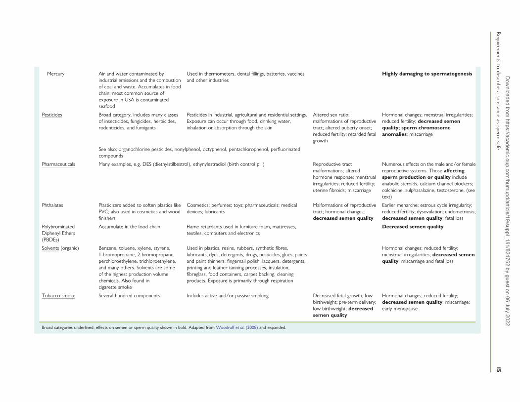
Table II Overview of substances and physico-chemical factors that have established direct effects on sperm functionin vitro.
Substances Effects on sperm Comments and established modesof actionMotility Capacitation Acrosome
reactionVitality DNA/
chromatin
Psychotropic drugs(anti-depressants,anti-epileptics, e.g. lithium,propranolol)
M
Opioid analgesics M
Calcium channel blockers M C A V Disturbed Ca2+ homeostasis
Phosphodiesterase inhibitors(e.g. caffeine, theophylline,pentoxifylline)
M C A V Can stimulate motility and capacitation ifused at lower doses, but induce metabolicstress (‘burn-out’) at higherconcentrations
Statins M C A V Deplete membrane cholesterol
Cannabis (recreational drug) asTHC
M A
Alcohols (e.g. ethanol,methanol, iso-propyl alcohol)
M V Membrane protein denaturation (fixation)
Tobacco smoke byproducts M V D ROS generation
Glycerol, propylene glycol M V D Membrane permeabilization (glycerol safeat low concentrations and if added slowly)
Spermotoxic products(e.g. nonoxynol-9)
M V Membrane permeabilization, primarily actas biocompatible detergents
Detergents and soaps M V Membrane permeabilization
Calcium chelators (e.g. EDTA) M Also likely effects on capacitationand acrosome reaction
Oils (e.g. ‘baby oil’, mineral oil,canola)
V Act on lipophilic membrane components,also contain pro-oxidative by-products
Lubricants/intimatemositurizers
M V Typically via hyperosmolarity, low/highpH, or membrane permeabilization,depending on constituents
Ultrasound coupling gels V Via hyperosmolarity, low/high pH, ormembrane permeabilization, dependingon constituents
Reactive oxygen species M C A V D
Endotoxin (pyrogens) M V D Via ROS generation
Electromagnetic radiation:UV light
M V Via ROS generation
Radio frequency emissions(e.g. mobile phones)
M V D Via ROS generation
Temperature (e.g. ‘cold shock’) M V
Osmolarity (outside thephysiological range)
M V
pH (outside the physiologicalrange)
M C A V
See text for details and bibliography.
i6 Mortimer et al.
Dow
nloaded from https://academ
ic.oup.com/hum
upd/article/19/suppl_1/i1/824762 by guest on 06 July 2022
such as lead and antimony, are all highly damaging to spermatogenesis,with cadmium salts specifically damaging the Sertoli cells; their acci-dental ingestion causes infertility (Boscolo et al., 1985; Benoff et al.,2000). The most toxic are cadmium and arsenic salts, which were his-torically used to protect roof timbers against dry rot and termite inva-sion (but are now banned in many countries for this purpose).Industrial exposure to lead can reduce the sperm count (Alexanderet al., 1996), and men heavily poisoned by lead can also suffer a reduc-tion in thyroid function as well as decreased cortisol production.Mercury also interferes with spermatogenesis and can damage the epi-didymal ducts. Heavy metals are also present in some welding fluxes, apossible explanation for the known relationship between welding andinfertility. Arsenic and antimony can be found in folk medicines, par-ticularly those from the Indian subcontinent and Traditional ChineseMedicine (Lynch and Braithwaite, 2005).
SolventsVarious organic solvents are also known to cause infertility, includingglycol ethers (Cherry et al., 2008) which are used in the printing industryand are also found in some paints (e.g. as used on naval vessels). Per-chloroethylene, used in the dry cleaning industry, can also cause subfer-tility, but its effects on sperm morphology and kinematics are subtle, andtheir impact on fertility remains unclear (Eskenazi et al., 1991).
PharmaceuticalsMany pharmaceuticals are known to have male reproductive sideeffects expressed through deleterious effects on either the productionof spermatozoa and/or their function (Drife, 1987). An excellent ref-erence book by Forman et al. (1996) has not been updated, and anup-to-date reference site does not exist—as witnessed by theregular appeals for information on drug side effects on the Androloglist server (www.andrology.org/?Links:13101201:Androlog_net).Some agents suppress gonadotrophins and thus secondarily cause in-fertility, others have a toxic effect upon the spermatogenic epitheliumand some have a direct effect on the spermatozoa themselves. Manyclasses of drugs or individual therapeutic agents beyond those witherectile dysfunction activity affect fertility or sexuality, only sometimeshaving adverse effects upon sperm production or sperm function asregistered side effects (Forman et al., 1996). These include psycho-tropic and central nervous system drugs (e.g. antidepressants, anti-epileptics), antihypertensives, cancer chemotherapy, therapeutic hor-mones, as well as recreational drugs and drugs of abuse such astobacco, marijuana and cocaine. Therapeutic drugs (as well as physicalagents and environmental toxicants) can affect the integrity of spermchromatin, inducing structural, genetic and/or epigenetic abnormal-ities (Delbes et al., 2010; Christensen and Marsit, 2011). While themechanisms that trigger such damage remain largely unresolved (de-pending on their nature, chemicals can directly target the DNA,induce oxidative stress, or modify epigenetic elements), a given indivi-dual’s susceptibility depends on their genetic background, lifestyle andexposure to various (other) insults.
Some specific examples of some common pharmaceuticals thataffect sperm production and/or sperm function are provided (in al-phabetical order based on their general purpose) to illustrate therisks that might be posed by such therapeutic agents and/or their bio-logical mechanisms of action.
AnalgesicsAnalgesic administration during pregnancy is a common occurrence;however, some of these drugs, as evidenced in animal experiments,exhibit anti-androgenic effects (Kristensen et al., 2011). Intrauterineexposure has been linked to abnormal reproductive development asthe sensitive reproductive programming window may be influencedby androgen deficiency, resulting in cryptorchidism, hypospadias andcompromised fertility (Welsh et al., 2008). Opioid analgesics, usedto treat acute pain, have a direct effect on human sperm motilityin vitro, by decreasing sperm motility and at higher concentrations, im-motile spermatozoa was observed (Xu et al., 2013).
Anti-depressants, anti-epileptics and other psychotropic drugsSeveral anti-epileptic drugs have been reported to adversely affecthuman sperm morphology and motility (Isojarvi et al., 2004) andlithium, used to treat bipolar affective illness, inhibits human spermmotility (Raoof et al., 1989). Various psychotropic antidepressantdrugs and the original b-blocker propranolol have also been shownto be potent inhibitors of sperm motility, at least in vitro—althoughwhether they have similar adverse effects in vivo remains unclear(Hong et al., 1981b, 1982; Levin et al., 1981).
Calcium channel blockersThese drugs disrupt the movement of calcium ions through calciumchannels, and have effects on many excitable cells such as cardiacmuscle, the smooth muscles of blood vessels and neurons. Theirmost widespread clinical application is as anti-hypertensives, butthey are also frequently used to control heart rate, prevent cerebralvasospasm and reduce chest pain due to angina. Because voltage-gated calcium channels are also involved in the regulation of spermcapacitation and hyperactivation, as well as the acrosome reaction(AR), these drugs can also impair sperm fertilizing ability, bothin vivo and during IVF (Benoff et al., 1994). As a corollary to this,the extreme elevation of intracellular calcium ions can adversely affectsperm vitality, even to the extent that the mechanism might havecontraceptive potential (Kumar et al., 2008). Therefore, any chemical,whether a pharmaceutical or naturally occurring, that affects thecalcium homeostasis of spermatozoa is likely to affect sperm function.
Chemotherapeutic agentsMost of the chemotherapeutic agents act as alkylating agents that co-valently join different molecules together and thus interfere with cellfunction and cell division, thereby damaging the spermatogenic epithe-lium and thus reduce the sperm count, often to azoospermia (Mitchellet al., 2009; Dohle, 2010). As there is no known way to block thiseffect upon the testis, and it is difficult to give specific informationregarding the possible return of spermatozoa to the ejaculate post-treatment (in many patients, sperm production is never resumed,yet in others, spermatozoa may unexpectedly be found in an ejacu-late), pretreatment sperm storage is essential (Lass et al., 2001;Tomlinson and Pacey, 2003). Others act as anti-folates and thustarget rapidly dividing cells; some also inhibit nucleic acid synthesis.The alkylating agent cyclophosphamide, which is probably the mostcommonly used chemotherapeutic drug, has the direst effect uponsperm production. The damage done to the testis is usually dose-dependent, and as cyclophosphamide is frequently used in combin-ation with other anti-mitotics, such therapy invariably decreases the
Requirements to describe a substance as sperm-safe i7D
ownloaded from
https://academic.oup.com
/humupd/article/19/suppl_1/i1/824762 by guest on 06 July 2022
sperm count. High-dose cyclophosphamide can also interfere withLeydig cell function and hence cause a reduction in testosterone secre-tion. With the increasing efficiency of these chemotherapeutic agents,the number of patients with this form of infertility is increasing and it isnot at all uncommon for such patients to present in infertility clinics.
The suggestion that pre-pubertal boys were less sensitive to the effectsof chemotherapy (Schalet, 1980) led to the use of gonadotrophin-releasing hormone (GnRH) analogues prior to chemotherapy. Whilethis possible method for protecting the testis from the effects of bothchemotherapy and irradiation is still used today (Wang et al., 2010), itremains controversial. GnRH analogues are also commonly used in thecontrol of prostate cancer, often administered in depot form, with theside effect of azoospermia. With some men in their 50s now attendinginfertility clinics, this situation does occasionally occur.
ColchicineCommonly used as a prophylactic treatment for gout, colchicine cancause a reversible impairment of sperm production in some men(Haimov-Kochman and Ben-Chetrit, 1998; Kirchin et al., 1999).
FinasterideThis anti-androgen, a synthetic 5a-reductase inhibitor (an inhibitor ofthe enzyme that converts testosterone to dihydrotestosterone), is fre-quently prescribed in the management of male pattern hair loss (e.g.Propecia, Merck, Sharpe & Dohme and various generic names; seewww.propecia.com, www.propeciacanada.org, www.propeciaaustralia.net, www.propeciauk.com), a practice that is becoming increasinglypopular with the creation of more clinics that specialize solely in treatingthis problem. While an early study suggested that finasteride had nodeleterious effect on sperm count (Overstreet et al., 1999), this hasbeen refuted in later reports (Glina et al., 2004; Amory et al., 2007;Collodel et al., 2007). It has also been known for some time thateven small doses of finasteride can interfere with sexual function (e.g.Uygur et al., 1998; Irwig and Kolukula, 2011), and can give rise toerectile dysfunction and disturbances in ejaculation. Finasteride is alsoused in the management of prostate cancer, also at doses that willreduce sperm numbers. Unfortunately, the adverse effect of finasterideon sperm count is often disregarded as a possible aetiology of reducedsperm counts in men attending an infertility clinic.
Phosphodiesterase inhibitorsThe idea of employing phosphodiesterase inhibitors such as caffeine,theophylline (dimethylxanthine), pentoxifylline and isobutylmethyl-xanthine (IBMX), as well as dibutyryl-cyclic adenosine monophosphate(db-cAMP), has been around since the 1970s (Matson et al., 1995;Henkel and Schill, 2003; Publicover and Barratt, 2011). These sub-stances certainly show in vitro stimulatory effects on motility and kine-matics in poorly motile spermatozoa from some men, as well as onvarious aspects of sperm function related to capacitation and theAR, but the effect can also be detrimental. For example, caffeine ad-versely affects the more motile spermatozoa (Serres et al., 1982),decreases longevity (Traub et al., 1982) and the use of caffeine stimu-lation of cryopreserved spermatozoa prior to insemination might bedeleterious (Vandeweghe et al., 1982). While pentoxifylline in particu-lar has seen extensive clinical use in ART with beneficial outcome inmany series of cases of asthenozoospermia, its indiscriminate usehas no benefit (Tournaye et al., 1995) and it can decrease sperm
longevity (Tournaye et al., 1994a), presumably due to metabolic‘burn out’ (Centola et al., 1995). Moreover, the inadvertent exposureof oocytes to pentoxifylline is clearly detrimental and must be avoided(Tournaye et al., 1994b). With the advent and widespread use of ICSI,clinical interest in such sperm stimulation therapies (which are, per-force, off-label use of these drugs) has waned somewhat, althoughpentoxifylline is still sometimes used to try and stimulate non-progressive or ‘twitching’ motility in testicular sperm to facilitatetheir selection for use in ICSI (Griveau et al., 2006).
StatinsWhile major adverse side effects of statins (used to treat hypercholes-terolaemia) on spermatozoa have not been identified, the questionhas been raised many times on bulletin boards and list servers,along with anecdotal reports of possible cases where statin treatmentmight have affected sperm fertilizing ability.
More powerful cholesterol sequestering drugs such as b-cyclodextrincan promote capacitation at low doses, since removal of cholesterolfrom the sperm membranes is an integral component of the capacitationprocess (Visconti et al., 1999), and can adversely affect sperm vitality afterlonger exposure following the induction of high levels of spontaneous,non-physiological, ARs (Cross, 1999). Consequently, excessive exposureto cyclodextrins, especially methyl-b-cyclodextrin, could lead to impairedsperm fertilizing ability as a result of premature ARs, leading to their pos-sible consideration as contraceptive agents (Visconti et al., 1999).
SulphasalazineSulphasalazine is a sulphonamide antibiotic that has been used to treat in-flammatory arthritis and some inflammatory bowel diseases such as ul-cerative colitis and Crohn’s disease, since the 1940s. In 1979, it wasrealized that it could cause oligozoospermia and even azoospermia, itsspermotoxic action being mediated through its metabolic breakdownproduct sulphapyridine (Levi et al., 1979; Toovey et al., 1981). Fortunately,all its effects, including reduced sperm production, decreased sperm mo-tility and increased abnormal forms, are reversible (Forman et al., 1996).
Testosterone and anabolic steroidsBeing ‘the male hormone’ testosterone is sometimes given mistakenly totry and treat a low sperm count. While exogenous testosterone ortestosterone-like agents can improve libido, its negative feedbackeffect will obviously reduce sperm production, making it an inappropri-ate therapy for suspected male subfertility. Treatment with the usualdoses of testosterone will totally ablate LH secretion (Amory et al.,2006) and cause a major reduction in the levels of intratesticular testos-terone: indeed testosterone forms the basis of the ‘male pill’ (Amory andBremner, 1998; Martin et al., 2000). While testosterone at typical dosesusually reduces the sperm count to zero, and hence can never be recom-mended for a man with subfertility, it can be administered to infertilemen in small doses without a reduction in the sperm count, providedit is given as an ‘add back’ preparation alongside hCG. This is oftenused in patients with hypogonadotrophic hypogonadism that can beassociated with a delay in pubertal development as is the case in patientswith disorders such as Kallman syndrome and similar disorders.
It must also be remembered that the secretion of naturally occurringtestosterone normally shows a marked diurnal change (Plymate et al.,1989; Waite et al., 2009), being high in the early morning and falling as
i8 Mortimer et al.
Dow
nloaded from https://academ
ic.oup.com/hum
upd/article/19/suppl_1/i1/824762 by guest on 06 July 2022
the day proceeds. Thus often among infertile men testosterone can below simply because it was measured at the wrong time of day: testoster-one is best measured between 08:00 and 10:00 h in all patients.
Anabolic steroids will also ablate LH secretion and cause azoosper-mia. Although these agents are very useful in the treatment of obesityand in terminally ill patients with clinical AIDS (Strawford et al., 1999),their abuse (typically illicit use) by bodybuilders, weight-lifters andother ‘power’ athletes such as sprinters results in marked adverseeffects upon male reproductive function and serious general healthside effects. Anabolic androgens replace the natural androgens and,like an excess of naturally occurring testosterone, suppress the pro-duction of LH via a negative feedback effect, substantially reducingthe production of natural testosterone.
Other drugs that raise prolactin and have a similar effect are someof the hypotensive agents as well as the long-term use of chlorpromaz-ine. Less commonly, the drug phenytoin, used in the treatment of epi-lepsy, also reduces the sperm count probably due to its action in thereduction of LH secretion (Murialdo et al., 1994), although phenytoinis little used nowadays.
Recreational drug use and abuseThe use of illicit drugs is a well-established cause of male infertility, withmarijuana being one of the most commonly used drugs (Fronczak et al.,2011). D-9-tetrahydrocannabinol (THC), the primary psychoactive can-nabinoid in marijuana, reduces gonadotrophin secretion, particularly LH(Smith and Asche, 1987), thereby depressing testosterone secretion bythe testis (Kolodny et al., 1974) and the sperm count falls as a conse-quence. THC markedly reduces progressive motility in human spermato-zoa, as well as their ability to undergo the AR (Whan et al., 2006). In mice,THC attenuates sperm motility and male fecundity (Morgan et al., 2012).
Opiates, including heroin and morphine, are another indirect cause ofgonadotrophin reduction. Most legal opiate use is limited in dosageand duration, being confined to some post-operative period or to ter-minally ill patients with a limited life span. Prolonged use of opiates isusually illicit, e.g. heroin addicts, and their intake is often excessive.Large doses of opiates have an anti-dopamine effect and raise thelevel of prolactin in serum that in turn reduces LH secretion (Torreand Falorni, 2007) and thus the level of testosterone secretion; asimilar effect may be seen in marijuana users. Since the testosteronelevel can be returned to normal by administration of hCG suggeststhat opiates do not have any effect on the testis itself.
Cocaine is a central nervous system stimulant and, in moderate doses,suppresses both serum LH and prolactin. There are no major studies ofthe effects of cocaine on human reproduction, but it has been suggestedthat abnormal sperm counts are significantly more common amongcocaine users than non-users (Bracken et al., 1990).
Alcohol has a mixed action, affecting both spermatozoa and thespermatogenic epithelium of the testis via reducing testosterone syn-thesis by the Leydig cells (Johnston et al., 1981). A further actionthat can affect consumers of large amounts of alcohol is damage tothe liver. Cirrhosis of the liver results in decreased metabolism ofthe steroid hormones, particularly estrogen, resulting in the develop-ment of gynecomastia and skin changes known as spider naevi. Asthe levels of estrogen rise, the gonadotrophin levels fall and thesperm count also falls. There are also direct effects of alcohol as acomponent of lotions or lubricants on sperm viability.
Smoking cigarettes reduces sperm production and increases oxida-tive stress, DNA damage and lipid peroxidation levels (Linschootenet al., 2011; Fariello et al., 2012). Spermatozoa from smokers havereduced fertilizing capacity, and embryos display lower implantationrates (Soares and Melo, 2008). Even in utero exposure to tobacco con-stituents leads to reduced sperm count in adult life (Jensen et al.,2004). Recent male smoking is associated with significantly decreasedlive birth rates even after adjusting for confounders (Fuentes et al.,2010). In vitro studies using cigarette smoke extract revealed suppres-sion of sperm motility in a concentration- and time-dependent manneras well as an increased number of spermatozoa with low mitochon-drial membrane potential (Calogero et al., 2009). In addition, cigarettesmoke extract has detrimental effects on sperm chromatin condensa-tion and apoptosis, inducing concentration- and time-dependentincreases in the number of spermatozoa with phosphatidylserine ex-ternalization (an early apoptotic sign) and fragmented DNA (a lateapoptotic sign) (Calogero et al., 2009). Given the adverse effects ofcigarette smoking by the male partner on assisted reproductive tech-niques and the transmission of smoking-induced sperm DNA altera-tions to preimplantation embryos, which may predispose offspringto a greater risk of malformations, cancer and genetic diseases, menseeking to become fathers should give up smoking.
Nicotine also has established adverse effects on fertility. It can causesexual dysfunction due to arteriosclerotic changes in the vessels of thepenis, and the consequent development of erectile failure. Nicotinecan also worsen infertility due to other causes (the so-called ‘co-factoreffect’) where smoking may exacerbate the action of a varicocele(Peng et al., 1990).
NutraceuticalsThere are many claims that various nutraceuticals such as acetyl-carnitine, zinc, folic acid, selenium, vitamins C and E and various anti-oxidants can improve sperm production and/or quality, primarilysperm motility, end-points that are all too often used as surrogatesfor actual fertility (Comhaire and Mahmoud, 2003; Showell et al.,2011). Prospective, randomized, placebo-controlled trials, especiallyones of sufficient size for confident interpretation of their results,are rare (Cavallini et al., 2004).
Antioxidant therapy is often proposed, and dietary antioxidantsmight be beneficial in reducing sperm DNA damage, particularlyhigh levels of DNA fragmentation, although their mechanism ofaction has not been established and most of the clinical studies aresmall (Zini and Al-Hathal, 2011; Zini et al., 2009). While in vitro anti-oxidant supplements have been shown to protect sperm DNA fromexogenous oxidants, effectiveness in protecting sperm from endogen-ous reactive oxygen species (ROS), sperm processing and cryopreser-vation has not been established. A recent Cochrane review concludedthat antioxidant supplementation in subfertile males might improve theoutcomes of live birth and pregnancy rate for subfertile couples under-going ART cycles (Showell et al., 2011).
Everyday products that can affectspermatozoaBeyond the wide range of environmental and workplace substances,pharmaceutical and similar compounds that affect sperm productionand sperm physiology in humans and other Eutheria, there are many
Requirements to describe a substance as sperm-safe i9D
ownloaded from
https://academic.oup.com
/humupd/article/19/suppl_1/i1/824762 by guest on 06 July 2022
other substances and products to which men are exposed within theireveryday lives that can have similar effects, even to the extent ofcausing subfertility or even sterility.
Food and beverage containers and packaging materialsMuch of our exposure to EDCs occurs through what we eat anddrink—chemicals such as plasticizers can migrate from food orbeverage packaging. Packaging can interact with the packaged foodby diffusion/migration processes usually depending on the chemicalproperties of the food contact material, temperature during treatmentand storage, exposure to UV light and storage time of the product(Muncke, 2009). Even though food packaging contributes significantlyto human EDC exposure, the role of food and beverage packagingas an additional source of EDC exposure received little attentionuntil recently.
The use of bottled water in the world has doubled in the last 8 years(Ceretti et al., 2010). The majority of mineral water brands (80%) aresold in plastic polyethylene terephthalate (PET) containers. To opti-mize the properties of packaging material, a variety of additives,such as stabilizers, antioxidants, coupling agents and pigments, areused in the formulation, e.g. di(2-ethylhexyl)phthalate (DEHP) is aplasticizer widely used in polyvinyl chloride (PVC) products as wellas in PET products (ATDSR, 2002). Although PET is a material char-acterized by elevated chemical inactivity, a number of studies indicatethat different storage conditions (sunlight, temperature and duration ofeach) can contribute to the migration of chemicals from bottles towater (Pinto and Reali, 2009). BPA is used extensively in many differenttypes of food packaging (Muncke, 2009), and BPAs and their deriva-tives with epoxy or chlorohydrin groups are known EDCs inhumans and are also potentially carcinogenic (Pocas and Hogg,2007; Vandenberg et al., 2007; Sheng and Zhu, 2011).
It has been reported that around 70% of overall consumer pack-aging consumption is used for food and beverage packaging. The ma-jority of metal cans have polymeric coatings, while paper or cartonpackaging is often coated or laminated with plastics (Castle, 2007;Muncke, 2009). Plastic food packaging films are used for domestic pur-poses to wrap foods and also to reheat in a microwave (Inoue et al.,2001). Food is a major exposure route for EDCs. Typical food con-taminants include pesticides, dioxins, PCBs, PBDEs, methylmercury,lead and arsenic, which are well characterized in food, with relativelyhigh international public and regulatory awareness (Muncke, 2009).
Phthalates are a group of industrial chemicals with many commercialuses which include paints, personal care products and most commonlyas plasticizers in medical devices and food packaging (ATDSR, 2002;Latini et al., 2006). These plasticizers are not covalently bound tothe plastic material and are consequently released into the environ-ment with time and use (Latini et al., 2006). Phthalates have substantialadverse effects upon reproductive health (Lambrot et al., 2009; Joblinget al., 2011), including semen quality (Huang et al., 2011). The migra-tion of plasticizers from plastics into food has been studied, in particu-lar some phthalates, e.g. di(2-ethylhexyl)adipate (DEHA), which wasfound in foods wrapped in PVC film (Inoue et al., 2001).
Cling wrap can quickly leach high amounts of p-nonylphenol intovegetable oil and thereby adversely affect the seminiferous tubules,sperm production and epididymides (Bornman et al., 1997; de Jageret al., 1999a). Maternal exposure resulted in impaired generalgrowth and male offspring had reduced testicular mass indicating a
direct toxic effect on the testis in animals exposed to p-nonylphenolduring fetal life, post-natal period and after weaning (de Jager et al.,1999b; McClusky et al., 2007).
Personal hygiene and skin care productsMoisturizers contain substances designed to add or retain water, andoften also to overcome friction. Typical combinations include smallmolecular weight alcohols such as glycerol or propylene glycol and/or oils, which make them hyper-osmotic (Richardson and Sadleir,1967; Critser et al., 1988; Gilmore et al., 1995; Katkov et al., 1998).Many oils and alcohols are toxic to spermatozoa and, like soaps anddetergents, permeabilize or dissolve the sperm plasma membrane(Ozgur et al., 1995; Otsuki et al., 2007; Morbeck et al., 2010).Although spermatozoa are not exposed to these products duringnormal reproductive activities, exposure can occur when couplesare either experiencing difficulty conceiving or are undergoing diagno-sis or treatment for subfertility. The major concern is when theseproducts are misused during intercourse by couples who are tryingto conceive, and during semen collection for fertility evaluation ortreatment (Mortimer, 1994; Bjorndahl et al., 2010; World HealthOrganization, 2010).
Reproductive aidsObviously, products intended for contraceptive purposes must beexpected to have severe deleterious effects upon spermatozoa, andhence must be avoided by couples who are trying to conceive.However, this is not always clear, and confusion certainly exists inthis area.
Spermotoxic productsContraceptives designed to kill spermatozoa on contact generallycontain detergents such as nonoxynol-9 that rupture or dissolve thesperm plasma membrane, leading to an immediate loss of sperm mo-tility and subsequent sperm death. Such compounds are delivered typ-ically either as condom lubricants or as contraceptive creams.Nonoxynol-9 (usually used at a concentration of 12.5%) can bepresent in a number of different forms such as a powder that linesthe inside of a condom as well as a solution. At one time, such a so-lution was used to wash out the distal portion of the vas deferens aftera vasectomy and thus attempt to reduce the time to azoospermia fol-lowing this procedure. Unfortunately, nonoxynol-9 at higher concen-trations produced inflammatory changes in the lower genital tractand the technique was abandoned (Donovan, 1995).
It is not just the chemical toxicity of a substance that is deleteriousto sperm survival and function but also the physical properties of thedelivery environment. The most effective format for nonoxynol-9 as acontraceptive is as a foam, used either alone as an intravaginal contra-ceptive or in combination with either a diaphragm or female condom.The foam prevents spermatozoa from reaching the cervix, and thejoint actions of the chemical and physical properties of this formatgive the combination a very good rating as a contraceptive. Eventoday when this substance is used both as a foam and in combinationwith an intravaginal barrier known as the ‘Sponge’ (Pharmatex or Pro-tectaid in USA and Canada, and the ‘Today Sponge’ in the USA), it isas effective as the oral contraceptive which nowadays is consideredthe ‘Gold Standard’ in contraception. These nonoxynol-9-containingcream and foams are also marketed under the name of ‘Delfen II’
i10 Mortimer et al.
Dow
nloaded from https://academ
ic.oup.com/hum
upd/article/19/suppl_1/i1/824762 by guest on 06 July 2022
(Ortho Pharmaceutical Corporation, USA) where the generation of afoam is very much part of the product’s contraceptive effect. Any sub-stance that forms a foam in the presence of spermatozoa will be verydeleterious to the normal function of those spermatozoa.
Lubricants/intimate moisturizersThis is a particularly interesting category of products that are specific-ally intended for use by couples who are trying to conceive and aresuffering from vaginal dryness: clearly, great care must be taken toensure that their formulations and manufacturing are ‘sperm safe’.
Personal (or vaginal) lubricants are designed to overcome friction,and enhance the comfort of intercourse. Commercial water-solublelubricants (even ones labelled as ‘non-spermicidal’) typically containglycerol and/or propylene glycol, with most products having osmolal-ity levels 5–10× the acceptable levels for normal sperm function(Begay et al., 2011; Vargas et al., 2011); elevated concentrations of gly-cerol have long been known to be spermotoxic (Richardson andSadleir, 1967; Critser et al., 1988; Gilmore et al., 1995; Katkovet al., 1998). Additionally, these products are often ‘balanced’ tothe vaginal pH with average pH levels between 4 and 6, and thereforeoutside the functional range for sperm (Bjorndahl et al., 2010; WorldHealth Organization, 2010; Begay et al., 2011). Preservatives used inthese products can also include calcium chelators (EDTA or citricacid) which can alter sperm function. Even ‘home remedies’ such asbaby oil, ‘mineral oil’ and canola oil are toxic to spermatozoa, e.g.by virtue of containing pro-oxidant by-products that can limit spermfunction, or from oils interacting with lipophilic membrane constitu-ents causing permeabilization or dissolution of the sperm plasmamembrane (Ozgur et al., 1995; Otsuki et al., 2007; Lee and Choe,2009; Morbeck et al., 2010). It is clear from the literature that allsuch products should be considered suspect (e.g. Goldenburg andWhite, 1975; Tulandi and McInnes, 1984; Boyers et al., 1987; Frish-man et al., 1992; Miller et al., 1994; Kutteh et al., 1996; Andersonet al., 1998; Agarwal et al., 2008a, b, c; Vargas et al., 2011).
It must be emphasized that ‘non-spermicidal’ is a drug classification,meaning that a product does not contain a spermicidal drug—it hasnothing to do with the sperm-safety of a product. It is of concernthat many fertility professionals, including gynaecologists, areunaware of studies showing that ‘non-spermicidal’ lubricants actuallyharm spermatozoa.
Only the few ‘intimate moisturizers’ specifically intended for use bycouples who are trying to conceive—and which have been specificallyformulated to be compatible with spermatozoa and sperm function,and proven safe by rigorous validated testing procedures, can be con-sidered as ‘sperm-safe’, e.g. Pre�Seed (INGfertility, Spokane, WA,USA; www.preseed.com).
Ultrasound gelsVaginal ultrasound examination is commonly used to monitor follicledevelopment in subfertile couples before intercourse or insemination.However, the gels used to ‘couple’ the ultrasound probe with thevaginal wall are known to be spermotoxic (Shimonovitz et al., 1994;Vargas et al., 2011), and clearly should either be used with greatcare for transvaginal procedures in fertility patients, or alternative pro-ducts found and used.
External physico-chemical factorsAlthough external physical and chemical factors are not ‘products’ thatcan affect sperm production or come into contact with spermatozoa,their abilities to adversely affect sperm production and/or functionmake it important that their effects are recognized, especially inregard to designing physiologically appropriate studies and avoidingartefacts.
Temperature and spermatozoaAll textbooks and laboratory guides for semen analysis stress the im-portance of keeping the ejaculate as close to body temperature aspossible during its transport to the laboratory, and the great majorityrecommend incubating the ejaculate at 378C to ensure efficient lique-faction (since it is an enzyme-driven process) (Mortimer, 1994;Bjorndahl et al., 2010; World Health Organization, 2010). Exposureof spermatozoa in semen to elevated temperatures greatly reducestheir survival, in terms of both motility and vitality, (Makler et al.,1981), exposure to cold, e.g. 48C, causes a loss of motility but notof vitality (Appel and Evans, 1977; Makler et al., 1981).
There is also the phenomenon of ‘cold shock’, which is due to acombination of irreversible changes in plasma membrane phospholi-pids and ionic imbalances as a result of reduced enzyme activity,notably calcium loading of the spermatozoa. Lipids can exist ineither an ordered ‘gel’ state or a less-ordered (more flexible) ‘fluid’state, with the transition between these states occurring over particu-lar temperature ranges, which in turn depend on the particular fattyacid composition of the membrane. This latter aspect is why coldshock affects the spermatozoa of some species (e.g. boar) morethan others (e.g. bull). The ‘melting point’ of most membranes ofmammalian cells is in the range 0–158C, but is not a ‘snap’ event,resulting in the co-existence of domains of phospholipids in bothstates, as a consequence of which the membrane undergoes conform-ational changes that lead to functional changes (White, 1993; Watson,2000; Sieme et al., 2008; Bjorndahl et al., 2010).
For the above reasons, semen samples to be used for any type ofstudy must be protected from exposure to temperatures outsidethe physiological range not just during their transport to the labora-tory, but also during their handling, analysis and clinical use.
Temperature and the testis/epididymisWhile there are no established adverse effects of cold upon the testisor epididymis until exposure causes frostbite, heating has long beenknown to have massive impacts on spermatogenesis and spermiogen-esis via cryptorchidism. Applying heat to the scrotum reduces bothsperm number and quality (Goldstein and Eid, 1989) and applyingheat to the testis in experimental animals may even reduce thequality of an embryo at IVF (Mieusset et al., 1992). Although it hasbeen suggested for many years that tight underpants may raise testicu-lar temperature and cause infertility (Brindley, 1982; Zorgniotti, 1982;Zorgniotti et al., 1982; Parazzini et al., 1995), and hence that coolingthe testes will restore fertility (Zorgniotti and Sealfon, 1984), there islimited clinical evidence to support such therapy (Zorgniotti et al.,1986). It has also been suggested that varicoceles cause infertility byincreasing intratesticular temperature, and varicocele ligation doesappear to lower testicular temperature (Hargreave, 1994b; Nieschlaget al., 2010b; Jequier, 2011). Nevertheless, there is a huge number of
Requirements to describe a substance as sperm-safe i11D
ownloaded from
https://academic.oup.com
/humupd/article/19/suppl_1/i1/824762 by guest on 06 July 2022
men with large varicoceles who have normal semen analyses (Zar-gooshi, 2007), so heat cannot be the only issue. Recently, concernhas been expressed regarding the use of laptop computers, scrotalheating and reduced sperm counts, although at least part of theeffect can be attributed to prolonged periods seated with ones legsclose together (Sheynkin et al., 2011) a situation analogous to theestablished problem for long distance drivers (Bujan et al., 2000).
Electromagnetic radiationVisible light. Under normal physiological conditions, mammalian sperm-atozoa are not exposed to light in the visible part of the electromag-netic spectrum. Sensitivity of spermatozoa to visible light seems tovary between species, with human spermatozoa not showing anymarked adverse effects during exposures of up to 24 h (Mann,1964; Makler et al., 1980).
Ultraviolet light. Marked adverse effects of ultraviolet (UV) light on bull,ram and mouse spermatozoa have been known for almost 50 years,based on the intracellular generation of ROS, but human spermatozoawere thought to be resistant to similar doses of irradiation within thispart of the spectrum, at least in terms of their motility and vitality(Makler et al., 1980). However, UV (254 nm) irradiation of humanspermatozoa caused decreases in sperm motility, progressive motility(including important kinematic measures), sperm vitality and, con-comitantly, an increase in the level of lipid peroxidation of thesperm membranes (Torres et al., 2010). Specific information on pos-sible risks to the sperm chromatin as a result of UV irradiation is as yetunavailable.
X-rays. It has long been known that exposure of the testes to evenrelatively small doses of X-rays results in sterility due to theextreme sensitivity of the seminiferous epithelium to such radiation,causing complete breakdown of spermatogenesis (MacLeod et al.,1964; Mann, 1964). Doses as low as 1–6 Gy can cause reductionsin sperm count (Rowley et al., 1974), and 15 Gy (the dose used totreat carcinoma-in situ of the testis) induces azoospermia. However,irradiation of mature ejaculated spermatozoa has usually little or noeffect on their motility, vitality, survival or metabolism, but this doesnot mean that their fertilizing ability, or capacity to generate a compe-tent embryo might not be compromised due to damage to the spermchromatin (Mann, 1964). Studies on human sperm motility in vitro fol-lowing X-ray irradiation cannot be taken as evidence that X-rays arenot harmful to spermatozoa (Makler et al., 1980). Sperm DNA frag-mentation as revealed by the Comet assay shows a positive relation-ship with increasing X-ray exposure over the range 0–2 Gy (Singhet al., 2003).
Radio frequencies. A deleterious effect of high-frequency radio waveson human spermatozoa in vitro was first described more than 30years ago (Makler et al., 1980) but has been of increasing concernover the past few years, since it was confirmed that electromagneticradiation (EMR) emitted by mobile cellular phones (Agarwal et al.,2008a, b, c), and more recently, wi-fi network signals (Avendanoet al., 2012) can affect semen analysis characteristics. A wide bodyof evidence now exists showing negative effects of exposure to EMRwithin this part of the spectrum on human health (Makker et al.,2009). The pathophysiological basis for the adverse effects on
spermatozoa has been elucidated as being EMR-induced increasedmitochondrial ROS generation causing decreases in both sperm motil-ity and vitality, while stimulating DNA base adduct formation leading,ultimately, to DNA fragmentation (De Iuliis et al., 2009). Notwith-standing the published evidence, the actual extent to which real-worldcellular phone usage impacts the health or fertility potential of anygiven man remains elusive—but it certainly remains a cause forconcern.
Whole body scanners. Magnetic resonance imaging scanners have beenconsidered not to affect spermatogenesis, based on data from mouse(Withers et al., 1985). There has also been no concern regarding riskto the testes or spermatozoa from computerized axial tomography(CAT or CT scans). However, concerns have been raised regardingthe new ‘whole body scanners’ now being used in airport securityscreening, which employ whole body X-ray back scatter imaging.While they operate at relatively low beam energies (28 keV), the ma-jority of the energy is delivered to the skin and the underlying tissue,and hence because of the proximity of the testes to the skin, there isconsidered to be a risk of sperm mutagenesis (Sedat et al., 2010).
OsmolarityAlthough the osmolarity of prostatic fluid is generally very similar toblood serum, i.e. between 280 and 300 mOsm, the osmolarity ofhuman seminal plasma is generally accepted to be higher thannormal body fluids, and increases with time after ejaculation due to li-quefaction, so that by 30–60 min at 378C, the typical range is 340–380 mOsm (Velazquez et al., 1977; Gopalkrishnan et al., 1989;Rossato et al., 2002). Exposure to values above 400 mOsm decreasesnormal sperm function, with a complete cessation of motility.600 mOsm (Rossato et al., 2002). Conditions of 600 mOsm havealso been reported to cause reorganization of the actin cytoskeletonin primate spermatozoa inducing sub-lethal flagellar defects (Correaet al., 2007). As noted already, many commercial vaginal lubricantsexceed 400 mOsm.
While spermatozoa are apparently readily able to adapt to mildhypertonic conditions (e.g. as in liquefied semen), exposure to hypo-tonic conditions causes them to take up water and swell, with onlysperm showing good metabolic function being able to osmoregulateand survive the swelling (Jeyendran et al., 1984). This osmoregulatoryfunction depends on aquaporins in the sperm plasma membrane, al-though the exact mechanisms involved remains the subject ofcurrent research (Yeung et al., 2010). More extreme stress, or poorosmoregulatory ability results in swelling until the sperm plasma mem-brane ruptures, killing the cell. It is clearly therefore essential thatspermatozoa must be maintained within the usual physiologicalrange of osmotic pressure if their function is to be maintained andprotected.
Acidity/alkalinity (pH)Human seminal plasma has a considerable buffering capacity and thepH of human semen is generally accepted as being within the rangeof 7.2–8.2, although semen pH does tend to increase with timeafter ejaculation (at least within periods consistent with non-deleterious exposure of spermatozoa to seminal plasma), so thatsemen pH values of 8.4 are not uncommon. Inflammatory disordersof the prostate or seminal vesicles can result in pH values outside
i12 Mortimer et al.
Dow
nloaded from https://academ
ic.oup.com/hum
upd/article/19/suppl_1/i1/824762 by guest on 06 July 2022
this range. On the whole, human spermatozoa in seminal plasma tol-erate elevated (alkaline) pH very well, but are highly sensitive to acidicpH and are killed rapidly when the pH is below 6.8, such as can beexperienced within the vaginal milieu following intercourse or pericer-vical artificial insemination (Mortimer, 1994).
Washed human spermatozoa in culture media are also killed rapidlyby acidic pH below 6.8. However, when incubated under capacitatingconditions, more spermatozoa undergo ARs when exposed to sul-phate ions at pH 6.8 compared with the generally accepted physio-logical pH range of 7.2–7.4, and elevated levels of ARs are alsoinduced at pH ≥ 8.0 (Mortimer, 1995a). Consequently, it is vitalthat when spermatozoa are being incubated under capacitating condi-tions, all necessary steps are taken to prevent the pH of the mediumexceeding the physiological range of 7.2–7.4. The pH can rise above7.4 very quickly when a bicarbonate-buffered medium is exposed toroom air for more than a few minutes, although the exact criticaltime will depend on the geometry of the specimen and its interfacewith the air, which will control the rate of loss of the dissolved CO2
from the medium (Swain, 2012). For this reason, density gradientsto be used for separating spermatozoa from seminal plasma mustbe prepared in zwitterion-buffered culture media, usually bufferedwith HEPES, sometimes MOPS (Mortimer, 2000a; Bjorndahl et al.,2010).
Culture media for assisted conception proceduresProducts intended for use in assisted reproductive technology (ART)applications such as sperm washing for IUI, IVF or ICSI are typicallytested using in vitro assays of sperm motility and survival, but oftennot for specific sperm functional potential in terms of fertilizingability. Two scenarios of concern exist: (i) the use of culture mediathat were not developed or intended for use in therapeutic proce-dures where sperm function has to be at least maintained, if not pro-moted; and (ii) media intended for ART applications, but which aresub-optimal in their formulations in respect of supporting spermfunction.
Sub-optimal ART media formulations. The great majority of human ARTmedia have been, and continue to be, developed based on efforts tooptimize the in vitro development of in vivo generated mouse zygotes(Mortimer, 2013). While such work typically gives benefit for humanembryo development in vitro, there have been several occasionswhere new products intended for the IVF step have had to be with-drawn/reformulated. Many culture media formulated for somaticcells have been used in human ART, although—in retrospect, andcompared with more modern formulations—with not necessarilyoptimum outcomes. For example, Edwards failed to achieve humanfertilization in vitro until he used Bavister’s modified Tyrode’smedium (Edwards et al., 1969; Edwards and Steptoe, 1980). More re-cently, Earle’s balanced salt solution, a medium that has been used inhuman IVF since the earliest days, was demonstrated to be significantlyless effective at capacitating human spermatozoa than a medium basedon the composition of human oviduct fluid intended for human IVF(Moseley et al., 2005).
Sperm capacitation requires the presence of serum albumin (usuallyhuman serum albumin, HSA) as a sterol acceptor, and low HSA levelscan impair capacitation, especially in subfertile men. In vivo, the proteincontent of oviductal fluid around the time of ovulation/fertilization is
30 mg/ml, and such a concentration provides excellent support forsperm function studies in vitro in a medium formulated to reflectoviduct fluid composition (Mortimer, 1986). Lower albumin concen-trations decrease sperm capacitation in vitro and hence diminish theability of spermatozoa to undergo the AR (Mortimer et al., 1989). Re-ducing the HSA concentration to 10 mg/ml provides adequatesupport for sperm capacitation in most men within current IVF labprotocols, but 5 mg/ml can lead to an increased prevalence of bothlow fertilization (,25% inseminated oocytes being fertilized) andfailed fertilization at IVF (Mortimer, 2013).
Unphysiological ionic constituents or balances. Spermatozoa can beexposed to these conditions when a non-physiological solution ormedium is used to resuspend washed spermatozoa, either followingdensity gradient centrifugation or swim-up migration. Unphysiologicalionic balances, especially due to missing essential ions such ascalcium or bicarbonate, and/or a metabolic substrate such asglucose, can have rapid, marked adverse impacts on sperm physiologythat will result in deleterious effects on not just in vitro studies of spermmetabolism, motility, vitality or the component processes of fertilizingability, such as capacitation or the AR, but also their subsequent func-tional potential if employed in ART treatments (Mortimer, 2013). Forsuch reasons, solutions such as ‘normal’ saline or phosphate-bufferedsaline are not appropriate for any clinical application or studies onhuman sperm physiology or functional competence in vitro. Similarly,media developed for somatic cells that include substances that cancause harm to spermatozoa, e.g. through promoting ROS generation(e.g. Ham’s F10 medium which contains iron, see Gomez and Aitken,1996).
Reactive oxygen speciesIt has been known for three decades that Eutherian spermatozoa gen-erate ROS, primarily in the mitochondria as a result of the monovalentreduction of molecular oxygen during oxidative phosphorylation(Alvarez and Storey, 1982; Holland et al., 1982). Because the spermplasma membrane has a relatively high content of polyunsaturatedfatty acids (PUFAs), they are particularly susceptible to lipid peroxida-tion by ROS (Alvarez and Storey, 1995). Docosahexanoic acid is oneof the main PUFAs, and plays an important role in regulating spermmembrane fluidity; it is also the main substrate for lipid peroxidation(Alvarez and Storey, 1995). Oxidation of docosahexanoic acid is themajor factor that determines the motile lifespan of spermatozoain vitro (Aitken and Clarkson, 1987), as well as membrane damageand DNA oxidation (Fraga et al., 1991, 1996).
In semen, most ROS are produced by immature spermatozoa andleucocytes, with the ROS generation capacity of activated leucocytesbeing several orders of magnitude greater than immature spermato-zoa. However, immature spermatozoa may produce comparablelevels of ROS in the presence of pro-inflammatory factors (Salehet al., 2002). In vivo, epithelial cells of the epididymis can also generateROS (Drevet, 2006). The cumulative effect of ROS derived from allsources is negatively correlated with normal sperm function, and dir-ectly correlated with male subfertility (Aitken et al., 1998; Agarwalet al., 2008a, b, c). ROS in human semen can be assayed quantitativelyusing luminol as a substrate (Bjorndahl et al., 2010).
The crucial role of ROS in the aetiology of human subfertility is amajor area of research, with major reviews being published regularly
Requirements to describe a substance as sperm-safe i13D
ownloaded from
https://academic.oup.com
/humupd/article/19/suppl_1/i1/824762 by guest on 06 July 2022
(e.g. Agarwal et al., 2008a, b, c; Aitken and De Iuliis, 2010; Aitkenet al., 2010; Henkel, 2011). Antioxidants can be efficacious both sys-temically (Kefer et al., 2009; Ross et al., 2010; Gharagozloo andAitken, 2011; Showell et al., 2011), and in vitro when added toculture media (Donnelly et al., 2000; Chi et al., 2008) and sperm cryo-preservation media (Taylor et al., 2009; Gadea et al., 2011).
EndotoxinEndotoxins are lipopolysaccharide (LPS) or lipooligosaccharide mole-cules found in the outer membrane of various Gram-negative bacteriaand are an important component of the ability of these bacteria tocause disease. LPS consists of a polysaccharide chain (which is highlyvariable between different bacterial species) and a lipid moiety, lipidA, which is mainly responsible for the toxic effects, although spermdeath can also be caused by a caspase-mediated apoptotic-likeeffect (Hakimi et al., 2006). Exposure of spermatozoa to LPSinduces excessive production of ROS, which have downstreamadverse effects on sperm motility (Urata et al., 2001). In vivo this isa sequela of genitourinary infection (Sikka et al., 2001), but can alsooccur in vitro when culture media or other products to whichspermatozoa are exposed contain endotoxins. Sterile manufactureor subsequent sterilization of a product does not preclude endotoxincontamination. For obvious reasons, ensuring low endotoxin contam-ination is a fundamental, essential requirement for all productsintended either for use in couples who are trying to conceive or forthe manipulation of spermatozoa during ART procedures.
Manufacturing and storage conditionsProducts that are designed to come into direct contact with spermato-zoa might be ‘safe’ when used in a research setting but might be modi-fied or contaminated during large-scale manufacturing. A bovinesperm motility assay has been used to screen biomaterials (Petzoldtet al., 1985). Plastics, used in production equipment and packaging,are especially prone to release of potentially spermotoxic compoundsupon heating, wear and light exposure. PVC is among the most notori-ous of plastics in this respect since it can leach phthalates, and is usedextensively in ‘medical grade’ tubing. Polycarbonate is used in drinkingbottles and other uses, and leaches BPA. Therefore, the design andcomponents of equipment used for production and packaging ofsperm-safe products must be carefully chosen and tested to avoid in-advertent spermotoxic contamination of the final product.
Regulatory aspects ofreproductive toxicology
Approval processesThe standard approach to regulating toxic compounds such as EDCs isbased on hazard assessment and risk assessment. Toxicology testingprotocols currently used by regulatory authorities are able to detectendocrine toxicity, and modes of action such as estrogenic or anti-androgenic are then confirmed using specific endocrine toxicologytests (Marx-Stoelting et al., 2011).
Hazard assessments to support the registration and safe human useof a particular type of chemical begins with necessary mammalian toxi-cology testing as per international consensus (Bars et al., 2012). Thesestudies usually include: acute single high-dose exposures; chronic
toxicity studies in rodents and non-rodents based on repeateddosing for up to 12 months; tests for carcinogenicity in two speciesof rodent; multi-generation reproductive toxicology; teratogenicitystudies in two species; in vitro and in vivo genotoxicity studies; andfurther studies as might be required for each particular substance,e.g. testing for neurotoxicity, or evaluation of endocrine effects suchas estrogenicity (Kortenkamp et al., 2012).
Regulatory bodies create specific guidelines that define the datarequired to support the registration of a chemical. The UnitedStates Environmental Protection Agency (US EPA) Office of ChemicalSafety and Pollution Prevention (OCSPP), previously the Office ofPrevention, Pesticides and Toxic Substances (OPPTS), regulates agro-chemicals, public health pesticides and large-scale industrial chemicals.The OCSPP provides harmonized guidelines and other documentsthat specify EPA-recommended protocols to ensure that data meetthe required quality standard (see http://www.epa.gov/ocspp/pubs/frs/home/guidelin.htm). Other international regulatory agenciessuch as the Organization for Economic Cooperation and Develop-ment (OECD) have produced similar guidelines and standardizedtoxicology testing protocols that specify the benchmark for designingacceptable toxicity testing data (see www.oecd.org on ‘reproductivetoxicity’). Protocols such as these include specific recommendationson selecting dosage levels (usually expressed as mg/kg bodyweight/day), test species, number and gender composition of treat-ment groups, exposure route and frequency, in vivo assessments (clin-ical observations, body weight, food consumption), haematology andblood chemistry, urine analysis, macroscopic anatomical findings andmicroscopic histopathology at necropsy. Repeat dose toxicitystudies, ranging from sub-acute to chronic exposures, can revealendocrine toxicity through organ weights, anatomy and histopath-ology, although standard protocols do not include hormoneevaluations.
The route of exposure employed in regulatory toxicity tests mustbe relevant to how humans might be expected to be exposed. Forchemicals, the majority of tests employ the oral route, with furthertesting following dermal or inhalational exposure as appropriate(BfR, 2011). Some extrapolation between routes of exposure interms of general systemic toxicity is possible, but significant human ex-posure to a chemical by a route other than oral necessitates specifictesting using that route (Bars et al., 2012). Because toxicity dependson total dose over time, valid regulatory testing requires exposureto the maximum tolerated dose (MTD), making appropriate selectionof dosing levels an important factor in study design (Harvey andEverett, 2006).
Regulatory endocrine toxicity testsBased on the US Food Quality Protection Act, which specifically iden-tified the risk that EDCs pose to human health, the US EPA has intro-duced a programme of additional endocrine toxicity testing. TheEndocrine Disruptor Screening and Testing Advisory Committee(EDSTAC) provides recommendations on identifying the endocrine-disrupting effects of chemicals (study designs, end-points, validation anddata interpretation: see http://epa.gov/endo/pubs/edspoverview/edstac.htm), and various protocols are now undergoing validation. TheEDSTAC recommended two-tier testing process has been designed tocombine both environmental effects and mammalian testing that can
i14 Mortimer et al.
Dow
nloaded from https://academ
ic.oup.com/hum
upd/article/19/suppl_1/i1/824762 by guest on 06 July 2022
be extrapolated to human safety. It will also provide a framework forexpanding toxicology databases to include the many thousands of chemi-cals that come under the US EPA’s regulatory remit (Harvey and Everett,2006).
Risk assessmentAssessment of a new chemical entity should involve a complete toxi-cology package comprising acute, sub-acute, sub-chronic and chronictoxicity studies in rodents and other mammals, together with carcino-genicity, repro- and genotoxicity studies (ECETOC, 2009). Data aresubmitted to a regulatory agency (e.g. US EPA) for evaluation and deter-mination of risk assessments that include: critical toxicity; the‘no-observed-adverse-effect-level’ (NOAEL: ‘the greatest concentrationor amount of a substance, found by experiment or observation, whichcauses no detectable adverse alteration of morphology, functional cap-acity, growth, development, or life span of the target organism underdefined conditions of exposure’); and the ‘lowest-observed-effectlevel’ (LOEL: ‘the lowest concentration or amount of a substance,found by experiment or observation, that causes any alteration inmorphology, functional capacity, growth, development, or life spanof target organisms distinguishable from normal/control organismsof the same species and strain under the same defined conditions ofexposure’) in each test species (Marx-Stoelting et al., 2011; Barset al., 2012; Kortenkamp et al., 2012).
The NOAEL dose (in mg/kg/day) can be extrapolated to definethe acceptable daily intake (ADI) for human exposure. For non-pharmaceuticals, a 100-fold safety factor is employed by convention,based on two 10-fold factors, one for the extrapolation from animaldata to humans, and another to compensate for variability in humansensitivity. Maximum residue limits are set independently, monitoredin all representative foodstuffs, and total exposure to residues infood should not exceed the ADI. For agrochemicals, the WHO andthe United Nations Food and Agriculture Organization operate theJoint Meeting on Pesticide Residues (WHO-FAO JMPR) to assess toxi-cology data and confirm ADI levels. This is very useful worldwide sincemany countries lack comparable regulatory resources (Harvey andEverett, 2006).
Device or drug?One interesting aspect when considering products that are intendedfor human exposure is whether they should considered (and hencedealt with from a regulatory perspective) as devices or drugs. Whilepharmaceuticals are considered to achieve their principal action bypharmacological, metabolic or immunological means, medicaldevices act by other modes of action such as physical, mechanical,physico-chemical or chemical means.
There is no single, precise definition, of a ‘drug’ as different mean-ings have been created within law (e.g. drug control law), governmentregulations, medicine and even colloquial usage. In pharmacology, adrug is ‘a chemical substance used in the treatment, cure, prevention,or diagnosis of disease or used to otherwise enhance physicalor mental well-being’ (http://dictionary.reference.com, accessed 14February 2013). Moreover, a drug can be a natural bodily productthat has been extracted with or without purification, or manufacturedexternally and then administered for medical purposes, e.g. insulin. Inregulatory considerations, a substance is typically considered to be a
‘drug’ if it alters normal bodily—or, by extension, cellular—function.One substance of particular interest in this regard for sperm physi-ology is hyaluronate, a.k.a. hyaluronic acid or hyaluronan, whichdoes interact with spermatozoa at the cellular level.
Of particular concern in the present context is when a drug is used‘off-label’, i.e. for a purpose or indication (or at a dose) other than forwhich it has received regulatory approval—and hence often in an areafor which, in all likelihood, no safety or side-effect data have beenestablished through controlled clinical trials.
How are substances identified as hazardous?Based on historical experience of adverse effects upon human repro-ductive health, a case could be made from the public health perspec-tive that any substance released (either intentionally or unintentionally)into the environment, encountered in the workplace or home, used asa food additive or as part of a personal hygiene or cosmetic productshould be tested for possible gonadotoxic, mutagenic, teratogenic orspermotoxic actions. However, it has not been possible for everyknown substance to be tested in advance. For the future, the financialburden of such testing must be weighed against the possible cost ofclean-ups, reparations and even punitive damages should a substanceaffect either human (reproductive) health at the population level orthe fertility of individual couples (e.g. via class action suits).
Cost-effective comprehensive screening methods will be requiredto protect future human reproductive health and fertility, and whilesuch protocols largely exist for gonadotoxicity, mutagenicity andteratogenicity, such is not the case for evaluating possible spermotoxi-city, especially in the rapidly growing markets for personal hygiene orcosmetic products (MarketResearch.com, 2011).
Lists of constituentsThere are thousands of chemicals with known EDC and/or toxiceffects. When considering a product, a complete list of its compo-nents must be known and, for any component known to have spermo-toxic activity, its concentration must also be known. Clearly, thismeans all constituents, not just the perceived ‘active’ component(s),need to be known—and also their metabolites and breakdown pro-ducts, since these could be more harmful than the actual constituentchemical. Several issues can confound the identification or understand-ing of potentially dangerous constituents.
NomenclatureTo avoid confusion, both inadvertent and intentional, arising from themultiple names for chemicals, agreement on the use of specific chem-ical nomenclature rather than ‘trivial’ names is needed. One eloquentexample of how different chemical nomenclature systems actually con-found understanding is phenoxyethanol, a constituent of some ‘per-sonal moisturizer’ products. But the same compound is also knownas ‘spermicide 741’ (see: www.chemindustry.com/chemicals/0599140.html, accessed 14 February 2013) and ethylene glycol monomethylether (EGME, see www.caslab.com/2-Phenoxyethanol_Ethylene_glycol_monophenyl_ether_CAS_122-99-6/, accessed 14 February2013), when it is readily recognized as having significant toxicity tohumans, including suspicions of reduced sperm counts, and affectingfemale offspring in future generations (Dart et al., 2003; Cherry et al.,2008; Weng et al., 2010).
Requirements to describe a substance as sperm-safe i15D
ownloaded from
https://academic.oup.com
/humupd/article/19/suppl_1/i1/824762 by guest on 06 July 2022
Same chemical, different purposeConfusion can also arise when a chemical is used for different pur-poses in different products. In one situation, the chemical mighthave a specific property that requires its use, while in another situ-ation, it can be deleterious, a good example being glycerol. While gly-cerol is a vital component of sperm freezing media, being an excellentpermeating cryoprotectant agent for spermatozoa, it is also known tohave deleterious effects upon sperm membranes and also on spermsurvival and longevity during prolonged exposure under physiologicalconditions, both in vivo and in vitro (Richardson and Sadleir, 1967;Critser et al., 1988; Gilmore et al., 1995; Katkov et al., 1998).Hence glycerol really should not be included in creams or lubricantsto which spermatozoa are exposed, especially if they might be usedby couples who are trying to conceive.
‘Generally regarded as safe’In the absence of specific information as to a chemical’s safety for usewith spermatozoa, all too often assumptions of presumed safety aremade on the basis of whether a chemical is ‘generally regarded assafe’ (‘GRAS’), e.g. presuming glycerol to be a safe component of avaginal lubricant on the grounds that (i) it is a commonly used inskin creams and other lotions, and (ii) it is used in sperm freezingmedia. This non sequitur has already been discussed, above.
Testing on both prototypes and production productsThere is a possibility that, during product development, a chemical isfound not to have a significant adverse effect upon sperm function, butwhen the final production version of the product is formulated, thesame chemical might now be a cause of sperm dysfunction. Thiscould arise from a change in the final formulation, where by anothercomponent has been added that now allows the expression of anadverse effect, or perhaps a change in the source or type of the ori-ginal chemical so that it is now of a different purity, or has other minorconstituents or contaminants. An example of the latter, a highly puri-fied form of hyaluronic acid might be used in research that leads to thedevelopment of a product, but when a commercial version goes intoproduction a cheaper form is used, many of which contain quite highlevels of other glycosaminoglycans such as chondroitin sulphates,which can induce artefactual ARs (Mortimer, 1995a).
Break-down productsThese could be the result of chemical modifications to one or moreconstituents due to improper storage in terms of mishandling, expos-ure to air or heat or the age of product. While a manufacturer shouldnot be held responsible for adverse effects that might arise under suchcircumstances, appropriate warnings should be stated. When develop-ing a product, an optimum formulation should take such eventualitiesinto account, and endeavour to minimize such possibilities.
Assessing male reproductivecapacityToxicants can affect the male reproductive system at a single site or atmultiple sites, including the testes, the accessory sex glands and thecentral nervous system, including the neuroendocrine system(Table I). There is no single all-encompassing marker of reproductive
capacity in men. Three methodologies with the greatest promise are ex-perimental toxicology, epidemiology and genetic toxicity (Moline et al.,2000), although integrating information from all three will lead to thebroadest understanding of impacts on human health (Woodruff, 2011).
Experimental toxicologyAnimal toxicity studies conducted for regulatory submission typicallyare conducted in rats, mice, rabbits and dogs with greater focus onrats; observations can be extended across a wide range of exposuresin animals, using any route of exposure and any specified dose versustime scenario (Moline et al., 2000). Testing guidelines generally requirethat common laboratory strains be used and employ at least threedose groups and a control group usually. For most toxicity tests, theUS EPA requires that the highest dose elicit signs of toxicity withoutcompromising survival.
Reproductive and developmental toxicity testing includes a broadercategory of end-points than other kinds of toxicity testing because ofthe multiple stages of exposure and the variability of possible effects.Exposure of sexually mature animals can cause sterility or decreasedfertility by depleting or affecting oocytes or spermatozoa, or by affect-ing endocrine functions of reproductive organs. If fertilization occurs,abnormalities in the gametes can lead to embryonic death, implant-ation failure, congenital malformations, embryonic growth retardation,genetic disease or cancer in the offspring (National Research Council,2006).
A rodent model is most commonly used for the study of reproduct-ive and developmental toxicity (Wyrobek et al., 1983a; Claudio et al.,1999). For toxicology data derived from animal studies to be used ad-vantageously in risk assessment, it is critical to identify and understandspecies-specific differences in physiology and metabolism that mightaffect the response to the particular toxicant. The genetic homogen-eity of laboratory rodents is advantageous by excluding potential con-founding factors, but it can confound the study of susceptiblesubpopulations unless different strains are studied. Nevertheless,studies in rodents are valuable for hazard identification and determin-ing dose–response and critical thresholds for fertility, and are oftenuseful surrogates for human studies, such as in investigating relation-ships between sperm-based end-points and function/fertility(Chapin et al., 1997).
The mouse sperm morphology test is highly sensitive to germ-cellmutagens and has potential use for identifying chemicals that inducespermatogenic dysfunction, and also perhaps heritable mutations.A comparison of 25 test chemicals on sperm counts, motility andmorphology in at least two species (including man, mouse and nineother mammals) demonstrated good agreement in response amongspecies (Wyrobek et al., 1983a).
Notwithstanding the development of new specific test guidelines forendocrine effects, the existing repertoire of OECD test guidelines willidentify likely endocrine effects pertinent to human health with reason-able certainty through:
† multiple dose testing on endocrine-related tissues ranging from sub-acute to chronic exposure (e.g. OECD test guideline 407, 409, 451and 452);
† prenatal toxicity testing in rat or rabbit (OECD test guideline 414)that can reveal disturbances in the development of reproductiveorgans;
i16 Mortimer et al.
Dow
nloaded from https://academ
ic.oup.com/hum
upd/article/19/suppl_1/i1/824762 by guest on 06 July 2022
† OECD-SIDS screening tests (e.g. OECD test guideline 421 and422) investigating endocrine-related tissues specifically designed todetect reproductive effects after relatively short exposure times;and
† one- and two-generation tests (OECD test guideline 415 and 416)which—especially the latter—constitute the most comprehensiveinvestigation of endocrine-related tissues and reproductive function(Gelbke et al., 2004).
Detecting a treatment that causes a statistically significant alteration insperm production depends on several factors, including the size of thetreatment response, the inherent variability in the end-point of interestamong normal, untreated males and the number of replicate animalsper treatment group (Berndtson, 2008).
After determining that a substance is toxic to the male reproductivesystem, further investigation must examine its mechanism(s) of tox-icity. Mechanistic information permits predictions regarding the poten-tial toxicity of individual compounds or complex mixtures in humans,better understanding of the windows of vulnerability in the develop-ment of the male reproductive system and for the development ofpossible preventive or curative measures.
Acute short-term exposure models combined with serial exposuremodels give a complete picture of the range of effects (Claudio et al.,1999). Exposing animals over a long period of time allows for thedetection of transgenerational effects from chemicals, such as male-mediated developmental effects. Multi-generation studies, in particu-lar continuous breeding studies, yield the most thorough assessmentof the many complex processes that result in reproductive and de-velopmental toxicity (Moline et al., 2000). Exposure at a criticalwindow of embryonic development can also establish a transgenera-tional epigenetic effect from a single exposure, as has been shownfor the fungicide vinclosolin (Anway et al., 2005, 2006), Dioxin(Mannikam et al., 2012a) and a mixture of the pyrethroid pesticidePermethrin and the common insect repellent DEET (Manikkamet al., 2012b).
Depending on the nature and purpose of a particular product, othertests of biocompatibility might also be necessary for it to be consid-ered safe for its intended use. For example, it is not only spermatozoathat can be affected by a vaginal lubricant, but also the vagina, thepenis and even non-genital skin. Consequently, evidence of all appro-priate aspects of biocompatibility must be obtained, using establishedvalidated methods such as the rabbit vaginal and penile irritationassays, in accordance with relevant regulatory authorities’ require-ments (International Organization for Standardization, 2010). Forexample, a mucosal irritation test using slugs has been shown to bemore sensitive than the rabbit vaginal irritation model in predictingthe vaginal toxicity caused by compounds such as nonoxynol-9(Dhondt et al., 2004).
Epidemiological approachesEpidemiological methods for assessing the impact of hazardous sub-stances on male reproductive health include questionnaires todetermine reproductive history and sexual function, reproductivehormone profiles and semen analysis (Buck et al., 2004; Tingenet al., 2004). Selection of appropriate methodologies for studying re-productive toxicants requires knowledge of key factors including thenature of the exposed population, the source, levels and known
routes of exposure, the organ systems in which a toxicant exerts itsactions, the hypothesized mechanisms of a toxicant’s actions andthe techniques available to assess the toxicity in the relevant organsystems (Wyrobek et al., 1997).
To establish the extent of testicular toxicity, researchers canmeasure the size of the testes, analyze semen samples or take testicularbiopsies, but basic semen analysis—subject to careful standardizationand quality control—has been the primary research tool for studyingthe effects of toxicants on the male reproductive system (Wyrobeket al., 1983b; Cohn et al., 2002; Brazil et al., 2004a, b). Semenquality has been used as a surrogate for fertility in epidemiologicalstudies (Slama et al., 2004; Olshan et al., 2007; Jurewicz et al., 2009;Merzenich et al., 2010). Rather than longitudinal studies that requiremultiple semen samples, cross-sectional epidemiological investigationsusing a single semen sample from each participant have been shown tobe sufficient if obtained under defined conditions and according to a setprotocol (Schrader et al., 1991; Stokes-Riner et al., 2007).
A toxicant or its metabolite may act directly on accessory sexglands, altering the quantity or quality of their secretions, or it canenter the seminal plasma and affect the spermatozoa, or it can becarried to the site of fertilization by the spermatozoa and affect theoocyte or embryo. The presence of toxicants or their metabolitesin seminal plasma can be analyzed using atomic absorption spectro-photometry or gas chromatography/mass spectrometry (Molineet al., 2000).
To establish the extent of endocrine dysfunction, hormone levelscan be measured in blood and urine. The NIOSH-recommendedprofile for evaluating endocrine dysfunction associated with reproduct-ive toxicity includes serum concentrations of FSH, LH, testosteroneand prolactin. Other indicators of central nervous system toxicityinclude alterations in sexual function, including libido, erection andejaculation, although literature on occupational exposures causingsexual dysfunction in men is limited because such outcomes are diffi-cult to assess due to a lack of objective measures and because sexualdysfunction can be attributed to, and affected by, psychological orphysiological factors (Schrader, 1997).
Genetic toxicityGenetics of male infertilityWhile certain cytogenetic abnormalities have long been known tocause male infertility (Martin, 2008), half of male infertility cases areof unknown aetiology. A recent review by McLachlan and O’Bryan(2010) summarized current knowledge as:
† Chromosomal anomalies are found in �7% of men with idiopathicspermatogenic failure: mainly numerical or structural anomalies incases of azoospermia and translocations/inversions in oligozoospermia.
† Microdeletions in the azoospermia factor (AZF) region of the longarm of the Y chromosome (Yq) are the most common genetic causeof male infertility, with a prevalence of �4% of men with ,5 millionsperm/ml. AZFc Yq deletions are the most common and result inlow sperm production; AZFa or AZFb + c deletions usually resultin failure of sperm production (no spermatozoa seen in testicularbiopsies).
† Mutations of the cystic fibrosis transmembrane receptor gene leadto congenital absence of the vasa deferentia (CBAVD), causingazoospermia.
Requirements to describe a substance as sperm-safe i17D
ownloaded from
https://academic.oup.com
/humupd/article/19/suppl_1/i1/824762 by guest on 06 July 2022
Until recently, the main technologies for examining chromosomal ab-normalities in spermatozoa were sperm karyotype analysis and fluor-escent in situ hybridization (FISH) (Moosani et al., 1995; Moline et al.,2000; Carrell, 2008; Martin, 2008). However, these techniques haverecently been superseded by micro-array-based techniques tomeasure the genetic impact of environmental factors, e.g. through ana-lysis of gene expression during spermatogenesis (Roy Choudhuryet al., 2010) and genome-wide association studies of candidategenes and their polymorphisms for male fertility traits (Kosova et al.,2012; Aston and Conrad, 2013). These assays provide promisingand sensitive approaches for investigating germinal and potentially her-itable effects of exposures to exogenous substances, and for confirm-ing epidemiological observations on smaller numbers of individuals.
Sperm function: physiology,regulation and factors affecting itTable III summarizes the location and possible methods for evaluatingthe various component processes leading to conception that involvesperm production and sperm function.
Sperm productionSpermatogenesis takes place in the germ-line cells that are embeddedin the Sertoli cells of the seminiferous epithelium, which lines the sem-iniferous tubules of the testis. This epithelium is formed by Sertoli cellswhich embrace and interact with the differentiating germ cells(Clermont, 1966; Hermo et al., 2010a; Phillips et al., 2010; Oatleyand Brinster, 2012). The entire process of spermatogenesis, whichincludes spermiogenesis, the differentiation of the round spermatidinto a ‘testicular spermatozoon’ (de Kretser, 1969; Hermo et al.,2010b, c), takes �70 days to complete in man, and culminates inthe release of the differentiated spermatozoa from the seminiferousepithelium or ‘spermiation’. These ‘testicular spermatozoa’ are notyet fully mature and must spend some 12–14 days undergoing matur-ation and then storage within the epididymis, although this can beshorter under some circumstances (Amann and Howards, 1980).Sperm maturation occurs in the head and body regions of the epididy-mis (caput epididymidis and corpus epididymidis, respectively), whilesperm storage is in the tail of the epididymis (cauda epididymidis), afunction that is shared with the ‘convoluted’ region of the vas deferensin men (Bedford, 1994; Jones, 1999). Epididymal sperm storage is nota very efficient process, especially in men, and spermatozoa becomesenescent after a relatively small number of days (although this canvary between individuals). Consequently, unless ejaculation occursevery few days, the spermatozoa in an ejaculate produced after a pro-longed period of sexual abstinence might well show poor function, andmany of them could be moribund or dead.
Regulation of the morphologically complex process of spermiogen-esis, as well as post-testicular maturation within the epididymis andeven in the female reproductive tract, involves molecular chaperones(Dun et al., 2012). This is currently an area of very active research, re-vealing that aberrant expression of molecular chaperones is associatedwith arrested spermatogenesis and sperm dysfunction (Dun et al.,2012). Various small RNA molecules, including small interferingRNAs (siRNAs) and micro RNAs (miRNAs), also play essentialroles in regulating genes during mammalian spermatogenesis (He
et al., 2009; McIver et al., 2012). While disruption of this controlcan contribute to the development of germ cell tumours (McIver etal., 2012), targeted knockdown of specific miRNA molecules couldform the basis for novel contraceptive technologies.
A normal fertile man ‘in his prime’ produces �100 million sperm-atozoa per day (�1000 per second) (Amann and Howards, 1980;Johnson et al., 1980).
Sperm structure and morphologyThe mature spermatozoon is a highly differentiated single cell thatcomprises head, midpiece and tail regions (Fig. 1A), although thehuman sperm form is more pleiomorphic than in almost any otherspecies (Fig. 1B). The human sperm head is a flattened pear shape,with the point anteriormost, �5 mm long, 3.5 mm wide and 1.5 mmthick. In plan view (after settling or drying down onto a flat surface),it has a smooth oval outline within which small ‘vacuoles’ are seenquite commonly in stained preparations; the presence of many orlarge vacuoles being considered abnormal (Fig. 2A and B). These‘vacuoles’ can be either small clear spaces within the condensed chro-matin of the sperm nucleus that are not membrane bound (‘nuclearvacuoles’, more correctly ‘nuclear spaces’), or else depressions inthe surface of the sperm head, often with a raised ridge aroundtheir rim, disrupting the underlying structures. ‘Craters’ are clearly dif-ferent from nuclear spaces, and are believed to have been created byprotrusions of the Sertoli cell into the round spermatid duringspermiogenesis.
In recent years, these ‘nuclear vacuoles’ have been of great interestin regard to selecting spermatozoa for an advanced version of ICSIknown as ‘IMSI’ (intracytoplasmic morphologically selected sperm in-jection), based on sperm normality as defined by ‘MSOME’ (motilesperm organelle morphology examination), i.e. the absence of‘nuclear vacuoles’ (Berkovitz et al., 2006; Kacem et al., 2010). Whileit has become clear that it is spermatozoa without ‘craters’ (whichare often described as structural defects of the acrosome, and can dis-appear following induction of the AR; Montjean et al., 2012) that arebeing selected, the functional damage appears to be abnormal chro-matin packaging leading to sperm DNA damage (Oliveira et al.,2010; Boitrelle et al., 2011; Perdrix et al., 2011; Franco et al., 2012)and also aneuploidy (Perdrix et al., 2011). During condensation ofthe chromatin the histones are replaced by protamines, althoughthe process is not always completed, resulting in heterogeneoussub-populations of spermatozoa with varying protamine : histoneratios that can be related to infertility (Carrell and Liu, 2001; Zhanget al., 2006; Carrell et al., 2007).
The sperm nuclear envelope is unique in that it does not have anynuclear pores except in the very limited region between the posteriorring, which marks the posterior boundary of the post-acrosomalsheath (a wide belt-like structure that wraps around most of the pos-terior 35–40% of the sperm head) and the basal plate, a thickenedstructure of the nuclear envelope that acts to create the implantationfossa for the connecting piece of the neck region, where the spermtail ‘articulates’ with the head (Fig. 2A and B). Human spermatozoain particular often have ‘redundant nuclear envelope’ that hangs likea folded curtain from this region, enclosing a small volume of nucleo-plasm (Pedersen, 1969), which has recently been identified as a prob-able calcium store important for sperm hyperactivation (Ho and
i18 Mortimer et al.
Dow
nloaded from https://academ
ic.oup.com/hum
upd/article/19/suppl_1/i1/824762 by guest on 06 July 2022
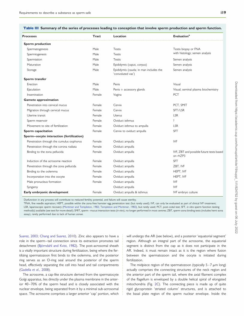
Suarez, 2003; Chang and Suarez, 2010). Zinc also appears to have arole in the sperm–tail connection since its extraction promotes taildetachment (Bjorndahl and Kvist, 1982). The post-acrosomal sheathis a vitally important structure during fertilization, being where the fer-tilizing spermatozoon first binds to the oolemma, and the posteriorring serves as an O-ring seal around the posterior of the spermhead, effectively separating the cell into head and tail compartments(Gadella et al., 2008).
The acrosome, a cap-like structure derived from the spermatocyteGolgi apparatus, lies directly under the plasma membrane in the anter-ior 40–70% of the sperm head and is closely associated with thenuclear envelope, being separated from it by a minimal sub-acrosomalspace. The acrosome comprises a larger anterior ‘cap’ portion, which
will undergo the AR (see below), and a posterior ‘equatorial segment’region. Although an integral part of the acrosome, the equatorialsegment is distinct from the cap as it does not participate in theAR. Indeed, it must remain intact as it is the region where fusionbetween the spermatozoon and the oocyte is initiated duringfertilization.
The midpiece region of the spermatozoon (typically 5–7 mm long)actually comprises the connecting structures of the neck region andthe anterior part of the sperm tail, where the axial filament complexof the flagellum is enveloped by a double helical spiral of elongatedmitochondria (Fig. 2C). The connecting piece is made up of quiterigid glycoprotein ‘striated column’ structures, and is attached tothe basal plate region of the sperm nuclear envelope. Inside the
.............................................................................................................................................................................................
Table III Summary of the series of processes leading to conception that involve sperm production and sperm function.
Processes Tract Location Evaluationa
Sperm production
Spermatogenesis Male Testis Testis biopsy or FNAwith histology; semen analysisSpermiogenesis Male Testis
Spermiation Male Testis Semen analysis
Maturation Male Epididymis (caput, corpus) Semen analysis
Storage Male Epididymis (cauda; in man includes the‘convoluted vas’)
Semen analysis
Sperm transfer
Erection Male Penis Visual
Ejaculation Male Penis + accessory glands Visual; seminal plasma biochemistry
Insemination Female Vagina PCT
Gamete approximation
Penetration into cervical mucus Female Cervix PCT, SMIT
Migration through cervical mucus Female Cervix SFT/LSR
Uterine transit Female Uterus LSR
Sperm reservoir Female Oviduct isthmus ?
Movement to site of fertilization Female Oviduct isthmus to ampulla LSR
Sperm capacitation Female Cervix to oviduct ampulla SFT
Sperm–oocyte interaction (fertilization)
Penetration through the cumulus oophorus Female Oviduct ampulla IVF
Penetration through the corona radiata Female Oviduct ampulla
Binding to the zona pellucida Female Oviduct ampulla IVF, ZBT and possible future tests basedon rhZP3
Induction of the acrosome reaction Female Oviduct ampulla SFT
Penetration through the zona pellucida Female Oviduct ampulla ZBT, IVF
Binding to the oolemma Female Oviduct ampulla HEPT, IVF
Incorporation into the oocyte Female Oviduct ampulla HEPT, IVF
Male pronucleus formation Female Oviduct ampulla IVF
Syngamy Oviduct ampulla IVF
Early embryonic development Female Oviduct ampulla & isthmus IVF embryo culture
Dysfunction in any process will contribute to reduced fertility potential, and failure will cause sterility.aFNA, fine needle aspiration; HEPT, possible within the zona-free hamster egg penetration test (but rarely used); IVF, can only be evaluated as part of clinical IVF treatment;LSR, laparoscopic sperm recovery (Mortimer and Templeton, 1982; Templeton and Mortimer, 1982), but rarely used; PCT, post-coital test; SFT, in vitro sperm function testingmethod(s) available (see text for more detail); SMIT, sperm–mucus interaction tests (in vitro), no longer performed in most centres; ZBT, sperm-zona binding tests (includes hemi-zonaassay), rarely performed due to lack of human zonae.
Requirements to describe a substance as sperm-safe i19D
ownloaded from
https://academic.oup.com
/humupd/article/19/suppl_1/i1/824762 by guest on 06 July 2022
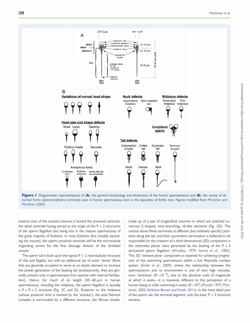
anterior part of the striated columns is buried the proximal centriole,the distal centriole having served as the origin of the 9 + 2 structuresof the sperm flagellum but being lost in the mature spermatozoa ofthe great majority of Eutheria. In most Eutheria (but notably exclud-ing the mouse), the sperm proximal centriole will be the microtubuleorganizing centre for the first cleavage division of the fertilizedoocyte.
The sperm tail is built upon the typical 9 + 2 microtubular structureof cilia and flagella, but with an additional set of outer ‘dense’ fibresthat are generally accepted to serve as an elastic element to increasethe power generation of the beating tail (evolutionarily, they are gen-erally present only in spermatozoa from species with internal fertiliza-tion). Hence, for much of its length (50–60 mm in humanspermatozoa), including the midpiece, the sperm flagellum is actuallya 9 + 9 + 2 structure (Fig. 2C and D). Posterior to the midpiece(whose posterior limit is marked by the ‘annulus’), the axial filamentcomplex is surrounded by a different structure, the fibrous sheath,
made up of a pair of longitudinal columns to which are attached nu-merous C-shaped, inter-branching, rib-like elements (Fig. 2D). Thevarious dense fibres terminate at different (but relatively specific) posi-tions along the tail, and their asymmetric termination is believed to beresponsible for the creation of a third-dimensional (3D) component inthe otherwise planar wave generated by the beating of the 9 + 2structured sperm flagellum (Woolley, 1979; Serres et al., 1983).This 3D ‘twisted plane’ component is essential for achieving progres-sion of the swimming spermatozoa within a low Reynolds numbersystem (Smith et al., 2009), where the relationship between thespermatozoon and its environment is one of very high viscosity,even ‘stickiness’ (R�10– 4), due to the absolute scale of magnitudeat which it exists—it is massively different to the perception of ahuman being or a fish swimming in water (R�104) (Purcell, 1977; Mor-timer, 2002; Kirkman-Brown and Smith, 2011). In the most distal partof the sperm tail, the terminal segment, only the basic 9 + 2 structureremains.
Figure 1 Diagrammatic representations of (A): the general morphology and dimensions of the human spermatozoon and (B): the variety of ab-normal forms (pleiomorphism) commonly seen in human spermatozoa even in the ejaculates of fertile men. Figures modified from Mortimer andMortimer (2005).
i20 Mortimer et al.
Dow
nloaded from https://academ
ic.oup.com/hum
upd/article/19/suppl_1/i1/824762 by guest on 06 July 2022
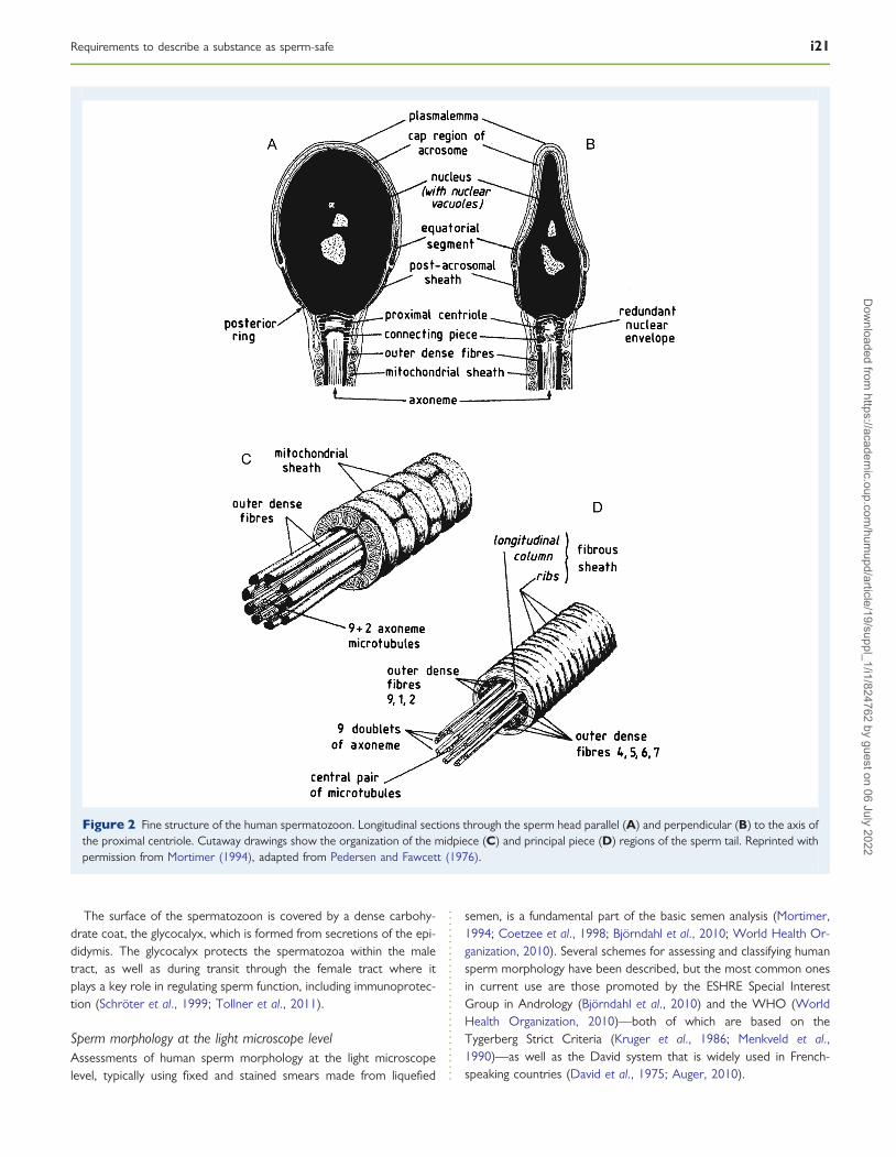
The surface of the spermatozoon is covered by a dense carbohy-drate coat, the glycocalyx, which is formed from secretions of the epi-didymis. The glycocalyx protects the spermatozoa within the maletract, as well as during transit through the female tract where itplays a key role in regulating sperm function, including immunoprotec-tion (Schroter et al., 1999; Tollner et al., 2011).
Sperm morphology at the light microscope levelAssessments of human sperm morphology at the light microscopelevel, typically using fixed and stained smears made from liquefied
semen, is a fundamental part of the basic semen analysis (Mortimer,1994; Coetzee et al., 1998; Bjorndahl et al., 2010; World Health Or-ganization, 2010). Several schemes for assessing and classifying humansperm morphology have been described, but the most common onesin current use are those promoted by the ESHRE Special InterestGroup in Andrology (Bjorndahl et al., 2010) and the WHO (WorldHealth Organization, 2010)—both of which are based on theTygerberg Strict Criteria (Kruger et al., 1986; Menkveld et al.,1990)—as well as the David system that is widely used in French-speaking countries (David et al., 1975; Auger, 2010).
Figure 2 Fine structure of the human spermatozoon. Longitudinal sections through the sperm head parallel (A) and perpendicular (B) to the axis ofthe proximal centriole. Cutaway drawings show the organization of the midpiece (C) and principal piece (D) regions of the sperm tail. Reprinted withpermission from Mortimer (1994), adapted from Pedersen and Fawcett (1976).
Requirements to describe a substance as sperm-safe i21D
ownloaded from
https://academic.oup.com
/humupd/article/19/suppl_1/i1/824762 by guest on 06 July 2022
Modern concepts of sperm normality are based on studies ofspermatozoa recovered from cervical mucus post-coitally (Menkveldet al., 1990; Mortimer and Menkveld, 2001) and supported by obser-vations of spermatozoa that bind to the zona pellucida in vitro (Liu andBaker, 1992).
There is growing opinion that the term ‘normal’ should be replacedby ‘ideal’ or ‘typical’, and that the per cent ideal spermatozoa—whichis usually very low—be supplemented by additional information on thedegree of malformation of the abnormal spermatozoa, which is anegative predictor of biological function (Auger, 2010; Bjorndahlet al., 2010).
In general terms, a ‘typical’ or ‘ideal’ human spermatozoon has anoval head form with a smooth contour and a clearly visible and well-defined acrosome. The tail is inserted symmetrically without any ab-normalities of the neck/midpiece and tail region, and there are nocytoplasmic residues in the neck/midpiece or tail regions. Thesperm head is between 3.0 and 5.0 mm in length and 2.0 and3.0 mm in width, with the acrosome covering 30–60% of its anteriorregion. The midpiece is no longer than 1.5× the length of a typicalhead, and �1 mm thick. The tail is 45–50 mm long with no sharpbends or kinks. Spermatozoa with any features that appear ‘border-line’ or slightly atypical are considered to be atypical (Bjorndahlet al., 2010).
Sperm motilityAs spermatozoa mature in the epididymis, they gain the capacity formotility, but do not express motility until they are ‘activated’ at ejacu-lation. Spermatozoa removed directly from the testis are capable,once washed into culture medium, of only twitching motility, althougha few progressively motile spermatozoa can sometimes be found(Bjorndahl et al., 2010). This twitching motility can be enhancedwith phosphodiesterase inhibitors such as pentoxifylline (see theFactors affecting male reproductive capacity—Pharmaceuticals—Phosphodiesterase inhibitors section). The percentage of testicularspermatozoa exhibiting motility may increase with overnight incuba-tion (Bjorndahl et al., 2010). Progressive motility requires ATP produc-tion, by both anaerobic glycolysis in the sperm tail and oxidativephosphorylation in the mitochondria of the sperm midpiece (Piomboniet al., 2012). Many compounds are known to inhibit these processes,and even more can cause outright sperm cell death by permeabilizingthe sperm membrane, preventing normal membrane function, or inhi-biting other critical processes.
Progressive motility is normally an attribute of, at a minimum,around half of the spermatozoa in an ejaculate; many spermatozoain semen exhibit non-progressive motility, meaning that their tailsare beating, but the cell is not moving from one place to another;other spermatozoa are non-motile (Mortimer, 1994, 1997; Bjorndahlet al., 2010; World Health Organization, 2010). Sperm motility isinduced by beating of the sperm flagellum, with waves being initiatedat the anterior end of the tail, inside the midpiece, where it joins theconnecting piece (reviews: Mortimer, 1997; Turner, 2006; Linde-mann and Lesich, 2010; Woolley, 2010; Inaba, 2011). Careful ana-lysis of swimming spermatozoa has revealed that each beat can bedefined in terms of beat initiation, wave development and wavepropagation. During the wave development phase, the curvature ofthe tail increases, and is dependent on intracellular Ca2+
concentration—so a delay in wave propagation will result in alarger amplitude wave being sent down the tail, generating greaterpropulsive force (Mortimer, 1997; Olson et al., 2010; Ishijima,2011). Larger amplitude waves lead to higher sperm velocity aswell as greater lateral displacement of the sperm head—a patternof movement that is essential for effective penetration into, and mi-gration within, cervical mucus and failure to develop such motility hasbeen established as a cause of infertility (Feneux et al., 1985). Thestudy of sperm motility patterns is termed ‘kinematics’ (Fig. 3) andhas been greatly facilitated since the development of computer-aidedsperm analysis technology (‘CASA’; Mortimer, 1994, 1997). Theactual human sperm trajectory in semen in 3D space is an ovoidhelix with a radius of �0.5–3.0 mm; a significant majority of sperm-atozoa (ca. 90%) preferring right-handed helices over left-handedones (Su et al., 2012).
Sperm survival/longevitySpermatozoa have a finite functional lifespan, a maximum of a few dayswithin the human female reproductive tract (reviews: Mortimer, 1983,1995b). Loss of motility is the most obvious indication of cell death,although a non-motile spermatozoon might still be alive in terms ofmembrane integrity (Bjorndahl et al., 2003). Studies of human sperm-atozoa removed from the cervix at different times after inseminationdemonstrated that spermatozoa lose their ability to acrosome reactand fuse with an oocyte in �2 days, but remain motile for anotherday or so (Zinaman et al., 1989).
As noted already, oxidation of docosahexanoic acid in the spermplasma membrane is the major factor that determines the motile life-span of spermatozoa in vitro (Aitken and Clarkson, 1987), as well asmembrane damage and DNA oxidation (Fraga et al., 1991, 1996).
Because of its ease of measurement, sperm motility is often usedby clinical laboratories as a ‘sperm survival assay’ as part of thequality control processes for plastic- or glass-ware and culturemedia (Claassens et al., 2000; Bjorndahl et al., 2010). Residual chemi-cals, detergents, plasticizers and endotoxins or other biological toxi-cants adversely affect sperm survival. As a clinical test, a survivalassay is a measure of sperm ‘hardiness’, although particular assay con-ditions can influence the outcome (e.g. a highly stimulatory mediumcan promote high ATP generation or ARs, either of which will leadto a shortened lifespan for spermatozoa). Human sperm motilityafter an overnight incubation in IVF medium is a major predictive com-ponent of pregnancy after intrauterine insemination (Branigan et al.,1999).
Sperm transfer: ejaculation, the ejaculateand impregnationEjaculation typically requires erection of the penis, and this is essentialfor impregnation of the female via intercourse. Erectile dysfunction cantherefore be a serious cause of subfertility, although many types oferectile dysfunction can now be treated using inhibitors of phospho-diesterase type 5, and electroejaculation can also be performed inextreme cases, e.g. those involving many types of spinal cord injury(van Ahlen and Kliesch, 2010). However, it is not known to whatextent electroejaculation mimics the normal ejaculation process,where spermatozoa are transferred from the cauda epididymidesand ‘convoluted vas’ and mixed with the secretion from the prostate
i22 Mortimer et al.
Dow
nloaded from https://academ
ic.oup.com/hum
upd/article/19/suppl_1/i1/824762 by guest on 06 July 2022
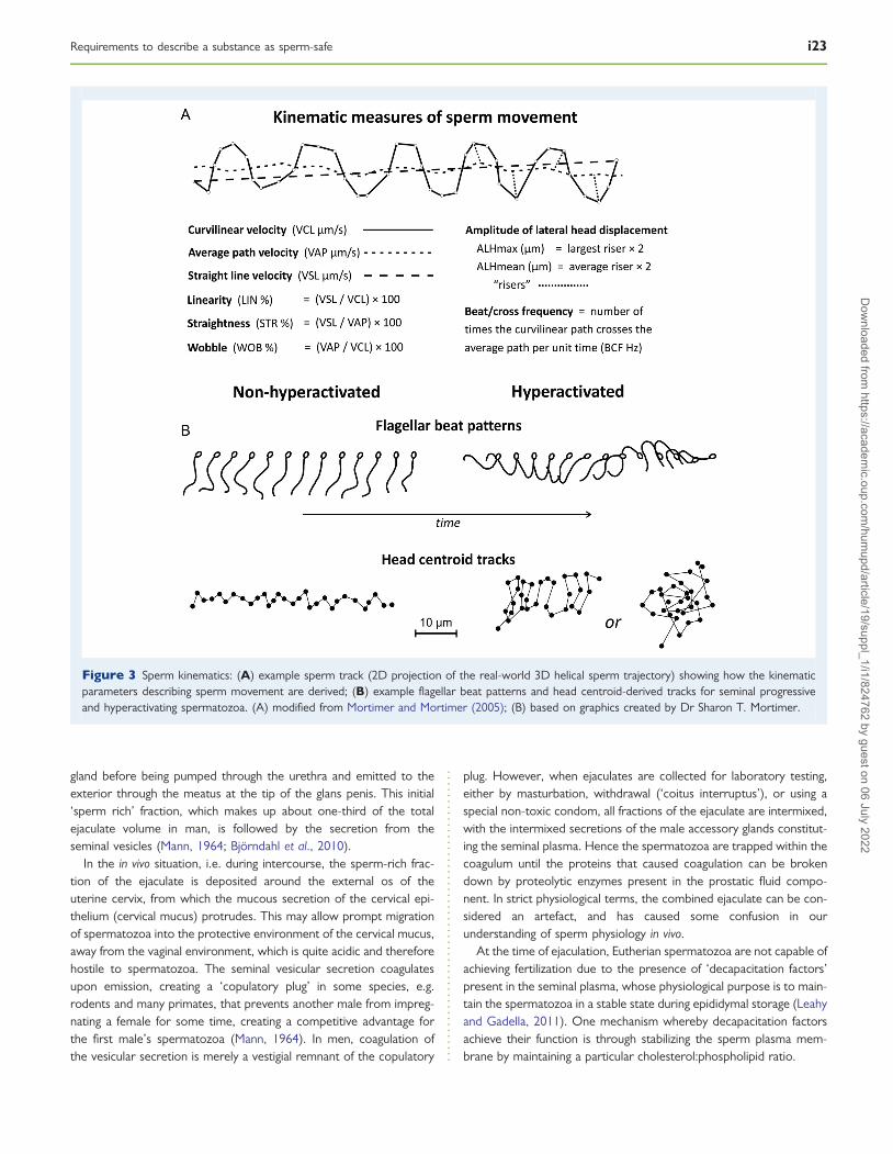
gland before being pumped through the urethra and emitted to theexterior through the meatus at the tip of the glans penis. This initial‘sperm rich’ fraction, which makes up about one-third of the totalejaculate volume in man, is followed by the secretion from theseminal vesicles (Mann, 1964; Bjorndahl et al., 2010).
In the in vivo situation, i.e. during intercourse, the sperm-rich frac-tion of the ejaculate is deposited around the external os of theuterine cervix, from which the mucous secretion of the cervical epi-thelium (cervical mucus) protrudes. This may allow prompt migrationof spermatozoa into the protective environment of the cervical mucus,away from the vaginal environment, which is quite acidic and thereforehostile to spermatozoa. The seminal vesicular secretion coagulatesupon emission, creating a ‘copulatory plug’ in some species, e.g.rodents and many primates, that prevents another male from impreg-nating a female for some time, creating a competitive advantage forthe first male’s spermatozoa (Mann, 1964). In men, coagulation ofthe vesicular secretion is merely a vestigial remnant of the copulatory
plug. However, when ejaculates are collected for laboratory testing,either by masturbation, withdrawal (‘coitus interruptus’), or using aspecial non-toxic condom, all fractions of the ejaculate are intermixed,with the intermixed secretions of the male accessory glands constitut-ing the seminal plasma. Hence the spermatozoa are trapped within thecoagulum until the proteins that caused coagulation can be brokendown by proteolytic enzymes present in the prostatic fluid compo-nent. In strict physiological terms, the combined ejaculate can be con-sidered an artefact, and has caused some confusion in ourunderstanding of sperm physiology in vivo.
At the time of ejaculation, Eutherian spermatozoa are not capable ofachieving fertilization due to the presence of ‘decapacitation factors’present in the seminal plasma, whose physiological purpose is to main-tain the spermatozoa in a stable state during epididymal storage (Leahyand Gadella, 2011). One mechanism whereby decapacitation factorsachieve their function is through stabilizing the sperm plasma mem-brane by maintaining a particular cholesterol:phospholipid ratio.
Figure 3 Sperm kinematics: (A) example sperm track (2D projection of the real-world 3D helical sperm trajectory) showing how the kinematicparameters describing sperm movement are derived; (B) example flagellar beat patterns and head centroid-derived tracks for seminal progressiveand hyperactivating spermatozoa. (A) modified from Mortimer and Mortimer (2005); (B) based on graphics created by Dr Sharon T. Mortimer.
Requirements to describe a substance as sperm-safe i23D
ownloaded from
https://academic.oup.com
/humupd/article/19/suppl_1/i1/824762 by guest on 06 July 2022
Sperm transport through the femalereproductive tractOverviewAmong Eutheria, there are numerous variations in mechanisms forsperm deposition within the female reproductive tract and their sub-sequent transport to, and storage at, the site of fertilization (Mortimer,1983, 1995b; Suarez and Pacey, 2006; Ikawa et al., 2010). Basically,there are two major strategies: ‘uterine’ and ‘vaginal’ insemination—with humans employing the latter.
Sperm–cervical mucus interactionOnce deposited around the external os, spermatozoa must penetrateinto the mucus secreted by the uterine cervix and which occludes thecervical canal. The cervical mucus is only receptive to spermatozoaaround mid-cycle (Mortimer, 1983, 1994, 1995b; Bjorndahl et al.,2010). Mid-cycle (periovulatory) cervical mucus is unique in itswater content and molecular arrangement, easily tested by its ‘Spinn-barkeit’ and ‘ferning’ properties, with its secretion being under thecontrol of estrogen. Penetration into, and migration within, cervicalmucus depends on effective progressive motility (Mortimer, 1983;Feneux et al., 1985); the older ‘insuck’ theory associated withfemale orgasm is no longer afforded any real credibility (Mortimer,1983, 1995b). Cervical mucus may harbour antisperm antibodies orother poorly defined molecules that immobilize spermatozoa.
There is no physiological evidence for a ‘cervical sperm reservoir’ asdescribed in older textbooks, nor is there evidence to supportrelationships between coital position and fecundity, coital practicesand the gender of resulting offspring, or between female orgasmand fertility (Levin, 2011). As spermatozoa migrate into cervicalmucus, they escape from the decapacitation factors-rich seminal envir-onment and the process of ‘capacitation’ commences. Spermatozoamust swim through the column of cervical mucus that occludes theuterine cervix.
Sperm migration through the uterus and oviductsOnce within the uterine lumen (which, in nulliparous women, is reallyonly a virtual cavity, with only a thin film of uterine fluid separating thequite closely apposed anterior and posterior walls of the uterus). Themost likely explanation for the transfer of spermatozoa from the in-ternal os of the cervix to the utero-tubal junctions is their beingspread by segmental contractions of the uterine wall, and migrationthrough the utero-tubal junction appears to depend on sperm motility(Mortimer, 1983, 1995b; Suarez and Pacey, 2006; Ikawa et al., 2010).In humans, the isthmus region of the oviduct (Fallopian tube) serves asthe site of physiological sperm storage, although solid evidence regard-ing this function has proven difficult to obtain in women (Williamset al., 1993; Mortimer, 1995b). The isthmic environment is capableof regulating sperm function during their storage there, with spermato-zoa usually interacting intimately with the lining of the isthmus. Duringisthmic sperm storage, the completion of capacitation seems to besuppressed, and release from the isthmic sperm reservoir seems tobe regulated via a portal blood supply linking the venous drainagefrom the ovary to the arterial supply of the ipsilateral oviduct(Suarez and Pacey, 2006; Suarez, 2007, 2008; Holt and Fazeli, 2010).
Sperm seem to continue to bind to oviductal epithelium afterleaving the isthmic reservoir, and motility is crucial in their migration
to the site of fertilization in the oviductal ampulla (Chang andSuarez, 2012). Regional gradations in oviduct fluid viscosity, perhapscombined with temperature gradients, could interact with sperm fla-gellar activity to reveal subtle differences in sperm functional potential(Hunter et al., 2011) and create environments for sperm selection(Kirkman-Brown and Smith, 2011).
Capacitation and hyperactivationAlthough ‘sperm capacitation’ was discovered 60 years ago (Chang,1951; Austin, 1952), and is defined as resulting in the spermatozoonhaving acquired the capacity to undergo the AR and fertilize anoocyte, its molecular mechanisms are still not fully understood(Yanagimachi, 1994; Fraser, 1998; Bedford, 2004; De Jonge, 2005;Salicioni et al., 2007). Several changes take place in different regionsof the spermatozoon, including changes in the sperm plasma mem-brane that involve the loss of decapacitation factors and cholesterol(Leahy and Gadella, 2011), phosphorylation of tyrosine residues inthe outer dense fibres and fibrous sheath of the tail (allowing thetail to become more flexible and exhibit different patterns of beatingto those seen in ejaculate or ‘seminal’ spermatozoa) (Yunes et al.,2003; Mariappa et al., 2006), the ability to generate calcium oscillations(Aitken and McLaughlin, 2007) and changes in the acrosome thatprepare it to undergo the AR (Yanagimachi, 2011). Capacitation isan essential requirement for fertilization, both in vivo and in vitro forIVF, and it must be adequately supported by any culture mediumthat is to be used in an IVF system.
The sperm head plasma membrane undergoes major reorganiza-tion, notably the formation of lipid ordered microdomains (lipidrafts) during capacitation with the dynamic redistribution of membranemolecules over its surface that create functionally heterogeneousregions such as the area overlying the acrosomal cap which partici-pates in the AR (Gadella et al., 2008; Leahy and Gadella, 2011). Cap-acitation can be promoted by substances such as albumin or minorcomponents such as cholesteryl ester transfer protein (Muller andRavnik, 1995; Ravnik et al., 1995), and bicarbonate (Gadella andVan Gestel, 2004; Visconti et al., 2011), and even to some degreeover-ridden by powerful agonists such as b-cyclodextrins, which se-quester cholesterol (Visconti et al., 1999), or by-passed by calciumionophores which can trigger the AR more-or-less directly (Bailey,2010). Capacitation is regulated by increased intracellular pH (pHi
having been maintained at a slightly acidic level during epididymalstorage) via the Hv1 voltage-sensitive proton channel (Lishko et al.,2012; Florman et al., 2010).
Hyperactivated motility‘Hyperactivated motility’ is expressed as a concomitant of capacita-tion: physiologically during the passage of spermatozoa through theupper parts of the female reproductive tract to the site of fertilizationin the oviductal ampulla, or under capacitating conditions in vitro. It ischaracterized by the development of high-amplitude flagellar waves,which causes a ‘whiplash-like’ movement of the head in free-swimmingspermatozoa (Yanagimachi, 1994; Mortimer, 1997). The vigorousbeating pattern serves to dislodge spermatozoa that have bound tothe oviductal epithelium, and later, to help drive the fertilizing sperm-atozoon through the zona pellucida.
Hyperactivation depends on increased flexibility of the tail struc-tures, combined with increased [Ca2+]i, presumably originating from
i24 Mortimer et al.
Dow
nloaded from https://academ
ic.oup.com/hum
upd/article/19/suppl_1/i1/824762 by guest on 06 July 2022
a calcium store located in the sperm neck and midpiece (Costelloet al., 2009; Buffone et al., 2012), and a delay in wave propagation,results in beats of greatly increased amplitude (Fig. 3B). If the delayin wave propagation is increased further, then the curvature developedin the proximal part of the tail can cause the sperm head to turn backon itself, so that when the wave is propagated (at much increased vel-ocity in hyperactivated spermatozoa), the spermatozoon ‘thrashes’,displaying a non-progressive ‘whiplash’ pattern of movement that isgenerally characteristic of hyperactivated Eutherian spermatozoaexamined in vitro (Yanagimachi, 1970; Mortimer, 1997), and can alsobeen seen in vivo in some species where suitable observations canbe made (Yanagimachi, 1970).
Hyperactivated spermatozoa show poor ability to traverse theutero-tubal junction (Shalgi et al., 1992), and hence prematuresperm capacitation (e.g. in sperm preparations intended for intrauter-ine insemination) could be severely disadvantageous, and precludeproper sperm transport to the site of fertilization.
In vitro, the proportion of spermatozoa with hyperactivated motilitycan be quantified using computer-aided motility analysis (Mortimer,1997; Bjorndahl et al., 2010). True hyperactivated motility does notoccur in seminal plasma. While a similar pattern of beating can beseen following ROS-induced damage (de Lamirande and Gagnon,1993; de Lamirande et al., 1997), it fails to meet proper kinematic criteriafor hyperactivation (because tyrosine phosphorylation has not occurredand the axial filament complex is not yet in its ‘capacitated’ state), andmust be seen as a pathological event that could lead to impaired fertility.
The role of calcium ion dynamics in the initiation and maintenance ofhyperactivated motility involves CatSper (cation channel of sperm), apH-activated weakly voltage-dependent calcium channel (Qi et al.,2007; Publicover et al., 2008; Olson et al., 2010; Brenker et al., 2012;Lishko et al., 2012), whose action can be integrated with hydrodynamicsand flagellar modelling to create a regulatory model for sperm hyperac-tivation (Olson et al., 2011a, b). CatSper also mediates progesterone-induced Ca2+ influx into human spermatozoa (Strunker et al., 2011),thereby serving as a non-genomic progesterone receptor in spermato-zoa that can modulate [Ca2+]i as the basis for chemotaxis (Publicoveret al., 2008; Yoshida and Yoshida, 2011). Hyperactivation and chemo-taxis have been combined into models for how spermatozoa locatethe oocyte within the 3D space of the oviduct (Guerrero et al., 2011).
Sperm–oocyte interaction, including thephysiological ARCumulus penetrationAfter reaching the site of fertilization in the ampulla of the oviduct (Fal-lopian tube), the fertilizing spermatozoon must migrate through thecumulus mass and corona radiata cells surrounding the oocyte(Ikawa et al., 2010; Kirkman-Brown and Smith, 2011). Although thematrix of the cumulus mass has a very high hyaluronate component,the traditional view that this migration was helped by hyaluronidasefrom the acrosome is now considered circumspect, it now beingunclear whether what was thought to be the enzyme hyaluronidaseactually has catalytic activity (Kang et al., 2010).
Sperm–zona pellucida bindingThe conventional view is that upon reaching the zona pellucida, thespermatozoon binds, in a more-or-less species-specific way at least
partly dependent on lectin-like (sugar–protein) interactions, toputative sperm receptors which then induce the physiological AR(Yanagimachi, 1994; Visconti and Florman, 2010). Although recentresearch in mice suggests the physiological AR is probably initiatedprior to binding to the zona pellucida (Jin et al., 2011; Yanagimachi,2011), in humans most of the spermatozoa recently bound to thezona pellucida are acrosome-intact (Cross et al., 1988) and acrosome-reacted spermatozoa are unable to bind to the zona pellucida(Liu et al., 2006).
Acrosome reactionThe AR is a complex event, involving extrinsic receptors, signaltransduction, influx of calcium, membrane fusion and vesiculationwith loss of parts of the plasma membrane and outer acrosomal mem-brane, and activation and release of enzymes from the acrosome(Yanagimachi, 2011). Prevention or disruption of any of these stepscan block the event, and thus prevent fertilization. In human sperm-atozoa, it is believed that the AR is induced following binding to a pu-tative sperm receptor integral to the zona pellucida, most probablyzona pellucida glycoprotein 3 (ZP3) and proceeds via a still poorlycharacterized cascade of intracellular biochemical pathways (Dean,2007; Visconti, 2009; Visconti and Florman, 2010). However, substan-tial elevation of [Ca2+]i is induced, perhaps involving a store in theacrosomal region (Costello et al., 2009; Buffone et al., 2012), and aCa2+-dependent Cl– current can be identified during ARs inducedby a recombinant ZP3 (rhZP3) molecule in vitro (Orta et al., 2012).
The ‘normal AR’ (Fig. 2A and B) involves dissociation of the tightlypackaged proenzymes stored inside the acrosome (mostlypro-acrosin), followed by localized fusions between the spermplasma membrane and the outer acrosomal membrane of the capregion of the acrosome creating openings through which enzymesare released (Zanetti and Mayorga, 2009). While acrosin may act tosoften the glycoprotein matrix of the zona pellucida, the fertilizingspermatozoon does not simply digest its way through the zona: itshyperactivated motility generates propulsive force 10× greater thanthat required to break covalent bonds (Drobnis et al., 1988), henceit pushes its way through the softened substance of the zona. Ultim-ately, these membranes are lost, leaving an acrosome-reacted sperm-atozoon whose anterior exterior surface is the inner acrosomalmembrane (Yanagimachi, 1994; Ikawa et al., 2010). In a normallyacrosome-reacted spermatozoon, the equatorial segment of the acro-some must remain intact to initiate fusion between the sperm andoocyte plasma membranes (spermalemma and oolemma) during fer-tilization; without the equatorial segment, a spermatozoon cannotachieve fertilization. Under in vivo conditions, the acrosome-reactedspermatozoon is held in place by virtue of being embedded withinthe corona radiata and cumulus mass, and so is ready to move intothe next stage of its penetration of the oocyte vestments.
Capacitated spermatozoa are in a highly labile state, and can alsoinitiate ‘acrosome loss’ via a ‘false AR’ in response to many physicaland chemical triggers (e.g. elevated pH: Mortimer, 1995a). Prematureloss of the acrosome (Tesarik, 1989; ESHRE Andrology SpecialInterest Group, 1996), as a result of either a normal or false ARbefore the spermatozoa reach the zona pellucida prevents theirbinding to it (Liu et al., 2006)—thereby impairing or perhaps prevent-ing fertilization.
Requirements to describe a substance as sperm-safe i25D
ownloaded from
https://academic.oup.com
/humupd/article/19/suppl_1/i1/824762 by guest on 06 July 2022
Sperm–oolemma binding and sperm–oocyte fusionOnce through the zona pellucida, the spermatozoon makes contactwith the oolemma and a specific binding process occurs betweenthe oolemma and the surface of the posterior sheath region of thespermatozoon, although the molecular details of this are still largelyunresolved (Ikawa et al., 2010). Based on old microcinematographicstudies (RJ Blandau, Seattle, WA, USA), sperm motility ceases atthis point and the remainder of the process of sperm incorporationinto the oocyte is effected by the oocyte. Sperm–oocyte fusion isinitiated between the remaining sperm plasma membrane over theequatorial segment and the oolemma, and the entire spermatozoon,including the midpiece and sperm tail structures, is incorporatedinto the oocyte and disassembled (Yanagimachi, 1994; Sutovsky andSchatten, 2000).
The sperm mitochondria, which have been tagged with ubiquitin,are destroyed, although remnants may be identifiable at the ultrastruc-tural level during the first one or two cleavage divisions (Sutovskyet al., 2000; Baska et al., 2008). The 9 + 9 + 2 axial filamentcomplex and the fibrous sheath of the sperm tail are all disassembledwithin the ooplasm. The sperm plasma membrane remains as a ‘patch’in the oolemma, it only enters the ooplasm during ICSI, and there isevidence that ROS-based oxidation of its phospholipid and cholesterolcontent (the latter is especially high, since spermatozoa used for ICSIare typically uncapacitated) can adversely affect embryonic develop-ment (Tateno and Kamiguchi, 2007).
Sperm contributions at fertilizationThe fertilizing spermatozoon brings three essential components to theoocyte: the ‘male haploid nucleus’, ‘centrosome’ (centriole), and an‘egg-activating factor’ (Sutovsky and Schatten, 2000; Barroso et al.,2009), any defects in any of these three elements can result in fertil-ization failure. After the fertilizing spermatozoon fuses with theoolemma and is drawn into the cytoplasm of the oocyte, the spermnucleus must decondense to form the male pronucleus. In somecases, the decondensation step occurs too early or not at all andcan result in failed fertilization, even following ICSI (Sakkas et al.,1996; Esterhuizen et al., 2002; Bjorndahl and Kvist, 2010). After themale and female pronuclei fuse, their respective chromosomes mustline up correctly and subsequently separate for the embryo’s firstcell division. In most Eutheria, this division is organized by the centriolebrought in by the spermatozoon (Schatten, 1994; Sathananthan et al.,1996, 2006; Sathananthan, 2009). Damaged centrioles will not preventfertilization, but will lead to the early demise of the embryo. The thirdcritical contribution is a signalling molecule, a sperm-specific phospho-lipase, PLCz (Kashir et al., 2010; Ramadan et al., 2012), that triggerscalcium oscillations and induces the resumption of the secondmeiotic division (Ajduk et al., 2008; Swann and Yu, 2008).
Syngamy and early embryonic developmentAfter the sperm nucleus has been incorporated, its nuclear envelope islost and a new one forms from oocyte-derived components as thesperm chromatin is decondensing and its protamines are replacedby histones. During this process, there is opportunity for somerepair to sperm DNA damage, but it is limited by time (de Boeret al., 2010). Once this process is complete, the structure is knownas the male pronucleus, which then merges with the female
pronucleus at syngamy, marking the completion of the fertilizationprocess and the creation of a new genetic individual.
Development of the early embryo is controlled by a pool of mRNAsthat was established in the ooplasm during the final stages of oocytematuration inside the Graafian follicle (Gosden, 2002; Krisher,2004). Timing of activation of the human embryonic genome (tran-scription) has generally been believed to begin between the 4- and8-cell stage, and translation at the 8-cell stage. However, a recentreport has described the presence of waves of transcriptional activitystarting as early as the 2-cell stage (as well as waves of degradation ofmaternal mRNA), although it remains unclear when these mRNAs aretranslated (Vassena et al., 2011). Hence the embryonic genome is re-sponsible for development beyond the 8-cell stage, i.e. from Day 3(Braude et al., 1988; Wong et al., 2010).
The paternal DNA is highly susceptible to fragmentation caused bychemotherapeutic drugs, radiation and other mechanisms that in-crease oxidative stress (Barratt et al., 2010). Fragmented spermDNA may or may not be able to be repaired by the oocyte orearly embryo, depending on the extent of damage. Also, small frag-ments of DNA that result from ‘double-double’ breaks might be inte-grated at the wrong place or inverted, resulting in spontaneous‘mutations’ that could lead to serious childhood disorders. Anumber of childhood diseases have already been linked to paternalage, and sperm DNA damage increases with age (Singh et al.,2003). There are numerous recent reviews on the role of spermDNA damage in male infertility (e.g. Barratt et al., 2010; Lewis andSimon, 2010; Pacey, 2010; Gharagozloo and Aitken, 2011), but al-though many published studies claim that sperm DNA damage/fragmentation has been assessed, the measurement methods areonly indirect—it remains unclear whether some of these ‘assays’detect actual endogenous DNA damage or just increased vulnerabilityof the sperm DNA (Barratt et al., 2010). Nonetheless, embryos withmassively damaged sperm-derived haploid genomes usually appearperfectly normal up until Day 3 of development, but typically have ex-tremely poor capacity to form normal blastocysts and implant (Seli andSakkas, 2005; Tesarik, 2005; Barroso et al., 2009; Sakkas and Alvarez,2010). Associations between altered epigenetic profiles in maturespermatozoa that reflect heritable epigenetic changes during sperm-atogenesis, perhaps induced via endocrine disruption, and infertilityhave recently been discussed (Carrell and Hammoud, 2010;McCarrey, 2012).
Assessments of sperm toxicity
Experimental and biological artefactsThere are two major sources of artefacts in many in vitro studies onsperm physiology: the impact of viscosity on sperm motility and thecollection of the ejaculate as a single specimen.
ViscosityChanges in the viscosity of the environment in which spermatozoa areswimming will affect their movement, and hence analyses of their pro-gressive motility and kinematics. A non-engineer’s perceptions ofhydrodynamics are based upon human perceptions of aquatic organ-isms, and their simple extrapolation to spermatozoa will lead to erro-neous concepts unless the Reynolds number (R: the ratio of the
i26 Mortimer et al.
Dow
nloaded from https://academ
ic.oup.com/hum
upd/article/19/suppl_1/i1/824762 by guest on 06 July 2022
inertial forces to the viscous forces) of the system is taken into account(Purcell, 1977; Mortimer, 2002; Smith et al., 2009; Gadelha et al.,2010). Sperm motility is far more complex than simple considerationof the ‘percentage motile’ or ‘progressive’ spermatozoa, and robustanalysis of sperm movement patterns (sperm ‘kinematics’) requiresconsideration of the interaction between flagellar beating patternsand the physical nature of the environment in which spermatozoaexist. Consequently, any studies in which the viscosity of products(e.g. sperm washing or culture medium or lubricant) is modifiedmust include proper controls to avoid artefacts, which can easily bemisconstrued as impairments (or enhancements) to sperm motility,and hence perceived functional ability.
The collected ejaculateThis artefactual situation, brought about by collecting the humanejaculate as a single specimen, results in several abnormal (non-physiological) circumstances that can have major impacts on sperm-atozoa (e.g. spermatozoa in the second fraction of ejaculates incases of asthenozoospermia and teratozoospermia show elevatedlevels of sperm DNA fragmentation (Kumar et al., 2011). However,for many practical reasons, it is difficult to avoid the routine collectionof the whole ejaculate in a single container, and this practice will mostlikely continue unless some diagnostic or therapeutic advantage toexamining or separating spermatozoa from the first fraction of a care-fully collected split ejaculate has been established. Hence collecting thecomplete human ejaculate for analysis remains the de facto standardfor the primary research tool for studying the effects of toxicants onthe male reproductive system (Cohn et al., 2002; Brazil et al.,2004a, b).
Exposure of spermatozoa to seminal vesicular secretion. Spermatozoa ini-tially exposed to seminal vesicular fluid rather than prostatic fluid areless motile and show impaired survival (Lindholmer, 1973; Lindholmerand Eliasson, 1974). Furthermore, due to extraction of nuclear zinc byseminal vesicle fluid, there is an initial increased accessibility of theDNA followed by a rapid super-stabilization of the chromatin.Increased DNA accessibility constitutes an opportunity for increasedDNA damage, especially in an oxidative environment such as theseminal vesicular fluid. Subsequently, after spermatozoa have beenexposed to the seminal plasma for more than a few minutes, accessto the DNA is reduced due to excessive disulphide bond formation(Bjorndahl and Kvist, 1985; Kvist and Bjorndahl, 1985; Kvist et al.,1985), a phenomenon that can influence sperm chromatin damagetest results by (i) concealing DNA damage due to reduced accessto the DNA (false-negative results) and (ii) inducing more damagein vitro beyond what already existed in vivo (false-positive results).Thus, a sperm-safe product should take into consideration possibleeffects on sperm chromatin structural stability as well as the risk foroxidative damage.
Prolonged exposure to seminal plasma. Eutherian ejaculated spermato-zoa are prevented from undergoing capacitation by decapacitationfactors present in the seminal plasma. Seminal plasma also containsone or more factors to which prolonged exposure adversely affectsperm function, including the ability to penetrate cervical mucus,undergo the AR in vitro and the fertilization process generally
(Yanagimachi, 1994; Mortimer, 2000a, b). Based on studies usingthe zona-free hamster oocyte penetration assay, exposure of humanspermatozoa to seminal plasma for more than 30 min after ejaculationcan permanently diminish their fertilizing capacity (Rogers et al., 1983),and contamination of prepared sperm populations with as little as0.01% (v/v) of seminal plasma (i.e. a 1:10 000 dilution) can decreasetheir fertilizing capacity (Kanwar et al., 1979). As discussed already, theexposure of human spermatozoa to the hypertonic conditions in lique-fied ejaculates can be considered unphysiological and might have sig-nificant implications for their subsequent handling and processing.Clearly, spermatozoa for clinical procedures (e.g. IUI or IVF) or la-boratory tests of sperm fertilizing ability must be removed from theseminal plasma as soon as possible after ejaculation (but which canbe delayed to allow for normal liquefaction at 378C), and why poorsperm washing protocols could adversely affect sperm functional po-tential as a result of carry-over of seminal plasma.
Human sperm motility and vitalityIn vitro assessment of sperm progression is the oldest way of assessingsperm function (Botella-Llusia, 1956) and for testing substances foradverse affects on sperm function, e.g. the transmembrane migrationtest (Hong et al., 1981a). Semen analysis sperm motility assessmentshave long been used in studies on reproductive toxicology(Wyrobek et al., 1983b; Schrader et al., 1991; Cohn et al., 2002),and the need for standardized training, robust protocols and qualitycontrol are well-recognized (Brazil et al., 2004a, b).
Visual assessments of sperm motility under a microscope are no-toriously subjective, and without rigorous training most observerscan only provide ‘guesstimates’ as to even the proportion of motility,or progressively motile, spermatozoa. This problem has been knownfor 60 years (MacLeod and Gold, 1951), but it can be eliminated, pro-vided that thorough, goal-orientated training using qualified referencematerials is undertaken (Mortimer, 1994). The issue of the quality ofsperm progressive motility has also been recognized since 1951, and itcan also be addressed via thorough training (Barratt et al., 2011). Beingable to determine the proportion of spermatozoa that show rapidprogression (defined as those with a space-gaining velocity of atleast 25 mm/s, at 378C) identifies the subpopulation of spermatozoathat are biologically—and hence clinically—important (Barratt et al.,2011).
CASA technology is still unable to provide reliable values for eithertotal sperm concentration or the proportions of motile or progres-sively motile spermatozoa in routine practice without a substantialuncertainty of measurement (Mortimer et al., 1995; ESHRE AndrologySpecial Interest Group, 1998; Mortimer and Mortimer, 1998).However, CASA can be used reliably to quantify specific subpopula-tions of spermatozoa with functional potential, e.g. those that showthe correct kinematics for penetrating into cervical mucus (ESHREAndrology Special Interest Group, 1998; Mortimer, 2000b; Mortimerand Mortimer, 2005; Bjorndahl et al., 2010).
When analyzing sperm motility as a marker of spermotoxicity, anassay can be either a single time end-point or else a longitudinalstudy over a period of time that is commensurate with physiology,e.g. the detection of spermotoxicity to spermatozoa in seminalplasma of supposedly ‘non-spermicidal’ lubricant and ultrasound gelsused in reproductive medicine (Agarwal et al., 2008a, b, c; Vargas
Requirements to describe a substance as sperm-safe i27D
ownloaded from
https://academic.oup.com
/humupd/article/19/suppl_1/i1/824762 by guest on 06 July 2022
et al., 2011). For spermatozoa in semen, this means no more than ashort-term incubation period since spermatozoa that migrate fromthe ejaculate into cervical mucus in vivo do so within a very shortperiod of time, making seminal plasma a non-physiological environ-ment for human spermatozoa beyond that time window (MacLeodand Gold, 1951; Mortimer, 1995b); longer incubation periods, e.g.several hours, can only be seen as a sort of unphysiological ‘stresstest’. Using an elevated incubation temperature, e.g. 398C, onlyexacerbates this issue. It should also be noted that the very highprotein content of seminal plasma can protect the spermatozoaagainst certain types of spermotoxicity.
The most common, and physiologically valid, sperm motility assaysare ‘sperm survival assays’ whereby spermatozoa are separated fromliquefied semen and resuspended in a physiological culture mediumcapable of supporting their motility. Such assays serve two purposes:(i) a bioassay for testing culture media and contact materials in an ARTlaboratory (Claasens et al., 2000; De Jonge et al., 2003); and (ii) toevaluate the longevity of a man’s spermatozoa (Coccia et al., 1997).While well-validated protocols are available for the former purpose(Bjorndahl et al., 2010), the latter concept raises an intriguingphysiological question as to whether a high sperm survival after 24 hincubation is ‘good’ or ‘bad’. This is because the prolonged incubationof human spermatozoa under capacitating conditions leads to ahigh level of spontaneous ARs due to the lability of capacitatedspermatozoa—but then having undergone the AR, the spermatozoadie (Mortimer et al., 1989). Consequently, a high proportion ofdead spermatozoa after 24 h in a medium optimized for spermcapacitation could be seen as revealing a high physiological responseof the man’s spermatozoa rather than ‘toxicity’, whereas in a sub-optimal capacitating medium, a high level of sperm survival wouldnot reflect a ‘good’ result. It should also be noted that in vivo storedspermatozoa lose their capacity to fertilize long before motilitydecreases: ‘aged’ but still motile spermatozoa lead to lower fertiliza-tion rates and cause chromosomal anomalies in embryos and hencean increased frequency of preimplantation losses (Tesh and Glover,1969; Martin-DeLeon et al., 1973; Martin-DeLeon and Boice, 1982).
In any sperm motility bioassay for toxicity, there should not only bean untreated control but also a known negative control, i.e. an aliquotof the sperm suspension should be exposed to a known toxic sub-stance to control for the relative ‘hardiness’ of the test spermpopulation.
Finally, it is essential that the method whereby the analyte spermpopulation is prepared must not expose the spermatozoa to anyadverse factors that could themselves affect the sperm motility or sur-vival. Of particular importance is using a known safe sperm prepar-ation method if using a washed sperm population as the analyte, i.e.either direct swim-up from semen or discontinuous density gradientcentrifugation (Mortimer, 2000a; Bjorndahl et al., 2010). While theformer method selects spermatozoa based on their motility, thelatter selects the most mature spermatozoa—and arguments can bemade for either as the preferred technique for spermotoxicity bio-assay applications, but either way the essential internal controls ofthe assay will ensure objective testing (e.g. Agarwal et al., 2008a, b, c).
Human sperm vitalityThese assays depend primarily on the quantification of the ‘live’ sub-population using a dye exclusion technique, e.g. eosin–nigrosin or
Hoechst 33258, but are more restricted to testing for sperm iso-and auto-immunity, or as extensions to assessments of sperm function(Mortimer et al., 1990a, b; Mortimer, 1994; ESHRE Andrology SpecialInterest Group, 1996; Bjorndahl et al., 2003).
Human sperm morphologyAssessment of sperm morphology has been used extensively in studieson reproductive toxicology, both using the mouse model (Wyrobeket al., 1983a) and human spermatozoa (Wyrobek et al., 1983b).Human sperm morphology has also been used successfully in epi-demiological studies on fertility (e.g. Jouannet et al., 1988; Slamaet al., 2002; Attaman et al., 2012).
Of the several assessment schemes in current clinical use, thosebased on strict application of classification criteria and the inclusionof an index of sperm abnormality as an additional dimension of infor-mation to complement the (typically very low) per cent ‘normal’ formsare likely to provide the most useful information (Mortimer andMenkveld, 2001; Auger, 2010). It is also recommended that theterm ‘normal’ be replaced by either ‘ideal’ or ‘typical’. The mostcommon schemes for classifying human sperm morphology arebased on either the Tygerberg Strict Criteria (Kruger et al., 1986;Menkveld et al., 1990) or the David system in France (David et al.,1975), although some centres still use the older WHO’92 scheme(World Health Organization, 1992). When assessments of sperm‘normality’ are made of the basis of strictness, there is no biologicallysignificant difference between the WHO’92 and Tygerberg schemes(Mortimer and Menkveld, 2001), and this is the basis for the currentscheme employed by the ESHRE Special Interest Group in Andrology(SIG-A: Bjorndahl et al., 2010; Barratt et al., 2011), which is similar to,but more informative than, the current WHO scheme (World HealthOrganization, 2010).
For the additional information on the degree of malformation of theabnormal spermatozoa, which is a negative predictor of biologicalfunction, the ESHRE Special Interest Group in Andrology scheme(Bjorndahl et al., 2010) uses the ‘teratozoospermia index’ (TZI) andthe French David scheme (Eustache and Auger, 2003) uses the ‘mul-tiple anomalies index’. The TZI is the average number of defectiveregions per abnormal spermatozoon: each abnormal cell havingfrom one to four defects, assessed in the head, neck/midpiece andtrail regions plus the presence of a cytoplasmic droplet.
As in all aspects of semen analysis, thorough training and qualitycontrol are essential to achieve robust standardization within andbetween laboratories (Auger et al., 2000; Franken et al., 2000;Bjorndahl et al., 2002; Eustache and Auger, 2003).
Human sperm function testsThere are numerous bioassays for evaluating different aspects ofhuman sperm function in vitro that can be applied in certain situationsfor evaluating spermotoxicity under various conditions. The primeconsideration when contemplating using such assays for this purposeis ensuring the physiological relevance of the end-point in regard tothe likely or potential circumstances under which exposure to thetoxicant might occur. Ensuring physiological appropriateness of themanipulations, the circumstances, concentrations and durations of ex-posure, and the post-exposure incubation conditions and duration areall vital to designing a valid spermotoxicity sperm function bioassay.
i28 Mortimer et al.
Dow
nloaded from https://academ
ic.oup.com/hum
upd/article/19/suppl_1/i1/824762 by guest on 06 July 2022
The omission or failure to achieve any one of these critical featurescould render a study meaningless—and hence allow a perceived‘safe’ substance or product into use with subsequent adverse out-comes and possible liability.
Sperm–mucus penetrationThe first obstacle that human spermatozoa must face in vivo is the cer-vical mucus, into which they must migrate from the ejaculate. Becauseonly the estrogenic mucus secreted during the periovulatory period isreceptive to penetration by spermatozoa, receptive cervical mucus isin very limited supply, with obvious opportunities for major biologicalvariability between samples. As a consequence, various researchershave attempted to use either animal (e.g. bovine) mucus (Schutte,1987; Barratt et al., 1992) or synthetic materials that mimic cervicalmucus in their penetrability by human spermatozoa such as hyaluronicacid (Mortimer et al., 1990a, b) or methylcellulose (Ivic et al., 2002).Although no objectively standardized protocols exist for currentlyavailable materials or products (Bjorndahl et al., 2010), such a bioassaywould be highly advantageous.
Sperm hyperactivationAlthough hyperactivation is a flagellar phenomenon, the only practicalmeans for identifying spermatozoa showing this pattern of motility in acapacitating population requires specialized CASA techniques (ESHREAndrology Special Interest Group, 1996). If all appropriate assay condi-tions are met, including careful separation of the spermatozoa fromseminal plasma, specific physiological requirements for the incubationmedium and an adequate analysis preparation depth so as not to con-strain flagellar movement, reliable analysis of human sperm hyperactiva-tion is possible (ESHRE Andrology Special Interest Group, 1998;Mortimer, 2000b), and can be used as the basis for a bioassay.However, the expression of spontaneous sperm hyperactivation ishighly variable over time, requiring multiple time point assessments. In-corporation of the agonists progesterone and pentoxifylline has allowedthe definition of a single time point bioassay, similar to the ‘ARIC’ test forstudying the human sperm AR (Bjorndahl et al., 2010), although its use-fulness in spermotoxicity testing remains to be ascertained.
Other markers of sperm capacitationVarious other markers of the capacitation process have beendescribed over the years, including binding of the lectin ConcanavalinA (Con A) to the sperm surface (Ahuja, 1985; Cross and Overstreet,1987), changes in chlortetracycline (CTC) fluorescence as a reflectionof intracellular calcium levels (Lee et al., 1987) and the activation ofphosphokinase A (PKA) and concomitant phosphorylation of tyrosineresidues within the structures of the sperm tail (Moseley et al., 2005).Neither Con A binding nor CTC fluorescence remain in commonusage, but the biochemical assays of PKA activation and tyrosine phos-phorylation would seem amenable to use in studying the effect of po-tential or known spermotoxic substances on aspects of the spermcapacitation process.
Acrosome reactionBecause the fertilizing spermatozoon undergoes its AR on the zonapellucida (Tesarik, 1989; Yanagimachi, 1994, 2011), the occurrenceof spontaneous ARs during in vitro incubation is actually related tosperm pathology rather than to sperm physiology. Assessments of
AR ability have focused on two areas of sperm pathophysiology:(i) AR prematurity and (ii) AR insufficiency (ESHRE AndrologySpecial Interest Group, 1996). The former identifies an instability ina man’s spermatozoa once they achieve the capacitated state, and isrelated to subfertility, compromised sperm–zona binding (Liu andBaker, 1994) and poor IVF performance, while the latter signifies animpaired ability of a man’s spermatozoa either to capacitate (fully)or for his capacitated spermatozoa to respond to the normal physio-logical trigger event and/or undergo the AR (Tesarik, 1989). Approxi-mately 5% of infertility patients have an AR problem, about half ARinsufficiency and half AR prematurity; intriguingly, both pathologiescan occur in the same man (ESHRE Andrology Special InterestGroup, 1996). From the fertility diagnostic perspective, comprehen-sive AR assessment for a patient involves assessing both the levelsof spontaneous ARs under optimized capacitating conditions, andthe occurrence of ARs following exposure to an appropriateagonist. In the absence of solubilized human zonae pellucidae, or bio-logically active rhZP3, the calcium ionophore A23187 remains thestandard agonist within the format of an ‘ARIC’ test (AR followingionophore challenge; Cummins et al., 1991), a standardized protocolfor which has been established (ESHRE Andrology Special InterestGroup, 1996; Bjorndahl et al., 2010).
Sperm–zona pellucida bindingAfter penetrating the cumulus oophorus, human spermatozoa bindtightly to the zona pellucida which induces a signal transductioncascade within the spermatozoon leading to the AR. Two types ofsperm–zona pellucida binding tests have been described: (i) the originalhemizona assay, exposing one half of a bisected oocyte to the test spermpopulation and the other half as the internal control (Burkman et al.,1988; Franken et al., 1989); and (ii) a competitive binding test usingintact zonae pellucidae in conjunction with mixtures of test andcontrol sperm populations with different fluorescent labels (Liu et al.,1988). Tight binding of the spermatozoa to the ZP is the primary end-point of these tests, which have a high predictive value for IVF successand a good ability to identify men at risk for failed or poor IVF fertilization(ESHRE Andrology Special Interest Group, 1996). The extremely limitedavailability of human ZPs precludes any routine use of these tests.
Zona-free hamster egg penetration testPopular during the 1980s and early 1990s, the ‘hamster egg penetra-tion test’ (HEPT), or the ‘sperm penetration assay’ (SPA) in the USliterature, is now rarely used as a clinical test. While the value ofthe test in assessing sperm function remains unquestioned, it is tech-nically complex and there are no widely accepted, standardized proto-cols for its application (ESHRE Andrology Special Interest Group,1996; Oehninger et al., 2000; Bjorndahl et al., 2010). Furthermore,in some jurisdictions, the mixing of human and animal gametes isnow prohibited by law.
Human IVFWhile clearly not amenable for experimental applications, clinicalhuman IVF might be seen as a valid trial study for certain clinical pro-ducts that have evolved from established routine clinical practices.However, this should be preceded by studies that have not identifiedany evidence for adverse effects upon sperm physiology or functionalpotential, including IVF in a suitable animal model.
Requirements to describe a substance as sperm-safe i29D
ownloaded from
https://academic.oup.com
/humupd/article/19/suppl_1/i1/824762 by guest on 06 July 2022
Human sperm DNA testingMany types of test for sperm DNA damage or DNA integrity havebeen described and are discussed elsewhere (Barratt et al., 2010;Bjorndahl et al., 2010; Anton and Krawetz, 2012). It should benoted that those tests which measure real DNA damage will bebetter predictors of pregnancy than tests which measure only the ‘po-tential’ for DNA damage, with the latter tests generally having lowerpredictive power. Because .90% of human DNA consists ofnon-protein-coding regions (introns), the probability of randomDNA damage affecting protein-coding regions (exons) is very low,and consequently, many spermatozoa bearing some DNA damagecan produce viable pregnancies. Although the Terminal deoxynucleo-tidyl transferase dUTP Nick End Labeling (TUNEL) test is often usedto determine apoptosis in cells, it can also be used to detect spermDNA fragmentation. Based on this, and the commercial availabilityof a test kit, it is currently recommended for measuring spermDNA fragmentation in routine laboratory use, and a standardizedprotocol has been proposed (Bjorndahl et al., 2010). Analysis of itsresults by flow cytometry is recommended, but methods using fluor-escence and light microscopy assessment are also available. Other re-cently described diagnostic tests of sperm DNA damage include theDNA breakage detection-FISH assay (Fernandez et al., 2011), thetwo-tailed Comet assay (Enciso et al., 2009) and the alkaline Cometassay—for which some clinical interpretation criteria exist (Simonet al., 2011). A modified comet assay ‘ReProComet’, based on theaddition of a protein extract from HeLa cells to agarose-embeddedbovine spermatozoa on microscopic slides so that DNA breaks pro-duced during adduct repair can be detected, has been reported forgerm cell genotoxicity testing (Cordelli et al., 2007).
In terms of clinical predictive value, the sperm chromatin structureassay (SCSA) has the most clinical data, and validated clinical interpret-ation guidelines are available: a low DNA fragmentation index (DFI ,
30%) being predictive for both natural fertility and pregnancy followingIUI (Evenson and Wixon, 2008). Since the SCSA is a ‘susceptibility’assay, its results can be influenced by, for example, manipulatingsperm zinc content. Because the DFI values used in the establishmentof the interpretation criteria for the SCSA were derived using propri-etary software to analyse the cytograms (SCSA Diagnostics Inc., Volga,SD, USA), only results obtained from a laboratory licensed to use thissoftware should be interpreted using these guidelines.
Non-human sperm bioassaysSperm motilityMultiple straws of cryopreserved bovine spermatozoa from an ejacu-late, or pool of ejaculates or fresh ejaculates that have either a largevolume or contain very large numbers of spermatozoa can be usedfor dose–response and/or multi-replicate studies (Petzoldt et al.,1985; Seibert et al., 1989). Cryopreserved material also permitsrepeat testing over time. Porcine spermatozoa have also been usedin this way (Andersson et al., 1998), and at least in regard to cereulidetoxin, a cause of food poisoning, were found to be 100 times moresensitive than bovine spermatozoa (Jaaskelainen et al., 2003). Whilesuch studies might be used as sensitive screening tools, given inter-species differences they cannot entirely replace testing with humanspermatozoa.
Sperm function testsSimilarly, tests of sperm function using material from domesticated orlaboratory species could be used as screening assays, although thereare few robust, established assays for non-human Eutherian spermato-zoa, and laboratories performing such tests on a routine basis are rela-tively scarce.
In vitro fertilizationIntegrated bioassays of sperm function based on animal IVF, e.g.bovine, represent a feasible, practical approach to screening forsperm toxicity (Lazzari et al., 2008) and could constitute a practicalsurrogate system since human IVF cannot be used as a general bio-assay on ethical and moral grounds. Because of the ease with whichmouse spermatozoa capacitate in vitro (Fraser, 1983), and becausemouse embryos are less sensitive to bacterial endotoxins thanhuman embryos (Randall and Gantt, 1990), this species is not wellsuited for such testing. Given the possibility of inter-species differ-ences, such a system still could not entirely replace testing withhuman spermatozoa, e.g. in vitro tests of sperm fertilizing ability.
Mouse embryo assayThe mouse embryo assay (MEA) is a common bioassay for toxicityand functionality testing of ART culture media or any device cominginto contact with gametes (Gardner et al., 2005; Punt-van der Zalmet al., 2009), and is required by various professional standards (e.g.ASRM and SART, 2008) regulatory authorities (e.g. US FDA) on aper batch, pre-release basis. While the MEA is often more sensitivethan a human sperm motility assay (Hughes et al., 2010), a negativeMEA result does not guarantee that a product or device might nothave some form of toxicity to spermatozoa, and hence andrologymedia and products are typically also tested using a sperm motility/survival assay.
Testing for sperm toxicityInternational standardized protocols already exist for investigatingmale reproductive toxicity using animal models, e.g. OECD 1- or2-generation reproductive toxicology studies, and should be appliedand strictly complied with when performing such testing (see the‘Regulatory aspects of reproductive toxicology’ section). Well-definedprinciples for designing studies on human male reproductive health(e.g. Buck Louis et al., 2008; Sharpe, 2009, 2010; Balabanic et al.,2011; Skakkebaek et al., 2011), and general human fertility in termsof time-to-pregnancy (e.g. Tingen et al., 2004), also exist. But, whilemany studies on human semen characteristics as indirect measuresof fertility potential have been published, and significant advanceshave been made in ensuring methodological and technical standardiza-tion (e.g. Schrader et al., 1992; Brazil et al., 2004a, b), debate con-tinues concerning difficulties of study design and subject recruitmentto avoid possible population bias (e.g. Cohn et al., 2002; Eustacheet al., 2004). It is hoped that recently proposed guidelines for the ap-praisal of studies based on semen quality end-points will help improvethe quality of research in this area (Sanchez-Pozo et al., 2013).
However, studies on direct sperm toxicity, typically using in vitroassessments of sperm fertilizing ability or DNA integrity, are uncom-mon and often difficult to interpret due to varied methodologiesand technical differences (Anton and Krawetz, 2012; Sanchez-Pozo
i30 Mortimer et al.
Dow
nloaded from https://academ
ic.oup.com/hum
upd/article/19/suppl_1/i1/824762 by guest on 06 July 2022
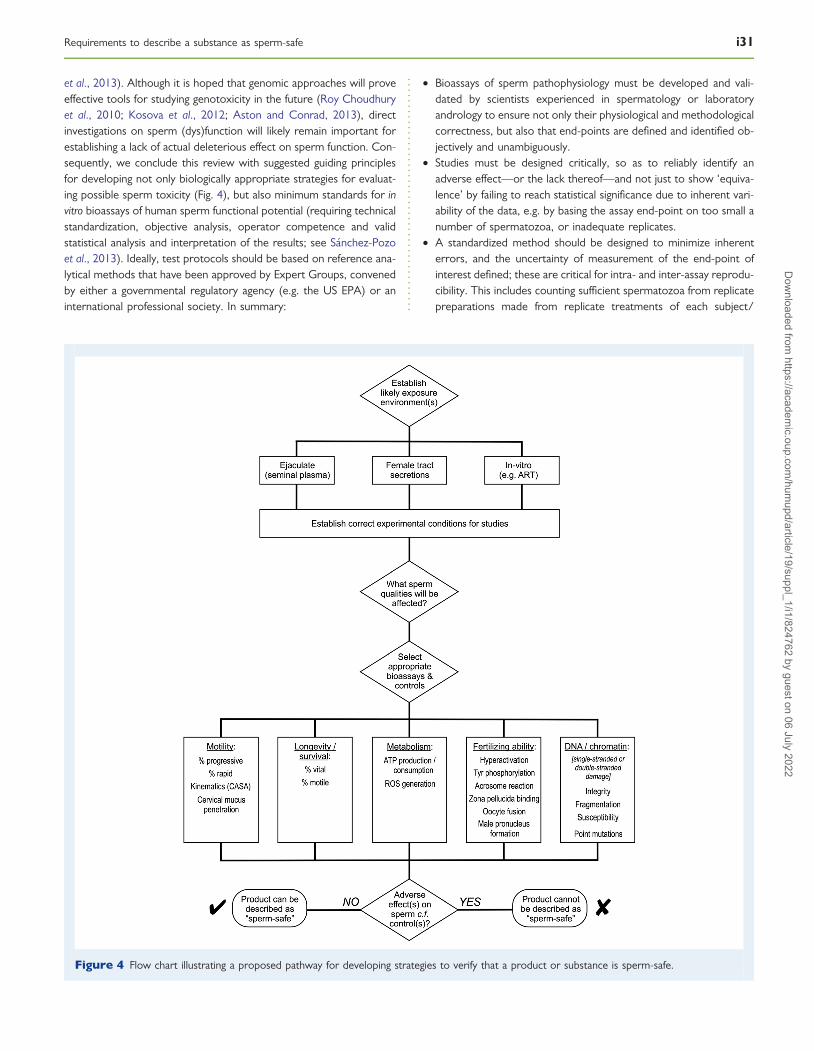
et al., 2013). Although it is hoped that genomic approaches will proveeffective tools for studying genotoxicity in the future (Roy Choudhuryet al., 2010; Kosova et al., 2012; Aston and Conrad, 2013), directinvestigations on sperm (dys)function will likely remain important forestablishing a lack of actual deleterious effect on sperm function. Con-sequently, we conclude this review with suggested guiding principlesfor developing not only biologically appropriate strategies for evaluat-ing possible sperm toxicity (Fig. 4), but also minimum standards for invitro bioassays of human sperm functional potential (requiring technicalstandardization, objective analysis, operator competence and validstatistical analysis and interpretation of the results; see Sanchez-Pozoet al., 2013). Ideally, test protocols should be based on reference ana-lytical methods that have been approved by Expert Groups, convenedby either a governmental regulatory agency (e.g. the US EPA) or aninternational professional society. In summary:
† Bioassays of sperm pathophysiology must be developed and vali-dated by scientists experienced in spermatology or laboratoryandrology to ensure not only their physiological and methodologicalcorrectness, but also that end-points are defined and identified ob-jectively and unambiguously.
† Studies must be designed critically, so as to reliably identify anadverse effect—or the lack thereof—and not just to show ‘equiva-lence’ by failing to reach statistical significance due to inherent vari-ability of the data, e.g. by basing the assay end-point on too small anumber of spermatozoa, or inadequate replicates.
† A standardized method should be designed to minimize inherenterrors, and the uncertainty of measurement of the end-point ofinterest defined; these are critical for intra- and inter-assay reprodu-cibility. This includes counting sufficient spermatozoa from replicatepreparations made from replicate treatments of each subject/
Figure 4 Flow chart illustrating a proposed pathway for developing strategies to verify that a product or substance is sperm-safe.
Requirements to describe a substance as sperm-safe i31D
ownloaded from
https://academic.oup.com
/humupd/article/19/suppl_1/i1/824762 by guest on 06 July 2022
concentration/dosage/time point permutation in order to minim-ize sampling and counting errors. Wherever practically possible,all assessments must be made ‘blind’, i.e. with the observerunaware of the provenance of each slide being read, to removethe risk of observer bias—both intentional and unintentional. Thisis especially important when relying on visual, subjective appraisalof the end-point.
Appropriate degree and durationof exposure to test substanceIt is essential that the manner of exposure of the analyte spermatozoato the test substance is appropriate; it needs to reflect physiology, themanner in which spermatozoa might be exposed to the substance inthe real world—including dosage and duration of exposure—and theenvironment in which that exposure occurs. For example, the initialevaluation of a potential vaginal lubricant product must expose sperm-atozoa still in seminal plasma to the actual product at a concentrationsimilar to which they might experience—or a high as they might pos-sibly be exposed—within the vagina during intercourse in vivo, e.g.10% (v/v) for perhaps 30 min (e.g. Agarwal et al., 2008a, b, c).Testing in a sperm motility assay could well be the first step, but effect-ive penetration from semen into cervical mucus post-exposure is alogical second stage, followed by testing for possible effects onsperm function ideally using spermatozoa prepared from semenpost-exposure.
However, when screening chemical compounds in isolation, it willbe difficult, if not impossible, to consider all possible synergistic andadditive effects that might arise in future mixtures containing the sub-stance. Therefore, a final product must be tested directly for spermo-toxic effects, rather than relying on previous absences of effect in testson its constituent materials.
Controls, test subjects/sperm donorsand replicatesEspecially when working with a biological material such as spermato-zoa as the analyte, it is essential that both positive and negative con-trols are included in all tests to control for sperm quality and toidentify non-specific effects. Sperm functional competence variesmarkedly between men, ranging from ‘super stud’ sperm donors tomen who are functionally sterile, and consequently epidemiologicalstudies on male fertility must pay close attention to avoiding subjectrecruitment bias (Cohn et al., 2002; Eustache et al., 2004; Sanchez-Pozo et al., 2013). Similarly, a laboratory performing in vitro tests ofsperm function must also avoid inappropriate bias in the provenanceof the sperm samples from which the analyte spermatozoa arebeing derived, e.g. a population of selected research donors whomight be drawn from a population of ‘retired’ therapeutic spermdonors). Compared with such men’s spermatozoa, those from menmore representative of the general population might show a greatersensitivity to a toxicant, and spermatozoa from subfertile men couldbe even more sensitive—and perhaps therefore more appropriateas test subjects. Because variable duration of exposure to seminalplasma influences both the functional properties and DNA vulnerabil-ity of spermatozoa, and because routines obviously vary between dif-ferent laboratories, it is likely that the unpredictable extent andvariability of such exposure in diagnostic laboratories could preclude
the use of diagnostic specimens in controlled scientific studies and vali-dations in many laboratories.
Therefore, rather than simply specifying a minimum number of sub-jects, it is essential that a spectrum of donors (e.g. selected researchdonors and general volunteers) as well as patients (male partners ofcouples attending an infertility clinic) be included to ensure broad-based investigation of a potential toxicant against a typical populationthat might be exposed to it. Combined with the possible magnitude ofpossible biological effect(s), this complicates establishing simplegeneric rules in advance, although a reasonable test population tosupport a claim that a substance or product does not show spermo-toxicity might be 20 men, at least half of whom are in the ‘at-risk’ in-fertility clinic or at least in the ‘trying-to-conceive’ category. For aproduct to which spermatozoa will specifically be exposed duringthe normal course of its use (e.g. a vaginal lubricant), then thisnumber should be considered a definite minimum.
When setting scientific limits between a ‘toxic effect’ and ‘no toxiceffect’, a general risk assessment of the nature of the actual effect mustbe included. For instance, a situation where a transgenerational effectmight be induced is far more serious than if no individual would beharmed—such as if exposed spermatozoa were simply unable to fer-tilize. The first situation would necessitate extensive assessments inorder to decrease the influence of random variation, while in thelatter case, cost–benefit and risk analysis would weigh the risk forsperm dysfunction against the possible benefits of the exposure.
Operator training and competenceBecause sperm-based bioassays typically rely on visual appraisal for theassessment of results, it is essential that—just like reliable diagnosticsemen analyses—they are only performed by properly trained, experi-enced laboratory andrology staff (MacLeod and Gold, 1951; Eliasson,1977; Mortimer, 1994; Bjorndahl et al., 2010; Barratt et al., 2011;Sanchez-Pozo et al., 2013). In order to achieve the necessary levelsof accuracy and precision, reflected in a suitably low uncertainty ofmeasurement (e.g. +10%; Bjorndahl et al., 2010), operator trainingmust be based on objectively defined assessment criteria and objectivedefinitions of competency. These must be followed by regular internalquality control exercises, and adequate performance in an externalquality assurance programme that incorporates an effective qualityimprovement capability (Bjorndahl et al., 2010; Sanchez-Pozo et al.,2013).
Statistical analysis and results reportingGiven the wide range of possible effects on sperm function, as well aslikely inter-individual variability in their magnitude and analyticaluncertainty of measurement, careful power calculations will berequired to establish appropriate subject and replicate numbers on astudy-by-study basis. Rigorous statistical analysis of assay data mustuse methods that will not obscure differences, e.g. not trying toconceal or reduce variability by expressing results as mean+ SEM(always use mean+ SD for suitable data), proper use of parametricand non-parametric testing, use of paired tests when valid, and avoid-ing the use of simplistic correlations when making comparisons (seeChapter 10 of Bjorndahl et al., 2010). Results must be interpreted ap-propriately: testing must show clear a lack of adverse effect (orperhaps a beneficial effect) for a substance or product to be
i32 Mortimer et al.
Dow
nloaded from https://academ
ic.oup.com/hum
upd/article/19/suppl_1/i1/824762 by guest on 06 July 2022
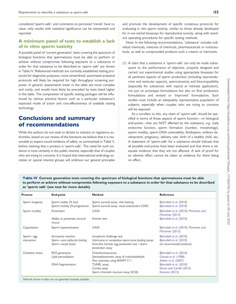
considered ‘sperm-safe’; and comments on perceived ‘trends’ have novalue, only results with statistical significance can be interpreted andreported.
A minimum panel of tests to establish a lackof in vitro sperm toxicityA possible panel of ‘current generation’ tests covering the spectrum ofbiological functions that spermatozoa must be able to perform orachieve without compromise following exposure to a substance inorder for that substance to be described as ‘sperm safe’ are shownin Table IV. Referenced methods are currently established testing pro-tocols for diagnostic purposes; more streamlined, automated analyticalprotocols will likely be required for high throughput screening pur-poses. In general, assessments lower in the table are more complexand costly, and would most likely be preceded by tests listed higherin the table. The composition of specific testing packages will be influ-enced by various practical factors such as a particular substance’sexpected mode of action and cost-effectiveness of available testingtechnology.
Conclusions and summaryof recommendationsWhile the authors do not seek to dictate to industry or regulatory au-thorities, based on our review of the literature we believe that it is rea-sonable to expect sound evidence of safety, as summarized in Table V,before claiming that a product is ‘sperm-safe’. The need for such evi-dence is most certainly in the public interest, especially that of coupleswho are trying to conceive. It is hoped that international andrology so-cieties or special interest groups will embrace our general principles
and promote the development of specific consensus protocols forevaluating in vitro sperm toxicity, similar to those already developedfor in vivo animal bioassays for reproductive toxicity, along with stand-ard operating procedures for specific testing methods.
Note: In the following recommendations, ‘substance’ includes indi-vidual chemicals, mixtures of chemicals, pharmaceuticals or nutraceu-ticals, as well as compounded products such a creams or lubricants.
(1) A claim that a substance is ‘sperm-safe’ can only be made subse-quent to the performance of objective, properly designed andcarried out experimental studies using appropriate bioassays forall pertinent aspects of sperm production (including neuroendo-crine and testicular aspects), spermotoxicity and biocompatibility(especially for substances with topical or intimate application),not just on prototype formulations but also on final productionformulations and revised or ‘improved’ formulations. Suchstudies must include an adequately representative population ofsubjects, especially when couples who are trying to conceivewill be exposed.
As a corollary to this, any claim of ‘sperm-safe’ should be spe-cified in terms of those aspects of sperm function—or biologicalend-points—that are NOT affected by the substance, e.g. maleendocrine function, sperm formation (number, morphology),sperm motility, sperm DNA vulnerability, fertilization, embryo de-velopment, pregnancy, delivery rate, birth of a healthy child, etc.A statement of ‘sperm-safe’ for a substance should indicate thatall possible end-points have been evaluated and that there is ad-equate evidence that they are not affected. A lack of proof foran adverse effect cannot be taken as evidence for there beingno effect.
.............................................................................................................................................................................................
Table IV Current generation tests covering the spectrum of biological functions that spermatozoa must be ableto perform or achieve without compromise following exposure to a substance in order for that substance to be describedas ‘sperm safe’ (see text for more details).
Process End-point Method Reference
Sperm longevity Sperm vitality (% live) Sperm survival assay: vital staining Bjorndahl et al. (2010)Sperm motility (% progressive) Sperm survival assay: visual assessment CASA Bjorndahl et al. (2010)
Sperm motility Kinematics CASA Bjorndahl et al. (2010); Mortimer andMortimer (2013)
Ability to penetrate cervicalmucus
Kremer test Bjorndahl et al. (2010)
Capacitation Sperm hyperactivation CASA Bjorndahl et al. (2010); Mortimer andMortimer (2013)
Sperm–egginteraction
Acrosome reaction Ionophore challenge test Bjorndahl et al. (2010)Sperm–zona pellucida binding Hemi-zona or competitive sperm-zona binding assays Bjorndahl et al. (2010)Sperm–oocyte fusion Zona-free hamster egg penetration test / sperm
penetration assay(no recommended protocol)
Oxidative stress ROS generation Chemiluminescence Bjorndahl et al. (2010)Lipid peroxidation Spectrophotometric assay of malondialdehyde Gomez et al. (1998)
Flow cytometry using BODIPY C11 Aitken et al. (2007)DNA fragmentation TUNEL assay Bjorndahl et al. (2010)
Comet assay Simon and Carrell (2013)Sperm chromatin structure assay (SCSA) Evenson (2013)
Methods shown in italics are not generally/routinely available.
Requirements to describe a substance as sperm-safe i33D
ownloaded from
https://academic.oup.com
/humupd/article/19/suppl_1/i1/824762 by guest on 06 July 2022
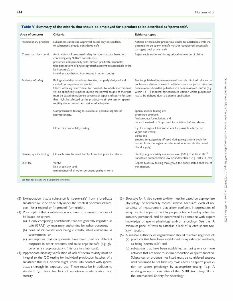
(2) Extrapolation that a substance is ‘sperm-safe’ from a predicatesubstance must be done only under the strictest of circumstances,even for a revised or ‘improved’ formulation.
(3) Presumption that a substance is not toxic to spermatozoa cannotbe based on either:(a) it only comprising constituents that are generally regarded as
safe (GRAS) by regulatory authorities for other purposes;(b) none of its constituents being currently listed elsewhere as
spermotoxic; or(c) assumptions that components have been used for different
purposes in other products and must ergo be safe (e.g. gly-cerol as a cryoprotectant c.f. its use in a lubricant).
(4) Appropriate bioassay verification of lack of sperm toxicity must beintegral to the QC testing for individual production batches of asubstance that will, or even might, come into contact with sperm-atozoa through its expected use. These must be in addition tostandard QC tests for lack of endotoxin contamination andsterility.
(5) Bioassays for in vitro sperm toxicity must be based on appropriatephysiology, be technically robust, achieve adequate levels of un-certainty of measurement that allow confident interpretation ofassay results, be performed by properly trained and qualified la-boratory personnel, and be interpreted by someone with expertknowledge of sperm physiology and/or andrology. See the ‘Aminimum panel of tests to establish a lack of in vitro sperm tox-icity’, section.
(6) A suitable authority or organization* should maintain registries of:(a) products that have been established, using validated methods,
as being ‘sperm-safe’; and(b) substances that have been established as having one or more
activities that are toxic to sperm production or sperm function.Substances or products not listed must be considered suspectuntil confirmed to not have any toxic effects on sperm produc-tion or sperm physiology by appropriate testing. *e.g. Aworking group or committee of the ESHRE Andrology SIG orthe International Society for Andrology.
.............................................................................................................................................................................................
Table V Summary of the criteria that should be employed for a product to be described as ‘sperm-safe’.
Area of concern Criteria Evidence types
Precautionary principle Substances cannot be approved based only on similarityto substances already considered safe
Actions or molecular properties similar to substances with thepotential to be sperm unsafe must be considered potentiallydamaging until proven safe
Claims must be sound Avoid claims of presumed safety for spermatozoa based on:containing only ‘GRAS’ constituents;presumed comparability with ‘similar’ predicate products;false perceptions of physiology (such as might be acceptable in thelay literature); orinvalid extrapolations from testing in other species
Reject such ‘evidence’ during critical evaluation of claims
Evidence of safety Biological validity based on objective, properly designed andcarried out experimental studies.Claims of being ‘sperm-safe’ for products to which spermatozoawill be specifically exposed during the normal course of their usemust be based on evidence covering all aspects of sperm functionthat might be affected by the product—a simple test on spermmotility alone cannot be considered adequate
Studies published in peer-reviewed journals. Limited reliance onconference abstracts: even if published—not subject to rigorouspeer-review. Should be published in a peer-reviewed journal (e.g.within 12–18 months) for continued citation unless publicationhas to be delayed due to a patent application
Comprehensive testing to exclude all possible aspects ofspermotoxicity.
Sperm-specific testing on:prototype products;final product formulation; andon each revised or ‘improved’ formulation before release
Other biocompatibility testing E.g. for a vaginal lubricant, check for possible effects on:vagina and cervix;penis; andembryo teratogenicity (if used during pregnancy it could becarried from the vagina into the uterine lumen via the portalblood supply)
General quality testing On each manufactured batch of product prior to release Sterility, e.g. a sterility assurance level (SAL) of at least 1023.Endotoxin contamination low or undetectable, e.g. ,0.5 EU/ml
Shelf life Verify:lack of toxicity; andmaintenance of all other pertinent quality criteria.
Repeat bioassay testing throughout the entire stated shelf life ofthe product.
See text for details and background evidence.
i34 Mortimer et al.
Dow
nloaded from https://academ
ic.oup.com/hum
upd/article/19/suppl_1/i1/824762 by guest on 06 July 2022
(7) The limited data available on human subjects, and in certaininstances, inconsistent data across studies, highlight the need forfurther epidemiological and reproductive toxicological researchon the different classes of EDCs which will contribute to ouroverall knowledge on ‘sperm-safe’ compounds.
(8) The relationship between EDC exposure and male reproductiveand developmental health includes the changes in exposurelevels among populations over time. The effects of complex envir-onmental EDC mixtures on spermatogenesis, as well as the trans-generational effects on the offspring of exposed males, includingtheir sperm function, should receive more attention.
(9) Innovations including improved biomarkers of exposure and moresophisticated statistical methods that deal with multiple exposuressimultaneously should be developed and implemented.
AcknowledgementsThe authors would like to express their gratitude to the reviewers ofthe original manuscript for their perceptive, supportive and construct-ive criticisms, and for their suggestions which contributed significantlyto the quality of the final article.
Authors’ rolesAll authors contributed substantively to the scope, writing and editingof the manuscript; D.M. acted as the coordinating author, otherco-authors are listed alphabetically.
FundingNo research funding was received by the authors in regard to thismanuscript. Publication of this Open Access Supplement was sup-ported financially by INGfertility, who had no input into the writingof the manuscript.
Conflict of interestNone of the authors have received any funding or other payments orgifts that have had any influence on the content of this manuscript.D.M. is employed full-time by Oozoa Biomedical which receives rev-enues from a wide range of commercial and clinical clients that variesover time.
ReferencesAgarwal A, Deepinder F, Cocuzza M, Short RA, Evenson DP. Effect of vaginal
lubricants on sperm motility and chromatin integrity: a prospective comparativestudy. Fertil Steril 2008a;89:375–379.
Agarwal A, Deepinder F, Sharma RK, Ranga G, Li J. Effect of cell phone usage onsemen analysis in men attending infertility clinic: an observational study. FertilSteril 2008b;89:124–128.
Agarwal A, Makker K, Sharma R. Clinical relevance of oxidative stress in male factorinfertility: an update. Am J Reprod Immunol 2008c;59:2–11.
Ahuja K. Carbohydrate determinants involved in mammalian fertilization. Am J Anat1985;174:207–223.
Aitken RJ, Clarkson JS. Cellular basis of defective sperm function and its associationwith the genesis of reactive oxygen species by human spermatozoa. J Reprod Fertil1987;81:459–469.
Aitken RJ, De Iuliis GN. On the possible origins of DNA damage in humanspermatozoa. Mol Hum Reprod 2010;16:3–13.
Aitken RJ, McLaughlin EA. Molecular mechanisms of sperm capacitation:progesterone-induced secondary calcium oscillations reflect the attainment of acapacitated state. Soc Reprod Fertil Suppl 2007;63:273–293.
Aitken RJ, Gordon E, Harkiss D, Twigg JP, Milne P, Jennings Z, Irvine DS. Relativeimpact of oxidative stress on the functional competence and genomic integrityof human spermatozoa. Biol Reprod 1998;59:1037–1046.
Aitken RJ, Koopman P, Lewis SEM. Seeds of concern. Nature 2004;432:48–52.Aitken RJ, Wingate JK, De Iuliis GN, McLaughlin EA. Analysis of lipid peroxidation in
human spermatozoa using BODIPY C11. Mol Hum Reprod 2007;13:203–211.Aitken RJ, Baker MA, De Iuliis GN, Nixon B. New insights into sperm physiology and
pathology. Handbook Exp Pharmacol 2010;198:99–115.Ajduk A, Malagocki A, Maleszewski M. Cytoplasmic maturation of mammalian
oocytes: development of a mechanism responsible for sperm-induced Ca2+
oscillations. Reprod Biol 2008;8:3–22.Akre O, Cnattingius S, Bergstrom R, Kvist U, Trichopoulos D, Ekbom A. Human
fertility does not decline: evidence from Sweden. Fertil Steril 1999;71:1066–1069.Alexander BH, Checkoway H, van Netten C, Muller CH, Ewers TG, Kaufman JD,
Mueller BA, Vaughan TL, Faustman EM. Semen quality of men employed at alead smelter. Occup Environ Med 1996;53:411–416.
Alvarez JG, Storey BT. Spontaneous lipid peroxidation in rabbit epididymalspermatozoa. Biol Reprod 1982;27:1102–1108.
Alvarez JG, Storey BT. Differential incorporation of fatty acids into and peroxidativeloss of fatty acids from phospholipids of human spermatozoa. Mol Reprod Dev1995;42:334–346.
Amann RP, Howards SS. Daily spermatozoal production and epididymalspermatozoal reserves of the human male. J Urol 1980;124:211–215.
Amory JK, Bremner WJ. The use of testosterone as a male contraceptive. BaillieresClin Endocrinol Metab 1998;12:471–484.
Amory JK, Page ST, Bremner WJ. Drug insight: recent advances in male hormonalcontraception. Nat Clin Pract Endocrinol Metab 2006;2:32–41.
Amory JK, Wang C, Swerdloff RS, Anawalt BD, Matsumoto AM, Bremner WJ,Walker SE, Haberer LJ, Clark RV. The effect of 5a-reductase inhibition withdutasteride and finasteride on semen parameters and serum hormones inhealthy men. J Clin Endocrinol Metab 2007;92:1659–1665.
Anderson L, Lewis SE, McClure N. The effects of coital lubricants on sperm motilityin vitro. Hum Reprod 1998;13:3351–3356.
Andersson MA, Mikkola R, Kroppenstedt RM, Rainey FA, Peltola J, Helin J,Sivonen K, Salkinoja-Salonen MS. The mitochondrial toxin produced byStreptomyces griseus strains isolated from an indoor environment is valinomycin.Appl Environ Microbiol 1998;64:4767–4773.
Anton E, Krawetz SA. Spermatozoa as biomarkers for the assessment of humanmale infertility and genotoxicity. Syst Biol Reprod Med 2012;58:41–50.
Anway MD, Cupp AS, Uzumcu M, Skinner MK. Epigenetic transgenerational actionsof endocrine disruptors and male fertility. Science 2005;308:1466–1469.
Anway MD, Memon MA, Uzumcu M, Skinner MK. Transgenerational effect of theendocrine disruptor vinclozolin on male spermatogenesis. J Androl 2006;27:868–879.
Appell RA, Evans PR. The effect of temperature on sperm motility and viability. FertilSteril 1977;28:1329–1332.
ASRM and SART. Revised guidelines for human embryology and andrologylaboratories. Fertil Steril 2008;90:S45–S59.
Aston KI, Conrad DF. A review of genome-wide approaches to study the geneticbasis for spermatogenic defects. In: Carrell DT, Aston KI (eds)Spermatogenesis: Methods and Protocols. Meth Mol Biol 2013;927:397–410.
ATDSR. Toxicological Profile for Di-(2-ethylhexyl) phthalate (DEHP). Atlanta:Department of Health and Human Services, Public Health Service, Agency forToxic Substances and Disease Registry, 2002.
Attaman JA, Toth TL, Furtado J, Campos H, Hauser R, Chavarro JE. Dietary fat andsemen quality among men attending a fertility clinic. Hum Reprod 2012;27:1466–1474.
Auger J. Assessing human sperm morphology: top models, underdogs or biometrics?Asian J Androl 2010;12:36–46.
Auger J, Eustache F, Ducot B, Blandin T, Daudin M, Diaz I, El Matribi S, Gony B,Keskes L, Kolbezen M et al. Intra- and inter-individual variability in humansperm concentration, motility and vitality assessment during a workshopinvolving ten laboratories. Hum Reprod 2000;15:2360–2368.
Requirements to describe a substance as sperm-safe i35D
ownloaded from
https://academic.oup.com
/humupd/article/19/suppl_1/i1/824762 by guest on 06 July 2022
Austin CR. The capacitation of the mammalian sperm. Nature 1952;170:326.Avendano C, Mata A, Sanchez Sarmiento CA, Doncel GF. Use of laptop computers
connected to internet through Wi-Fi decreases human sperm motility andincreases sperm DNA fragmentation. Fertil Steril 2012;97:39–45.
Bailey JL. Factors regulating sperm capacitation. Syst Biol Reprod Med 2010;56:334–348.
Balabanic D, Rupnik M, Klemencic AK. Negative impact of endocrine-disruptingcompounds on human reproductive health. Reprod Fertil Dev 2011;23:403 – 416.
Barratt CL, McLeod ID, Dunphy BC, Cooke ID. Prognostic value of two putativesperm function tests: hypo-osmotic swelling and bovine sperm mucuspenetration test (Penetrak). Hum Reprod 1992;7:1240–1244.
Barratt CL, Aitken RJ, Bjorndahl L, Carrell DT, de Boer P, Kvist U, Lewis SE,Perreault SD, Perry MJ, Ramos L et al. Sperm DNA: organization, protectionand vulnerability: from basic science to clinical applications—a position report.Hum Reprod 2010;25:824–838.
Barratt CLR, Bjorndahl L, Menkveld R, Mortimer D. The ESHRE Special InterestGroup for Andrology Basic Semen Analysis Course: a continued focus onaccuracy, quality, efficiency and clinical relevance. Hum Reprod 2011;26:3207–3212.
Barroso G, Valdespin C, Vega E, Kershenovich R, Avila R, Avendano C, Oehninger S.Developmental sperm contributions: fertilization and beyond. Fertil Steril 2009;92:835–848.
Bars R, Fegert I, Gross M, Lewis D, Weltje L, Weyers A, Wheeler JR,Galay-Burgos M. Risk assessment of endocrine active chemicals: identifyingchemicals of regulatory concern. Regul Toxicol Pharmacol 2012;64:143–154.
Baska KM, Manandhar G, Feng D, Agca Y, Tengowski MW, Sutovsky M, Yi YJ,Sutovsky P. Mechanism of extracellular ubiquitination in the mammalianepididymis. J Cell Physiol 2008;215:684–696.
Bedford JM. The status and the state of the human epididymis. Hum Reprod 1994;9:2187–2199.
Bedford JM. Enigmas of mammalian gamete form and function. Biol Rev 2004;79:429–460.
Begay O, Jean-Pierre N, Abraham CJ, Chudolij A, Seidor S, Rodriguez A, Ford BE,Henderson M, Katz D, Zydowsky T et al. Identification of personal lubricants thatcan cause rectal epithelial cell damage and enhance HIV Type 1 replication in vitro.AIDS Res Hum Retroviruses 2011;27:1019–1024.
Beischlag TV, Luis MJ, Hollingshead BD, Perdew GH. The aryl hydrocarbon receptorcomplex and the control of gene expression. Crit Rev Euraryot Gene Expr 2008;18:207–250.
Benoff S, Cooper GW, Hurley I, Mandel FS, Rosenfeld DL, Scholl GM, Gilbert BR,Hershlag A. The effect of calcium ion channel blockers on sperm fertilizationpotential. Fertil Steril 1994;62:606–617.
Benoff S, Jacob A, Hurley IR. Male infertility and environmental exposure to lead andcadmium. Hum Reprod Update 2000;6:107–121.
Berkovitz A, Eltes F, Ellenbogen A, Peer S, Feldberg D, Bartoov B. Does thepresence of nuclear vacuoles in human sperm selected for ICSI affectpregnancy outcome? Hum Reprod 2006;21:1787–1790.
Berndtson EB. Comparative reliability and sensitivity of different methods forassessing treatment effects on sperm production. Anim Reprod Sci 2008;105:5–22.
BfR (Bundesinstitut fur Risikobewertung). Final joint DE-UK position. Regulatorydefinition of an endocrine disrupter in relation to potential threat to humanhealth. 2011. www.bfr.bund.de (14 February 2013, date last accessed).
Bjorndahl L, Kvist U. Importance of zinc for human sperm head–-tail connection.Acta Physiol Scand 1982;116:51–55.
Bjorndahl L, Kvist U. Loss of an intrinsic capacity for human sperm chromatindecondensation. Acta Physiol Scand 1985;124:189–194.
Bjorndahl L, Kvist U. Human sperm chromatin stabilization: a proposed modelincluding zinc bridges. Mol Hum Reprod 2010;16:23–29.
Bjorndahl L, Barratt CL, Fraser LR, Kvist U, Mortimer D. ESHRE basic semen analysiscourses 1995–1999: immediate beneficial effects of standardized training. HumReprod 2002;17:1299–1305.
Bjorndahl L, Soderlund I, Kvist U. Evaluation of the one-step eosin–nigrosin stainingtechnique for human sperm vitality assessment. Hum Reprod 2003;18:813–816.
Bjorndahl L, Mortimer D, Barratt CLR, Castilla JA, Menkveld R, Kvist U, Alvarez JG,Haugen TB. A Practical Guide to Basic Laboratory Andrology. Cambridge, UK:Cambridge University Press, 2010.
Boitrelle F, Ferfouri F, Petit JM, Segretain D, Tourain C, Bergere M, Bailly M,Vialard F, Albert M, Selva J. Large human sperm vacuoles observed in motilespermatozoa under high magnification: nuclear thumbprints linked to failure ofchromatin condensation. Hum Reprod 2011;26:1650–1658.
Bonde JP. Male reproductive organs are at risk from environmental hazards. Asian JAndrol 2010;12:152–156.
Bornman MS, de Jager C, Vorster H, Burger AEC, Apps PJA. p-Nonylphenol in foodpackaging material: an absorption study.: Congress on Current Aspects ofReproductive Biology. 10th Biennial Meeting of SASRSS, August 1997, pp. 22–25.
Bornman MS, Barnhoorn IEJ, de Jager C, Veeramachaneni DNR. Testicularmicrolithiasis and neoplastic lesions in wild eland (Tragelaphus oryx): possibleeffects of exposure to environmental pollutants? Environ Res 2010;110:327–333.
Boscolo P, Sacchattoni-Longrocino G, Ranelletti FO, Gioia A, Carmignani M. Effectsof long term cadmium exposure on the testes of rabbits: ultrastructural study.Toxicol Lett 1985;24:145–149.
Botella-Llusia J. Measurement of linear progression of the human spermatozoon asan index of male fertility. Int J Fertil 1956;1:113–130.
Boyers SP, Corrales MD, Huszar G, DeCherney AH. The effects of Lubrin on spermmotility in vitro. Fertil Steril 1987;47:882–884.
Bracken MB, Eskanazi B, Sachse K, McSharry J-E, Hellenbrand K, Leo-Summers L.Association of cocaine use with sperm concentration, motility morphology.Fertil Steril 1990;53:315–322.
Branigan EF, Estes MA, Muller CH. Advanced semen analysis: a simple screening testto predict intrauterine insemination success. Fertil Steril 1999;71:547–551.
Braude P, Bolton V, Moore S. Human gene expression first occurs between the four-and eight-cell stages of preimplantation development. Nature 1988;332:459–461.
Braw-Tal R. Endocrine disruptors and timing of human exposure. Pediatr EndocrinolRev 2010;8:41–46.
Brazil C, Swan SH, Drobnis EZ, Liu F, Wang C, Redmon JB, Overstreet JW; Studyfor Future Families Research Group. Standardized methods for semen evaluationin a multicenter research study. J Androl 2004a;25:635–644.
Brazil C, Swan SH, Tollner CR, Treece C, Drobnis EZ, Wang C, Redmon JB,Overstreet JW; Study for Future Families Research Group. Quality control oflaboratory methods for semen evaluation in a multicenter research study.J Androl 2004b;25:645–656.
Brenker C, Goodwin N, Weyand I, Kashikar ND, Naruse M, Krahling M, Muller A,Kaupp UB, Strunker T. The CatSper channel: a polymodal chemosensor in humansperm. EMBO J 2012;31:1654–1665.
Brindley GS. Deep scrotal temperature and the effect on it of clothing, airtemperature, activity, posture and paraplegia. Br J Urol 1982;54:49–55.
Buck GM, Lynch CD, Stanford JB, Sweeney AM, Schieve LA, Rockett JC, Selevan SG,Schrader SM. Prospective pregnancy study designs for assessing reproductive anddevelopmental toxicants. Environ Health Perspect 2004;112:79–86.
Buck Louis GM, Gray LE Jr, Marcus M, Ojeda SR, Pescovitz OH, Witchel SF,Sippell W, Abbottn DH, Soto A, Tyl RW et al. Environmental factors andpuberty timing: expert panel research needs. Pediatrics 2008;112:192–207.
Buffone MG, Ijiri TW, Cao W, Merdiushev T, Aghajanian HK, Gerton GL. Heads ortails? Structural events and molecular mechanisms that promote mammaliansperm acrosomal exocytosis and motility. Mol Reprod Dev 2012;79:4–18.
Bujan L, Daudin M, Charlet JP, Thonneau P, Mieusset R. Increase in scrotaltemperature in car drivers. Hum Reprod 2000;15:1355–1357.
Burkman LJ, Coddington CC, Franken DR, Krugen TF, Rosenwaks Z, Hodgen GD.The hemizona assay (HZA): development of a diagnostic test for binding ofhuman spermatozoa to the human zona pellucida to predict fertilizationpotential. Fertil Steril 1988;49:688–693.
Calogero A, Polosa R, Perdichizzi A, Guarino F, La Vignera S, Scarfia A,Fratantonio E, Condorelli R, Bonanno O, Barone N et al. Cigarette smokeextract immobilizes human spermatozoa and induces sperm apoptosis. ReprodBiomed Online 2009;19:564–571.
Carlsen E, Giwercman A, Keiding N, Skakkebaek NE. Evidence for decreasing qualityof semen during past 50 years. BMJ 1992;305:609–613.
Carrell DT. Contributions of spermatozoa to embryogenesis: assays to evaluatetheir genetic and epigenetic fitness. Reprod Biomed Online 2008;16:474–484.
Carrell DT, Hammoud SS. The human sperm epigenome and its potential role inembryonic development. Mol Hum Reprod 2010;16:37–47.
Carrell DT, Liu L. Altered protamine 2 expression is uncommon in donors of knownfertility, but common among men with poor fertilizing capacity, and may reflectother abnormalities of spermiogenesis. J Androl 2001;22:604–610.
i36 Mortimer et al.
Dow
nloaded from https://academ
ic.oup.com/hum
upd/article/19/suppl_1/i1/824762 by guest on 06 July 2022
Carrell DT, Emery BR, Hammoud S. Altered protamine expression and diminishedspermatogenesis: what is the link? Hum Reprod Update 2007;13:313–327.
Casswall TH, Bjorndahl L, Kvist U. Cadmium interacts with the zinc-dependentstability of the human sperm chromatin. J Trace Elem Electrolytes Health Dis1987;1:85–87.
Castle L. Chemical migration into food: an overview. In: Castle L (ed). ChemicalMigration and Food Contact Materials. Cambridge, UK: CRC Press/WoodeheadPublishing Ltd, 2007, 1–13.
Cavallini G, Ferraretti AP, Gianaroli L, Biagiotti G, Vitali G. Cinnoxicam andL-carnitine/acetyl-L-carnitine treatment for idiopathic and varicocele-associatedoligoasthenospermia. J Androl 2004;25:761–770.
Centola GM, Cartie RJ, Cox C. Differential responses of human sperm to varyingconcentrations of pentoxyfylline with demonstration of toxicity. J Androl 1995;16:136–142.
Ceretti E, Zani C, Zerbini I, Guzzella L, Scaglia M, Berna V, Donato F, Monarca S,Feretti D. Comparative assessment of genotoxicity of mineral water packed inpolyethylene terephthalate (PET) and glass bottles. Water Res 2010;44:1462–1470.
Chang MC. Fertilizing capacity of spermatozoa deposited into the fallopian tubes.Nature 1951;168:697–698.
Chang H, Suarez SS. Rethinking the relationship between hyperactivation andchemotaxis in mammalian sperm. Biol Reprod 2010;83:507–513.
Chang H, Suarez SS. Unexpected flagellar movement patterns and epithelial bindingbehavior of mouse sperm in the oviduct. Biol Reprod 2012;86:140, 1–8.
Chapin RE, Sloane RA, Haseman JK. The relationships among reproductiveendpoints in Swiss mice, using the reproductive assessment by continuousbreeding database. Fundam Appl Toxicol 1997;38:129–142.
Cherry N, Moore H, McNamee R, Pacey A, Burgess G, Clyma JA, Dippnall M,Baillie H, Povey A. Occupation and male infertility: glycol ethers and otherexposures. Occup Environ Med 2008;65:708–714.
Chi HJ, Kim JH, Ryu CS, Lee JY, Park JS, Chung DY, Choi SY, Kim MH, Chun EK,Roh SI. Protective effect of antioxidant supplementation in sperm-preparationmedium against oxidative stress in human spermatozoa. Hum Reprod 2008;23:1023–1028.
Christensen BC, Marsit CJ. Epigenomics in environmental health. Front Genet 2011;2:84.
Claassens OE, Wehr JB, Harrison KL. Optimizing sensitivity of the human spermmotility assay for embryo toxicity testing. Hum Reprod 2000;15:1586–1591.
Claudio L, Bearer CF, Wallinga D. Assessment of the U.S. Environmental ProtectionAgency methods for identification of hazards to developing organisms. Part I: Thereproduction and fertility testing guidelines. Am J Ind Med 1999;35:543–553.
Clermont Y. Spermatogenesis in man. A study of the spermatogonial population.Fertil Steril 1966;17:705–721.
Coccia ME, Becattini C, Criscuoli L, Fuzzi B, Scarselli G. A sperm survival test andin-vitro fertilization outcome in the presence of male factor infertility. HumReprod 1997;12:1969–1973.
Coetzee K, Kruge TF, Lombard CJ. Predictive value of normal sperm morphology: astructured literature review. Hum Reprod Update 1998;4:73–82.
Cohn BA, Overstreet JW, Fogel RJ, Brazil CK, Baird DD, Cirillo PM. Epidemiologicstudies of human semen quality: considerations for study design. Am J Epidemiol2002;155:664–671.
Colborn T, vom Saal FS, Soto AM. Developmental effects of endocrine-disruptingchemicals in wildlife and humans. Environ Health Perspect 1993;101:378–384.
Colborn T, Dumanoski D, Myers JP. Our Stolen Future: Are We Threatening OurFertility, Intelligence, and Survival?—A Scientific Detective Story. New York, USA:Plume/Penguin Books USA, 1997. http://www.ourstolenfuture.org/basics/chemlist.htm (14 February 2013, date last accessed).
Collodel G, Scapigliati G, Moretti E. Spermatozoa and chronic treatment withfinasteride: a TEM and FISH study. Arch Androl 2007;53:229–233.
Comhaire FH, Mahmoud A. The role of food supplements in the treatment of theinfertile man. Reprod Biomed Online 2003;7:385–391.
Cordelli E, Fresegna AM, D’Alessio A, Eleuteri P, Spano M, Pacchierotti F, Villani P.ReProComet: a new in vitro method to assess DNA damage in mammaliansperm. Toxicol Sci 2007;99:545–552.
Correa LM, Thomas A, Meyers SA. The macaque sperm actin cytoskeletonreorganizes in response to osmotic stress and contributes to morphologicaldefects and decreased motility. Biol Reprod 2007;77:942–953.
Costello S, Michelangeli F, Nash K, Lefievre L, Morris J, Machado-Oliveira G,Barratt C, Kirkman-Brown J, Publicover S. Ca2+-stores in sperm: their identitiesand functions. Reproduction 2009;138:425–437.
Coutts SM, Fulton N, Anderson RA. Environmental toxicant-induced germ cellapoptosis in the human fetal testis. Hum Reprod 2007;22:2912–2918.
Critser JK, Huse-Benda AR, Aaker DV, Arneson BW, Ball GD. Cryopreservation ofhuman spermatozoa. III. The effect of cryoprotectants on motility. Fertil Steril1988;50:314–320.
Cross NL. Effect of methyl-beta-cyclodextrin on the acrosomal responsiveness ofhuman sperm. Mol Reprod Dev 1999;53:92–98.
Cross NL, Overstreet JW. Glycoconjugates of the human sperm surface:distribution and alterations that accompany capacitation in vitro. Gamete Res1987;16:23–35.
Cross NL, Morales P, Overstreet JW, Hanson FW. Induction of acrosome reactionsby the human zona pellucida. Biol Reprod 1988;38:235–244.
Cummins JM, Pember SM, Jequier AM, Yovich JL, Hartmann PE. A test of the humansperm acrosome reaction following ionophore challenge. Relationship to fertilityand other seminal parameters. J Androl 1991;12:98–103.
Damstra T, Barlow S, Bergman A, Kavlock R, Van der Kraak G. Global Assessment ofthe State-of-the-Science of Endocrine Disruptors. WHO publication no. WHO/PCS/EDC/02.2. Geneva, Switzerland: World Health Organization, 2002.
Danish Environmental Protection Agency. Male Reproductive Health andEnvironmental Chemicals with Estrogenic Effects. Miljoproject 290. Copenhagen,Denmark: Ministry of Environment and Energy, Danish EnvironmentalProtection Agency, 1995.
Dart RC. (ed). Medical Toxicology, 3rd edn. Philadelphia, USA: Lippincott Williams &Wilkins, 2003.
David G, Bisson JP, Czyglik F, Jouannet P, Gernigon C. Anomalies morphologiquesdu spermatozoıde humain. 1) Propositions pour un systeme de classification.J Gyn Obstet Biol Reprod 1975;4(Suppl. 1):17–36.
Dean J. The enigma of sperm–egg recognition in mice. Soc Reprod Fertil Suppl 2007;63:359–365.
de Boer P, Ramos L, de Vries M, Gochhait S. Memoirs of an insult: sperm as apossible source of transgenerational epimutations and genetic instability. MolHum Reprod 2010;16:48–56.
De Iuliis DN, Newey RJ, King BV, Aitken RJ. Mobile phone radiation indices reactiveoxygen species production and DNA damage in human spermatozoa in vitro. PLoSOne 2009;4:e6446.
De Jager C, Bornman MS, van der Horst G I. The effect of p-nonylphenol, anenvironmental toxicant with oestrogenic properties, on fertility potentila inadult male rats. Andrologia 1999a;31:99–106.
De Jager C, Bornman MS, Oosthuizen JMC II. The effect of p-nonylphenol on thefertility potential of male rats after gestational, lactational and direct exposure.Andrologia 1999b;31:107–113.
De Jonge C. Biological basis for human capacitation. Hum Reprod Update 2005;11:204–215.
De Jonge CJ, Centola GM, Reed ML, Shabanowitz RB, Simon SD, Quinn P. Humansperm survival assay as a bioassay for the assisted reproductive technologieslaboratory. J Androl 2003;24:16–18.
de Kretser DM. Ultrastructural features of human spermiogenesis. Z ZellforschMikrosk Anat 1969;98:477–505.
de Lamirande E, Gagnon C. Human sperm hyperactivation in whole semen and itsassociation with low superoxide scavenging capacity in seminal plasma. Fertil Steril1993;59:1291–1295.
de Lamirande E, Jiang H, Zini A, Kodama H, Gagnon C. Reactive oxygen species andsperm physiology. Rev Reprod 1997;2:48–54.
Delbes G, Levacher C, Duquenne C, le Racine C, Pakarinen P, Habert R.Endogenous estrogens inhibit mouse fetal leydig cell development via estrogenreceptor alpha. Endocrinology 2005;146:2454–2461.
Delbes G, Hales BF, Robaire B. Toxicants and human sperm chromatin integrity. MolHum Reprod 2010;16:14–22.
Deng F, Tao FB, Li DY, Xu YY, Hao JH, Sun Y. Effects of growth environments andtwo environmental endocrine disruptors on children with idiopathic precociouspuberty. Eur J Endocrinol 2012;166:803–809.
Dhondt MMM, Adriaens E, Remon JP. The evaluation of the local tolerance ofvaginal formulations, with or without nonoxynol-9, using the slug mucosalirritation test. Sex Transm Dis 2004;31:229–235.
Requirements to describe a substance as sperm-safe i37D
ownloaded from
https://academic.oup.com
/humupd/article/19/suppl_1/i1/824762 by guest on 06 July 2022
Diamanti-Kandarakis E, Bourguignon JP, Giudice LC, Hauser R, Prins GS, Soto AM,Zoeller RT, Gore AC. Endocrine-disrupting chemicals: an Endocrine Societyscientific statement. Endocr Rev 2009;30:293–342.
Dohle GR. Male infertility in cancer patients: review of the literature. Int J Urol 2010;17:327–331.
Donnelly ET, McClure N, Lewis SE. Glutathione and hypotaurine in vitro: effects onhuman sperm motility, DNA integrity and production of reactive oxygen species.Mutagenesis 2000;15:61–68.
Donovan B. Barriers to conception and disease. Ann Acad Med Singapore 1995;24:608–614.
Drevet JR. The antioxidant glutathione peroxidase family and spermatozoa: acomplex story. Mol Cell Endocrinol 2006;250:70–79.
Drife JO. The effects of drugs on sperm. Drugs 1987;33:610–622.Drobnis EZ, Yudin AI, Cherr GN, Katz DF. Hamster sperm penetration of the zona
pellucida: kinematic analysis and mechanical implications. Dev Biol 1988;130:311–323.
Dun MD, Aitken RJ, Nixon B. The role of molecular chaperones in spermatogenesisand the post-testicular maturation of mammalian spermatozoa. Hum ReprodUpdate 2012;18:420–435.
Dunbar B, Patel M, Fahey J, Wira C. Endocrine control of mucosal immunity in thefemale reproductive tract: impact of environmental disruptors. Mol Cell Endocrinol2012;354:85–93.
ECETOC. Technical report 106: Guidance on identifying endocrine disrupting effects.Brussels: European Centre for Ecotoxicology and Toxicology of Chemicals, 2009.http://bit.ly/ecetoc-tr106 (14 February 2013, date last accessed).
Edwards RG, Bavister BD, Steptoe PC. Early stages of fertilization in vitro of humanoocytes matured in vitro. Nature 1969;221:632–635.
Edwards R, Steptoe P. A Matter of Life. London, UK: Hutchinson and Co., 1980.Edwards TM, Moore BC, Guillette LJ Jr. Reproductive dysgenesis in wildlife: a
comparative view. Int J Androl 2006;29:109–121.Eliasson R. Semen analysis and laboratory workup. In: Cockett ATK, Urry RL (eds).
Male Infertility. Workup, Treatment and Research. New York, USA: Grune &Stratton, 1977, 169–188.
Enciso M, Sarasa J, Agarwal A, Fernandez JL, Gosalvez J. A two-tailed Comet assayfor assessing DNA damage in spermatozoa. Reprod Biomed Online 2009;18:609–616.
ESHRE Andrology Special Interest Group. Consensus workshop on advanceddiagnostic andrology techniques. Hum Reprod 1996;11:1463–1479.
ESHRE Andrology Special Interest Group. Guidelines on the application of CASAtechnology in the analysis of spermatozoa. Hum Reprod 1998;13:142–145.
Eskenazi B, Wyrobek AJ, Fenster L, Katz DF, Sadler M, Lee J, Hudes M, Rempel DM.A study of the effect of perchloroethylene exposure on semen quality in drycleaning workers. Am J Ind Med 1991;20:575–591.
Esterhuizen AD, Franken DR, Becker PJ, Lourens JG, Muller II, van Rooyen LH.Defective sperm decondensation: a cause for fertilization failure. Andrologia2002;34:1–7.
European Science Foundation. Male Reproductive Health. Its impacts in relation togeneral wellbeing and low European fertility rates. Science Policy Briefing 40,2010. http://www.esf.org/publications/science-policy-briefings.html (14February 2013, date last accessed).
Eustache F, Auger J. Inter-individual variability in the morphological assessment ofhuman sperm: effect of the level of experience and the use of standardmethods. Hum Reprod 2003;18:1018–1022.
Eustache F, Auger J, Cabrol D, Jouannet P. Are volunteers delivering semen samplesin fertility studies a biased population? Hum Reprod 2004;19:2831–2837.
Evenson DP. Sperm chromatin strcuture assay (SCSAw). In Carrell DT and Aston KI(eds) Spermatogenesis: Methods and Protocols. Methods Mol Biol 2013;927:147–164.
Evenson DP, Wixon R. Data analysis of two in vivo fertility studies using SpermChromatin Structure Assay-derived DNA fragmentation index vs. pregnancyoutcome. Fertil Steril 2008;90:1229–1231.
Fariello RM, Pariz JR, Spaine DM, Gozzo FC, Pilau EJ, Fraietta R, Bertolla RP,Andreoni C, Cedenho AP. Effect of smoking on the functional aspects ofsperm and seminal plasma protein profiles in patients with varicocele. HumReprod 2012;27:3140–3149.
Feneux D, Serres C, Jouannet P. Sliding spermatozoa: a dyskinesia responsible forhuman infertility? Fertil Steril 1985;44:508–511.
Fernandez JL, Cajigal D, Gosalvez J. Simultaneous labeling of single- anddouble-strand DNA breaks by DNA breakage detection-FISH (DBD-FISH).Methods Mol Biol 2011;682:133–147.
Florman HM, Jungnickel MK, Sutton KA. Shedding light on sperm pHertility. Cell2010;140:310–312.
Forman R, Gilmour-White S, Forman N. Drug-Induced Infertility and SexualDysfunction. Cambridge, UK: Cambridge University Press, 1996.
Fraga CG, Motchnik PA, Shigenaga MK, Helbock HJ, Jacob RA, Ames BN. Ascorbicacid protects against endogenous oxidative DNA damage in human sperm. ProcNatl Acad Sci USA 1991;88:11003–11006.
Fraga CG, Motchnik PA, Wyrobek AJ, Rempel DM, Ames BN. Smoking and lowantioxidant levels increase oxidative damage to sperm DNA. Mutat Res 1996;351:199–203.
Franco JG Jr, Mauri AL, Petersen CG, Massaro FC, Silva LF, Felipe V, Cavagna M,Pontes A, Baruffi RL, Oliveira JB et al. Large nuclear vacuoles are indicative ofabnormal chromatin packaging in human spermatozoa. Int J Androl 2012;35:46–51.
Franken DR, Oehninger S, Burkman LJ, Coddington CC, Kruger TF, Rosenwaks Z,Acosta AA, Hodgen GD. The hemizona assays (HZA): a predictor of humansperm fertilizing potential in in vitro fertilization (IVF) treatment. J In Vitro FertilEmbryo Transf 1989;6:44–49.
Franken DR, Smith M, Menkveld R, Kruger TF, Sekadde-Kigondu C, Mbizvo M,Akande EO. The development of a continuous quality control programme forstrict sperm morphology among sub-Saharan African laboratories. Hum Reprod2000;15:667–671.
Fraser LR. Mouse sperm capacitation assessed by kinetics and morphology offertilization in vitro. J Reprod Fertil 1983;69:419–428.
Fraser LR. Sperm capacitation and the acrosome reaction. Hum Reprod 1998;13(Suppl. 1):9–19.
Frishman GN, Luciano AA, Maier DB. Evaluation of Astroglide, a new vaginallubricant: effects of length of exposure and concentration on sperm motility.Fertil Steril 1992;58:630–632.
Fronczak CM, Kim ED, Barqawi AB. The insults of recreational drug abuse on malefertility. J Androl 2012;33:515–528.
Fuentes A, Munoz A, Barnhart K, Arguello B, Dıaz M, Pommer R. Recent cigarettesmoking and assisted reproductive technologies outcome. Fertil Steril 2010;93:89–95.
Gadea J, Molla M, Selles E, Marco MA, Garcia-Vazquez FA, Gardon JC. Reducedglutathione content in human sperm is decreased after cryopreservation: effectof the addition of reduced glutathione to the freezing and thawing extenders.Cryobiology 2011;62:40–46.
Gadelha H, Gaffney EA, Smith DJ, Kirkman-Brown JC. Nonlinear instability inflagellar dynamics: a novel modulation mechanism in sperm migration? J R SocInterface 2010;7:1689–1697.
Gadella BM, Van Gestel RA. Bicarbonate and its role in mammalian sperm function.Anim Reprod Sci 2004;82–83:307–319.
Gadella BM, Tsai PS, Boerke A, Brewis IA. Sperm head membrane reorganisationduring capacitation. Int J Dev Biol 2008;52:473–480.
Gardner DK, Reed L, Linck D, Sheehan C, Lane M. Quality control in human in vitrofertilization. Semin Reprod Med 2005;23:319–324.
Gelbke HP, Kayser M, Poole A. OECD test strategies and methods for endocrinedisruptors. Toxicology 2004;205:17–25.
Gharagozloo P, Aitken RJ. The role of sperm oxidative stress in male infertilityand the significance of oral antioxidant therapy. Hum Reprod 2011;26:1628–1640.
Gilmore JA, McGann LE, Liu J, Gao DY, Peter AT, Kleinhans FW, Critser JK. Effect ofcryoprotectant solutes on water permeability of human spermatozoa. Biol Reprod1995;53:985–995.
Giwercman A. Estrogens and phytoestrogens in male infertility. Curr Opin Urol 2011;21:519–526.
Glina S, Neves PA, Saade R, Netto NR Jr, Soares JB, Galuppo AG.Finasteride-associated male infertility. Rev Hosp Clin Fac Med Sao Paulo 2004;59:203–205.
Goldenburg RL, White R. The effect of vaginal lubricants on sperm motility in vitro.Fertil Steril 1975;26:872–873.
Goldstein M, Eid JE. Elevation of intra-testicular temperature and scrotal skin surfacetemperature in men with varicocele. J Urol 1989;142:743–745.
i38 Mortimer et al.
Dow
nloaded from https://academ
ic.oup.com/hum
upd/article/19/suppl_1/i1/824762 by guest on 06 July 2022
Gomez E, Aitken J. Impact of in vitro fertilization culture media on peroxidativedamage to human spermatozoa. Fertil Steril 1996;65:880–882.
Gomez E, Irvine DS, Aitken RJ. Evaluation of a spectrophotometric assay for themeasurement of malondialdehyde and 4-hydroxyalkenals in humanspermatozoa: relationships with semen quality and sperm function. Int J Androl1998;21:81–94.
Gopalkrishnan K, Hinduja IN, Anand Kumar TC. Determining the osmolality ofseminal fluid aids in the rapid diagnosis of the fertilizing potential ofspermatozoa. Assist Reprod Genet 1989;6:119–121.
Gore AC. Endocrine-Disrupting Chemicals: From Basic Research to Clinical Practice.Totowa, NJ: Humana Press, 2007.
Gore AC, Walker DM, Zama AM, Armenti AE, Uzumcu M. Early life exposure toendocrine-disrupting chemicals causes lifelong molecular reprogramming of thehypothalamus and premature reproductive aging. Mol Endocrinol 2011;25:2157–2168.
Gosden RG. Oogenesis as a foundation for embryogenesis. Mol Cell Endocrinol 2002;186:149–153.
Gottardo F, Kliesch S. Ejakulatdiagnostik. Spermiogramm nach WHO-Kriterien2010. Der Urologe 2011;50:101–108.
Griveau JF, Lobel B, Laurent MC, Michardiere L, Le Lannou D. Interest ofpentoxifylline in ICSI with frozen-thawed testicular spermatozoa from patientsin non-obstructive azoospermia. Reprod Biomed Online 2006;12:14–18.
Gruber CJ, Gruber DM, Gruber IM, Wieser F, Huber JC. Anatomy of the estrogenresponse element. Trends Endocrinol Metab 2004;15:73–78.
Guerrero A, Carneiro J, Pimentel A, Wood CD, Corkidi G, Darszon A. Strategiesfor locating the female gamete: the importance of measuring sperm trajectoriesin three spatial dimensions. Mol Hum Reprod 2011;17:511–523.
Haimov-Kochman R, Ben-Chetrit E. The effect of colchicine treatment on spermproduction and function: a review. Hum Reprod 1998;13:360–362.
Hakimi H, Geary I, Pacey A, Eley A. Spermicidal activity of bacteriallipopolysaccharide is only partly due to lipid A. J Androl 2006;27:774–779.
Hamlin HJ, Guillette LJ. Birth defects in wildlife: the role of environmentalcontaminants as inducers of reproductive and developmental dysfunction. SysBiol Reprod Med 2010;56:113–121.
Hargreave TB (ed). Male Infertility, 2nd edn. Berlin, Germany: Springer-Verlag,1994a.
Hargreave TB. Varicocele. In Hargreave TB (ed). Male infertility, 2nd edn. Berlin,Germany: Springer-Verlag, 1994b, 249–267.
Harris RM, Waring RH. Diethylstilboestrol—a long-term legacy. Maturitas 2012;72:108–112.
Harvey PW, Everett DJ. Regulation of endocrine-disrupting chemicals: Criticaloverview and deficiencies in toxicology and risk assessment for human health.Best Pract Res Clin Endocrinol Metab 2006;20:145–165.
He Z, Kokkinaki M, Pant D, Gallicano GI, Dym M. Small RNA molecules in theregulation of spermatogenesis. Reproduction 2009;137:901–911.
Henkel RR. Leukocytes and oxidative stress: dilemma for sperm function and malefertility. Asian J Androl 2011;13:43–52.
Henkel RR, Schill WB. Sperm preparation for ART. Reprod Biol Endocrinol 2003;1:108.
Hermo L, Pelletier RM, Cyr DG, Smith CE. Surfing the wave, cycle, life history, andgenes/proteins expressed by testicular germ cells. Part 1: background tospermatogenesis, spermatogonia, and spermatocytes. Microsc Res Tech 2010a;73:241–278.
Hermo L, Pelletier RM, Cyr DG, Smith CE. Surfing the wave, cycle, life history, andgenes/proteins expressed by testicular germ cells. Part 2: changes in spermatidorganelles associated with development of spermatozoa. Microsc Res Tech2010b;73:279–319.
Hermo L, Pelletier RM, Cyr DG, Smith CE. Surfing the wave, cycle, life history, andgenes/proteins expressed by testicular germ cells. Part 3: developmental changesin spermatid flagellum and cytoplasmic droplet and interaction of sperm with thezona pellucida and egg plasma membrane. Microsc Res Tech 2010c;73:320–363.
Ho HC, Suarez SS. Characterization of the intracellular calcium store at the base ofthe sperm flagellum that regulates hyperactivated motility. Biol Reprod 2003;68:1590–1596.
Holland MK, Alvarez JG, Storey BT. Production of superoxide and activity ofsuperoxide dismutase in rabbit epididymal spermatozoa. Biol Reprod 1982;27:1109–1118.
Holt WV, Fazeli A. The oviduct as a complex mediator of mammalian spermfunction and selection. Mol Reprod Dev 2010;77:934–943.
Hong CY, Chaput de Saintonge DM, Turner P. A simple method to measure drugeffects on human sperm motility. Br J Clin Pharmacol 1981a;11:385–387.
Hong CY, Chaput de Saintonge DM, Turner P. The inhibitory action of procaine,(+)-propranolol and (+/2)-propranolol on human sperm motility: antagonismby caffeine. Br J Clin Pharmacol 1981b;12:751–753.
Hong CY, Chaput de Saintonge DM, Turner P. Effects of chlorpromazine and otherdrugs acting on the central nervous system on human sperm motility. Eur J ClinPharmacol 1982;22:413–416.
Huang LP, Lee CC, Hsu PC, Shih TS. The association between semen quality inworkers and the concentration of di(2-ethylhexyl) phthalate in polyvinylchloride pellet plant air. Fertil Steril 2011;96:90–94.
Hughes PM, Morbeck DE, Hudson SB, Fredrickson JR, Walker DL, Coddington CC.Peroxides in mineral oil used for in vitro fertilization: defining limits of standardquality control assays. J Assist Reprod Genet 2010;27:87–92.
Hunter RH, Coy P, Gadea J, Rath D. Considerations of viscosity in the preliminariesto mammalian fertilisation. J Assist Reprod Genet 2011;28:191–197.
Ikawa M, Inoue N, Benham AM, Okabe M. Fertilization: a sperm’s journey to andinteraction with the oocyte. J Clin Invest 2010;120:984–994.
Inaba K. Sperm flagella: comparative and phylogenetic perspectives of proteincomponents. Mol Hum Reprod 2011;17:524–538.
Inoue K, Kondo S, Yoshie Y, Kato K, Yoshimura Y, Horie M, Nakazawa H. Migrationof 4-nonylphenol chloride food packaging films into food simulants and foods.Food Addit Contam 2001;18:157–164.
International Organization for Standardization. ISO 10993-10:2010 BiologicalEvaluation of Medical Devices Part 10: Tests for Irritation and Delayed-typeHypersensitivity. Geneva, Switzerland: International Organization forStandardization, 2010.
Irwig MS, Kolukula S. Persistent sexual side effects of finasteride for male pattern hairloss. J Sex Med 2011;8:1747–1753.
Ishijima S. Dynamics of flagellar force generated by a hyperactivated spermatozoon.Reproduction 2011;142:409–415.
Isobe T. New method to estimate the possibility of natural pregnancy usingcomputer-assisted sperm analysis. Syst Biol Reprod Med 2012;58:339–347.
Isojarvi JI, Lofgren E, Juntunen KS, Pakarinen AJ, Paivansalo M, Rautakorpi I,Tuomivaara L. Effect of epilepsy and antiepileptic drugs on male reproductivehealth. Neurology 2004;62:247–253.
Ivic A, Onyeaka H, Girling A, Brewis IA, Ola B, Hammadieh N, Papaioannou S,Barratt CL. Critical evaluation of methylcellulose as an alternative medium insperm migration tests. Hum Reprod 2002;17:143–149.
Jaaskelainen EL, Teplova V, Andersson MA, Andersson LC, Tammela P,Andersson MC, Pirhonen TI, Saris NE, Vuorela P, Salkinoja-Salonen MS. Invitro assay for human toxicity of cereulide, the emetic mitochondrial toxinproduced by food poisoning Bacillus cereus. Toxicol In Vitro 2003;17:737–744.
Jensen TK, Jørgensen N, Punab M, Haugen TB, Suominen J, Zilaitiene B, Horte A,Andersen AG, Carlsen E, Magnus Ø et al. Association of in utero exposure tomaternal smoking with reduced semen quality and testis size in adulthood: across-sectional study of 1,770 young men from the general population in fiveEuropean countries. Am J Epidemiol 2004;159:49–58.
Jequier AM. Male Infertility: A Clinical Guide. Cambridge, UK: Cambridge UniversityPress, 2011.
Jeyendran RS, Van der Ven HH, Perez-Pelaez M, Crabo BG, Zaneveld LJ.Development of an assay to assess the functional integrity of the human spermmembrane and its relationship to other semen characteristics. J Reprod Fertil1984;70:219–228.
Jin M, Fujiwara E, Kakiuchi Y, Okabe M, Satouh Y, Baba SA, Chiba K, Hirohashi N.Most fertilizing mouse spermatozoa begin their acrosome reaction before contactwith the zona pellucida during in vitro fertilization. Proc Natl Acad Sci USA 2011;108:4892–4896.
Jobling MS, Hutchison GR, van den Driesche S, Sharpe RM. Effects of di(n-butyl)phthalate exposure on foetal rat germ-cell number and differentiation:identification of age-specific windows of vulnerability. Int J Androl 2011;34:e386–396.
Joffe M. What has happened to human fertility? Hum Reprod 2010;25:295–307.Johnson L, Petty CS, Neaves WB. A comparative study of daily sperm production
and testicular composition in humans and rats. Biol Reprod 1980;22:1233–1243.
Requirements to describe a substance as sperm-safe i39D
ownloaded from
https://academic.oup.com
/humupd/article/19/suppl_1/i1/824762 by guest on 06 July 2022
Johnston DE, Chiao Y, Gaveler JS, Van Weil DH. Inhibition of testosterone synthesisby ethanol and acetaldehyde. Biochem Pharmacol 1981;30:1827–1831.
Jones RC. To store or mature spermatozoa? The primary role of the epididymis. Int JAndrol 1999;22:57–67.
Jouannet P, Ducot B, Feneux D, Spira F. Male factors and the likelihood of pregnancyin infertile couples. I. Study of sperm characteristics. Int J Androl 1988;11:379–394.
Jurewicz J, Hanke W, Radwan M, Bonde JP. Environmental factors and semen quality.Int J Occup Med Environ Health 2009;22:305–329.
Kabuto H, Amakawa M, Shishibori T. Exposure to bisphenol A during embryonic/fetal life and infancy increases oxidative injury and causes underdevelopment ofthe brain and testis in mice. Life Sci 2004;74:2931–2940.
Kacem O, Sifer C, Barraud-Lange V, Ducot B, De Ziegler D, Poirot C, Wolf J. Spermnuclear vacuoles, as assessed by motile sperm organellar morphological examination,are mostly of acrosomal origin. Reprod Biomed Online 2010;20:132–137.
Kalfa N, Philibert P, Sultan C. Is hypospadias a genetic, endocrine or environmentaldisease, or still an unexplained malformation? Int J Androl 2009;32:187–197.
Kang W, Zhou C, Koga Y, Baba T. Hyaluronan-degrading activity of mouse spermhyaluronidase is not required for fertilization? J Reprod Dev 2010;56:140–144.
Kanwar KC, Yanagimachi R, Lopata A. Effects of human seminal plasma on fertilizingcapacity of human spermatozoa. Fertil Steril 1979;31:321–327.
Kashir J, Heindryckx B, Jones C, De Sutter P, Parrington J, Coward K. Oocyteactivation, phospholipase C zeta and human infertility. Hum Reprod Update2010;16:690–703.
Katkov II, Katkova N, Critser JK, Mazur P. Mouse spermatozoa in highconcentrations of glycerol: chemical toxicity vs osmotic shock at normal andreduced oxygen concentrations. Cryobiology 1998;37:325–338.
Kefer JC, Agarwal A, Sabanegh E. Role of antioxidants in the treatment of maleinfertility. Int J Urol 2009;16:449–457.
Keinan-Boker L, van Der Schouw YT, Grobbee DE, Peeters PH. Dietaryphytoestrogens and bresat cancer risk. Am J Clin Nutr 2004;79:282–288.
Kirchin VS, Southgate HJ, Beard RC. Colchicine: an unusual cause of reversibleazoospermia. BJU Int 1999;83:156.
Kirkman-Brown JC, Smith DJ. Sperm motility: is viscosity fundamental to progress?Mol Hum Reprod 2011;17:539–544.
Klinefelter GR, Laskey JW, Winnik WM, Suarez JD, Roberts NL, Strader LF,Riffle BW, Veeramachaneni DN. Novel molecular targets associated withtesticular dysgenesis induced by gestational exposure to diethylhexyl phthalatein the rat: a role for estradiol. Reproduction 2012;144:747–761.
Kolodny RC, Master WH, Kolodney AB, Toro G. Depression of plasmatestosterone levels after chronic intensive marijuana use. N Engl J Med 1974;290:872–874.
Kortenkamp A. Ten years of mixing cocktails: a review of combination effects ofendocrine-disrupting chemicals. Environ Health Perspect 2007;115(Suppl. 1):98–105.
Kortenkamp A, Faust M, Scholze M, Backhaus T. Low-level exposure to multiplechemicals: reason for human health concerns? Environ Health Perspect 2007;115(Suppl. 1):106–114.
Kortenkamp A, Martin O, Faust M, Evans R, McKinlay R, Orton F, Rosivatz E. Stateof the art assessment of endocrine disrupters. European Commission Report2012. http://ec.europa.eu/environment/endocrine/documents/4_SOTA%20EDC%20Final%20Report%20V3%206%20Feb%2012.pdf (14 February 2013, date lastaccessed).
Kosova G, Scott NM, Niederberger C, Prins GS, Ober C. Genome-wide associationstudy identifies candidate genes for male fertility traits in human. Am J Hum Genet2012;90:950–961.
Krisher RL. The effect of oocyte quality on development. J Anim Sci 2004;82(E-Suppl):E14–E23.
Kristensen DM, Hass U, Lesne L, Lottrup G, Jacobsen PR, Desdoits-Lethimonier C,Boberg J, Petersen JH, Toppari J, Jensen TK et al. Intrauterine exposure to mildanalgesics is a risk factor for development of male reproductive disorders inhuman and rat. Hum Reprod 2011;26:235–244.
Kruger TF, Menkveld R, Stander FSH, Lombard CJ, Van Der Merwe JP, Van Zyl JA,Smith K. Sperm morphologic features as a prognostic factor in in vitro fertilization.Fertil Steril 1986;46:1118–1123.
Kuhl AJ, Ross SM, Gaido KW. CCAAT/enhancer binding protein beta, but notsteroidogenic factor-1, modulates the phthalate-induced dysregulation of ratfetal testicular steroidogenesis. Endocrinology 2007;148:5851–5864.
Kumar N, Jain S, Gupta A, Tiwary AK. Spermicidal activity of sulfonylureas andmeglitinide analogues: role of intrasperm Ca2+ elevation. J Pharm Pharmacol2008;60:323–330.
Kumar D, Kalthur G, Mascarenhas C, Kumar P, Adiga SK. Ejaculate fractions ofasthenozoospermic and teratozoospermic patients have differences in thesperm DNA integrity. Andrologia 2011;43:416–421.
Kutteh WH, Choe CH, Ritter JO, Byrd W. Vaginal lubricants for the infertile couple:effect on sperm activity. Int J Fertil Menopause Stud 1996;41:400–404.
Kvist U, Bjorndahl L. Zinc preserves an inherent capacity for human spermchromatin decondensation. Acta Physiol Scand 1985;124:195–200.
Kvist U, Bjorndahl L, Roomans GM, Lindholmer C. Nuclear zinc in humanepididymal and ejaculated spermatozoa. Acta Physiol Scand 1985;125:297–303.
Laier P, Metzdorff SB, Borch J, Hagen ML, Hass U, Christiansen S, Axelstad M,Kledal T, Dalgaard M, McKinnell C et al. Mechanisms of action underlying theantiandrogenic effects of the fungicide prochloraz. Toxicol Appl Pharmacol 2006;213:160–171.
Lambrot R, Muczynski V, Lecureuil C, Angenard G, Coffigny H, Pairault C,Moison D, Frydman R, Habert R, Rouiller-Fabre V. Phthalates impair germ celldevelopment in the human fetal testis in vitro without change in testosteroneproduction. Environ Health Perspect 2009;117:32–37.
Lass A, Akagbosu F, Brinsden P. Sperm banking and assisted reproduction treatmentfor couples following cancer treatment of the male partner. Hum Reprod Update2001;7:370–377.
Latini G, Del Vecchio A, Massaro M, Verrotti A, De Felice C. Phthalate exposureand male infertility. Toxicology 2006;226:90–98.
Lazzari G, Tessaro I, Crotti G, Galli C, Hoffmann S, Bremer S, Pellizzer C.Development of an in vitro test battery for assessing chemical effects onbovine germ cells under the ReProTect umbrella. Toxicol Appl Pharmacol 2008;233:360–370.
Leahy T, Gadella BM. Sperm surface changes and physiological consequencesinduced by sperm handling and storage. Reproduction 2011;142:759–778.
Lee J, Choe E. Effects of phosphatidylcholine and phosphatidylethanolamine on thephotooxidation of canola oil. J Food Sci 2009;74:C481–486.
Lee MA, Trucco GS, Bechtol KB, Wummer N, Kopf GS, Blasco L, Storey BT.Capacitation and acrosome reactions in human spermatozoa monitored by achlortetracycline fluorescence assay. Fertil Steril 1987;48:649–658.
Levi AJ, Fisher AM, Hughes L, Hendry WF. Male infertility due to sulphasalazine.Lancet 1979;ii:276–278.
Levin RJ. Can the controversy about the putative role of the human female orgasm insperm transport be settled with our current physiological knowledge of coitus?J Sex Med 2011;8:1566–1578.
Levin RM, Amsterdam JD, Winokur A, Wein AJ. Effects of psychotropic drugs onhuman sperm motility. Fertil Steril 1981;36:503–506.
Lewis SE, Simon L. Clinical implications of sperm DNA damage. Hum Fertil 2010;13:201–207.
Lindemann CB, Lesich KA. Flagellar and ciliary beating: the proven and the possible.J Cell Sci 2010;123:519–528.
Lindholmer C. Survival of human spermatozoa in different fractions of split ejaculate.Fertil Steril 1973;24:521–526.
Lindholmer C, Eliasson R. The effects of albumin, magnesium, and zinc on humansperm survival in different fractions of split ejaculates. Fertil Steril 1974;25:424–431.
Linschooten JO, Laubenthal J, Cemeli E, Baumgartner A, Anderson D, Sipinen VE,Brunborg G, Haenen GR, Fthenou E, Briede JJ et al. Incomplete protection ofgenetic integrity of mature spermatozoa against oxidative stress. Reprod Toxicol2011;32:106–111.
Lishko PV, Kirichok Y, Ren D, Navarro B, Chung JJ, Clapham DE. The controlof male fertility by spermatozoan ion channels. Ann Rev Physiol 2012;74:453 – 475.
Liu DY, Baker HWG. Morphology of spermatozoa bound to the zona pellucidaof human oocytes that failed to fertilize in vitro. J Reprod Fertil 1992;94:71–84.
Liu DY, Baker HWG. Tests for human sperm zona pellucida binding andpenetration. In: Tesarik J (ed). Male Factor in Human Infertility. Frontiers inEndocrinology, Vol. 8. Rome: Ares Serono Symposia, 1994, 169–185.
Liu DY, Lopata A, Johnston WIH, Baker HWG. A human sperm–zona pellucidabinding test using oocytes that failed to fertilize in vitro. Fertil Steril 1988;50:782–788.
i40 Mortimer et al.
Dow
nloaded from https://academ
ic.oup.com/hum
upd/article/19/suppl_1/i1/824762 by guest on 06 July 2022
Liu K, Lehmann KP, Sar M, Young SS, Gaido KW. Gene expression profilingfollowing in utero exposure to phthalate esters reveals new gene targets in theetiology of testicular dysgenesis. Biol Reprod 2005;73:180–192.
Liu DY, Garrett C, Baker HW. Acrosome-reacted human sperm in inseminationmedium do not bind to the zona pellucida of human oocytes. Int J Androl 2006;29:475–481.
Lynch E, Braithwaite R. A review of the clinical and toxicological aspects of‘traditional’ (herbal) medicines adulterated with heavy metals. Expert Opin DrugSaf 2005;4:769–778.
MacLeod J, Gold RZ. The male factor in fertility and infertility. III. An analysis ofmotile activity in the spermatozoa of 1000 fertile men and 1000 men ininfertile marriage. Fertil Steril 1951;2:187–204.
MacLeod J, Hotchkiss RS, Sitterson BW. Recovery of male infertility after sterilisationby nuclear radiation. JAMA 1964;187:637–642.
Mahmoud AM, Depoorter B, Piens N, Comhaire FH. The performance of 10different methods for the estimation of spermconcentration. Fertil Steril 1997;68:340–345.
Makker K, Varghese A, Desai NR, Mouradi R, Agarwal A. Cell phones: modernman’s nemesis?. Reprod Biomed Online 2009;18:148–157.
Makler A, Tatcher M, Vilensky A, Brandes JM. Factors affecting sperm motility. III.Influence of visible light and other electromagnetic radiations on human spermvelocity and survival. Fertil Steril 1980;33:439–444.
Makler A, Deutch M, Vilensky A, Palti Y. Factors affecting sperm motility VIII.Velocity and survival of human spermatozoa as related to temperatures abovezero. Int J Androl 1981;4:559–569.
Manikkam M, Tracey R, Guerrero-Bosagna C, Skinner MK. Dioxin (TCDD) inducesepigenetic transgenerational inheritance of adult onset disease and spermepimutations. PLoS One 2012a;7:e46249.
Manikkam M, Tracey R, Guerrero-Bosagna C, Skinner MK. Pesticide and insectrepellent mixture (Permethrin and DEET) induces epigenetic transgenerationalinheritance of disease and sperm epimutations. Reprod Toxicol 2012b;34:708–719.
Mann T. The Biochemistry of Semen and of the Male Reproductive Tract. London, UK:Methuen and Co., 1964.
Mariappa D, Siva AB, Shivaji S, Seshagiri PB. Tyrphostin-A47 inhibitable tyrosinephosphorylation of flagellar proteins is associated with distinct alteration ofmotility pattern in hamster spermatozoa. Mol Reprod Dev 2006;73:215–225.
MarketResearch.com. Cosmetics & Personal Care Market Research Reports. www.marketresearch.com/Consumer-Goods-c1596/Consumer-Goods-Retailing-c80/Cosmetics-Personal-Care-c113/ (14 February 2013, date last accessed).
Martin RH. Cytogenetic determinants of male fertility. Hum Reprod Update 2008;14:379–390.
Martin CW, Anderson RA, Cheng L. Potential impact of hormonal malecontraception: cross-cultural implications for the development of novelpreparations. Hum Reprod 2000;15:637–645.
Martin-DeLeon PA, Boice ML. Sperm aging in the male and cytogenetic anomalies.An animal model. Hum Genet 1982;62:70–77.
Martin-Deleon PA, Shaver EL, Gammal EB. Chromosome abnormalities in rabbitblastocysts resulting from spermatozoa aged in the male tract. Fertil Steril 1973;24:212–219.
Marx-Stoelting P, Pfeil R, Solecki R, Ulbrich B, Grote K, Ritz V, Banasiak U,Heinrich-Hirsch B, Moeller T, Chahoud I et al. Assessment strategies anddecision criteria for pesticides with endocrine disrupting properties relevant tohumans. Reprod Toxicol 2011;31:574–584.
Marzec-Wroblewska U, Kaminski P, Lakota P. Influence of chemical elements onmammalian spermatozoa. Folia Biol (Praha) 2012;58:7–15.
Matson PL, Yovich JM, Edirisinghe WR, Junk SM, Yovich JL. An argument for the pastand continued use of pentoxifylline in assisted reproductive technology. HumReprod 1995;10(Suppl. 1):67–71.
McCarrey JR. The epigenome as a target for heritable environmental disruptions ofcellular function. Mol Cell Endocrinol 2012;354:9–15.
McClusky LM, de Jager C, Bornman MS. Stage-related increase in the proportion ofapoptotic germ cells and altered frequencies of stages in the spermatogenic cyclefollowing gestational, lactational and direct exposure of male rats top-nonylphenol. Toxicol Sci 2007;95:249–256.
McIver SC, Roman SD, Nixon B, McLaughlin EA. miRNA and mammalian male germcells. Hum Reprod Update 2012;18:44–59.
McLachlan RI, O’Bryan MK. State of the art for genetic testing of infertile men. J ClinEndocrinol Metab 2010;95:1013–1024.
Menkveld R, Stander FSH, Kotze TJvW, Kruger TF, Van Zyl JA. The evaluation ofmorphological characteristics of human spermatozoa according to strictercriteria. Hum Reprod 1990;5:586–592.
Merzenich H, Zeeb H, Blettner M. Decreasing sperm quality: a global problem? BMCPublic Health 2010;10:24.
Mieusset R, Quintna Casares P, Sanchez Partida LP, Sowerbutts SF, Zipp JL,Setchell BP. Effects of heating the epididymides of rams by scrotal insulation onfertility and embryonic mortality in ewes inseminated with frozen semen.J Reprod Fertil 1992;94:337–343.
Miller B, Klein TA, Opsahl MS. The effect of surgical lubricant on in vivo spermpenetration of cervical mucus. Fertil Steril 1994;6:1171–1173.
Mitchell RT, Saunders PT, Sharpe RM, Kelnar CJ, Wallace WH. Male fertility andstrategies for fertility preservation following childhood cancer treatment. EndocrDev 2009;15:101–134.
Mnif W, Hassine AID, Bouaziz A, Bartegi A, Thomas O, Roig B. Effect ofendocrine disruptor pesticides: a review. Int J Environ Res Public Health 2011;8:2265–2303.
Moline J, Golden A, Bar-Chama N, Smith E, Rauch ME, Chapin RE, Perreault SD,Schrader SM, Suk WA, Landrigan PJ. Exposure to hazardous substances andmale reproductive health: a research framework. Environ Health Perspect 2000;108:803–813.
Montjean D, Belloc S, Benkhalifa M, Dalleac A, Menezo Y. Sperm vacuoles arelinked to capacitation and acrosomal status. Hum Reprod 2012;27:2927–2932.
Moosani N, Pattinson HA, Carter MD, Cox DM, Rademaker AW, Martin RH.Chromosomal analysis of sperm from men with idiopathic infertility usingsperm karyotyping and fluorescence in situ hybridization. Fertil Steril 1995;64:811–817.
Morbeck DE, Khan Z, Barnidge DR, Walker DL. Washing mineral oil reducescontaminants and embryotoxicity. Fertil Steril 2010;94:2747–2752.
Morgan DJ, Muller CH, Murataeva NA, Davis BJ, Mackie K.Delta-9-tetrahydrocannabinol (D(9)-THC) attenuates mouse sperm motility andmale fecundity. Br J Pharmacol 2012;165:2575–2583.
Mortimer D. Sperm transport in the human female reproductive tract. In: Finn CA(ed). Oxford Reviews of Reproductive Biology, Vol. 5. Oxford, UK: OxfordUniversity Press, 1983, 30–61.
Mortimer D. Elaboration of a new culture medium for physiological studies onhuman sperm motility and capacitation. Hum Reprod 1986;1:247–250.
Mortimer D. Practical Laboratory Andrology. New York, USA: Oxford UniversityPress, 1994.
Mortimer D. In vitro induction of the human sperm acrosome reaction. In: Fenichel P,Parinaud J (eds). Human Sperm Acrosome Reaction. Montrouge: ColloqueINSERM/John Libbey Eurotext Ltd, 1995a, 236:295–313.
Mortimer D. Sperm transport in the female genital tract. In Grudzinskas JG,Yovich JL (eds). Cambridge Reviews in Human Reproduction, Volume 2:Gametes—The Spermatozoon. Cambridge, UK: Cambridge University Press,1995b, 157–174.
Mortimer ST. A critical review of the physiological importance and analysis of spermmovement in mammals. Hum Reprod Update 1997;3:403–439.
Mortimer D. Sperm preparation methods. J Androl 2000a;21:357–666.Mortimer ST. CASA—practical aspects. J Androl 2000b;21:515–524.Mortimer D. Sperm form and function: beauty is in the eye of the beholder. In: van
der Horst G, Franken D, Bornman R, de Jager T, Dyer S (eds). Proceedings of 9thInternational Symposium on Spermatology. Bologna, Italy: Monduzzi Editore, 2002,257–262.
Mortimer D. Essential features of media development for spermatozoa, oocytes andembryos. In: Quinn P (ed). Culture Media, Solutions & Systems in Human ART.Cambridge, UK: Cambridge University Press, 2013, in press.
Mortimer D, Menkveld R. Sperm morphology assessment—historical perspectivesand current opinions. J Androl 2001;22:192–205.
Mortimer D, Mortimer ST. Value and reliability of CASA systems. In: Ombelet W,Bosmans E, Vandeput H, Vereecken A, Renier M, Hoomans E (eds). Modern ARTin the 2000s—Andrology in the Nineties. New York/London: Parthenon, 1998,73–89.
Mortimer D, Mortimer ST. Laboratory investigation of the infertile male. InBrinsden PR (ed). Textbook of in Vitro Fertilization and Assisted Reproduction. TheBourn Hall Guide to Clinical and Laboratory Practice. Abingdon, UK: Taylor &Francis, 2005, 61–91.
Requirements to describe a substance as sperm-safe i41D
ownloaded from
https://academic.oup.com
/humupd/article/19/suppl_1/i1/824762 by guest on 06 July 2022
Mortimer D, Mortimer ST. Computer-aided sperm analysis (CASA) of spermmotility and hyperactivation. In Carrell DT and Aston KI (eds)Spermatogenesis: Methods and Protocols. Methods Mol Biol 2013;927:77–87.
Mortimer D, Templeton AA. Sperm transport in the human female reproductivetract in relation to semen analysis characteristics and time of ovulation. J ReprodFertil 1982;64:401–408.
Mortimer D, Curtis EF, Camenzind AR, Tanaka S. The spontaneous acrosomereaction of human spermatozoa incubated in vitro. Hum Reprod 1989;4:57–62.
Mortimer D, Curtis EF, Camenzind AR. Combined use of fluorescent peanutagglutinin lectin and Hoechst 33258 to monitor the acrosomal status andvitality of human spermatozoa. Hum Reprod 1990a;5:99–103.
Mortimer D, Mortimer ST, Shu MA, Swart R. A simplified approach tosperm-cervical mucus interaction using a hyaluronate migration test. HumReprod 1990b;5:835–841.
Mortimer D, Aitken RJ, Mortimer ST, Pacey AA. Clinical CASA—the quest forconsensus. Reprod Fertil Dev 1995;7:951–959.
Moseley FL, Jha KN, Bjorndahl L, Brewis IA, Publicover SJ, Barratt CL, Lefievre L.Protein tyrosine phosphorylation, hyperactivation and progesterone-inducedacrosome reaction are enhanced in IVF media: an effect that is not associatedwith an increase in protein kinase A activation. Mol Hum Reprod 2005;11:523–529.
Mouritsen A, Aksglaede L, Sørensen K, Mogensen SS, Leffers H, Main KM,Frederiksen H, Andersson AM, Skakkebaek NE, Juul A. Hypothesis: exposureto endocrine-disrupting chemicals may interfere with timing of puberty. Int JAndrol 2010;33:346–359.
Muller CH, Ravnik SE. Lipid Transfer Protein: a natural stimulator of the spermcapacitation process. In: Fenichel P, Parinaud J (eds). Human Sperm AcrosomeReaction. Montrouge: Colloque INSERM/John Libbey Eurotext Ltd,1995;236:67–84.
Muncke J. Exposure to endocrine disrupting compounds via the food chain: Ispackaging a relevant source? Sci Total Environ 2009;407:4549–4559.
Murialdo G, Galimberti CA, Fonzi S, Manni R, Costelli P, Parodi C, Torre F,Solinas GP, Polleri A, Tartara A. Sex hormones, gonadotropins and prolactin inmale epileptic subjects in remission: role of the epileptic syndrome and ofantiepileptic drugs. Neuropsychobiology 1994;30:29–36.
Mutoh J, Taketoh J, Okamura K, Kagawa T, Ishida T, Ishii Y, Yamada H. Fetalpituitary gonadotropin as an initial target of dioxin in its impairment ofcholesterol transportation and steroidogenesis in rats. Endocrinology 2006;147:927–936.
National Research Council (U.S.). Committee on Toxicity Testing and Assessmentof Environmental Agents. Toxicity testing for assessment of environmentalagents interim report. Washington, DC, USA: National Academies Press (U.S.),2006.
Ndebele K, Graham B, Tchounwou PB. Estrogenic activity of coumestrol, DDT, andTCDD in human cervical cancer cells. Int J Environ Res Public Health 2010;7:2045–2056.
Newbold RR. Developmental exposure to endocrine-disrupting chemicals programsfor reproductive tract alterations and obesity later in life. Am J Clin Nutr 2011;94:1939S–1942S.
Nieschlag E, Behre HM, Nieschlag S. (eds). Andrology: Male Reproductive Health andDysfunction, 3rd edn. Berlin, Germany: Springer-Verlag, 2010a.
Nieschlag E, Behre HM, Wieacker P, Meschede D, Kamischke A, Kliesch S.Disorders at the testicular level. In: Nieschlag E, Behre HM, Nieschlag S (eds).Andrology: Male Reproductive Health and Dysfunction, 3rd edn. Berlin, Germany:Springer-Verlag, 2010b, 193–238.
Oatley JM, Brinster RL. The germline stem cell niche unit in mammalian testes.Physiol Rev 2012;92:577–595.
Oehninger S, Franken DR, Sayed E, Barroso G, Kolm P. Sperm function assays andtheir predictive value for fertilization outcome in IVF therapy: a meta-analysis.Hum Reprod Update 2000;6:160–168.
Oliveira JB, Massaro FC, Baruffi RL, Mauri AL, Petersen CG, Silva LF, Vagnini LD,Franco JG Jr. Correlation between semen analysis by motile sperm organellemorphology examination and sperm DNA damage. Fertil Steril 2010;94:1937–1940.
Olshan AF, Perreault SD, Bradley L, Buus RM, Strader LF, Jeffay SC, Lansdell L,Savitz DA, Herring A. The healthy men study: design and recruitmentconsiderations for environmental epidemiologic studies in male reproductivehealth. Fertil Steril 2007;87:554–564.
Olson SD, Suarez SS, Fauci LJ. A model of CatSper channel mediated calciumdynamics in mammalian spermatozoa. Bull Math Biol 2010;72:1925–1946.
Olson SD, Fauci LJ, Suarez SS. Mathematical modeling of calcium signaling duringsperm hyperactivation. Mol Hum Reprod 2011a;17:500–510.
Olson SD, Suarez SS, Fauci LJ. Coupling biochemistry and hydrodynamics captureshyperactivated sperm motility in a simple flagellar model. J Theor Biol 2011b;283:203–216.
Orta G, Ferreira G, Jose O, Trevino CL, Beltran C, Darszon A. Human spermatozoapossess a calcium-dependent chloride channel that may participate in theacrosomal reaction. J Physiol 2012;590:2659–2675.
Otsuki J, Nagai Y, Chiba K. Peroxidation of mineral oil used in droplet culture isdetrimental to fertilization and embryo development. Fertil Steril 2007;88:741–743.
Overstreet JW, Fuh VL, Gould J, Howards SS, Lieber MM, Hellstrom W, Shapiro S,Carroll P, Corfman RS, Petrou S et al. Chronic treatment with finasteride dailydoes not affect spermatogenesis or semen production in young men. J Urol1999;162:1295–1300.
Ozgur K, Franken DR, Kaskar K, Gabriel LK, Kruger TF. The influence of a mineraloil overlay on the zona pellucida binding potential of human spermatozoa.Andrologia 1995;27:155–159.
Pacey AA. Environmental and lifestyle factors associated with sperm DNA damage.Hum Fertil 2010;13:189–193.
Parazzini F, Marchini M, Luchini L, Tozzi L, Mezzopane R, Fedele L. Tight underpantsand trousers and risk of dyspermia. Int J Androl 1995;18:137–140.
Patton PE, Battaglia DE. (eds). Office Andrology. Totowa, NJ: Humana Press, 2005.Pedersen H. Ultrastructure of the ejaculated human sperm. Z Zellforsch Mikrosk Anat
1969;94:542–554.Pedersen H, Fawcett DW. Functional anatomy of the human spermatozoon. In:
Hafez ESE (ed). Human Semen and Fertility Regulation in Men. St Louis, USA: CVMosby Co., 1976, 65–75.
Peng BC, Tomashevsky P, Nagler HM. The co-factor effect: varicocele and infertility.Fertil Steril 1990;54:143–148.
Perdrix A, Travers A, Chelli MH, Escalier D, Do Rego JL, Milazzo JP, Mousset-Simeon N, Mace B, Rives N. Assessment of acrosome and nuclear abnormalitiesin human spermatozoa with large vacuoles. Hum Reprod 2011;26:47–58.
Perry MJ. Effects of environmental and occupational pesticide exposure on humansperm: a systematic review. Hum Reprod Update 2008;14:233–242.
Perry MJ, Venners SA, Chen X, Liu X, Tang G, Xing H, Barr DB, Xu X.Organophosphorous pesticide exposures and sperm quality. Reprod Toxicol2011;31:75–79.
Petzoldt R, Wolf H, Reefschlager J, Karge M, Berger G. A rapid quantitative methodbased on motility of bull sperm cells for in vitro toxicity testing of biomaterials.Biomaterials 1985;6:105–109.
Phillips KP, Foster WG. Key Developments in endocrine disrupter research andhuman health. J Toxicol Environ Health B Crit Rev 2008;11:322–344.
Phillips KP, Tanphaichitr N. Human exposure to endocrine disrupters and semenquality. J Toxicol Environ Health B Crit Rev 2008;11:188–220.
Phillips BT, Gassei K, Orwig KE. Spermatogonial stem cell regulation andspermatogenesis. Phil Trans R Soc Lond B Biol Sci 2010;365:1663–1678.
Pinto B, Reali D. Screening of estrogen-like activity of mineral water stored in PETbottles. Int J Hyg Environ Health 2009;212:228–232.
Piomboni P, Focarelli R, Stendardi A, Ferramosca A, Zara V. The role ofmitochondria in energy production for human sperm motility. Int J Androl 2012;35:109–124.
Plymate JS, Tenover JS, Bremner WJ. Circadian variation in testosterone, sexhormone binding globulin and calculated non-sex hormone binding globulinbound testosterone secretion in healthy young and elderly men. J Androl 1989;10:366–371.
Pocas MdF, Hogg T. Exposure assessment of chemicals from packaging materials infoods: a review. Trends Food Sci Tech 2007;18:219–230.
Publicover SJ, Barratt CL. Sperm motility: things are moving in the lab! Mol HumReprod 2011;17:453–456.
Publicover SJ, Giojalas LC, Teves ME, de Oliveira GS, Garcia AA, Barratt CL,Harper CV. Ca2+ signalling in the control of motility and guidance inmammalian sperm. Front Biosci 2008;13:5623–5637.
Punt-van der Zalm JP, Hendriks JC, Westphal JR, Kremer JA, Teerenstra S,Wetzels AM. Toxicity testing of human assisted reproduction devices using themouse embryo assay. Reprod Biomed Online 2009;18:529–535.
i42 Mortimer et al.
Dow
nloaded from https://academ
ic.oup.com/hum
upd/article/19/suppl_1/i1/824762 by guest on 06 July 2022
Purcell EM. Life at low Reynolds number. Am J Phys 1977;45:3–11.Qi H, Moran MM, Navarro B, Chong JA, Krapivinsky G, Krapivinsky L, Kirichok Y,
Ramsey IS, Quill TA, Clapham DE. All four CatSper ion channel proteins arerequired for male fertility and sperm cell hyperactivated motility. Proc Natl AcadSci USA 2007;104:1219–1223.
Ramadan WM, Kashir J, Jones C, Coward K. Oocyte activation and phospholipase Czeta (PLCz): diagnostic and therapeutic implications for assisted reproductivetechnology. Cell Commun Signal 2012;10:12.
Randall GW, Gantt PA. Preimplantation murine embryos are more resistant thanhuman embryos to bacterial endotoxins. J In Vitro Fert Embryo Transf 1990;7:280–282.
Raoof NT, Pearson RM, Turner P. Lithium inhibits human sperm motility in vitro. Br JClin Pharmacol 1989;28:715–717.
Ravnik SE, Albers JJ, Muller CH. Stimulation of human sperm capacitation by purifiedlipid transfer protein. J Exp Zool 1995;272:78–83.
Rhomberg LR, Goodman JE. Low-dose effects and nonmonotonic dose–responsesof endocrine disrupting chemicals: Has the case been made? Regul ToxicolPharmacol 2012;64:130–133.
Richardson DW, Sadleir RM. The toxicity of various non-electrolytes to humanspermatozoa and their protective effects during freezing. J Reprod Fertil 1967;14:439–444.
Rogers BJ, Perreault SD, Bentwood BJ, McCarville C, Hale RW, Soderdahl DW.Variability in the human-hamster in vitro assay for fertility evaluation. Fertil Steril1983;39:204–11.
Ross C, Morriss A, Khairy M, Khalaf Y, Braude P, Coomarasamy A, El-Toukhy T. Asystematic review of the effect of oral antioxidants on male infertility. ReprodBiomed Online 2010;20:711–723.
Rossato M, Balercia G, Lucarelli G, Foresta C, Mantero F. Role of seminal osmolarityin the regulation of human sperm motility. Int J Androl 2002;25:230–235.
Rowley MJ, Leach DR, Warner GA, Heller CG. Effect of graded doses of ionisingradiation on the human testis. Radiat Res 1974;59:665–678.
Roy Choudhury D, Small C, Wang Y, Mueller PR, Rebel VI, Griswold MD,McCarrey JR. Microarray-based analysis of cell-cycle gene expression duringspermatogenesis in the mouse. Biol Reprod 2010;83:663–675.
Sakkas D, Alvarez JG. Sperm DNA fragmentation: mechanisms of origin, impact onreproductive outcome, and analysis. Fertil Steril 2010;93:1027–1036.
Sakkas D, Urner F, Bianchi PG, Bizzaro D, Wagner I, Jaquenoud N, Manicardi G,Campana A. Sperm chromatin anomalies can influence decondensation afterintracytoplasmic sperm injection. Hum Reprod 1996;11:837–843.
Saleh RA, Agarwal A, Kandirali E, Sharma RK, Thomas AJ, Nada EA, Evenson DP,Alvarez JG. Leukocytospermia is associated with increased reactive oxygenspecies production by human spermatozoa. Fert Steril 2002;78:1215–1224.
Salicioni AM, Platt MD, Wertheimer EV, Arcelay E, Allaire A, Sosnik J, Visconti PE.Signalling pathways involved in sperm capacitation. Soc Reprod Fertil Suppl 2007;65:245–259.
Sanchez-Pozo MC, Mendiola J, Serrano M, Mozas J, Bjorndahl L, Menkveld R,Lewis SE, Mortimer D, Jørgensen N, Barratt CL et al. ; on behalf of the SpecialInterest Group in Andrology (SIGA) of the European Society of HumanReproduction and Embryology. Proposal of guidelines for the appraisal ofSEMen QUAlity studies (SEMQUA). Hum Reprod 2013;28:10–21.
Sathananthan AH. Human centriole: origin, & how it impacts fertilization,embryogenesis, infertility & cloning. Indian J Med Res 2009;129:348–350.
Sathananthan AH, Ratnam SS, Ng SC, Tarın JJ, Gianaroli L, Trounson A. The spermcentriole: its inheritance, replication and perpetuation in early human embryos.Hum Reprod 1996;11:345–356.
Sathananthan AH, Selvaraj K, Girijashankar ML, Ganesh V, Selvaraj P, Trounson AO.From oogonia to mature oocytes: inactivation of the maternal centrosome inhumans. Microsc Res Tech 2006;69:396–407.
Schalet SM. Effects of cancer chemotherapy on gonadal function in the human.Cancer Treatment Rev 1980;7:141–152.
Schatten G. The centrosome and its mode of inheritance: the reduction of thecentrosome during gametogenesis and its restoration during fertilization. DevBiol 1994;165:299–335.
Scheike TH, Rylander L, Carstensen L, Keiding N, Jensen TK, Stromberg U, Joffe M,Akre O. Time trends in human fecundability in Sweden. Epidemiology 2008;19:191–196.
Schill WB, Comhaire FH, Hargreave TB. (eds). Andrology for the Clinician. Berlin,Germany: Springer-Verlag, 2006.
Schrader SM. Male reproductive toxicity. In Massaro EJ (ed). Handbook of HumanToxicology. Boca Raton, USA: CRC Press, 1997, 962–980.
Schrader SM, Turner TW, Simon SD. Longitudinal study of semen quality ofunexposed workers. Sperm motility characteristics. J Androl 1991;12:126–131.
Schrader SM, Chapin RE, Clegg ED, Davis RO, Fourcroy JL, Katz DF, Rothmann SA,Toth G, Turner TW, Zinaman M. Laboratory methods for assessing humansemen in epidemiologic studies: a consensus report. Reprod Toxicol 1992;6:275–279.
Schroter S, Osterhoff C, McArdle W, Ivell R. The glycocalyx of the sperm surface.Hum Reprod Update 1999;5:302–313.
Schutte B. Penetration ability of human spermatozoa into standardized bovinecervical mucus (Penetrak) in patients with normal and pathological semensamples. Andrologia 1987;19 Spec No; 217–224.
Sedat J, Agard D, Shuman M, Stroud R. Letter of concern, 2010. www.npr.org/assets/news/2010/05/17/concern.pdf (14 February 2013, date last accessed).
Seibert H, Kolossa M, Wassermann O. Bovine spermatozoa as an in vitro model forstudies on the cytotoxicity of chemicals: effects of chlorophenols. Cell Biol Toxicol1989;5:315–330.
Seli E, Sakkas D. Spermatozoal nuclear determinants of reproductive outcome:implications for ART. Hum Reprod Update 2005;11:337–349.
Serres C, Feneux D, David G. Microcinematographic analysis of the motility ofhuman spermatozoa incubated with caffeine. Andrologia 1982;14:454–460.
Serres C, Escalier D, David G. Ultrastructural morphometry of the human spermflagellum with a stereological analysis of the lengths of the dense fibres. Biol Cell1983;149:153–162.
Shalgi R, Smith TT, Yanagimachi R. A quantitative comparison of the passage ofcapacitated and uncapacitated hamster spermatozoa through the uterotubaljunction. Biol Reprod 1992;46:419–424.
Sharpe RM. Pathways of endocrine disruption during male sexual differentiation andmasculinisation. Best Pract Res Clin Endocrinol Metab 2006;20:91–110.
Sharpe RM. Male Reproductive Health Disorders and the Potential Role of Exposure toEnvironmental Chemicals. Bishops Lydeard, UK: CHEM Trust, 2009. www.chemtrust.org.uk/documents/ProfRSHARPE-MaleReproductiveHealth-CHEMTrust09.pdf (14 February 2013, date last accessed).
Sharpe RM. Environmental/lifestyle effects on spermatogenesis. Phil Trans R Soc LondB Biol Sci 2010;365:1697–1712.
Sharpe RM. Sperm counts and fertility in men: a rocky road ahead. Science & SocietySeries on Sex and Science. EMBO Rep 2012;13:398–403.
Sharpe RM, Skakkebaek NE. Are oestrogens involved in falling sperm count anddisorders of the male reproductive tract? Lancet 1993;341:1392–1395.
Sharpe RM, Skakkebaek NE. Male reproductive disorders and the role of endocrinedisruption: Advances in understanding and identification of areas for futureresearch. Pure Appl Chem 2003;75:2023–2038.
Sheiner EK, Sheiner E, Hammel RD, Potashnik G, Carel R. Effect of occupationalexposures on male fertility: literature review. Industrial Health 2003;41:55–62.
Sheng ZG, Zhu BZ. Low concentrations of bisphenol A induce mousespermatogonial cell proliferation by G protein-coupled receptor 30 andestrogen receptor-a? Environ Health Perspect 2011;119:1775–1780.
Sheynkin Y, Welliver R, Winer A, Hajimirzaee F, Ahn H, Lee K. Protection fromscrotal hyperthermia in laptop computer users. Fertil Steril 2011;95:647–651.
Shimonovitz S, Yagel S, Zacut D, Ben Chetrit A, Ever-Hadani P, Har-Nir R, Ron M.Ultrasound transmission gel in the vagina can impair sperm motility. Hum Reprod1994;9:482–483.
Showell MG, Brown J, Yazdani A, Stankiewicz MT, Hart RJ. Antioxidants for malesubfertility. Cochrane Database Syst Rev 2011;19:CD007411.
Sieme H, Harrison RA, Petrunkina AM. Cryobiological determinants of frozensemen quality, with special reference to stallion. Anim Reprod Sci 2008;107:276–292.
Sikka SC, Champion HC, Bivalacqua TJ, Estrada LS, Wang R, Rajasekaran M,Aggarwal BB, Hellstrom WJ. Role of genitourinary inflammation in infertility:synergistic effect of lipopolysaccharide and interferon-gamma on humanspermatozoa. Int J Androl 2001;24:136–141.
Silva E, Kabil A, Kortenkamp A. Cross-talk between non-genomic and genomicsignalling pathways—distinct effect profiles of environmental estrogens. ToxicolAppl Pharmacol 2010;245:160–170.
Simon L, Carrell DT. Sperm DNA damage measured by Comet assay. In: CarrellDT, Aston KI (eds). Spermatogenesis: Methods and Protocols. Methods MolBiol 2013;927:137–146.
Requirements to describe a substance as sperm-safe i43D
ownloaded from
https://academic.oup.com
/humupd/article/19/suppl_1/i1/824762 by guest on 06 July 2022
Simon L, Lutton D, McManus J, Lewis SE. Sperm DNA damage measured by thealkaline Comet assay as an independent predictor of male infertility and in vitrofertilization success. Fertil Steril 2011;95:652–657.
Singh NP, Muller CH, Berger RE. Effects of age on DNA double strand breaks andapoptosis in human sperm. Fertil Steril 2003;80:1420–1430.
Skakkebaek NE, Rajpert-De-Meyts E, Main KM. Testicular dysgenesis syndrome: anincreasingly common developmental disorder with environmental aspects. HumReprod 2001;16:972–978.
Skakkebaek NE, Toppari J, Soder O, Gordon M, Divall S, Draznin M. The exposureof fetuses and children to endocrine disrupting chemicals: a European Society forPaediatric Endocrinology (ESPE) and Pediatric Endocrine Society (PES) call toaction statement. J Clin Endocrinol Metab 2011;96:3056–3058.
Slama R, Eustache F, Ducot B, Jensen TK, Jørgensen N, Horte A, Irvine S,Suominen J, Andersen AG, Auger J et al. Time to pregnancy and semenparameters: a cross-sectional study among fertile couples from four Europeancities. Hum Reprod 2002;17:503–515.
Slama R, Kold-Jensen T, Scheike T, Ducot B, Spira A, Keiding N. How would adecline in sperm concentration over time influence the probability ofpregnancy? Epidemiology 2004;15:458–465.
Smith CG, Asche RH. Drug abuse and reproduction. Fertil Steril 1987;48:355–373.Smith DJ, Gaffney EA, Gadelha H, Kapur N, Kirkman-Brown JC. Bend propagation in
the flagella of migrating human sperm, and its modulation by viscosity. Cell MotilCytoskeleton 2009;66:220–236.
Soares SR, Melo MA. Cigarette smoking and reproductive function. Curr Opin ObstetGynecol 2008;20:281–291.
Stokes-Riner A, Thurston SW, Brazil C, Guzick D, Liu F, Overstreet JW, Wang C,Sparks A, Redmon JB, Swan SH. One semen sample or 2? Insights from a study offertile men. J Androl 2007;28:638–643.
Strawford A, Barbieri T, Neese R, Van Loan M, Christiansen M, Hoh R, Sathyan G,Skowronski R, King J, Hellerstein M. Effects of nandrolone decanoate therapy inborderline hypogonadal men with HIV-associated weight loss. J Acquir ImmuneDefic Syndr Hum Retrovirol 1999;20:137–146.
Strunker T, Goodwin N, Brenker C, Kashikar ND, Weyand I, Seifert R, Kaupp UB.The CatSper channel mediates progesterone-induced Ca2+ influx in humansperm. Nature 2011;471:382–386.
Su TW, Xue L, Ozcan A. High-throughput lensfree 3D tracking of human sperms revealsrare statistics of helical trajectories. Proc Natl Acad Sci USA 2012;109:16018–16022.
Suarez SS. Interactions of spermatozoa with the female reproductive tract: inspirationfor assisted reproduction. Reprod Fertil Dev 2007;19:103–110.
Suarez SS. Control of hyperactivation in sperm. Hum Reprod Update 2008;14:647–57.Suarez SS, Pacey AA. Sperm transport in the female reproductive tract. Hum Reprod
Update 2006;12:23–37.Sutovsky P, Schatten G. Paternal contributions to the mammalian zygote: fertilization
after sperm–egg fusion. Int Rev Cytol 2000;195:1–65.Sutovsky P, Moreno RD, Ramalho-Santos J, Dominko T, Simerly C, Schatten G.
Ubiquitinated sperm mitochondria, selective proteolysis, and the regulation ofmitochondrial inheritance in mammalian embryos. Biol Reprod 2000;63:582–590.
Sutton P, Woodruff TJ, Perron J, Stotland N, Conry JA, Miller MD, Giudice LC.Toxic environmental chemicals: the role of reproductive health professionals inpreventing harmful exposures. Am J Obstet Gynecol 2012;207:164–173.
Swain JE. Is there an optimal pH for culture media used in clinical IVF? Hum ReprodUpdate 2012;18:333–339.
Swann K, Yu Y. The dynamics of calcium oscillations that activate mammalian eggs.Int J Dev Biol 2008;52:585–594.
Tas S, Lauwerys R, Lison D. Occupational hazards for the male reproductive system.Crit Rev Toxicol 1996;26:261–307.
Tateno H, Kamiguchi Y. Evaluation of chromosomal risk following intracytoplasmicsperm injection in the mouse. Biol Reprod 2007;77:336–342.
Taylor K, Roberts P, Sanders K, Burton P. Effect of antioxidant supplementation ofcryopreservation medium on post-thaw integrity of human spermatozoa. ReprodBiomed Online 2009;18:184–189.
Teede H, Deeks A, Moran L. Polycystic ovary syndrome: a complex condition withpsychological, reproductive and metabolic manifestations that impacts on helathacross the lifespan. BMC Med 2010;8:41–51.
Templeton AA, Mortimer D. The development of a clinical test of sperm migrationto the site of fertilization. Fertil Steril 1982;37:410–415.
Tesarik J. Appropriate timing of the acrosome reaction is a major requirement forthe fertilizing spermatozoon. Hum Reprod 1989;4:957–961.
Tesarik J. Paternal effects on cell division in the human preimplantation embryo.Reprod Biomed Online 2005;10:370–375.
Tesh JM, Glover TD. Ageing of rabbit spermatozoa in the male tract and its effect onfertility. J Reprod Fertil 1969;20:287–297.
Tingen C, Stanford JB, Dunson DB. Methodologic and statistical approaches tostudying human fertility and environmental exposure. Environ Health Perspect2004;112:87–93.
Tollner TL, Venners SA, Hollox EJ, Yudin AI, Liu X, Tang G, Xing H, Kays RJ, Lau T,Overstreet JW et al. A common mutation in the defensin DEFB126 causesimpaired sperm function and subfertility. Sci Transl Med 2011;3:92ra65.
Tomlinson MJ, Pacey AA. Practical aspects of sperm banking for cancer patients.Hum Fertil 2003;6:100–105.
Toovey S, Hudson E, Hendry WF, Levi AJ. Sulphasalazine and male infertility:reversibility and possible mechanism. Gut 1981;22:445–451.
Toppari J, Larsen J, Christiansen P, Giwercman A, Grandjean P, Guillette LJ Jr, Jegou B,Jensen TK, Jouannet P, Keiding N et al. Male Reproductive Health andEnvironmental Xenoestrogens. Environ Health Perspec 1996;104(Suppl. 4):741–803.
Torre DL, Falorni A. Pharmacological causes of hyperprolactinemia. Ther Clin RiskManag 2007;3:929–951.
Torres ER, Abad C, Pinero S, Proverbio T, Marın R, Proverbio F, Camejo MI. Effect ofultraviolet C irradiation on human sperm motility and lipideroxidation. Int J RadiatBiol 2010;86:187–193.
Tournaye H, Janssens R, Devroey P, van Steirteghem A. The influence ofpentoxifylline on motility and viability of spermatozoa from normozoospermicsemen samples. Int J Androl 1994a;17:1–8.
Tournaye H, Van der Linden M, Van den Abbeel E, Devroey P, Van Steirteghem A.The effect of pentoxifylline on mouse in-vitro fertilization and early embryonicdevelopment. Hum Reprod 1994b;9:1903–1908.
Tournaye H, Devroey P, Camus M, Van der Linden M, Janssens R, Van Steirteghem A.Use of pentoxifylline in assisted reproductive technology. Hum Reprod 1995;10(Suppl. 1):72–79.
Traub AI, Earnshaw JC, Brannigan PD, Thompson W. A critical assessment of theresponse to caffeine of human sperm motility. Fertil Steril 1982;37:436–437.
Tulandi T, McInnes RA. Vaginal lubricants: effect of glycerin and egg white on spermmotility and progression in vitro. Fertil Steril 1984;41:151–153.
Turner RM. Moving to the beat: a review of mammalian sperm motility regulation.Reprod Fertil Dev 2006;18:25–38.
Urata K, Narahara H, Tanaka Y, Egashira T, Takayama F, Miyakawa I. Effect ofendotoxin-induced reactive oxygen species on sperm motility. Fertil Steril 2001;76:163–166.
Uygur MC, Gur E, Arik AI, Altug U, Erol D. Erectile dysfunction following treatmentsof benign prostatic hyperplasia: a prospective study. Andrologia 1998;30:5–10.
van Ahlen H, Kliesch S. Disorders of Erection, Cohabitation, and Ejaculation. In:Nieschlag E, Behre HM, Nieschlag S (eds). Andrology: Male Reproductive Healthand Dysfunction, 3rd edn. Berlin, Germany: Springer-Verlag, 2010, 279–321.
Vandenberg LN, Hauser R, Marcus M, Olea N, Welshons WV. Human exposure tobisphenol A (BPA). Reprod Toxicol 2007;24:139–177.
Vandeweghe M, Vermeulin L, Comhaire F. Adverse effect of caffeine on fertilizingcapacity of cryopreserved humans sperm? In Spira A and Jouannet P (eds)Human Fertility Factors. Ed INSERM, Paris 1982;103:515–516.
Van Waeleghem K, De Clercq N, Vermeulen L, Schoonjans F, Comhaire F.Deterioration of sperm quality in young healthy Belgian men. Hum Reprod 1996;11:325–329.
Vargas J, Crausaz M, Senn A, Germond M. Sperm toxicity of ‘nonspermicidal’lubricant and ultrasound gels used in reproductive medicine. Fertil Steril 2011;95:835–836.
Vassena R, Boue S, Gonzalez-Roca E, Aran B, Auer H, Veiga A, Izpisua Belmonte JC.Waves of early transcriptional activation and pluripotency program initiationduring human preimplantation development. Development 2011;138:3699–709.
Velazquez A, Pedron N, Delgado NM, Rosado A. Osmolality and conductance ofnormal and abnormal human seminal plasma. Int J Fertil 1977;22:92–97.
Visconti PE. Understanding the molecular basis of sperm capacitation through kinasedesign. Proc Natl Acad Sci USA 2009;106:667–668.
Visconti PE, Florman HM. Mechanisms of sperm–egg interactions: between sugarsand broken bonds. Sci Signal 2010;3:pe35.
Visconti PE, Galantino-Homer H, Ning X, Moore GD, Valenzuela JP, Jorgez CJ,Alvarez JG, Kopf GS. Cholesterol efflux-mediated signal transduction inmammalian sperm. b-cyclodextrins initiate transmembrane signaling leading to
i44 Mortimer et al.
Dow
nloaded from https://academ
ic.oup.com/hum
upd/article/19/suppl_1/i1/824762 by guest on 06 July 2022
an increase in protein tyrosine phosphorylation and capacitation. J Biol Chem1999;274:3235–3242.
Visconti PE, Krapf D, de la Vega-Beltran JL, Acevedo JJ, Darszon A. Ion channels,phosphorylation and mammalian sperm capacitation. Asian J Androl 2011;13:395–405.
Waite E, Kershaw Y, Spiga F, Lightman SL. A glucocorticoid sensitive biphasic rhythmof testosterone secretion. J Neuroendocrinol 2009;21:737–741.
Wang G, Shao SH, Weng CC, Wei C, Meistrich M. Hormonal suppression restoresfertility in irradiated mice from both endogenous and donor derived stemspermatogonia. Toxicol Sci 2010;117:225–237.
Watson PF. The causes of reduced fertility with cryopreserved semen. Anim ReprodSci 2000;60–61:481–492.
Welsh M, Saunders PTK, Fisken M, Scott HM, Hutchison GR, Smith LB, Sharpe RM.Identification in rats of a programming window for reproductive tractmasculization, disruption of which leads to hypospadias and cryptorchidism.J Clin Invest 2008;118:1479–1490.
Weng SP, Wu TC, Chen SU, Wu J, Lin CC, Yang YC, Chen PC. The impact ofethylene glycol monomethyl ether on ovarian function may extend to the nextgeneration in female mice: a preliminary study. Reprod Toxicol 2010;29:452–457.
Whan LB, West MC, McClure N, Lewis SE. Effects of delta-9-tetrahydrocannabinol,the primary psychoactive cannabinoid in marijuana, on human sperm function invitro. Fertil Steril 2006;85:653–660.
White IG. Lipids and calcium uptake of sperm in relation to cold shock andpreservation: a review. Reprod Fertil Dev 1993;5:639–658.
Williams M, Hill CJ, Scudamore I, Dunphy B, Cooke ID, Barratt CL. Sperm numbersand distribution within the human fallopian tube around ovulation. Hum Reprod1993;8:2019–2026.
Wirth JJ, Mijal RS. Adverse effects of low level heavy metal exposure on malereproductive function. Syst Biol Reprod Med 2010;56:147–167.
Withers HR, Mason KA, Davis CA. MR effect on murine spermatogenesis. Radiology1985;156:741–742.
Wong CC, Loewke KE, Bossert NL, Behr B, De Jonge CJ, Baer TM, Reijo Pera RA.Non-invasive imaging of human embryos before embryonic genome activationpredicts development to the blastocyst stage. Nat Biotechnol 2010;28:1115–1121.
Woodruff TJ. Bridging epidemiology and model organisms to increase understandingof endocrine disrupting chemicals and human health effects. J Steroid Biochem MolBiol 2011;127:108–117.
Woodruff TJ, Carlson A, Schwartz JM, Guidice LC. Proceedings of the summit onenvironmental challenges to reproductive health and fertility: executivesummary. Fertil Steril 2008;89:281–300.
Woolley DM. Interpretations of the pattern of sperm tail movements. In:Fawcett DW, Bedford JM (eds). The Spermatozoon. Baltimore, USA: Urban &Schwarzenberg, 1979, 69–79.
Woolley DM. Flagellar oscillation: a commentary on proposed mechanisms. Biol RevCamb Philos Soc 2010;85:453–470.
World Health Organization. WHO Laboratory Manual for the Examination of HumanSemen and Sperm–Cervical Mucus Interaction, 3rd edn. Cambridge, UK:Cambridge University Press, 1992.
World Health Organization. WHO Laboratory Manual for the Examination and Processingof Human Semen. Geneva, Switzerland: World Health Organization, 2010.
Wyrobek AJ, Gordon LA, Burkhart JG, Francis MW, Kapp RW Jr, Letz G,Malling HV, Topham JC, Whorton MD. An evaluation of the mouse spermmorphology test and other sperm tests in nonhuman mammals. A report ofthe U.S.: Environmental Protection Agency Gene-Tox Program. Mutat Res1983a;115:1–72.
Wyrobek AJ, Gordon LA, Burkhart JG, Francis MW, Kapp RW Jr, Letz G,Malling HV, Topham JC, Whorton MD. An evaluation of human sperm asindicators of chemically induced alterations of spermatogenic function. A reportof the U.S.: Environmental Protection Agency Gene-Tox Program. Mutat Res1983b;115:73–148.
Wyrobek AJ, Schrader SM, Perreault SD, Fenster L, Huszar G, Katz DF, Osorio AM,Sublet V, Evenson D. Assessment of reproductive disorders and birth defects incommunities near hazardous chemical sites. III: Guidelines for field studies of malereproductive disorders. Reprod Toxicol 1997;11:243–259.
Xu B, Wang ZP, Wang YJ, Lu PH, Wang LJ, Wang XH. The toxic effect of opioidanalgesics on human sperm motility in vitro. Drug Chem Toxicol 2013;39:205–208.
Yanagimachi R. The movement of golden hamster spermatozoa before and aftercapacitation. J Reprod Fertil 1970;23:193–196.
Yanagimachi R. Mammalian fertilization. In: Knobil E, Neill JD (eds). The Physiology ofReproduction, 2nd edn. New York: Raven Press, 1994, 189–317.
Yanagimachi R. Mammalian sperm acrosome reaction: where does it begin beforefertilization? Biol Reprod 2011;85:4–5.
Yeung CH, Callies C, Tuttelmann F, Kliesch S, Cooper TG. Aquaporins in the humantestis and spermatozoa—identification, involvement in sperm volume regulationand clinical relevance. Int J Androl 2010;33:629–641.
Yiee JH, Baskin LS. Environmental factors in genitourinary development. J Urol 2010;180:34–41.
Yoshida M, Yoshida K. Sperm chemotaxis and regulation of flagellar movement byCa2+. Mol Hum Reprod 2011;17:457–465.
Yunes R, Doncel GF, Acosta AA. Incidence of sperm-tail tyrosine phosphorylationand hyperactivated motility in normozoospermic and asthenozoospermichuman sperm samples. Biocell 2003;27:29–36.
Zanetti N, Mayorga LS. Acrosomal swelling and membrane docking are required forhybrid vesicle formation during the human sperm acrosome reaction. Biol Reprod2009;81:396–405.
Zargooshi J. Sperm count and sperm motility in incidental high-grade varicocele.Fertil Steril 2007;88:1470–1473.
Zhang X, San Gabriel M, Zini A. Sperm nuclear histone to protamine ratio in fertileand infertile men: evidence of heterogeneous subpopulations of spermatozoa inthe ejaculate. J Androl 2006;27:414–420.
Zhu QX, Huang YF. Impact of endocrine disrupting chemicals on semen quality.National J Androl 2011;17:943–946. Article in Chinese: Zhonghua Nan Ke Xue2011;17:943–946.
Zinaman M, Drobnis EZ, Morales P, Brazil C, Kiel M, Cross NL, Hanson FW,Overstreet JW. The physiology of sperm recovered from the human cervix:acrosomal status and response to inducers of the acrosome reaction. BiolReprod 1989;41:790–797.
Zini A, Al-Hathal N. Antioxidant therapy in male infertility: fact or fiction? Asian JAndrol 2011;13:374–381.
Zini A, San Gabriel M, Baazeem A. Antioxidants and sperm DNA damage: a clinicalperspective. J Assist Reprod Genet 2009;26:427–432.
Zorgniotti AW. Elevated intrascrotal temperature. I: A hypothesis for poor semen ininfertile men. Bull NY Acad Med 1982;58:535–540.
Zorgniotti AW, Sealfon AI. Scrotal hypothermia: new therapy for poor semen.Urology 1984;23:439–441.
Zorgniotti A, Reiss H, Toth A, Sealfon A. Effect of clothing on scrotal temperature innormal men and patients with poor semen. Urology 1982;19:176–178.
Zorgniotti AW, Cohen MS, Sealfon AI. Chronic scrotal hypothermia: results in 90infertile couples. J Urol 1986;135:944–947.
Requirements to describe a substance as sperm-safe i45D
ownloaded from
https://academic.oup.com
/humupd/article/19/suppl_1/i1/824762 by guest on 06 July 2022