Embed Size (px)
Citation preview

Heterogeneous Genetic Background of the Associationof Pheochromocytoma/Paraganglioma and PituitaryAdenoma: Results From a Large Patient Cohort
Judit Dénes, Francesca Swords, Eleanor Rattenberry, Karen Stals, Martina Owens,Treena Cranston, Paraskevi Xekouki, Linda Moran, Ajith Kumar, Christopher Wassif,Naomi Fersht, Stephanie E. Baldeweg, Damian Morris, Stafford Lightman, Amar Agha,Aled Rees, Joan Grieve, Michael Powell, Cesar Luiz Boguszewski, Pinaki Dutta,Rajesh V. Thakker, Umasuthan Srirangalingam, Chris J. Thompson, Maralyn Druce,Claire Higham, Julian Davis, Rosalind Eeles, Mark Stevenson, Brendan O’Sullivan,Phillipe Taniere, Kassiani Skordilis, Plamena Gabrovska, Anne Barlier, Susan M. Webb,Anna Aulinas, William M. Drake, John S. Bevan, Cristina Preda, Nadezhda Dalantaeva,Antônio Ribeiro-Oliveira Jr, Isabel Tena Garcia, Galina Yordanova, Violeta Iotova,Jane Evanson, Ashley B. Grossman, Jacqueline Trouillas, Sian Ellard,Constantine A. Stratakis, Eamonn R. Maher, Federico Roncaroli, and Márta Korbonits*Context: Pituitary adenomas and pheochromocytomas/paragangliomas (pheo/PGL) can occur inthe same patient or in the same family. Coexistence of the two diseases could be due to either acommon pathogenic mechanism or a coincidence.
Objective: The objective of the investigation was to study the possible coexistence of pituitaryadenoma and pheo/PGL.
Design: Thirty-nine cases of sporadic or familial pheo/PGL and pituitary adenomas were investigated.Known pheo/PGL genes (SDHA-D, SDHAF2, RET, VHL, TMEM127, MAX, FH) and pituitary adenoma genes(MEN1,AIP,CDKN1B)weresequencedusingnextgenerationorSangersequencing.Lossofheterozygositystudy and pathological studies were performed on the available tumor samples.
Setting: The study was conducted at university hospitals.
Patients: Thirty-nine patients with sporadic of familial pituitary adenoma and pheo/PGL partici-pated in the study.
Outcome: Outcomes included genetic screening and clinical characteristics.
Results: Eleven germline mutations (five SDHB, one SDHC, one SDHD, two VHL, and two MEN1) andfour variants of unknown significance (two SDHA, one SDHB, and one SDHAF2) were identified inthe studied genes in our patient cohort. Tumor tissue analysis identified LOH at the SDHB locus inthree pituitary adenomas and loss of heterozygosity at the MEN1 locus in two pheochromocyto-mas. All the pituitary adenomas of patients affected by SDHX alterations have a unique histologicalfeature not previously described in this context.
Conclusions: Mutations in the genes known to cause pheo/PGL can rarely be associated with pituitaryadenomas,whereasmutationinagenepredisposingtopituitaryadenomas(MEN1)canbeassociatedwithpheo/PGL.Ourfindingssuggestthatgenetictestingshouldbeconsideredinallpatientsorfamilieswiththeconstellation of pheo/PGL and a pituitary adenoma. (J Clin Endocrinol Metab 100: E531–E541, 2015)
ISSN Print 0021-972X ISSN Online 1945-7197Printed in U.S.A.This article has been published under the terms of the Creative Commons AttributionLicense (CC-BY: https://creativecommons.org/licenses/by/4.0/), which permits unrestricteduse, distribution, and reproduction in any medium, provided the original author and sourceare credited. Copyright for this article is retained by the author(s). Author(s) grant(s) theEndocrine Society the exclusive right to publish the article and identify itself as the originalpublisher.Received August 26, 2014. Accepted December 9, 2014.First Published Online December 12, 2014
* Author Affiliations are shown at the bottom of the next page.Abbreviations: AIP, aryl hydrocarbon receptor interacting protein; ER, endoplasmic reticulum; ER-LEC1, ER lectin 1; FH, fumarate hydratase; FIPA, familial isolated PA; H&E, hematoxylin and eosin;LOH, loss of heterozygosity; MAX, MYC associated factor X; MEN, multiple endocrine neoplasia;MLPA, multiplex ligation-dependent probe amplification; NFPA, nonfunctioning pituitary adeno-ma; PAS, periodic acid-Schiff; PRL, prolactin; SDHA, succinate dehydrogenase subunit A; SDHAF2,succinate dehydrogenase complex assembly factor 2; SDHB, succinate dehydrogenase subunit B;SDHC, succinate dehydrogenase subunit C; SDHD, succinate dehydrogenase subunit D; PA, pitu-itary adenoma; pheo/PGL, pheochromocytomas/paragangliomas; RET, rearranged during trans-fection tyrosinekinase receptor; TMEM127, transmembraneprotein127;VHL, vonHippel-Lindau.
J C E M O N L I N E
A d v a n c e s i n G e n e t i c s
doi: 10.1210/jc.2014-3399 J Clin Endocrinol Metab, March 2015, 100(3):E531–E541 jcem.endojournals.org E531
Dow
nloaded from https://academ
ic.oup.com/jcem
/article/100/3/E531/2840010 by guest on 18 September 2022
The prevalence of symptomatic pituitary adenomas(PAs) in the general population is 1:1063 to 1:1282
(1, 2), whereas the prevalence of clinically diagnosedpheochromocytomas/paragangliomas (pheo/PGL) is1:2500 to 1:6667 (3, 4). Although both are relatively rarediseases, PAs and pheo/PGL can sometimes occur in thesame patient or in the same family. Coexistence of the twodiseases could be due to pure coincidence, but it is possiblethat in some cases the two conditions share a commonpathogenic mechanism. Since the first description of a pa-tient with acromegaly and pheochromocytoma in 1952(5), 70 cases have been published with this rare diseasecombination (Supplemental Tables 1–5). The simultane-ous occurrence of these two tumor types might be ex-plained by the following: 1) a pheo/PGL-related gene mu-tation, which, in addition to the pheo/PGL, also causes PA,as suggested for the SDHX mutation being involved in PAformation (6–8); 2) a mutation in a familial PA gene thatalso causes pheo/PGL; 3) a digenic disease, ie, two geneabnormalities are present in the same patient or familycausing the two diseases; 4) a single, possibly novel, genecausing both diseases; 5) ectopic hypothalamic hormone-secreting adrenal tumors causing pituitary enlargementmimicking PA; or 6) the development of a pituitary ade-noma and a pheo/PGL in the same patient or same familydue to pure coincidence.
In the current study, we describe 39 cases of sporadic orfamilial pheo/PGL and PA in which a germline geneticanalysis, loss of heterozygosity (LOH), and pathologicalstudies were performed. Eleven germline mutations in fivedifferent genes (five SDHB, one SDHC, one SDHD, two
VHL, and two MEN1) and four germline variants of un-known significance in three different genes (two SDHA,one SDHB, and one SDHAF2) were identified in the stud-ied genes in our patient cohort. Tumor tissue analysis iden-tified LOH at the SDHB locus in three pituitary adenomasand LOH at the MEN1 locus in two pheochromocytomas.We have also identified a novel histological feature ofSDHX-related PAs.
Materials and Methods
PatientsWe collected clinical data, genomic DNA, and tumor tissue,
when available, from 39 patients with pheo/PGL and PA in asporadic (n � 19) or familial (n � 20) setting. Probands from 23aryl hydrocarbon receptor interacting protein (AIP) mutationnegative familial isolated PA (FIPA) families (defined as two ormore subjects with pituitary adenomas but no syndromic fea-tures of other diseases such as multiple endocrine neoplasia(MEN)-1 or Carney complex) served as controls. Neurofibro-matosis was ruled out based on clinical criteria according pub-lished guidelines (9). The study was approved by the local ethicscommittee and all subjects gave written informed consent.
Genetic screening
Nucleic acid extractionGenomic DNA was extracted from peripheral blood using a
BACC2 DNA extraction kit (RPN-8502; GE Healthcare) ac-cording to the manufacturer’s protocol. DNA extraction fromformalin-fixed, paraffin-embedded pituitary or pheo/PGL tis-sue was performed using a QIAamp DNA FFPE tissue kit(QIAGEN). Representative tumor tissue was marked by a pa-
Department of Endocrinology (J.D., U.S., M.D., P.G., W.M.D., M.K.), Barts and the London School of Medicine, Queen Mary University of London, London EC1M 6BQ, United Kingdom;Semmelweis University, School of PhD studies, Doctoral School of Clinical Medicine, Budapest, Hungary (J.D.), Endocrinology Directorate (F.S.), Norfolk and Norwich University Hospital,Norwich NR4 7UZ, United Kingdom; Department of Medical and Molecular Genetics (E.R., E.R.M.), University of Birmingham, Birmingham B15 2TT, United Kingdom; Department ofMolecular Genetics (K.S., M.O., S.E.), Royal Devon and Exeter National Health Service Foundation Trust, Exeter EX2 5DW, United Kingdom; University of Exeter Medical School (S.E.), ExeterEX4 4PY, United Kingdom; Oxford Medical Genetics Laboratories (T.C.), Oxford University Hospitals National Health Service Trust, The Churchill Hospital, Oxford OX3 7LJ, United Kingdom;Section on Endocrinology and Genetics (P.X., C.A.S.) and Section on Molecular Dysmorphology (C.W.), Eunice Kennedy Shriver Institute of Child Health and Human Development, NationalInstitutes of Health, Bethesda, Maryland 20892; Electron Microscopy Unit (L.M.), Department Histopathology, Charing Cross Hospital, Imperial College Healthcare National Health ServiceTrust, London W6 8RF, United Kingdom; Department of Clinical Genetics (A.K.), Great Ormond Street Hospital, London WC1N 1LE, United Kingdom; Departments of Oncology (N.F.)and Endocrinology (S.E.B.), University College London Hospitals, London WC1E 6BT, United Kingdom; Department of Diabetes and Endocrinology (D.M.), The Ipswich Hospital NationalHealth Service Trust, Ipswich IP4 5PD, United Kingdom; Henry Wellcome Laboratories for Integrative Neuroscience and Endocrinology (S.L.), University of Bristol, Bristol BS1 3NY, UnitedKingdom; Department of Endocrinology (A.Ag., C.J.T.), Beaumont Hospital, Dublin 9, Ireland; Institute of Molecular and Experimental Medicine (A.R.), Cardiff University, Cardiff CF103US, United Kingdom; Department of Neurosurgery (J.G., M.P.), National Hospital for Neurology and Neurosurgery, London WC1N 3BG, United Kingdom; Servico de Endocrinologia eMetabologia (C.L.B.), Hospital de Clinicas, Universidade Federal do Parana, 80210 Curitiba, Brazil; Department of Endocrinology (P.D.), Post Graduate Institute of Medical Education andResearch, Chandigarh 160012, India; Academic Endocrine Unit (R.V.T., M.S.), University of Oxford, Oxford OX1 3QX, United Kingdom; Christie Hospital National Health Service FoundationTrust (C.H.), Manchester M20 4BX, United Kingdom; Centre for Endocrinology and Diabetes (J.D.), Institute of Human Development, Faculty of Medical and Human Sciences, Universityof Manchester, Manchester M13 9PT, United Kingdom; Department of Endocrinology (J.D.), Manchester Royal Infirmary, Central Manchester University Hospitals National Health ServiceFoundation Trust, Manchester Academic Health Science Centre, Manchester M13 9WL, United Kingdom; Division of Genetics and Epidemiology (R.E.), The Institute of Cancer Research,London SW7 3RP, United Kingdom; Department of Histopathology (B.O., P.T., K.S.), University Hospitals Birmingham National Health Service Foundation Trust, Birmingham B29 6JD,United Kingdom; APHM Conception (A.B.), Laboratory of Molecular Biology, and Aix-Marseille Université, Centre National de la Recherche Scientifique, CRN2M-Unité Mixte de Recherche,7286 Marseille, France; Departments of Endocrinology and Medicine (S.M.W., A.Au.), Hospital Sant Pau, IIB-Sant Pau, Centro de Investigación Biomédica en Red de Enfermedades Raras,Unit 747, Instituto de Salud Carlos III, 28029 Madrid, Spain; Universitat Autònoma de Barcelona, 08035 Barcelona, Spain; The JJR Macleod Centre for Diabetes, Endocrinology, andMetabolism (J.S.B.), Aberdeen Royal Infirmary, Aberdeen AB25 2ZB, Scotland, United Kingdom; Department of Endocrinology (C.P.), “Gr.T.Popa” University of Medicine and Pharmacy,700115 Iasi, Romania; Endocrinology Research Centre (N.D.), Lomonosov Moscow State University, Moscow 115478, Russian Federation; Department of Internal Medicine (A.R.-O.),Federal University of Minas Gerais, 30330-120 Belo Horizonte, Brazil; Department of Medical Oncology and Cancer Genetics (I.T.G.), Castellon Provincial Hospital, 12002 Spain;Department of Pediatrics and Medical Genetics (G.Y., V.I.), University Multiprofile Hospital for Active Treatment “St. Marina,” 2010 Varna, Bulgaria; Department of Radiology (J.E.), StBartholomew’s Hospital, London E1 4NS, United Kingdom; Oxford Centre for Diabetes, Endocrinology and Metabolism (A.B.G.), University of Oxford, Oxford OX3 7LJ, United Kingdom;INSERM Unité 1028 (J.T.), Centre de Pathologie Est, Hospices Civils de Lyon, University of Lyon, 69622 Lyon, France; Department of Medical Genetics (E.R.M.), University of Cambridge,Cambridge CB2 1TN, United Kingdom; and Division of Brain Sciences (F.R.), Imperial College, London SW7 2AZ, United Kingdom
E532 Dénes et al Pheochromocytoma/Paraganglioma & Pituitary Adenoma J Clin Endocrinol Metab, March 2015, 100(3):E531–E541
Dow
nloaded from https://academ
ic.oup.com/jcem
/article/100/3/E531/2840010 by guest on 18 September 2022

thologist to avoid areas showing suboptimal preservation andcontamination with normal tissue.
Mutation testingSequence analysis of the AIP gene (NM_003977.2), MEN
type 1 gene (MEN1; NM_130799.2), cyclin-dependent kinaseinhibitor 1B gene (CDKN1B; coding region NM_004064.3, up-stream open reading frame NM_004064.2) was performed usingSanger sequencing and multiplex ligation-dependent probe am-plification (MLPA), as previously described (10–12). Genes im-plicated in pheo/PGL [MYC associated factor X (MAX;NM_002382.3), rearranged during transfection tyrosine kinasereceptor gene (RET; NM_020975.4), succinate dehydrogenasesubunit A (SDHA; NM_004168.2), succinate dehydrogenasecomplex assembly factor 2 (SDHAF2; NM_017841.2), succi-nate dehydrogenase subunit B (SDHB; NM_003000.2), succi-nate dehydrogenase subunit C (SDHC; NM_003001.3), su-ccinate dehydrogenase subunit D (SDHD; NM_003002.2),transmembrane protein 127 (TMEM127; NM_017849.3), andvon Hippel-Lindau gene (VHL; NM_000551.3)] were analyzedusing a combination of next-generation sequencing, Sanger se-quencing and MLPA, as previously described (13, 14). In addi-tion, fumarate hydratase (NM_000143) was studied in a subsetof patients. Tissue DNA analysis with PCR and sequencing wascarried out according to standard protocols (Applied Biosys-tems). The sequences were analyzed using Mutation Surveyor(version 4.0.6; Softgenetics). In silico analysis of variants wasperformed using the Polyphen2 (http//:genetics.bwh.harvard-.edu) and ALAMUT 2.2.0 (http://www.interactive-biosoftware.com/) softwares.
Loss of heterozygosity analysisMicrosatellites D1S170 and D1S3669 for the SDHB locus
were identified on the National Center for Biotechnology Infor-mation website (http://www.ncbi.nlm.nih.gov/) and the Univer-sity of California, Santa Cruz Genome Browser website (http://genome.ucsc.edu/). Details of the microsatellites at the 11q13locus (for MEN1) were previously described (15). Simple repeatswere identified using the University of California, Santa Cruzwebsite and designed accordingly for the specific region (15).The NCBI36/hg18 assembly of the human genome was used forthe localization of the markers. Fragment analysis was carriedout using standard protocols on an ABI 3730 (Applied Biosys-tems) and analyzed using GeneMarker (version 2.20; SoftGen-etics). All primer sequences are available on request.
ImmunohistochemistryImmunostaining for GHRH was performed using GHRH an-
tibody 451–7 (Lyon, France), 1:2000 dilution, as previously de-scribed (16, 17). Pheochromocytomas of patients with theMEN1 mutation were stained for menin using a rabbit poly-clonal antimenin antibody (Abcam; ab2605, dilution 1:500), aspreviously described (18). Mouse pancreas showing islets andpheochromocytomas of patients without any known germlinemutation were used as a positive control. SDHA and SDHB im-munostaining was performed using a mouse monoclonal anti-SDHA antibody (2E3GC12FB2AE2, ab147159, dilution 1:200;Abcam) and a rabbit polyclonal anti-SDHB antibody(HPA002867, dilution 1:200; Sigma-Aldrich), as previously de-scribed (19). Further immunostaining was performed using theantimitochondrial antibody 113-1 recognizing a 60- to 65-kDa
nonglycosylated membrane protein (Merck Millipore; dilution1:150) and an antibody directed against the endoplasmic retic-ulum lectin 1 (ERLEC1; dilution 1:100; Novus Biological). Im-munoreactions were performed using the automated Leica BondIII system. For antigen unmasking, EDTA at pH 8 was used foranti-113-1 and sodium citrate buffer (10 mM sodium citrate,0.05% Tween 20, at pH 6) for anti-ERLEC1. The primary an-tibody binding was visualized with the SuperSentitive immuno-histochemistry detection system from BioGenex. Sections werecounterstained with Mayer’s hemalum before being dehydratedand coverslipped.
Statistical analysisThe statistical analysis was performed using StatsDirect soft-
ware (Addison-Wesley-Longman). Normal distribution of thedata was tested by the Shapiro-Wilk test. The Student t test wasused to compare numerical variables. The �2 or Fisher’s exacttests were used to compare categorical variables. The results arereported as mean � SD. Values of P � .05 were consideredstatistically significant.
Results
Clinical dataWe identified 39 patients with sporadic (n � 19) or
familial (n � 20 from eight families) pheo/PGL and PA.The gender distribution did not differ significantly (P � .6)in our cohort (18 males, 21 females) compared with thecontrol group (12 males, 11 females). The mean age atdiagnosis was 43.7 � 18.2 years (mean � SD) for PA and47.2 � 15.6 years for pheo/PGL (Supplemental Table 6).There was no significant difference in age of onset of PAscompared with the control group (35 � 15.4; P � .08). Inthe PA-pheo/PGL cohort, comparing patients with andwithout mutation, no difference was identified in the ageat diagnosis of the PA [mutation positive group (n � 12)43.4 � 18.9 y vs mutation negative group (n � 16) 44.8 �
17.1 y, P � .8] or in the age of diagnosis of the pheo/PGL[mutation positive group (n � 15) 46.7 � 14.3 y vs mu-tation negative group (n � 14) 48.4 � 19.7 y, P � .8].
Nineteen patients had both pheo/PGL and PA, whereasa further 20 patients had pheo/PGL or PA in a settingdetailed below. In two families (families 1 and 6), the pro-band had both PA and pheo/PGL, whereas other familymembers had either PA or pheo/PGL. In five families thepituitary and pheo/PGL tumors occurred in the same fam-ily but not in the same individual. One patient with a VHLmutation and a family history of clear-cell renal tumor andmultiple hemangioblastomas had a PA presenting at 15years (no typical VHL manifestations at this stage) (20).Two patients with MEN1 mutations had a pheochromo-cytoma. One patient had acromegaly due to a GHRH-secreting pheochromocytoma (21).
doi: 10.1210/jc.2014-3399 jcem.endojournals.org E533
Dow
nloaded from https://academ
ic.oup.com/jcem
/article/100/3/E531/2840010 by guest on 18 September 2022
Most PAs were lactotroph adenomas (n � 15), but so-matotroph (n � 6), clinically nonfunctioning (n � 5, fourof them showing positive FSH, LH or �-subunit immu-nostaining), and corticotroph (n � 1) adenomas were alsoseen. Twenty patients had macroadenomas and four pa-tients had a microadenoma (for three patients PA size wasnot available). There was no significant difference (P � .8)in the pituitary adenoma size compared with the controlgroup. Therapeutic modalities for pituitary disease in-cluded surgery, medical therapy (cabergoline or bro-mocriptine and somatostatin analogues), or radiotherapy.Twelve patients needed only one therapeutic intervention,and four patients needed two, three patients needed three,three patients needed four, and one patient needed fivedifferent therapeutic interventions (for three patients in-formation on treatment modality was not available). Onepatient developed pituitary apoplexy.
Sixteen patients had pheochromocytomas and 14 pa-tients had PGLs, of which 12 were head and neck PGLsand two were abdominal (retroperitoneal) PGLs.
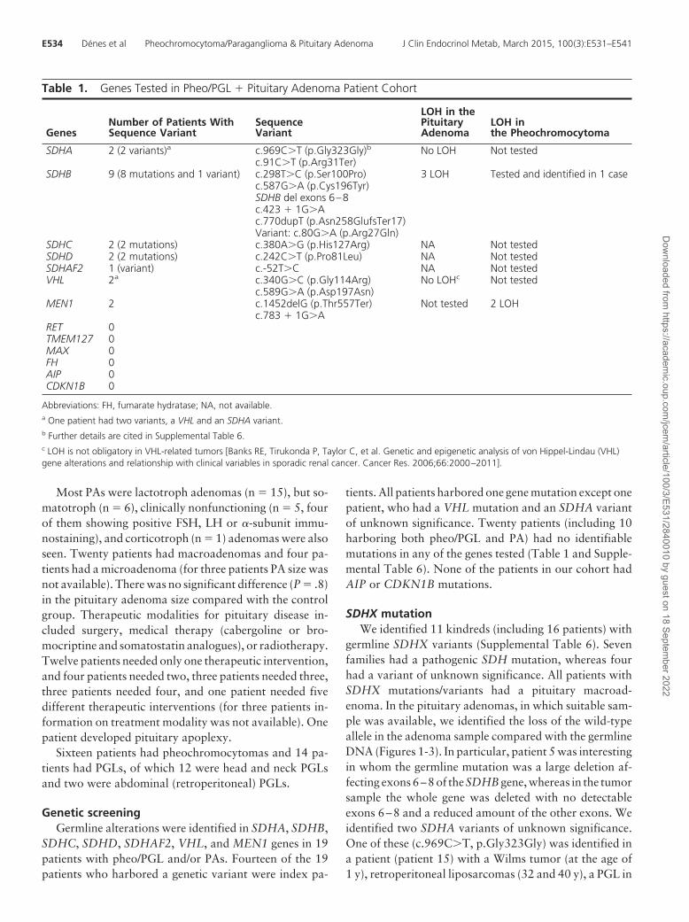
Genetic screeningGermline alterations were identified in SDHA, SDHB,
SDHC, SDHD, SDHAF2, VHL, and MEN1 genes in 19patients with pheo/PGL and/or PAs. Fourteen of the 19patients who harbored a genetic variant were index pa-
tients. All patients harbored one gene mutation except onepatient, who had a VHL mutation and an SDHA variantof unknown significance. Twenty patients (including 10harboring both pheo/PGL and PA) had no identifiablemutations in any of the genes tested (Table 1 and Supple-mental Table 6). None of the patients in our cohort hadAIP or CDKN1B mutations.
SDHX mutationWe identified 11 kindreds (including 16 patients) with
germline SDHX variants (Supplemental Table 6). Sevenfamilies had a pathogenic SDH mutation, whereas fourhad a variant of unknown significance. All patients withSDHX mutations/variants had a pituitary macroad-enoma. In the pituitary adenomas, in which suitable sam-ple was available, we identified the loss of the wild-typeallele in the adenoma sample compared with the germlineDNA (Figures 1-3). In particular, patient 5 was interestingin whom the germline mutation was a large deletion af-fecting exons 6–8 of the SDHB gene, whereas in the tumorsample the whole gene was deleted with no detectableexons 6–8 and a reduced amount of the other exons. Weidentified two SDHA variants of unknown significance.One of these (c.969C�T, p.Gly323Gly) was identified ina patient (patient 15) with a Wilms tumor (at the age of1 y), retroperitoneal liposarcomas (32 and 40 y), a PGL in
Table 1. Genes Tested in Pheo/PGL � Pituitary Adenoma Patient Cohort
GenesNumber of Patients WithSequence Variant
SequenceVariant
LOH in thePituitaryAdenoma
LOH inthe Pheochromocytoma
SDHA 2 (2 variants)a c.969C�T (p.Gly323Gly)b No LOH Not testedc.91C�T (p.Arg31Ter)
SDHB 9 (8 mutations and 1 variant) c.298T�C (p.Ser100Pro) 3 LOH Tested and identified in 1 casec.587G�A (p.Cys196Tyr)SDHB del exons 6–8c.423 � 1G�Ac.770dupT (p.Asn258GlufsTer17)Variant: c.80G�A (p.Arg27Gln)
SDHC 2 (2 mutations) c.380A�G (p.His127Arg) NA Not testedSDHD 2 (2 mutations) c.242C�T (p.Pro81Leu) NA Not testedSDHAF2 1 (variant) c.-52T�C NA Not testedVHL 2a c.340G�C (p.Gly114Arg) No LOHc Not tested
c.589G�A (p.Asp197Asn)MEN1 2 c.1452delG (p.Thr557Ter) Not tested 2 LOH
c.783 � 1G�ARET 0TMEM127 0MAX 0FH 0AIP 0CDKN1B 0
Abbreviations: FH, fumarate hydratase; NA, not available.a One patient had two variants, a VHL and an SDHA variant.b Further details are cited in Supplemental Table 6.c LOH is not obligatory in VHL-related tumors [Banks RE, Tirukonda P, Taylor C, et al. Genetic and epigenetic analysis of von Hippel-Lindau (VHL)gene alterations and relationship with clinical variables in sporadic renal cancer. Cancer Res. 2006;66:2000–2011].
E534 Dénes et al Pheochromocytoma/Paraganglioma & Pituitary Adenoma J Clin Endocrinol Metab, March 2015, 100(3):E531–E541
Dow
nloaded from https://academ
ic.oup.com/jcem
/article/100/3/E531/2840010 by guest on 18 September 2022

the retroperitoneum(50y), a renaloncocytoma (50y), anda nonfunctioning pituitary adenoma (NFPA; 53 y). Hisfather had an NFPA operated at 44 years and again at 74years. His mother (no known tumors) carried thec.969C�T variant. The other SDHA variant was identi-fied in a patient with a VHL mutation and PA (patient 21).We have also identified an SDHB variant (c.80G�Ap.Arg27Gln, patient 17) of unknown significance. Wehave tested the proband’s pheochromocytoma andshowed LOH at the SDHB locus; however, the SDHBstaining of the pheochromocytoma did not show loss ofSDHB expression. No pituitary tissue was available fortesting in this family. An SDHAF2 variant c.-52T�C wasidentified in a patient with somatotroph macroadenomaand head and neck PGL. The patient was not operatedupon and therefore no tissue is available.
We identified two families with SDH mutations inwhich a family member with a PA did not carry the germ-line SDHX mutation: family 6 with two SDHC mutation-positive siblings had PA and/or PGL, whereas a first cousinhad an NFPA but no SDHC mutation; and family 7 inwhom the parent and child both had SDHD mutation-positive PGL and another child had a microprolactinomabut no SDHD mutation (Supplemental Figure 1). Thesecases are either phenocopies or could, theoretically, be
explained by a digenic disease pat-tern in which the second disease-causing gene has not been identified.
VHL mutationAn18-year-oldpatientwithapatho-
genic VHL mutation [c.340G�C, amissense mutation affecting a surfaceamino-acid (22)], had an invasive GH-and prolactin (PRL)-positive PA asshown in Supplemental Table 6 andSupplemental Figure 2 (20).
MEN1 mutationWe identified two patients (pa-
tients 22 and 23) with a germlineMEN1 mutation and pheochromo-cytoma, whereas all the other testedgenes were normal (SupplementalTable 6). Both pheochromocytomasshowed LOH in the MEN1 gene,supporting, although not proving,the pathogenic role of MEN1 inthese tumors (see Figure 4, A and B).Although the association of pheo/PGLs and an MEN1-like syndromehas been described in the literature in13 cases, in only four of these have
MEN1 mutations been identified (23–25), and none ofthem has been studied for LOH in the pheochromocytomatissue.
Control patientsWe studied 23 MEN1-, AIP-, and CDKN1B-negative
FIPA family probands without features of Carney com-plex or a personal or family history of pheo/PGL (Supple-mental Table 7). We analyzed their DNA for all the pheo/PGL-related genes included in our panel to investigate therole of these genes in FIPA families. No pheo/PGL-relatedgene mutations were found in these families.
Pathological featuresThe PAs of patients with SDHX mutations (patients 1
and 2 from family 1, patient 4, and patient 5) were char-acterized by intracytoplasmic vacuoles. The extent of vac-uolization was not related to the histological type (pro-lactinoma or NFPA) of the tumor (Figures 1–3). Thenumber of vacuolated cells varied from about 50% to80% of the neoplastic cell population. Vacuoles rangedfrom small and multiple (Figure 3C) to large, occupyingmost of the cytoplasm and mimicking signet-ring cells(Figure 2C). None of the vacuoles indented the nucleus as
Figure 1. Pedigree (A) and LOH (B) at the SDHB locus in the pituitary adenoma of patient 1 infamily 1 is shown. C, H&E staining of the pituitary adenoma of the proband (patient 1 in family1) shows predominant trabecular architecture (�20). D, Vacuoles at times filling the entirecytoplasm characterize this case (arrow) (H&E, �40). E, H&E staining (�20) of the pituitaryadenoma of the proband’s mother (patient 2 in family 1) also shows similar intracytoplasmicvacuoles. F, The immunoreaction with the anti-113-1 antibody (immunoperoxidase, �20) showsthe mitochondria content. G, MRI imaging of proband’s mother’s pituitary adenoma. H, MRIimaging of the proband’s pituitary adenoma and glomus vagale tumor. MRI, magnetic resonanceimaging.
doi: 10.1210/jc.2014-3399 jcem.endojournals.org E535
Dow
nloaded from https://academ
ic.oup.com/jcem
/article/100/3/E531/2840010 by guest on 18 September 2022

commonly seen with accumulation of lipids. One caseshowed focal oncocytic changes identifiable on the hema-toxylin and eosin (H&E)-stained sections. The histochem-ical stain periodic acid-Schiff (PAS)/diastase-resistant pe-riodic acid of Schiff did not reveal any glycogenaccumulation. Vacuoles were not seen in the PA of thepatient with the germline VHL mutation (without SDHmutation) (Supplemental Figure 2). The SDHB staining ofPAs with the SDHB mutation showed either a loss of ex-pression of SDHB or a faint expression (Figures 2D and3E).
Because SDHX mutations are known to alter mito-chondrial function, immunostaining was performed for amitochondrial membrane protein with the anti-113-1 an-tibody. This staining documented variable accumulationof mitochondria in SDHX mutation-positive PA cells.Some adenomas in particular showed increased immuno-staining compared with the other cases (Figures 1F and3D) in keeping with the focal oncocytic changes observedin the H&E-stained sections. Vacuoles did not appear tobe rimmed by this protein, suggesting that vacuolization isnot secondary to dilatation of mitochondria. To under-stand whether vacuoles were the result of swelling of theendoplasmic reticulum (ER), we immunostained our sam-ples for the ER marker ERLEC1. None of the vacuoles waslined by this protein, indicating that they were not relatedto the ER (Supplemental Figure 3).
Menin staining of the pheochro-mocytoma samples of the patientswith MEN1 mutations showed ei-ther no menin-positive cells orweakly positive staining nuclei (Fig-ure 4).
Discussion
Syndromic presentation of PA andpheo/PGL is rare, and it is not part ofthe classical multiple endocrine tu-mor syndromes. This study de-scribes, we believe, the largest cohortof patients with PAs and pheo/PGLs.Systematic testing of this populationfor alterations of the known pitu-itary and pheo/PGL-related genessuggest that SDH mutations play apathogenic role in the developmentof PAs in some of these patients.Cases of other pheo/PGL genes asso-ciated with PA, VHL and RET, areexceptionally rare. On the otherhand, the MEN1 mutations can
sometimes lead to pheo/PGLs, as suggested previously(23–25), and here we present supporting LOH and im-munostaining findings. An endocrine rather than geneticassociation occurs when pheochromocytomas secrete hy-pothalamic-releasing hormones (GHRH or CRH) mim-icking the PA and pheo/PGL syndrome, described previ-ously in eight cases (Supplemental Table 2). Although inthese cases only the adrenal gland harbors a tumorwhereas the pituitary usually displays hyperplasia in re-sponse to the ectopic hormone secretion, this is a relevantclinical differential diagnostic scenario and should be keptin mind in patients with pituitary disease and pheo/PGLs.In approximately half of our cases, no germline abnor-malities were seen, suggesting either the presence of otherdisease-causing genes or the coincidental occurrence of thepituitary and pheo/PGL tumors.
Because this is a multicentric study with a patient co-hort from all over the world, with a heterogeneous geneticbackground, it is difficult to estimate whether the coinci-dence of these two tumors occurred randomly, or other,not-yet-specified genetic factors could be playing a role.Using the ranges of the available prevalence data for PAsand pheo/PGLs in the general population (1–4), the co-incidental chance for the two diseases occurring in thesame patient ranges between 1 in 2.5 million and 1 in 8.5million subjects. In our single center (Barts), we reviewed
Figure 2. Pedigree (A) and LOH (B) at the SDHB locus in the pituitary adenoma of patient 4; themicrosatellite upstream of the mutation has also shown to be lost. C, H&E-stained section (�20)of this adenoma shows prominent vacuolar changes in most neoplastic cells; the cytoplasmotherwise appears weakly eosinophilic. D, SDHB staining suggesting lack of strong granularstaining of the pituitary adenoma of the proband (immunoperoxidase, �20) (inset: positive SDHBstaining as positive control in a paraganglioma).
E536 Dénes et al Pheochromocytoma/Paraganglioma & Pituitary Adenoma J Clin Endocrinol Metab, March 2015, 100(3):E531–E541
Dow
nloaded from https://academ
ic.oup.com/jcem
/article/100/3/E531/2840010 by guest on 18 September 2022

828 patients with pituitary tumors and 150 with pheo/PGL (26, 27). Assuming a maximum population fre-quency of pheo/PGL of 1 in 2500, we predict that 0.33cases in a population-based series of 828 pituitary ade-noma patients would have a pheo/PGL, whereas the actualfrequency in patients seen at our center was 2 in 828 (P �.048; Fisher’s exact test on single proportions). Likewise,assuming the maximum population frequency of PA of 1in 1000, we expect 0.06 cases in a population-based seriesof 150 pheo/PGL patients would have a PA, whereas theactual frequency is 2 in 150 (P � .01). Both of these datasets suggest an increased incidence.
Of the six suggested explanations for the coexistence ofPA and pheo/PGL that we outlined in the introductorytext, we could confirm the following options: 1) a pheo/PGL-related gene causes PA, 2) a pituitary gene causespheo/PGL, 5) ectopic hypothalamic hormone synthesis ina pheochromocytoma, and probably one or more familiesin our cohort match option, and 6) representing pure co-incidence. Regarding option 3, we have not found anypatients with mutations in two genes, such as a classicalpheo/PGL and a pituitary tumor gene. In addition, wefound LOH at the SDH locus in pituitary adenomas andat the MEN1 locus in pheochromocytomas, suggesting,although not proving, that in these patients a single gene
is responsible for both tumors. Exome or whole-genomesequencing studies in the future might find novel genescausing both diseases (option 4). In our cohort 19 patients(48%) had a germline alteration, among them 17 (43%)with a genetic variant in the pheo/PGL genes. Large studiesshowed that about one-third of pheo/PGL patients (mostfamilial cases and 10%–20% of the sporadic cases) carrya germline mutation in RET, VHL, NF1, SDHA, SDHB,SDHC, SDHD, SDHAF2, MAX, or TMEM127 genes(28, 29), suggesting that our cohort may have a slightlyhigher percentage of germline alterations.
The clinical features of the published cases of the asso-ciation of pituitary disease and pheo/PGLs are summa-rized in the Supplemental Material (Supplemental Tables1–5). More recently, three screening studies have been per-formed. One of them screened a group of patients (26 PGLpatients and eight carriers) with a particular SDHD mu-tation due to a founder effect for the presence of a PA. OneGH-secreting macroadenoma and three nonfunctioningmicroadenomas (suggested to be incidentalomas) were di-agnosed in this patient cohort. No LOH was found at theSDHD locus in the GH-secreting PA (30). In the secondstudy, 309 PAs were screened for SDH mutations and amacroprolactinoma with two different somatic SDHAmutations with normal sequence in the germline (31) was
Figure 3. Pedigree (A) and sagittal and coronal magnetic resonance images of the pituitary adenoma (B) are shown. C, H&E-stained section (�20)shows that the tumor of patient 5 contains multiple vacuoles. D, The immunoreaction with the anti-113-1 antibody (immunoperoxidase, �20)highlights the mitochondria content. E, SDHB immunostaining shows loss of expression in neoplastic cells, whereas endothelial cells (arrow) retainthe expression (immunoperoxidase, �20). Loss of the SDHB gene in germline and pituitary tumor tissue in patient 5. F, Germline DNA shows adeletion affecting MLPA SDHB probes 6–8 in DNA derived from leukocytes. G, In pituitary adenoma tissue, a complete loss of genetic material atthe SDHB probes 6–8 area and heterozygous loss of SDHB probes 1–5.
doi: 10.1210/jc.2014-3399 jcem.endojournals.org E537
Dow
nloaded from https://academ
ic.oup.com/jcem
/article/100/3/E531/2840010 by guest on 18 September 2022

found. In the third study, screening has been performed inSDHX-mutated patients for nonpheo/PGL tumors. Twopatients with SDHD mutations were found to have a PA,and in one of these cases, LOH at the SDHD locus wasshown in the macroprolactinoma (32). Whether it is costeffective to measure prolactin in patients with pheo/PGLsneeds to be studied further.
Summarizing our cases combined with the cases avail-able in the literature (altogether 109 cases since 1952), wehave tried to identify any particular features for each genealteration for the tumor not classically associated withthat gene. Twenty cases have a confirmed SDHX mutationwith pituitary adenoma [(two SDHA (8, 31), eight SDHB(33, 34), two SDHC (35), and eight SDHD (30, 32, 36,37)]. The patients with an SDH mutation had various PAtypes (Supplemental Tables 3 and 6): nine macroprolacti-nomas, three somatotroph adenomas, and five NFPAshave been described. In three cases the PA subtypes couldnot be classified. All the PAs were macroadenomas, exceptfor three nonfunctioning microadenomas (possibly inci-dentalomas). The patients needed one to four therapeuticinterventions. Five patients needed a single therapeutic
intervention, five patients needed two, one patient neededthree, and two patients needed four therapeutic interven-tions. Of the 109 patients, five patients had RET muta-tions (38–41); two cases with acromegaly, two cases withprolactinoma, and one NFPA (one macroadenoma andone microadenoma, and in three cases the adenoma size isnot available). Four patients needed one therapeutic in-tervention (three surgeries and one medical treatment),whereas one patient needed medical therapy after trans-sphenoidal resection of the pituitary tumor. Two patientshad a VHL mutation (20), one with a PRL and one witha GH- and PRL-secreting adenoma. Six patients had aconfirmed MEN1 mutation and pheo/PGL (23–25): fivepatients with pheochromocytoma and one head and neckPGL.
We have identified a novel feature of the PAs of patientsharboring SDHX variants. The adenoma tissues show ex-tensive vacuolization of cytoplasm with features reminis-cent of signet-ring cells or physalipherous cells (42). Theorigin of vacuoles remains unclear. Lipid and glycogenaccumulation was suggested in the literature, but none ofthe vacuoles indented the nucleus as commonly seen in
Figure 4. A, LOH analysis at the MEN1 locus of the pheochromocytoma of patient 22 and patient 23 (B). Underlined microsatellite results identifymarkers that show a reduction in peak height in the pheochromocytoma sample compared with blood, indicating LOH but suggesting that somenontumoral tissue was also retained in the operated samples. C, Pheochromocytoma of patient 22 shows a loss of menin staining (inset: positivemenin staining in mouse Langerhans islet). D, The menin staining of the pheochromocytoma of patient 23 shows some weakly positive stainingnuclei (inset: positive menin staining in a sporadic pheochromocytoma used as a positive control).
E538 Dénes et al Pheochromocytoma/Paraganglioma & Pituitary Adenoma J Clin Endocrinol Metab, March 2015, 100(3):E531–E541
Dow
nloaded from https://academ
ic.oup.com/jcem
/article/100/3/E531/2840010 by guest on 18 September 2022
cells with accumulation of lipids and the histochemicalstain PAS/diastase-resistant periodic acid of Schiff did notreveal any glycogen accumulation. The vacuoles also donot resemble particle-rich cytoplasmic structures, de-scribed in epithelial neoplasms (43). Vacuolization of thenontumorous adenohypophyseal cells has been describedin cases of fatal hypothermia in two separate studies (44,45). Ishikawa et al (44) suggested that the vacuoles aredifferent from dilated cisternae of rough ER and fromdistended Golgi apparatus, which are the result of castra-tion or gonadal dysfunction and raised the possibility thatthey are lipid droplets due to metabolic dysfunction ini-tiated by the hypothermia. Doberentz et al (45) also notedcytoplasmic vacuolation of the anterior pituitary cells inthe case of hypothermia, and they suggested that this couldbe due to gradually developing tissue hypoxia. OncocyticPAs have recently been identified to contain somatic mu-tations affecting mitochondrial respiratory chain complexI, but these tumors do not show the vacuolar changes wehave identified in the SDH-related samples (46).
Inactivation of succinate dehydrogenase or VHL canlead to activation of the hypoxia inducible factor pathwayand a pseudohypoxic state. Indeed, we have shown in-creased hypoxia inducible factor-1� in an SDHD-mutatedcase linked to pituitary adenoma (37). It is not knownwhether the vacuoles seen in the SDH-related tumors aredue to the pseudohypoxic state, but we did not observe thisphenomenon in the VHL mutation-related PA (Supple-mental Figure 2).
Immunostaining for a mitochondrial membrane pro-tein or for an ER marker did not prove that the vacuolesarise from these organelles. We attempted electron mi-croscopy to identify the nature of the vacuoles, but thiswas inconclusive due to the poor preservation of formalin-fixed tissue recovered from paraffin (data not shown).These vacuoles were not specifically described in the stud-ies of recently published SDHX mutations associated withPAs, but based on the available histological pictures, thepresence of vacuoles cannot be ruled out (8, 31, 37). Vac-uoleshavebeendescribed in SDHBmutation-related renalcarcinoma and were attributed to giant mitochondria(47), but the clear cytoplasm observed in these tumors canalso represent glycogen or fat (48). Large cytoplasmic vac-uoles suggested to be mitochondria based on electron mi-croscopy have previously been described in PAs (49), pos-sibly due to ischemia. Acidophil stem cell adenomas canalso contain paranuclear vacuoles resulting from giant mi-tochondria (50).
The activity of certain mitochondrial enzymes involvedin oxidative phosphorylation is decreased in cancer cellscompared with normal tissue (51). Taking into accountthat succinate dehydrogenase enzymes, being part of the
mitochondrial complex II, play an important role in mi-tochondrial function, mutations that affect the activity ofthese enzymes might have a role in mitochondria dysfunc-tion (52). We believe that the vacuoles represent a hall-mark of PA in patients with the SDHX variant, but theirnature remains to be further investigated. In addition, fur-ther study of the metabolic pathways in SDH-related en-docrine tumors are awaited.
Our study has several shortcomings. First of all, beinga specialist pituitary and adrenal center with an interest infamilial pituitary adenomas, our center might attract moreunusual genetic conditions, therefore representing ahigher prevalence of these cases. In a significant portion ofthe patients, tumor samples were not available, often dueto the lack of surgical intervention; therefore, no appro-priate material was available for LOH or to study in fur-ther detail the unusual histological phenotype in the PAs.
In summary, germline mutations were identified in thestudied genes in 11 of 27 kindreds with the combinationof pheo/PGL and PAs. LOH at the SDHB locus in the PAsamples and LOH at the MEN1 locus in the pheochro-mocytoma samples was demonstrated, suggesting, al-though not proving, the pathogenic role of these genes inthese nonclassically disease-specific tissues. In addition,we noted intracytoplasmic vacuoles in PAs of patients af-fected by SDH mutations. Together with the single casereports available in the literature, this large cohort sup-ports the hypothesis that in some families SDH mutationsmay have a role in PA formation and MEN1 mutationsmay have a role in the development of pheochromocy-toma. Whether screening for PAs in SDHX patients iswarranted needs to be studied in the future, but our find-ings suggest that genetic testing for germline mutations inSDHX and MEN1 should be considered in patients withthe constellation of pheo/PGLs and PAs.
Acknowledgments
We are grateful to Professor Kalman Kovacs (Toronto, Canada)for the advice on the histological features of the SDH-relatedpituitary adenoma tissue and to Michael Weedon (Exeter, UK)for the statistical help.
Address all correspondence and requests for reprints to:Professor Márta Korbonits, Department of Endocrinology,Barts and the London School of Medicine and Dentistry, Char-terhouse Square, London EC1M 6BQ, United Kingdom. E-mail:[email protected].
N.D. was supported by a grant from the Royal Society.Disclosure Summary: S.E. is a Wellcome Trust Senior Inves-
tigator. The other authors have no conflicting interests.
doi: 10.1210/jc.2014-3399 jcem.endojournals.org E539
Dow
nloaded from https://academ
ic.oup.com/jcem
/article/100/3/E531/2840010 by guest on 18 September 2022

References
1. Daly AF, Rixhon M, Adam C, Dempegioti A, Tichomirowa MA,Beckers A. High prevalence of pituitary adenomas: a cross-sectionalstudy in the province of Liege, Belgium. J Clin Endocrinol Metab.2006;91:4769–4775.
2. Fernandez A, Karavitaki N, Wass JA. Prevalence of pituitary ade-nomas: a community-based, cross-sectional study in Banbury (Ox-fordshire, UK). Clin Endocrinol (Oxf). 2010;72:377–382.
3. Mazzaglia PJ. Hereditary pheochromocytoma and paraganglioma.J Surg Oncol. 2012;106:580–585.
4. Eisenhofer G, Pacak K, Maher ER, Young WF, de Krijger RR. Pheo-chromocytoma. Clin Chem. 2013;59:466–472.
5. Iversen K. Acromegaly associated with phaeochromocytoma. ActaMed Scand. 1952;142:1–5.
6. Beckers A. Means, motive, and opportunity: SDH mutations aresuspects in pituitary tumors. J Clin Endocrinol Metab. 2013;98:2274–2276.
7. Xekouki P, Stratakis CA. Succinate dehydrogenase (SDHx) muta-tions in pituitary tumors: could this be a new role for mitochondrialcomplex II and/or Krebs cycle defects? Endocr Relat Cancer. 2012;19:C33–C40.
8. Dwight T, Mann K, Benn DE, et al. Familial SDHA mutation as-sociated with pituitary adenoma and pheochromocytoma/paragan-glioma. J Clin Endocrinol Metab. 2013;98:E1103–E1108.
9. Ferner RE, Huson SM, Thomas N, et al. Guidelines for the diagnosisand management of individuals with neurofibromatosis 1. J MedGenet. 2007;44:81–88.
10. Korbonits M, Storr H, Kumar AV. Familial pituitary adenomas—who should be tested for AIP mutations? Clin Endocrinol (Oxf).2012;77:351–356.
11. Owens M, Stals K, Ellard S, Vaidya B. Germline mutations in theCDKN1B gene encoding p27 Kip1 are a rare cause of multiple en-docrine neoplasia type 1. Clin Endocrinol (Oxf). 2009;70:499–500.
12. Occhi G, Regazzo D, Trivellin G, et al. A novel mutation in theupstream open reading frame of the CDKN1B gene causes a MEN4phenotype. PLoS Genet. 2013;9:e1003350.
13. Schouten JP, McElgunn CJ, Waaijer R, Zwijnenburg D, Diepvens F,Pals G. Relative quantification of 40 nucleic acid sequences by mul-tiplex ligation-dependent probe amplification. Nucleic Acids Res.2002;30:e57.
14. Rattenberry E, Vialard L, Yeung A, et al. A comprehensive nextgeneration sequencing-based genetic testing strategy to improve di-agnosis of inherited pheochromocytoma and paraganglioma. J ClinEndocrinol Metab. 2013;98:E1248–E1256.
15. Chahal HS, Stals K, Unterlander M, et al. AIP mutation in pituitaryadenomas in the 18th century and today. N Engl J Med. 2011;364:43–50.
16. Sassolas G, Chayvialle JA, Partensky C, et al. [Acromegaly, clinicalexpression of the production of growth hormone releasing factor inpancreatic tumors]. Ann Endocrinol (Paris). 1983;44:347–354.
17. Berger G, Trouillas J, Bloch B, et al. Multihormonal carcinoid tu-mor of the pancreas. Secreting growth hormone-releasing factor asa cause of acromegaly. Cancer. 1984;54:2097–2108.
18. Harding B, Lemos MC, Reed AA, et al. Multiple endocrine neopla-sia type 1 knockout mice develop parathyroid, pancreatic, pituitaryand adrenal tumours with hypercalcaemia, hypophosphataemiaand hypercorticosteronaemia. Endocr Relat Cancer. 2009;16:1313–1327.
19. Gill AJ, Benn DE, Chou A, et al. Immunohistochemistry for SDHBtriages genetic testing of SDHB, SDHC, and SDHD in paragangli-oma-pheochromocytoma syndromes. Hum Pathol. 2010;41:805–814.
20. Tudorancea A, Francois P, Trouillas J, et al. Von Hippel-Lindaudisease and aggressive GH-PRL pituitary adenoma in a young boy.Ann Endocrinol (Paris). 2012;73:37–42.
21. Mumby C, Davis JR, Trouillas J, Higham CE. Phaeochromocytoma
and acromegaly: a unifying diagnosis. Endocrinol Diabetes MetabCase Rep. 2014;2014:140036.
22. Ong KR, Woodward ER, Killick P, Lim C, Macdonald F, Maher ER.Genotype-phenotype correlations in von Hippel-Lindau disease.Hum Mutat. 2007;28:143–149.
23. Langer P, Cupisti K, Bartsch DK, et al. Adrenal involvement inmultiple endocrine neoplasia type 1. World J Surg. 2002;26:891–896.
24. Dackiw AP, Cote GJ, Fleming JB, et al. Screening for MEN1 mu-tations in patients with atypical endocrine neoplasia. Surgery. 1999;126:1097–1103; discussion 1103–1104.
25. Jamilloux Y, Favier J, Pertuit M, et al. A MEN1 syndrome with aparaganglioma. Eur J Hum Genet. 2014;22:283–285.
26. Herincs M, Owusu-Antwi S, Chahal HS, et al. Prevalence of familialisolated pituitary adenomas. Endocrine Abstracts. 2013;31:P258.
27. Srirangalingam U, Khan F, Gunganah K, et al. SDHB surveillanceregime: a single UK institution experience. Endocrine Abstracts.2014;34:P188.
28. Almeida MQ, Stratakis CA. Solid tumors associated with multipleendocrine neoplasias. Cancer Genet Cytogenet. 2010;203:30–36.
29. Gimenez-Roqueplo AP, Dahia PL, Robledo M. An update on thegenetics of paraganglioma, pheochromocytoma, and associated he-reditary syndromes. Horm Metab Res. 2012;44:328–333.
30. Dematti S, Branz G, Casagranda G, et al. Pituitary tumors in SDHmutation carriers. Paper presented at: 12th Annual Meeting of Eu-ropean Network for the Study of Adrenal Tumors ENSAT; 2013;Abstract Book:P29.
31. Gill AJ, Toon CW, Clarkson A, et al. Succinate dehydrogenase de-ficiency is rare in pituitary adenomas. Am J Surg Pathol. 2014;38:560–566.
32. Papathomas TG, Gaal J, Corssmit EP, et al. Non-pheochromocy-toma (PCC)/paraganglioma (PGL) tumors in patients with succinatedehydrogenase-related PCC-PGL syndromes: a clinicopathologicaland molecular analysis. Eur J Endocrinol. 2014;170:1–12.
33. Benn DE, Gimenez-Roqueplo AP, Reilly JR, et al. Clinical presen-tation and penetrance of pheochromocytoma/paraganglioma syn-dromes. J Clin Endocrinol Metab. 2006;91:827–836.
34. Majumdar S, Friedrich CA, Koch CA, Megason GC, Fratkin JD,Moll GW. Compound heterozygous mutation with a novel splicedonor region DNA sequence variant in the succinate dehydrogenasesubunit B gene in malignant paraganglioma. Pediatr Blood Cancer.2010;54:473–475.
35. Lopez-Jimenez E, de Campos JM, Kusak EM, et al. SDHC mutationin an elderly patient without familial antecedents. Clin Endocrinol(Oxf). 2008;69:906–910.
36. Varsavsky M, Sebastian-Ochoa A, Torres Vela E. Coexistence of apituitary macroadenoma and multicentric paraganglioma: a strangecoincidence. Endocrinol Nutr. 2013;60:154–156.
37. Xekouki P, Pacak K, Almeida M, et al. Succinate dehydrogenase(SDH) D subunit (SDHD) inactivation in a growth-hormone-pro-ducing pituitary tumor: a new association for SDH? J Clin Endo-crinol Metab. 2012;97:E357–E366.
38. Brauer VF, Scholz GH, Neumann S, Lohmann T, Paschke R, KochCA. RET germline mutation in codon 791 in a family representing3 generations from age 5 to age 70 years: should thyroidectomy beperformed? Endocr Pract. 2004;10:5–9.
39. Saito T, Miura D, Taguchi M, Takeshita A, Miyakawa M, TakeuchiY. Coincidence of multiple endocrine neoplasia type 2A with acro-megaly. Am J Med Sci. 2010;340:329–331.
40. Heinlen JE, Buethe DD, Culkin DJ, Slobodov G. Multiple endocrineneoplasia 2a presenting with pheochromocytoma and pituitarymacroadenoma. ISRN Oncol. 2011;2011:732452.
41. Lugli F, Leone E, Iacovazzo D, et al. DM 2012 FMTC and prolacti-noma: a casual association or a new genetic syndrome? Paper pre-sented at: 13th International Workshop on Multiple EndocrineNeoplasia Final Program and Abstract Book: 77; P43.
42. Klijanienko J, Lagace R. Particular aspects. In: Klijanienko J, LagaceR, eds. Soft Tissue Tumors: A Multidisciplinary Decisional Diag-
E540 Dénes et al Pheochromocytoma/Paraganglioma & Pituitary Adenoma J Clin Endocrinol Metab, March 2015, 100(3):E531–E541
Dow
nloaded from https://academ
ic.oup.com/jcem
/article/100/3/E531/2840010 by guest on 18 September 2022

nostic Approach. Hoboken, NJ: John Wiley Blackwell; 2011:121–411.
43. Necchi V, Sommi P, Vanoli A, Manca R, Ricci V, Solcia E. Protea-some particle-rich structures are widely present in human epithelialneoplasms: correlative light, confocal and electron microscopystudy. PLoS One. 2011;6:e21317.
44. Ishikawa T, Miyaishi S, Tachibana T, Ishizu H, Zhu BL, Maeda H.Fatal hypothermia related vacuolation of hormone-producing cellsin the anterior pituitary. Leg Med (Tokyo). 2004;6:157–163.
45. Doberentz E, Preuss-Wossner J, Kuchelmeister K, Madea B. Histo-logical examination of the pituitary glands in cases of fatal hypo-thermia. Forensic Sci Int. 2011;207:46–49.
46. Kurelac I, MacKay A, Lambros MB, et al. Somatic complex I dis-ruptive mitochondrial DNA mutations are modifiers of tumorigen-esis that correlate with low genomic instability in pituitary adeno-mas. Hum Mol Genet. 2013;22:226–238.
47. Housley SL, Lindsay RS, Young B, et al. Renal carcinoma with giantmitochondria associated with germ-line mutation and somatic lossof the succinate dehydrogenase B gene. Histopathology. 2010;56:405–408.
48. Srigley JR, Delahunt B, Eble JN, et al. The International Society ofUrological Pathology (ISUP) Vancouver Classification of RenalNeoplasia. Am J Surg Pathol. 2013;37:1469–1489.
49. Horoupian DS. Large mitochondria in a pituitary adenoma withhyperprolactinemia. Cancer. 1980;46:537–542.
50. Horvath E, Kovacs K, Singer W, et al. Acidophil stem cell adenomaof the human pituitary: clinicopathologic analysis of 15 cases. Can-cer. 1981;47:761–771.
51. Kroemer G. Mitochondria in cancer. Oncogene. 2006;25:4630–4632.
52. Zhan X, Desiderio DM. Signaling pathway networks mined fromhuman pituitary adenoma proteomics data. BMC Med Genomics.2010;3:13.
doi: 10.1210/jc.2014-3399 jcem.endojournals.org E541
Dow
nloaded from https://academ
ic.oup.com/jcem
/article/100/3/E531/2840010 by guest on 18 September 2022