Embed Size (px)
Citation preview
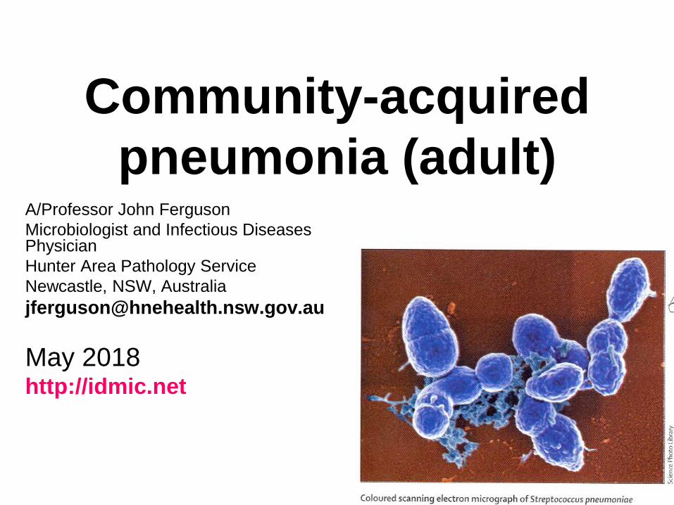
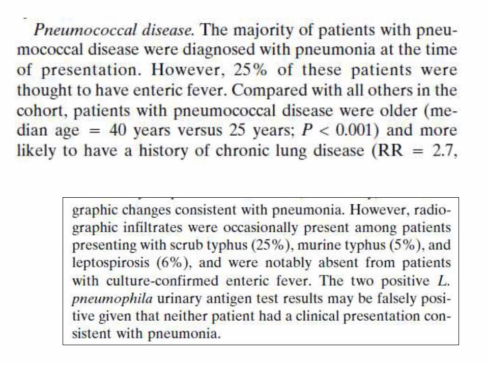
Community-acquired
pneumonia (adult) A/Professor John Ferguson
Microbiologist and Infectious Diseases Physician
Hunter Area Pathology Service
Newcastle, NSW, Australia
May 2018 http://idmic.net
Overview
1. Epidemiology
2. Prevention
3. Clinical assessment
4. Laboratory diagnostics
5. Antimicrobial therapeutics
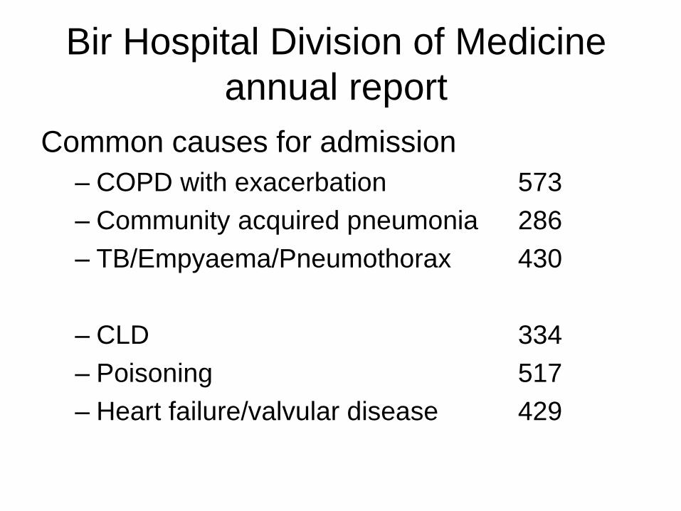
Bir Hospital Division of Medicine
annual report
Common causes for admission
– COPD with exacerbation 573
– Community acquired pneumonia 286
– TB/Empyaema/Pneumothorax 430
– CLD 334
– Poisoning 517
– Heart failure/valvular disease 429
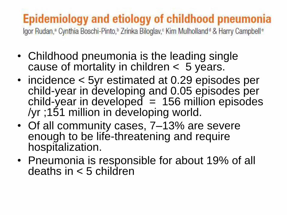
Epidemiology
• Childhood pneumonia is the leading single cause of mortality in children < 5 years.
• incidence < 5yr estimated at 0.29 episodes per child-year in developing and 0.05 episodes per child-year in developed = 156 million episodes /yr ;151 million in developing world.
• Of all community cases, 7–13% are severe enough to be life-threatening and require hospitalization.
• Pneumonia is responsible for about 19% of all deaths in < 5 children
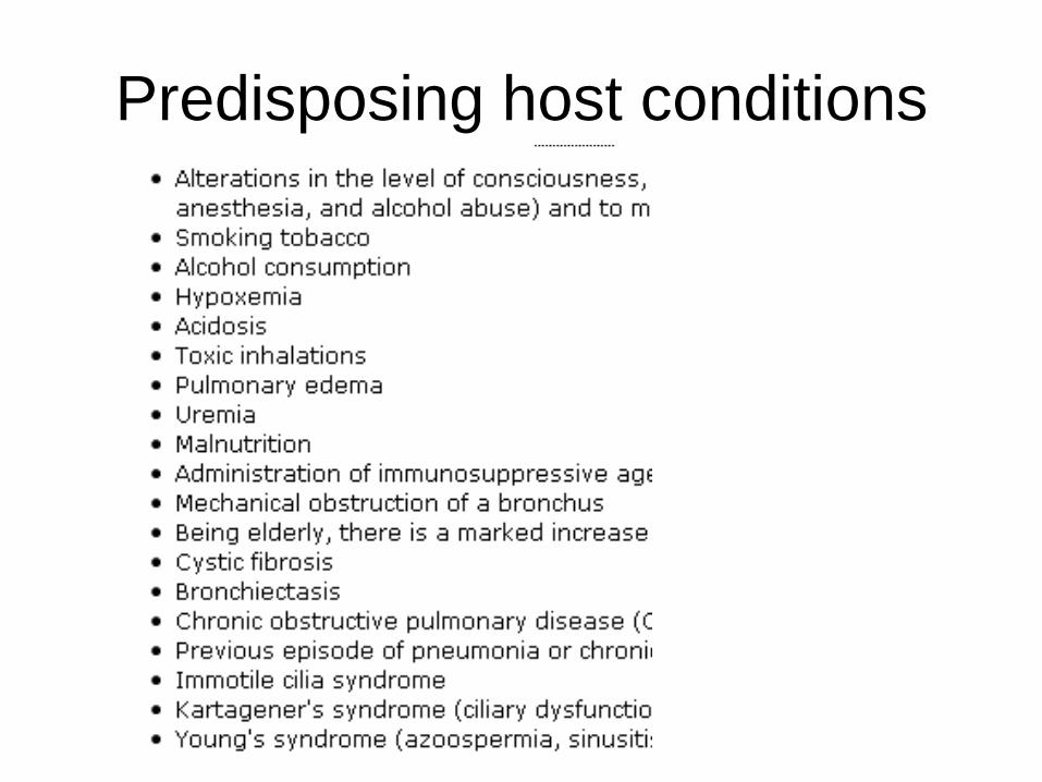
Predisposing host conditions
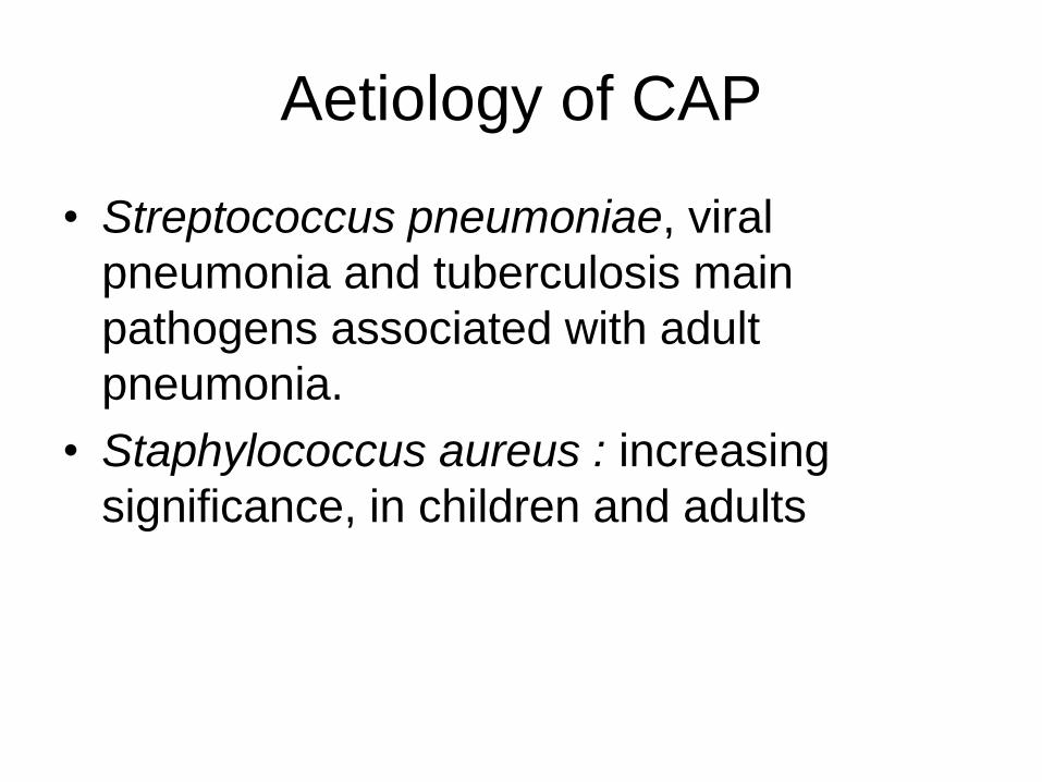
Aetiology of CAP
• Streptococcus pneumoniae, viral
pneumonia and tuberculosis main
pathogens associated with adult
pneumonia.
• Staphylococcus aureus : increasing
significance, in children and adults
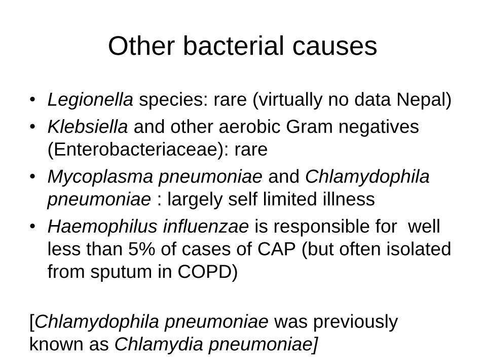
Other bacterial causes
• Legionella species: rare (virtually no data Nepal)
• Klebsiella and other aerobic Gram negatives
(Enterobacteriaceae): rare
• Mycoplasma pneumoniae and Chlamydophila
pneumoniae : largely self limited illness
• Haemophilus influenzae is responsible for well
less than 5% of cases of CAP (but often isolated
from sputum in COPD)
[Chlamydophila pneumoniae was previously
known as Chlamydia pneumoniae]
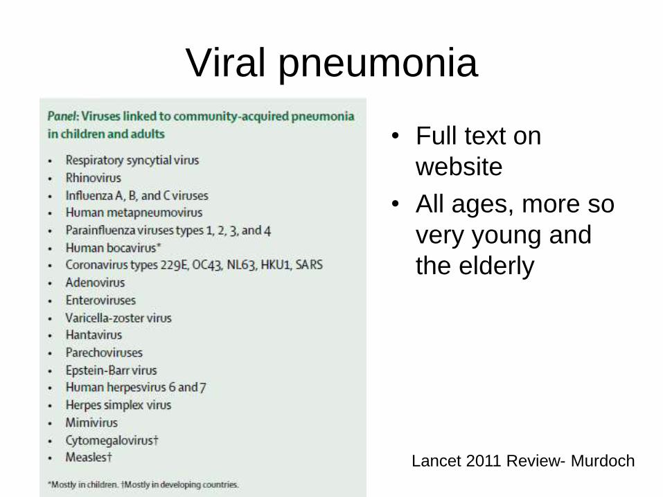
Viral pneumonia
• Full text on
website
• All ages, more so
very young and
the elderly
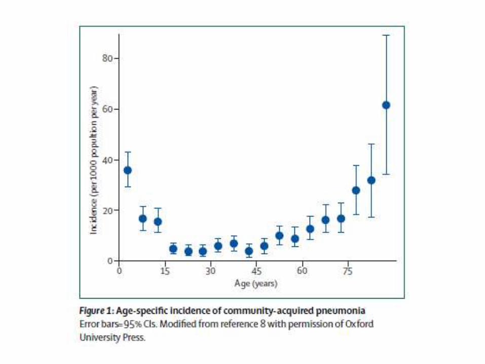
Lancet 2011 Review- Murdoch
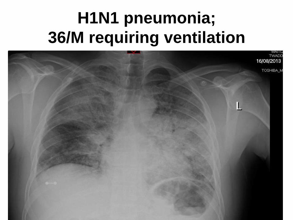
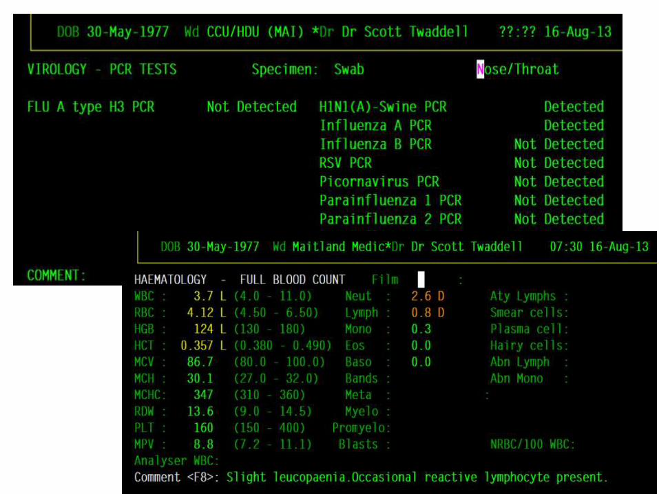
H1N1 pneumonia;
36/M requiring ventilation
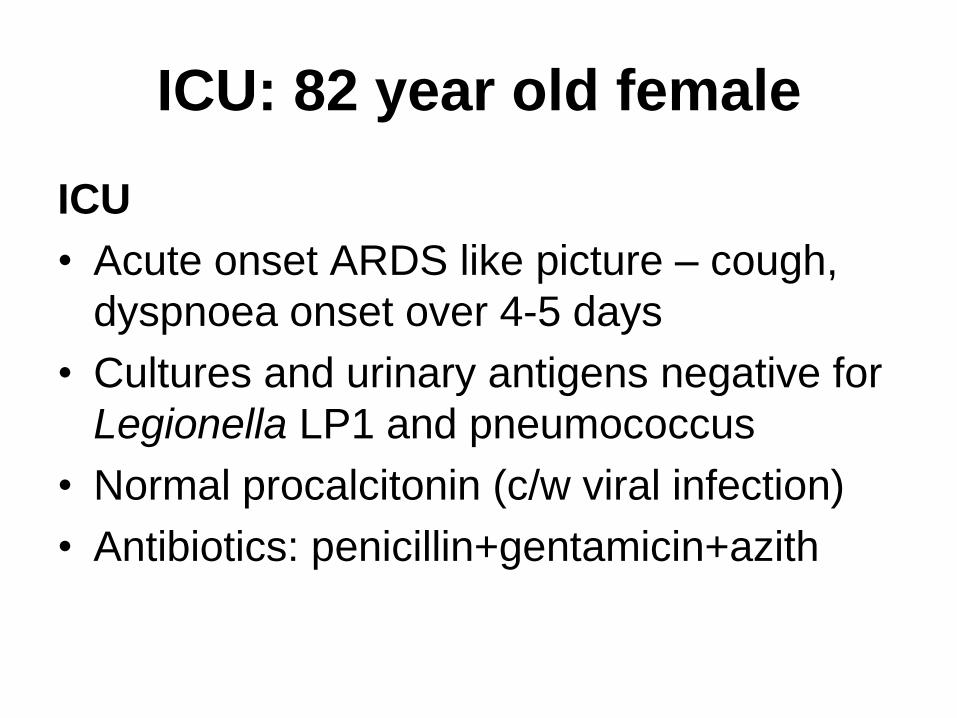
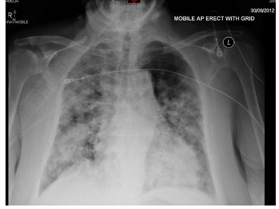
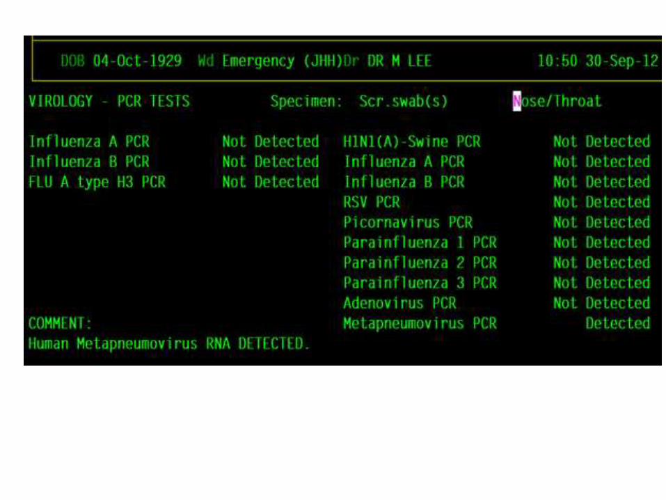
ICU: 82 year old female
ICU
• Acute onset ARDS like picture – cough,
dyspnoea onset over 4-5 days
• Cultures and urinary antigens negative for
Legionella LP1 and pneumococcus
• Normal procalcitonin (c/w viral infection)
• Antibiotics: penicillin+gentamicin+azith
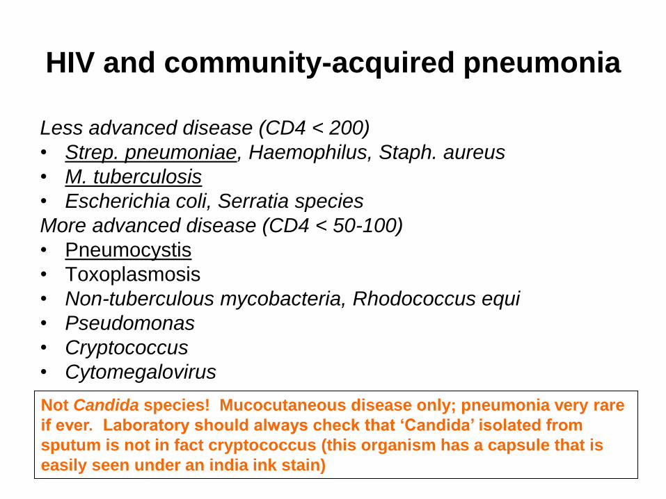
HIV and community-acquired pneumonia
Less advanced disease (CD4 < 200)
• Strep. pneumoniae, Haemophilus, Staph. aureus
• M. tuberculosis
• Escherichia coli, Serratia species
More advanced disease (CD4 < 50-100)
• Pneumocystis
• Toxoplasmosis
• Non-tuberculous mycobacteria, Rhodococcus equi
• Pseudomonas
• Cryptococcus
• Cytomegalovirus
Not Candida species! Mucocutaneous disease only; pneumonia very rare
if ever. Laboratory should always check that ‘Candida’ isolated from
sputum is not in fact cryptococcus (this organism has a capsule that is
easily seen under an india ink stain)
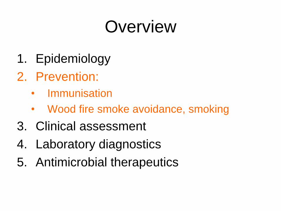
Overview
1. Epidemiology
2. Prevention:
• Immunisation
• Wood fire smoke avoidance, smoking
3. Clinical assessment
4. Laboratory diagnostics
5. Antimicrobial therapeutics
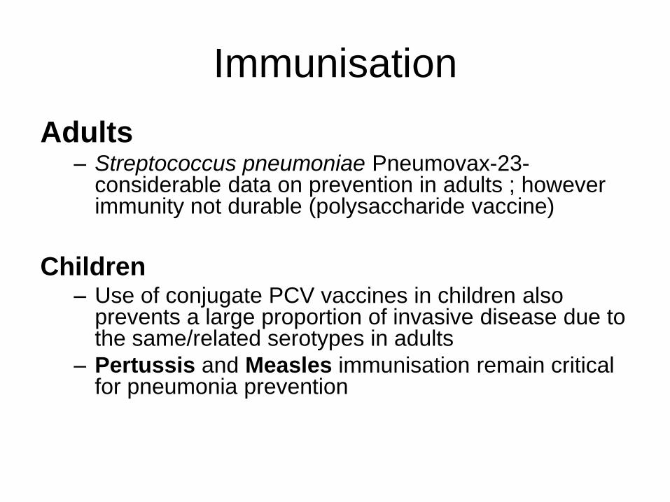
Immunisation
Adults – Streptococcus pneumoniae Pneumovax-23-
considerable data on prevention in adults ; however immunity not durable (polysaccharide vaccine)
Children – Use of conjugate PCV vaccines in children also
prevents a large proportion of invasive disease due to the same/related serotypes in adults
– Pertussis and Measles immunisation remain critical for pneumonia prevention
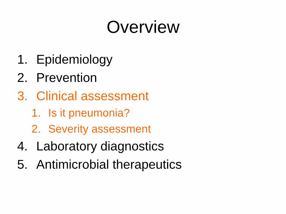
Overview
1. Epidemiology
2. Prevention
3. Clinical assessment
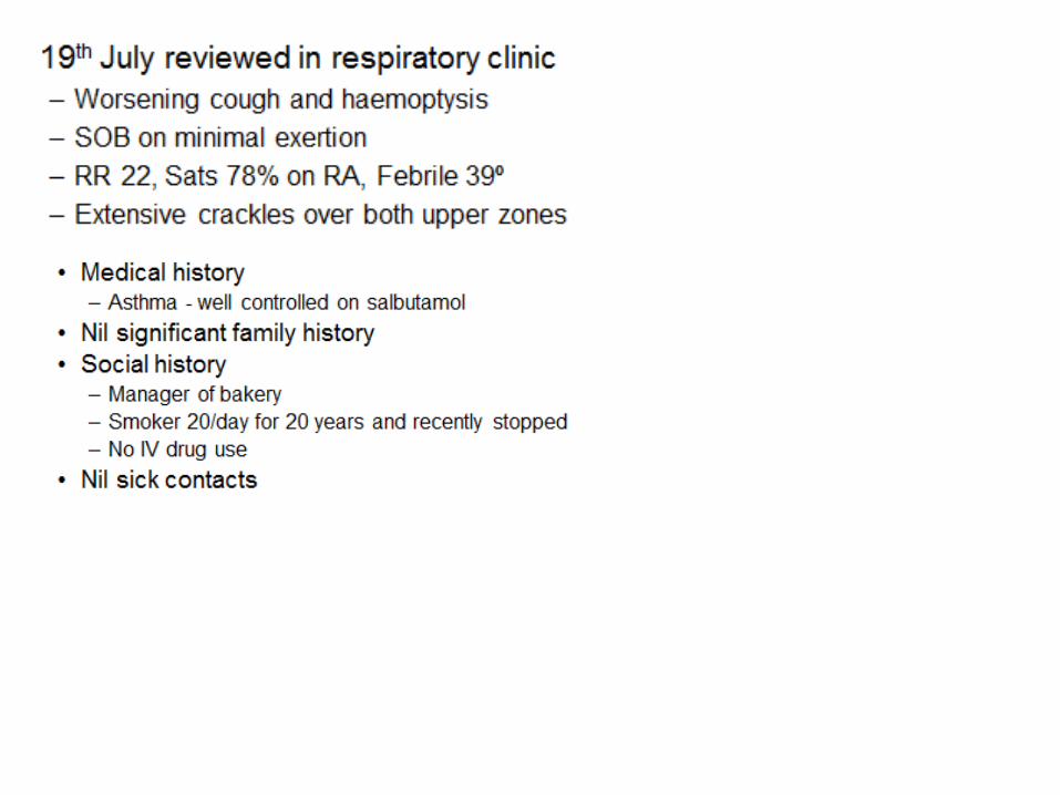
1. Is it pneumonia?
2. Severity assessment
4. Laboratory diagnostics
5. Antimicrobial therapeutics
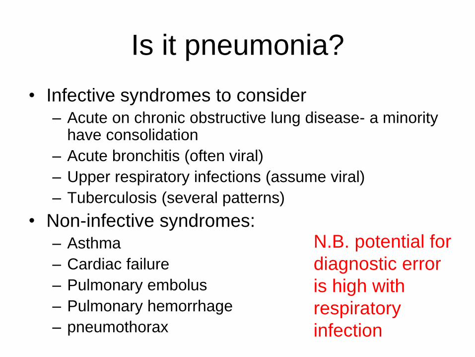
Is it pneumonia?
• Infective syndromes to consider – Acute on chronic obstructive lung disease- a minority
have consolidation
– Acute bronchitis (often viral)
– Upper respiratory infections (assume viral)
– Tuberculosis (several patterns)
• Non-infective syndromes: – Asthma
– Cardiac failure
– Pulmonary embolus
– Pulmonary hemorrhage
– pneumothorax
N.B. potential for
diagnostic error
is high with
respiratory
infection
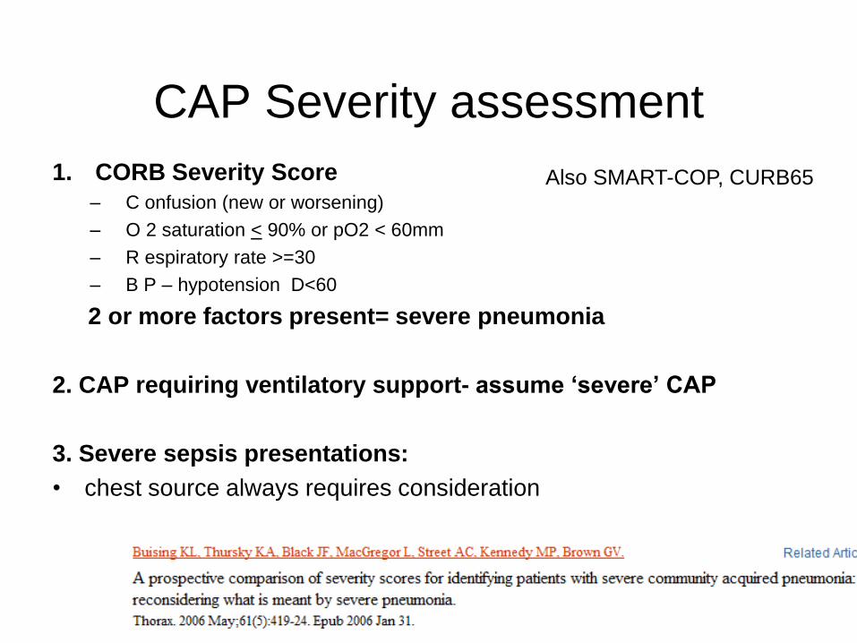
CAP Severity assessment
1. CORB Severity Score
– C onfusion (new or worsening)
– O 2 saturation < 90% or pO2 < 60mm
– R espiratory rate >=30
– B P – hypotension D<60
2 or more factors present= severe pneumonia
2. CAP requiring ventilatory support- assume ‘severe’ CAP
3. Severe sepsis presentations:
• chest source always requires consideration
Also SMART-COP, CURB65
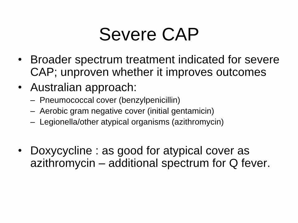
Severe CAP
• Broader spectrum treatment indicated for severe CAP; unproven whether it improves outcomes
• Australian approach: – Pneumococcal cover (benzylpenicillin)
– Aerobic gram negative cover (initial gentamicin)
– Legionella/other atypical organisms (azithromycin)
• Doxycycline : as good for atypical cover as azithromycin – additional spectrum for Q fever.
Overview
1. Epidemiology
2. Prevention
3. Clinical assessment
4. Laboratory diagnostics
5. Antimicrobial therapeutics
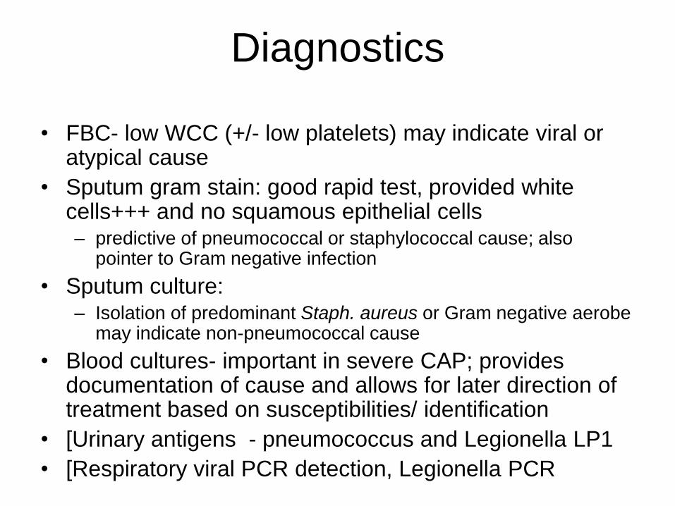
Diagnostics
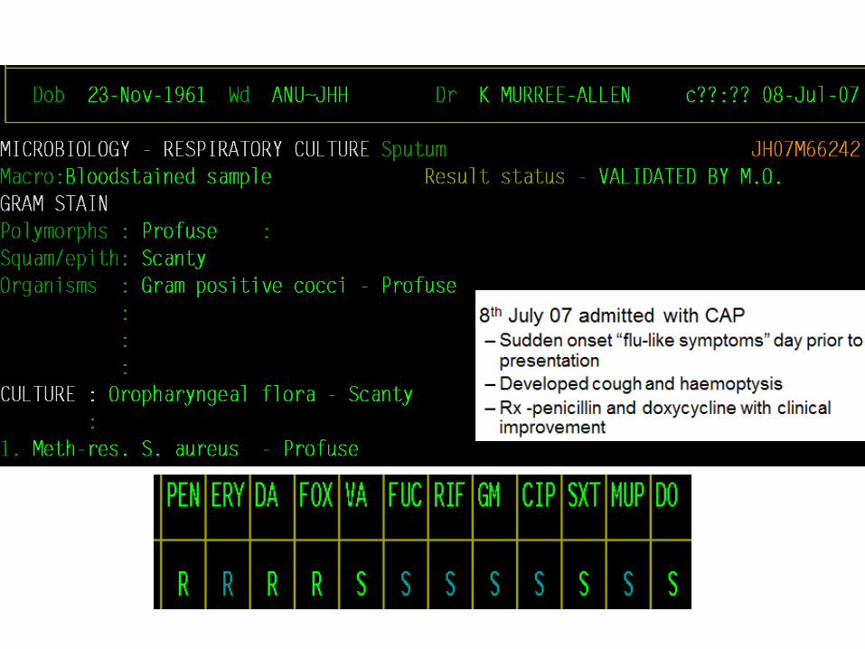
• FBC- low WCC (+/- low platelets) may indicate viral or atypical cause
• Sputum gram stain: good rapid test, provided white cells+++ and no squamous epithelial cells – predictive of pneumococcal or staphylococcal cause; also
pointer to Gram negative infection
• Sputum culture: – Isolation of predominant Staph. aureus or Gram negative aerobe
may indicate non-pneumococcal cause
• Blood cultures- important in severe CAP; provides documentation of cause and allows for later direction of treatment based on susceptibilities/ identification
• [Urinary antigens - pneumococcus and Legionella LP1
• [Respiratory viral PCR detection, Legionella PCR
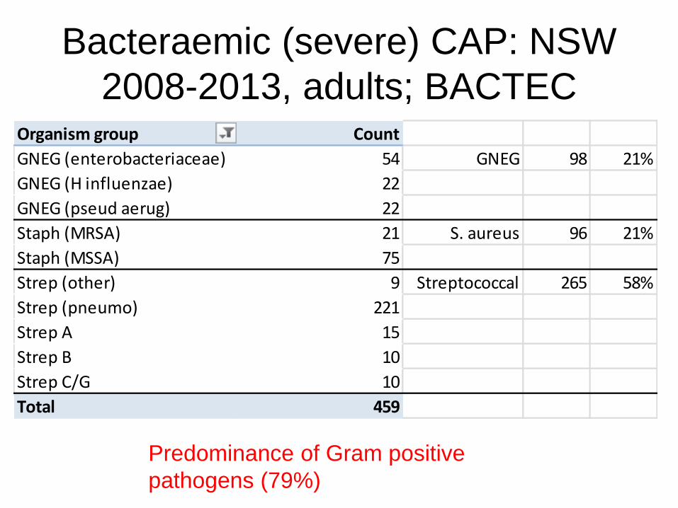
Bacteraemic (severe) CAP: NSW
2008-2013, adults; BACTEC
Organism group Count
GNEG (enterobacteriaceae) 54 GNEG 98 21%
GNEG (H influenzae) 22
GNEG (pseud aerug) 22
Staph (MRSA) 21 S. aureus 96 21%
Staph (MSSA) 75
Strep (other) 9 Streptococcal 265 58%
Strep (pneumo) 221
Strep A 15
Strep B 10
Strep C/G 10
Total 459
Predominance of Gram positive
pathogens (79%)
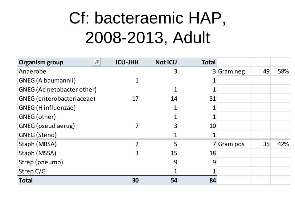
Cf: bacteraemic HAP,
2008-2013, Adult
Organism group ICU-JHH Not ICU Total
Anaerobe 3 3 Gram neg 49 58%
GNEG (A baumannii) 1 1
GNEG (Acinetobacter other) 1 1
GNEG (enterobacteriaceae) 17 14 31
GNEG (H influenzae) 1 1
GNEG (other) 1 1
GNEG (pseud aerug) 7 3 10
GNEG (Steno) 1 1
Staph (MRSA) 2 5 7 Gram pos 35 42%
Staph (MSSA) 3 15 18
Strep (pneumo) 9 9
Strep C/G 1 1
Total 30 54 84
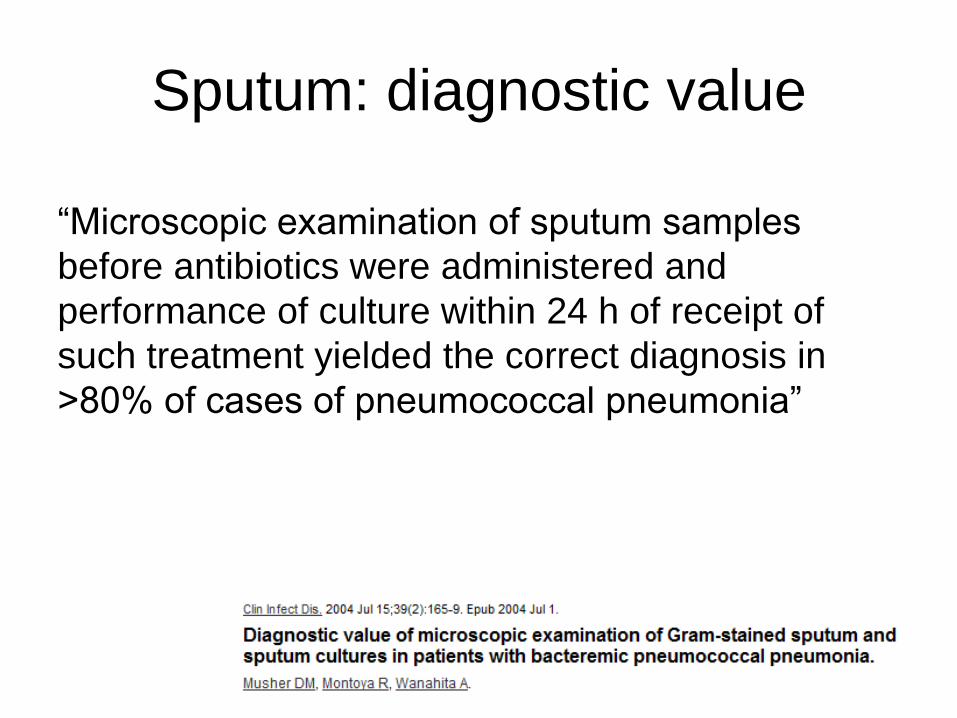
Sputum: diagnostic value
“Microscopic examination of sputum samples
before antibiotics were administered and
performance of culture within 24 h of receipt of
such treatment yielded the correct diagnosis in
>80% of cases of pneumococcal pneumonia”
Potential problems with sputum
• Specimen not possible- early pneumonia or poorly cooperation/illness
• Delay in transport to lab or high temperatures – Particularly a problem for pneumococcal or TB
isolation
• Poor quality samples- shown by presence of squamous epithelial cells and absence of neutrophils
• Growth of colonising respiratory flora in chronic lung disease patients- e.g. Haemophilus, Moraxella, Pseudomonas, E. coli etc. creates diagnostic confusion
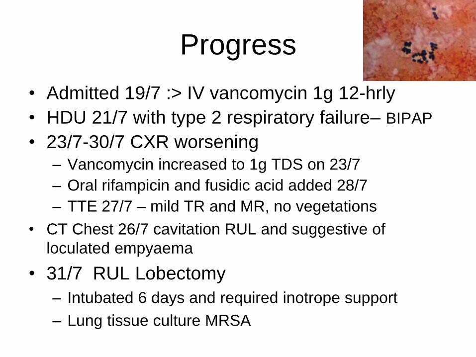
Progress
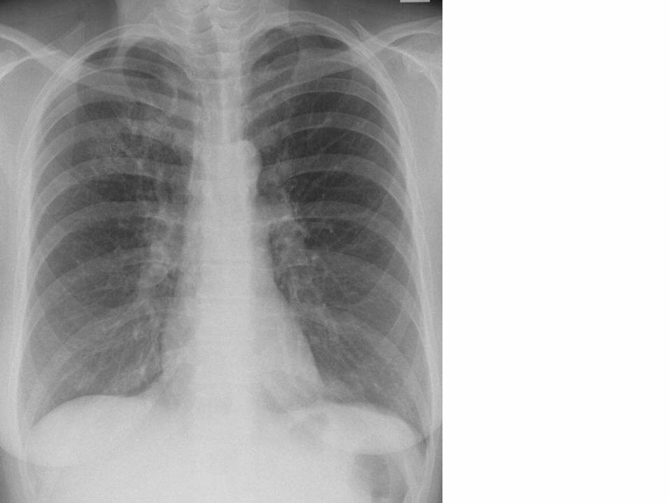
• Admitted 19/7 :> IV vancomycin 1g 12-hrly
• HDU 21/7 with type 2 respiratory failure– BIPAP
• 23/7-30/7 CXR worsening – Vancomycin increased to 1g TDS on 23/7
– Oral rifampicin and fusidic acid added 28/7
– TTE 27/7 – mild TR and MR, no vegetations
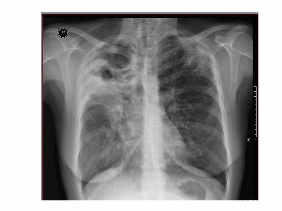
• CT Chest 26/7 cavitation RUL and suggestive of
loculated empyaema
• 31/7 RUL Lobectomy
– Intubated 6 days and required inotrope support
– Lung tissue culture MRSA
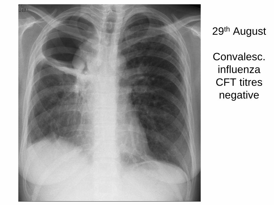
29th August
Convalesc.
influenza
CFT titres
negative
Overview
1. Epidemiology
2. Prevention
3. Clinical assessment
4. Laboratory diagnostics
5. Antimicrobial therapeutics
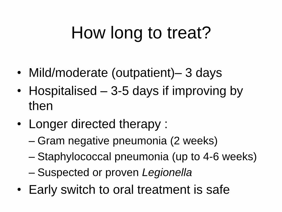
How long to treat?
• Mild/moderate (outpatient)– 3 days
• Hospitalised – 3-5 days if improving by
then
• Longer directed therapy :
– Gram negative pneumonia (2 weeks)
– Staphylococcal pneumonia (up to 4-6 weeks)
– Suspected or proven Legionella
• Early switch to oral treatment is safe
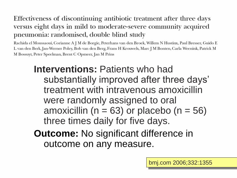
Interventions: Patients who had substantially improved after three days’ treatment with intravenous amoxicillin were randomly assigned to oral amoxicillin (n = 63) or placebo (n = 56) three times daily for five days.
Outcome: No significant difference in outcome on any measure.
bmj.com 2006;332:1355
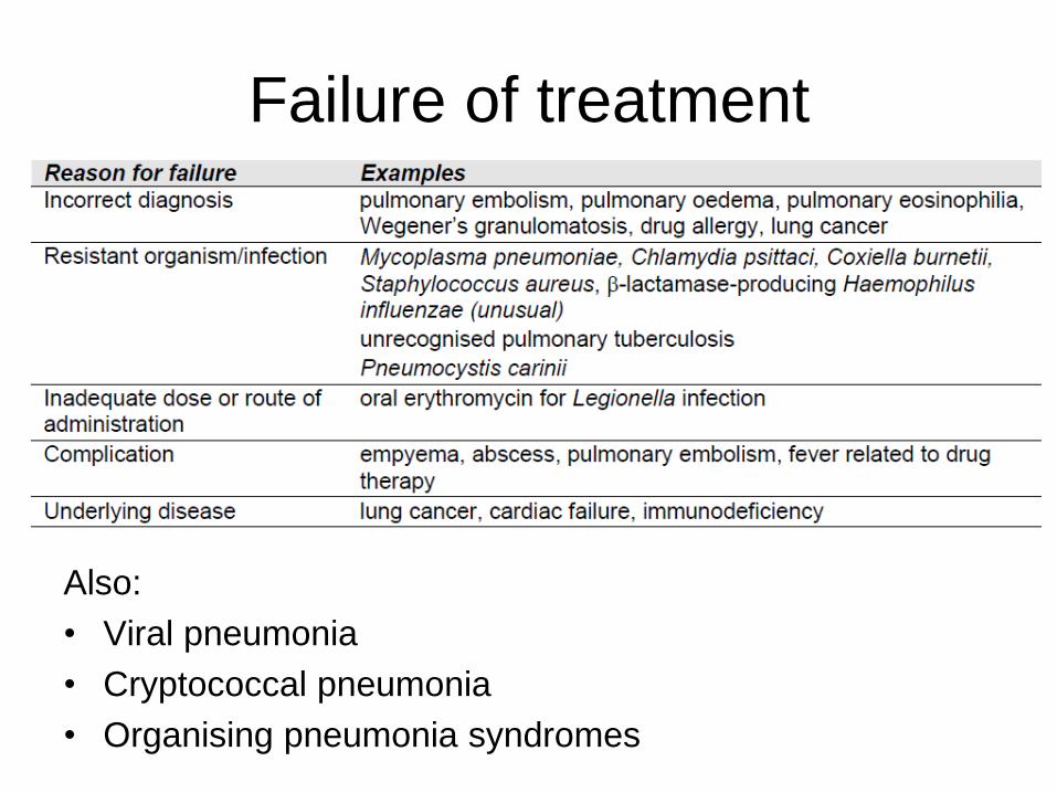
Failure of treatment
Also:
• Viral pneumonia
• Cryptococcal pneumonia
• Organising pneumonia syndromes
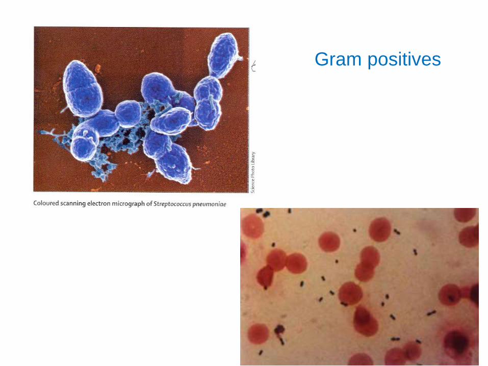
Gram positives
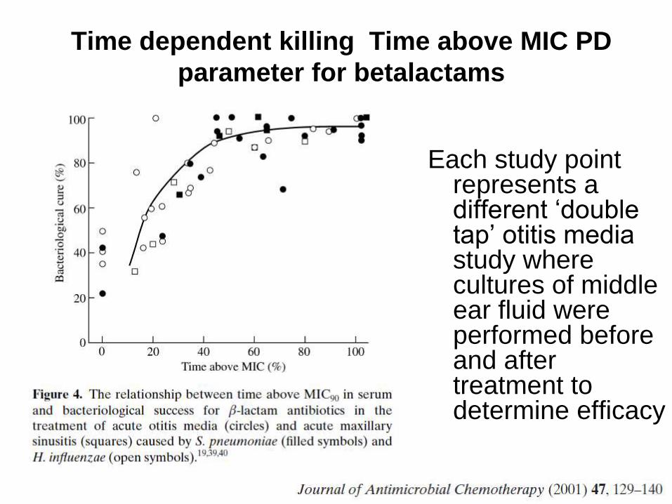
Time dependent killing Time above MIC PD
parameter for betalactams
Each study point represents a different ‘double tap’ otitis media study where cultures of middle ear fluid were performed before and after treatment to determine efficacy
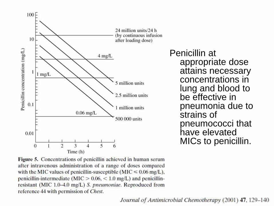
Penicillin at appropriate dose attains necessary concentrations in lung and blood to be effective in pneumonia due to strains of pneumococci that have elevated MICs to penicillin.
Streptococcus pneumoniae:
betalactam therapy • Community-acquired pneumonia
– evidence that treatment failures do NOT occur with benzylpenicillin or amoxycillin provided dosing is appropriate.
– Trial evidence fails to indicate superiority of IV over oral
• Ampicillin/amoxycillin
• Penicillin-G (benzylpenicillin).
• Oral penicillin-V (phenoxymethyl penicillin) NOT recommended
AVOID ceftriaxone in CAP- unless documented penicillin allergy- cefuroxime sufficient then
AVOID cephalexin/cephalothin/cefazolin –high level pneumococcal resistance possible
Other potential pneumococcal agents:
rates of (high-level) resistance*
• Doxycycline: 23%
• Erythomycin: 15%
• Co-trimoxazole: 89% (52% in another study)
• Vancomycin : all pneumococci susceptible;
reserve for MRSA pneumonia – high dosing
required (loading dose)
AVOID quinolones for CAP – best to reserve
their use for proven Gram negative infection or
MDR-TB etc
* Trop Med Int Health. 2009 Sep;14(9):1025-33. Study of carriage
of Strep. pneumoniae in rural children of Nepal.
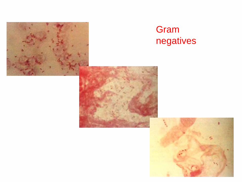
Gram
negatives
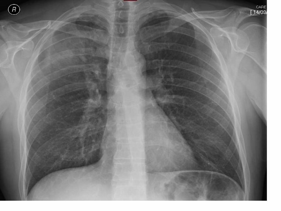
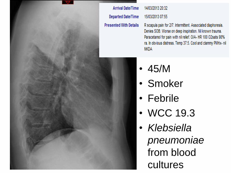
• 45/M
• Smoker
• Febrile
• WCC 19.3
• Klebsiella
pneumoniae
from blood
cultures
Aminoglycosides in Gram negative lung
infection
• Represent a good empiric option for pneumonia
where Gram negatives are suspected, provided
local susceptibility is high (above 75%) e.g.
– Neonate
– HIV infected
– Severe pneumonia case – until re-evaluation 48 hrs
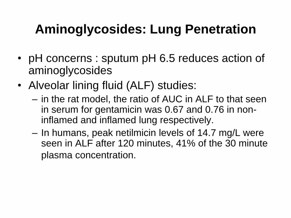
Aminoglycosides: Lung Penetration
• pH concerns : sputum pH 6.5 reduces action of aminoglycosides
• Alveolar lining fluid (ALF) studies: – in the rat model, the ratio of AUC in ALF to that seen
in serum for gentamicin was 0.67 and 0.76 in non-inflamed and inflamed lung respectively.
– In humans, peak netilmicin levels of 14.7 mg/L were seen in ALF after 120 minutes, 41% of the 30 minute
plasma concentration.
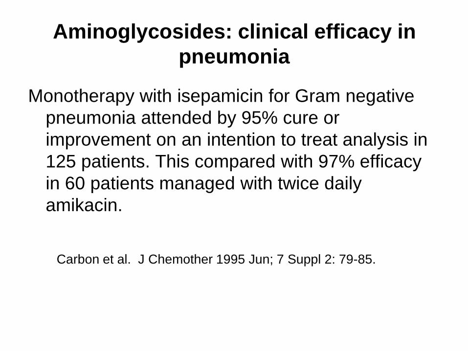
Aminoglycosides: clinical efficacy in
pneumonia
Monotherapy with isepamicin for Gram negative
pneumonia attended by 95% cure or
improvement on an intention to treat analysis in
125 patients. This compared with 97% efficacy
in 60 patients managed with twice daily
amikacin.
Carbon et al. J Chemother 1995 Jun; 7 Suppl 2: 79-85.
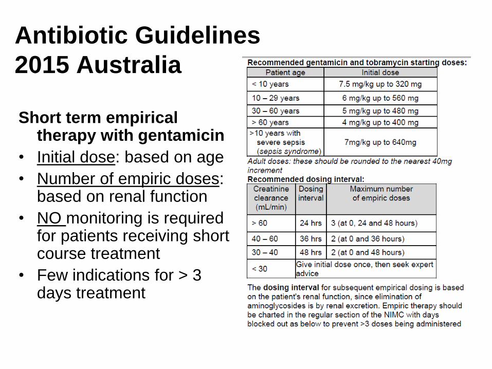
Antibiotic Guidelines
2015 Australia
Short term empirical therapy with gentamicin
• Initial dose: based on age
• Number of empiric doses: based on renal function
• NO monitoring is required for patients receiving short course treatment
• Few indications for > 3 days treatment
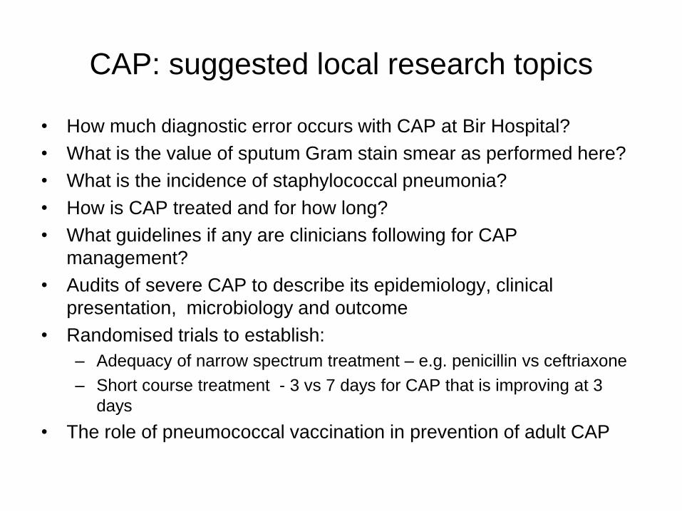
CAP: suggested local research topics
• How much diagnostic error occurs with CAP at Bir Hospital?
• What is the value of sputum Gram stain smear as performed here?
• What is the incidence of staphylococcal pneumonia?
• How is CAP treated and for how long?
• What guidelines if any are clinicians following for CAP
management?
• Audits of severe CAP to describe its epidemiology, clinical
presentation, microbiology and outcome
• Randomised trials to establish:
– Adequacy of narrow spectrum treatment – e.g. penicillin vs ceftriaxone
– Short course treatment - 3 vs 7 days for CAP that is improving at 3
days
• The role of pneumococcal vaccination in prevention of adult CAP
COPD patients
• Usually colonised with Haemophilus influenzae (non-
typeable) +/- Moraxella +/- pneumococcus
• Exacerbations – majority if not all are due to prior
respiratory viral infections; most do not exhibit CXR
consolidation
• During a viral exacerbation, overgrowth of colonisers
occurs- lab reports these and it drives unnecessary
antibiotic treatment
• Exacerbations have a self limited time course – up to 2
weeks; placebo controlled antibiotic trials indicate that
antibiotics provide minimal additional benefit
Approach for acute on chronic
airflow patients
• In admitted cases, AVOID prolonged antibiotics- MAX 3
days; chose penicillin/amoxycillin OR doxycycline to
target pneumococcus and maximise supportive care
• In presence of CXR-proven consolidation, treat as per
CAP – narrow spectrum – penicillin +/- doxycycline
• Outpatient cases – AVOID antibiotics
• Maximise non-antibiotic management measures- e.g.
treat coincident asthma, oxygen, physiotherapy, cease
smoking
COPD: suggested local research topics
• How much diagnostic error occurs with COPD exacerbations at Bir
Hospital? E.g. missed cardiac failure, asthma, other diagnosis
• How are outpatient COPD exacerbations treated and for how long?
• What guidelines if any are clinicians following for COPD
management?
• Audits of inpatients with exacerbation of COPD to describe
management and outcome
• Microbiological studies of prior viral causes of exacerbations
• Randomised trials to establish:
– Value of antibiotics in admitted cases – narrow spectrum vs placebo.
– Value of antibiotics in non-admitted cases – narrow spectrum vs placebo
– Value of certain interventions to reduce smoking or other causative factors
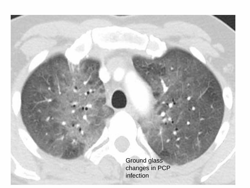
Ground glass
changes in PCP
infection
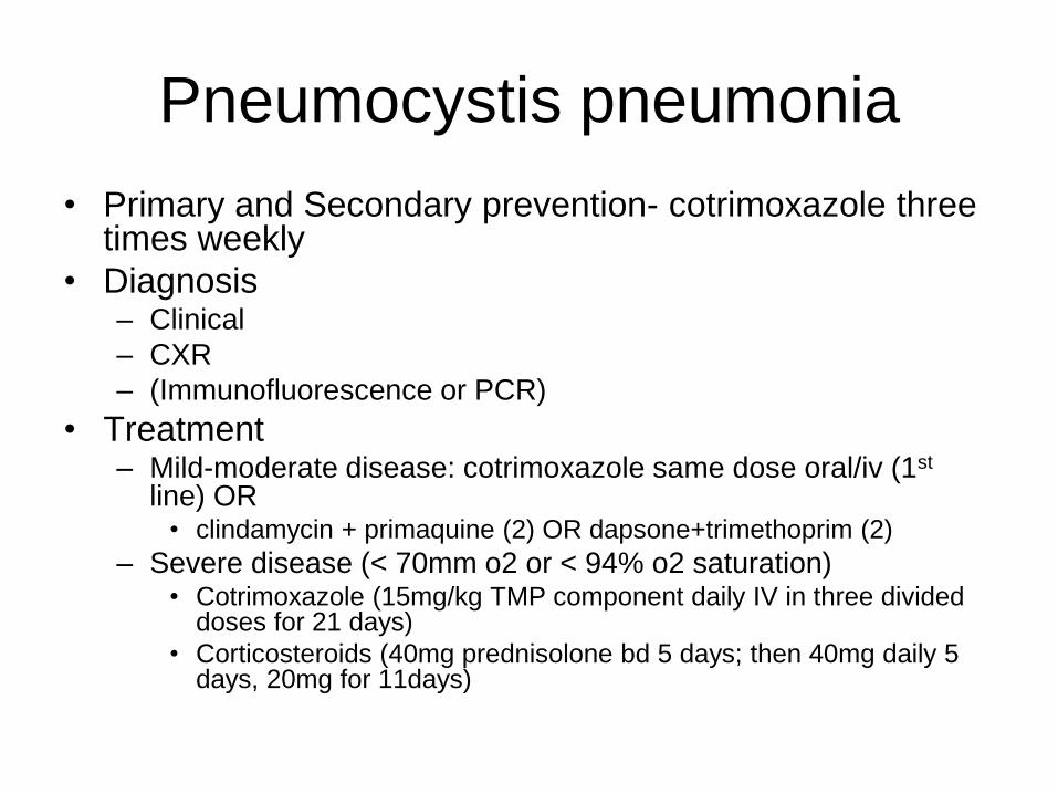
Pneumocystis pneumonia
• Primary and Secondary prevention- cotrimoxazole three times weekly
• Diagnosis – Clinical
– CXR
– (Immunofluorescence or PCR)
• Treatment – Mild-moderate disease: cotrimoxazole same dose oral/iv (1st
line) OR • clindamycin + primaquine (2) OR dapsone+trimethoprim (2)
– Severe disease (< 70mm o2 or < 94% o2 saturation) • Cotrimoxazole (15mg/kg TMP component daily IV in three divided
doses for 21 days)
• Corticosteroids (40mg prednisolone bd 5 days; then 40mg daily 5 days, 20mg for 11days)
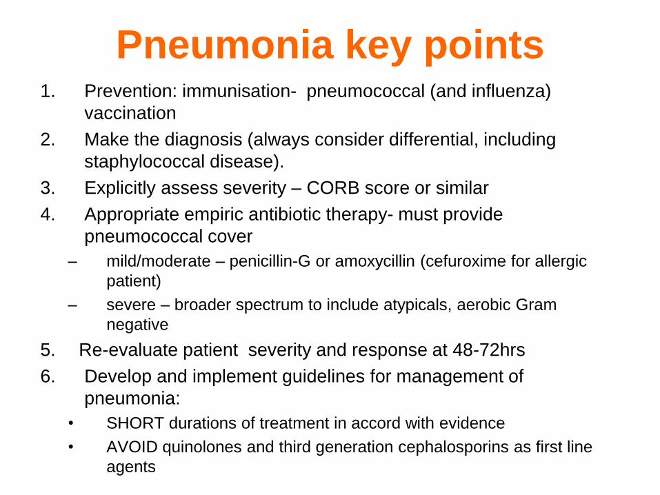
Pneumonia key points 1. Prevention: immunisation- pneumococcal (and influenza)
vaccination
2. Make the diagnosis (always consider differential, including
staphylococcal disease).
3. Explicitly assess severity – CORB score or similar
4. Appropriate empiric antibiotic therapy- must provide
pneumococcal cover
– mild/moderate – penicillin-G or amoxycillin (cefuroxime for allergic
patient)
– severe – broader spectrum to include atypicals, aerobic Gram
negative
5. Re-evaluate patient severity and response at 48-72hrs
6. Develop and implement guidelines for management of
pneumonia:
• SHORT durations of treatment in accord with evidence
• AVOID quinolones and third generation cephalosporins as first line
agents