Embed Size (px)
Citation preview
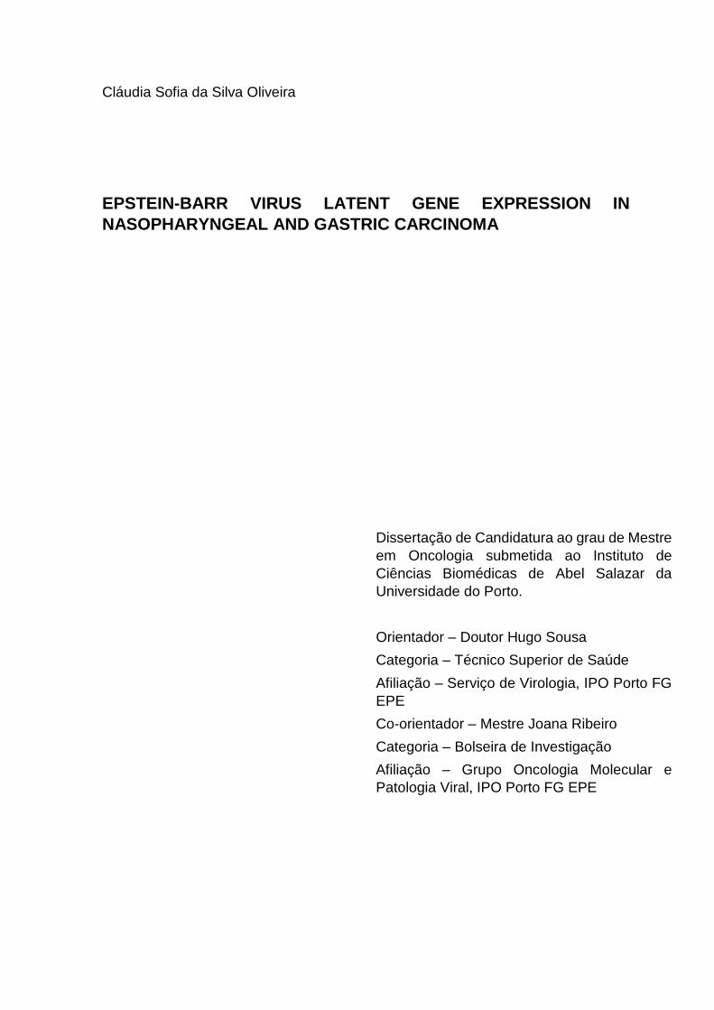
Cláudia Sofia da Silva Oliveira
EPSTEIN-BARR VIRUS LATENT GENE EXPRESSION IN
NASOPHARYNGEAL AND GASTRIC CARCINOMA
Dissertação de Candidatura ao grau de Mestre
em Oncologia submetida ao Instituto de
Ciências Biomédicas de Abel Salazar da
Universidade do Porto.
Orientador – Doutor Hugo Sousa
Categoria – Técnico Superior de Saúde
Afiliação – Serviço de Virologia, IPO Porto FG
EPE
Co-orientador – Mestre Joana Ribeiro
Categoria – Bolseira de Investigação
Afiliação – Grupo Oncologia Molecular e
Patologia Viral, IPO Porto FG EPE

Cláudia Oliveira | MSc Oncology
Epstein-Barr virus latent gene expression in nasopharyngeal and gastric carcinoma | I
PREFACE
This study was realized in the Molecular Oncology & Viral Pathology Group of
the Portuguese Oncology Institute of Porto (IPO Porto).
A systematic review of literature is being prepared to be published: Ribeiro J,
Oliveira C, Sousa H. EBV LATENCY PATTERN IN GASTRIC CARCINOMAS: A
SYSTEMATIC REVIEW.
The results obtained in this study are being prepared to be included in different
publications to be submitted in the near future.

Cláudia Oliveira | MSc Oncology
Epstein-Barr virus latent gene expression in nasopharyngeal and gastric carcinoma | III
AGRADECIMENTOS
Esta tese não teria certamente sido possível, sem o apoio, motivação e
encorajamento, que fui tendo ao longo da sua realização. Deixo aqui os meus
agradecimentos a todos, que de uma forma ou de outra, contribuíram para a sua execução.
Em primeiro lugar gostaria de agradecer ao meu orientador, Doutor Hugo Sousa
por me ter aceite no Grupo de Oncologia Molecular e Patologia Viral do Instituto Português
de Oncologia do Porto (IPO Porto FG EPE), pelos novos conhecimentos adquiridos, por
toda a ajuda e disponibilidade fornecida e por ter deste modo ter enriquecido o meu
percurso académico. Não menos importante, á minha co-orientadora, Mestre Joana
Ribeiro, por ter de igual forma ter contribuído para que este projeto fosse possível.
Queria também agradecer ao serviço de Anatomia Patológica do IPO-PORTO, em
especial ao Dr. Luis Pedro, á Dra. Ana Gallaghar, ao Dr. Manuel Jácome, á Técnica
Fernanda Silva e a Técnica Ana tavares, por toda a ajuda e disponibilidade que forneceram,
que tornaram este trabalho possível.
Às minhas companheiras de laboratório, Nádia Neto e Ana Bela Campos, pelos
momentos de desespero passados em conjunto, pelas horas infindáveis a moldar os sofás
da entrada, mas principalmente pelas gargalhadas e por todas as situações de
descontração que tornaram este ano muito mais fácil de aguentar. Um especial
agradecimento também á Mariana Malta, que na altura do aperto, foi uma ajuda preciosa
no laboratório.
Aos meus amigos, pelos convívios, pelas saídas, por todos os bons momentos, mas
mais importante do que isso, por todo o apoio que ao longo destes anos todos nunca falhou.
Aos meus pais e família, por terem permitido que eu realizasse todo este percurso
sem nunca terem duvidado de mim.
Por último, quero agradecer àqueles que ao longo deste ano aturaram todo o meu
mau humor e, que á sua maneira, sempre souberam sempre como o mudar.
A todos, um sincero obrigada!

Cláudia Oliveira | MSc Oncology
Epstein-Barr virus latent gene expression in nasopharyngeal and gastric carcinoma | V
RESUMO O EBV é um vírus associado a cancro que infecta cerca de 90% da população
mundial, sem causar sintomas na maioria dos portadores ao longo da vida. A infeção por
EBV pode adotar quatro diferentes padrões de latência, a latência 0, I, II, e III, que parecem
estar correlacionados com os diferentes tipos de doenças associadas ao EBV. . No entanto,
há algumas dúvidas sobre a expressão de genes latentes do EBV em diferentes doenças
tais como o carcinoma nasofaríngeo e o carcinoma gástrico.
Foi realizado um estudo retrospetivo com 23 casos de carcinoma da nasofaringe e
9 casos de carcinoma gástrico associados ao EBV, a fim de avaliar a expressão de
proteínas do EBV nos diferentes tumores e tentar estabelecer uma correlação clínica entre
a latência viral e a malignidade. A deteção do EBV foi feita por EBER-IHS e a deteção de
proteínas do EBV (LMP1 e LMP2a) foi realizada por imuno-histoquímica. Todos os casos
utilizados neste estudo foram EBV positivos.
LMP1 esteve presente em 95,5% dos casos de carcinoma da nasofaringe, mas
nenhum dos casos de carcinoma gástrico mostrou ter expressão LMP1. Em relação á
LMP2a, ela foi expressa em 100% dos casos de carcinoma da nasofaringe, enquanto no
carcinoma gástrico estava presente em 44,4%. A expressão proteica de cada um dos
tumores levou a diferentes tipos de latência. No carcinoma da nasofaringe, 21 casos
(95,5%), apresentaram expressão de LMP1 e LMP2a, o que corresponde a um padrão de
latência II, e apenas 1 caso (4,5%) expressou um padrão de latência distinto semelhante à
latência II, mas sem a expressão de LMP1. No carcinoma gástrico, 5 casos (55,5%) não
apresentavam expressão de LMP1 e LMP2a, que corresponde a um padrão de latência I.
Os restantes 4 casos, tiveram o mesmo padrão de latência que um caso de carcinoma da
nasofaringe onde há expressão de LMP2a, mas não de LMP1. Esses dados confirmam os
padrões de latência associados ao carcinoma da nasofaringe (latência II) e com o
carcinoma gástrico (latência I). Identificou-se também um padrão de latência novo e ainda
não aceite (latency II-like) que é especialmente importante no carcinoma gástrico, e é
caracterizado por a expressão de EBNA 1 e LMP2a.
Este foi o primeiro estudo realizado em Portugal para caracterizar os padrões de
latência do EBV em diferentes neoplasias. Em conclusão, mesmo com a confirmação que
o carcinoma da nasofaringe expressa tipicamente um padrão Latência II e o carcinoma
gástrico a latência I, observou-se também que alguns casos expressam um perfil de
latência diferente. Portanto, há uma necessidade de uma determinação correta da
expressão de proteínas do EBV para um conhecimento correto sobre os mecanismos de

Cláudia Oliveira | MSc Oncology
VI | Epstein-Barr virus latent gene expression in nasopharyngeal and gastric carcinoma
transformação. A literatura suporta a necessidade de reestruturação dos padrões de
latência, considerando a expressão de todos os tipos de expressão de proteínas.
Cláudia Oliveira | MSc Oncology
Epstein-Barr virus latent gene expression in nasopharyngeal and gastric carcinoma | VII
ABSTRACT
EBV is a human cancer-associated virus that infects about 90% of the global
population, without causing major symptoms in the majority of lifelong carriers trough the
establishment of a viral latent state. EBV infection can adopt four different patterns of
latency, named type 0, I, II, and III, which seem to be correlated with the different types of
EBV associated disease. Nevertheless, there is some doubt on the differential expression
of EBV latent genes in different diseases such as Nasopharyngeal Carcinoma and Gastric
Carcinoma.
We have performed a retrospective study with 23 NPC and 9 EBVaGC cases, in
order to evaluate the expression of EBV proteins in different tumors and try to establish a
clinical correlation between the viral latency and the malignances. The identification of EBV
was made by EBER-ISH and the detection of EBV proteins (LMP1 and LMP2a) was
performed by immunohistochemistry. All cases used in this study were EBV positive
LMP1 was present in 95.5% of NPC cases, but in none EBVaGC was shown LMP1
expression. Regarding the LMP2a, it was expressed in 100% of NPC cases, while in GC it
was present in 44.4%. The protein expression that each of the tumour presented led to
different latency types. In NPC, 21 cases (95.5%), had the expression of both LMP1 and
LMP2a, which corresponds to a latency II pattern, and only 1 case (4.5%) expressed a
distinct latency pattern similar to latency II, but without the expression of LMP1. In GC, 5
cases (55.5%) had no expression of LMP1 and LMP2a, wich corresponds to a latency I
pattern. The remaining 4 cases, had the same latency pattern as one case of NPC were
there is expression of LMP2a, but not LMP1. These data confirm the latency patterns
associated with NPC (latency II) and with EBVaGC (latency I). We also identified a novel
and yet not accepted latency (latency II-like) that is especially important in EBVaGC,
characterized by the expression of EBNA1 and LMP2a.
This was the first study in Portugal to characterize the latency patterns of EBV in
different diseases. In conclusion, even with the confirmation of NPC as typically expressing
a Latency II pattern and EBVaGC a latency I, we observed that some cases express a
different latency profile. Hence, there is an urge in the correct determination of EBV profile
expression for a correct knowledge on the mechanisms of transformation. The literature
supports the need for restructuring the latency patterns, considering the expression of all
types of protein expression.

Cláudia Oliveira | MSc Oncology
Epstein-Barr virus latent gene expression in nasopharyngeal and gastric carcinoma | IX
ABREVIATIONS LIST
BARTs - BamHI A rightward transcripts
B-CLL - B-Chronic Lymphocytic Leukemia
BL- Burkitt’s Lymphoma
CSF - colony stimulating factor
EBNAs - Epstein Barr Nuclear Antigens
EBERs - Epstein-Barr Virus-encoded RNAs
eBL - Endemic BL
EBV - Epstein-Barr virus
EBVaGC - Epstein-Barr virus associated gastric carcinoma
GC - gastric cancer
gH - glycoprotein H
GIST - gastrointestinal stromal tumours
gM - glycoprotein M
gN - glycoprotein N
HHV4 - Human Herpesvirus 4
HIV - human immunodeficiency vírus
HL - Hodgkin lymphoma
HSV - herpes simplex virus
IHC - immunohistochemistry
IM - Infectious mononucleosis
IRs - internal repeat sequences
Kb - kilobase pairs
LELC - lymphoepithelioma-like gastric carcinomas
Cláudia Oliveira | MSc Oncology
X | Epstein-Barr virus latent gene expression in nasopharyngeal and gastric carcinoma
LMPs - Latent Membrane Proteins
LPHL - lymphocyte-predominant
M-CSF - macrophage colony-stimulating factor
MHC - Major histocompatibility complex
miR-BARTs – EBV microRNAs
miRNAs - microRNAs
NET - neuroendocrine tumours
NPC - nasopharyngeal carcinoma
ori-lyt - lytic origin of replication
PML - Promyelocytic Leukemia gene
PTLD - Post-transplant lymphoproliferative disease
sBL - Sporadic Burkitt’s lymphoma
STAT1 - activation of transcription 1
TF - transcription factor
TLR3 - Toll-like receptor 3
TRs - terminal direct repeats
VCA - viral capsid antigen
WHO - World Health Organization
ZEBRA - Z Epstein–Barr replication activator
Cláudia Oliveira | MSc Oncology
Epstein-Barr virus latent gene expression in nasopharyngeal and gastric carcinoma | XI
FIGURE LIST
Figure 1: EBV virion structure
Figure 2: Map of EBV genome
Figure 3: Interactions between Epstein–Barr virus and host cells
Figure 4: EBV lytic cycle activation
Figure 5: EBV latent genes target cancer hallmarks
Figure 6: Map of Africa showing the 'lymphoma belt' in which eBL occurs at high
incidence.
Figure 7: Hodgkin Lymphoma incidence worldwide, both sexes, all ages,
Figure 8: Hodgkin Lymphoma body locations
Figure 9: Nasopharynx location
Figure 10: Nasopharyngeal carcinoma incidence worldwide, both sexes, all ages,
Figure 11: Gastric cancer incidence worldwide, both sexes, all ages,
Figure 12: Illustrative scheme of the stomach regions
Figure 13: EBV cycle and latent states
Figure 14: EBER-IH results
Figure 15: LMP1results
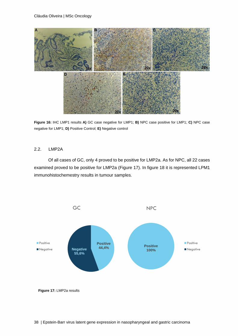
Figure 16: IHC LMP1 results
Figure 17: LMP2a results
Figure 18: IHC LMP2a results

Cláudia Oliveira | MSc Oncology
Epstein-Barr virus latent gene expression in nasopharyngeal and gastric carcinoma | XIII
TABLE LIST
Table 1: Overview of Burkitt’s lymphoma clinical variants.
Table 2: Reported incidence of PTLD by organ system and recipient age
Table 3: Comparison of Lauren’s and WHO classification systems
Table 4: EBV gene latency programmes
Table 5: Characterization of Nasopharingeal carcinoma cases
Table 6: Characterization of gastric cancer cases
Table 7: Antibodies and conditions used for the detection of different EBV proteins
Table 8: Description of nasopharyngeal cancer cases
Table 9: Description of gastric cancer cases
Table 10: Frequency of EBV latent proteins observed in different studies
Table 11: Frequency of EBV latency patterns observed in different studies

Cláudia Oliveira | MSc Oncology
Epstein-Barr virus latent gene expression in nasopharyngeal and gastric carcinoma | XV
INDEX
PREFACE _______________________________________________________ I
AGRADECIMENTOS ______________________________________________ III
RESUMO _______________________________________________________ V
ABSTRACT ____________________________________________________ VII
ABREVIATIONS LIST _____________________________________________ IX
FIGURE LIST ____________________________________________________ XI
TABLE LIST ____________________________________________________ XIII
INDEX ________________________________________________________ XV
I. INTRODUCTION ______________________________________________ 1
1. Epstein-Barr Virus ________________________________________________ 3
1.1. Historical Background__________________________________________________ 3 1.2. EBV Structure and Genome _____________________________________________ 3 1.3. EBV Infection ________________________________________________________ 5 1.4. Viral gene expression __________________________________________________ 6
1.4.1. Lytic genes ______________________________________________________ 6 1.4.1.1. Immediate-Early Lytic Proteins _____________________________________ 6 1.4.1.2. Early Lytic Proteins ______________________________________________ 7 1.4.1.3. Late Lytic Proteins ______________________________________________ 8
1.4.2. Latent genes _____________________________________________________ 8 1.4.2.1 EBV-nuclear antigens _________________________________________ 9 1.4.2.1. Latent membrane proteins ____________________________________ 10
1.4.3. Other transcripts __________________________________________________ 11
2. EBV-Associated Malignancies ______________________________________ 13
2.1. Burkitt’s Lymphoma __________________________________________________ 13 2.2. Hodgkin’s Lymphoma _________________________________________________ 15 2.3. Post-transplant lymphoproliferative disease _______________________________ 16 2.4. Nasopharyngeal Carcinoma ____________________________________________ 17 2.5. Gastric Carcinoma ___________________________________________________ 19
3. EBV Latency ____________________________________________________ 21
3.1. Latency 0 __________________________________________________________ 21 3.2. Latency I ___________________________________________________________ 21 3.3. Latency II __________________________________________________________ 22 3.4. Latency III __________________________________________________________ 22 3.5. Other types of latency_________________________________________________ 22
II. AIMS OF THE STUDY _________________________________________ 25
III. MATERIAL AND METHODS __________________________________ 29
1. Population and Type of study ______________________________________ 29
2. Sample collection and processing ____________________________________ 30
3. EBV detection (EBER-ISH) __________________________________________ 30
4. EBV proteins expression analysis _____________________________________ 32
Cláudia Oliveira | MSc Oncology
XVI | Epstein-Barr virus latent gene expression in nasopharyngeal and gastric carcinoma
5. Quality control ____________________________________________________ 33
6. Data analysis ____________________________________________________ 33
IV. RESULTS _________________________________________________ 37
1. EBER-ISH _______________________________________________________ 37
2. Protein expression _______________________________________________ 37
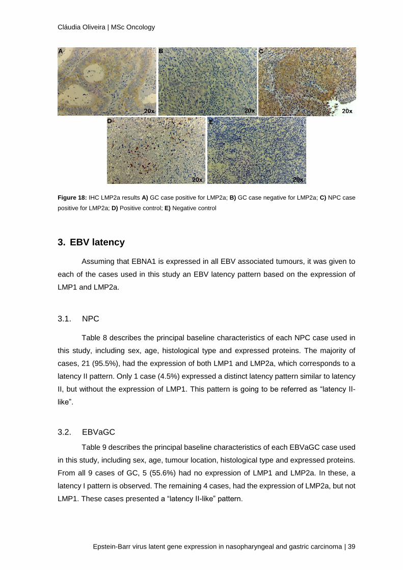
2.1. LMP1 _____________________________________________________________ 37 2.2. LMP2A ____________________________________________________________ 38
3. EBV latency _____________________________________________________ 39
3.1. NPC ______________________________________________________________ 39 3.2. EBVaGC ___________________________________________________________ 39
V. DISCUSSION ______________________________________________ 39
VI. CONCLUSION _____________________________________________ 47
VII. REFERENCES _____________________________________________ 51
VIII. APPENDIX ________________________________________________ 63

I. INTRODUCTION

Cláudia Oliveira | MSc Oncology
Epstein-Barr virus latent gene expression in nasopharyngeal and gastric carcinoma | 3
1. Epstein-Barr Virus
1.1. Historical Background
In 1958, Denis Burkitt identified a frequent cancer among children in Equatorial
Africa [1, 2]. This tumour was dependent on climatic and geographical conditions, which led
to the possibility that it could be associated with a vector-borne agent [1]. Later, in 1964,
Anthony Epstein, Yvonne Barr and Bert Achong using electronic microscopy discovered
typical herpesvirus particles in biopsies of the named Burkitt’s Lymphoma (BL), which they
have called Epstein-Barr Virus (EBV). In the late 1960s, studies revealed that BL patients
had higher antibody titers to EBV antigens [3, 4] These serological assays also allowed the
identification of EBV as the etiological agent of infectious mononucleosis (IM) and
nasopharyngeal carcinoma (NPC) [5, 6].
1.2. EBV Structure and Genome
EBV also recognized as Human Herpesvirus 4 (HHV4), belongs to the Herpesviridae
family, Gammaherpesvirinae subfamily and is the only human virus from the
Lymphocryptovirus genus [7]. The virion of EBV is about 120-300 nm in diameter and
consists of a toroid shaped protein core wrapped with linear double stranded DNA with
approximately 172 kilobase pairs (kb) in an icosahedral nucleocapsid with 162 capsomeres,
an outer envelope with external glycoprotein spikes and a tegument protein between the
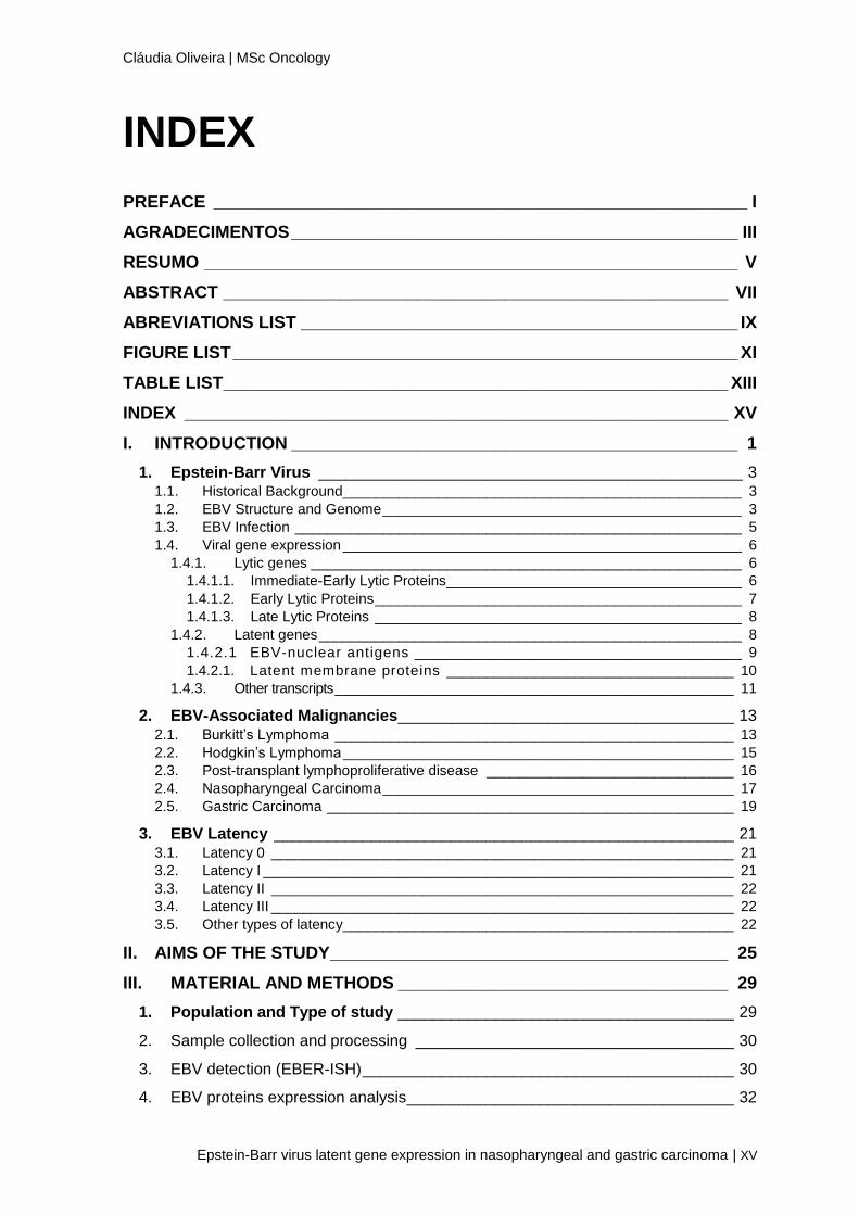
nucleocapsid and envelope (Figure 1) [8-11].
Figure 1: EBV virion structure (adapted from http://viralzone.expasy.org/viralzone/all_by_species/185.html)
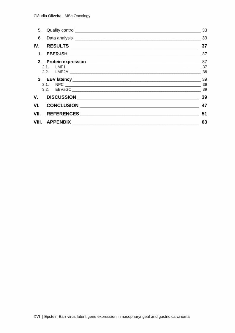
EBV genome has several terminal repeats (TRs) and internal repeat sequences (IRs)
that divide the genome into short and long unique sequence domains (Figure 2) [12]. The
EBV genome is linear, but once it reaches the nucleus of the infected cell, it adopts an
Cláudia Oliveira | MSc Oncology
4 | Epstein-Barr virus latent gene expression in nasopharyngeal and gastric carcinoma
episomal structure through the binding of the TRs, which is required for replication of the
viral genome [13].
Only a very small fraction of the EBV genome that enters the cell is able to reach the
nucleus and an even smaller fraction is able to circularize. During cellular proliferation, linear
viral genomes are gradually lost while circular DNA is maintained [14, 15] Indeed, during
latency, EBV DNA acts like the celular DNA, being associated with histones and replicating
once during the S phase, depending only on the cellular machinery and being equally
transmitted to daughter cells [13].
By gene sequencing of different EBV isolates it was possible to identify two different
EBV subtypes, which were classified as type 1 and type 2 (or type A and type B,
respectively) [16]. The difference between these two subtypes are mainly on genetic
polymorphism in the Epstein Barr Nuclear Antigens (EBNAs), but other single base changes
may be observed in other regions of the genome [9, 17-19]. Type 1 is observed in almost
all populations, being predominant in Europe, America, South America and Asia; while type
2 is mainly found in Central Africa, New Guinea and in Alaskan Eskimos [20]. The influence
of these EBV subtypes in disease development is not yet understood, but type 1 virus
seems to prevail in most EBV positive associated diseases, whereas type 2 is mainly
associated with immunocompromised patients [21].
Figure 2: Map of EBV genome (Straus, 1993)

Cláudia Oliveira | MSc Oncology
Epstein-Barr virus latent gene expression in nasopharyngeal and gastric carcinoma | 5
1.3. EBV Infection
EBV infection is restricted to humans, and is transmitted almost exclusively by saliva
[5]. The primary infection usually occurs early in life and the family is frequently responsible
for the transmission, although in developed countries this infection can be delayed until
adolescence or adulthood and here it can lead to the development of a strong immune
response medically known as IM [5, 22, 23]. After controlled, the infection is asymptomatic
due to the establishment of a viral latency within the memory B lymphocytes [24]. In fact,
EBV establishes a persistent lifelong infection, in most cases without consequences, in
more than 90% of the world population [25, 26].
It is assumed that the primary infection occurs in the oropharynx, due to the interaction
of the viral membrane glycoprotein gp85/42, with a Major histocompatibility complex (MHC)
class II receptor on the surface of lymphoepithelial cells of the Waldeyer's ring [27].
Subsequently, EBV starts a short period of lytic replication, after which it is released into the
saliva and infects adjacent epithelial cells (Figure 3) [27].
Figure 3: Interactions between Epstein–Barr virus and host cells (Young, 2004)
Cláudia Oliveira | MSc Oncology
6 | Epstein-Barr virus latent gene expression in nasopharyngeal and gastric carcinoma
Then, after the infection of epithelial cells in the oropharynx, EBV infects immature B
lymphocytes that are nearby, process only possible due to the binding of EVB's membrane
glycoprotein gp350 / 220 with the CD21 molecule of B lymphocytes [28]. The infected B-
lymphocytes will be transformed into lymphoblastoid cells with latent EBV, proliferating
without control. Many of these proliferating cells are killed by cytotoxic T-lymphocyte
response, nevertheless some escape through downregulation of antigen expression and
the establishment of a stable reservoir of memory B lymphocytes where viral antigen
expression is almost nonexistent [29].
1.4. Viral gene expression
As all other herpesviruses, EBV has a life cycle with different viral gene expression
programs: a latent one, on which no viral particles are formed; and a lytic one, on which
new infectious viruses are produced [16]. There are more than 90 viral proteins coded by
the EBV genome and the great majority has no known function [30].
1.4.1. Lytic genes
EBV establishes a latent persistent lifelong infection in most cases, nevertheless, for
reasons not yet understood, the virus can reactivate and restart its lytic cycle in specific
conditions [24]. By analogy with other herpesviruses, the proteins expressed in this phase
are classified as immediate-early, early, and late lytic proteins: Immediate-early genes are
transcribed after infection in the presence of protein synthesis inhibitors; Early genes are
expressed in the presence of viral DNA synthesis inhibitors; and late genes are not
transcribed when these inhibitors are present.
EBV lytic genes are named by BamHI fragment within which they are located, whether
they are expressed in a leftward (L) or rightward direction (R), and the number of their
position in the BamHI fragment. For example, BZLF1 is the first transcript expressed in the
leftward direction in the BamHI Z fragment of EBV (Figure 4) [31].
1.4.1.1. Immediate-Early Lytic Proteins
Immediate-early genes are important for regulating gene expression in the virus.
The major immediate-early proteins of EBV are encoded by BZLF1 and BRLF1, also named
by Z Epstein–Barr replication activator (ZEBRA) or Zta and Rta, respectively. BZLF1 and
BRLF1 proteins activate transcription of viral early genes [32, 33]. BZLF1 protein inhibits
transcription from the EBNA Cp promoter and may facilitate the switch from latent to lytic
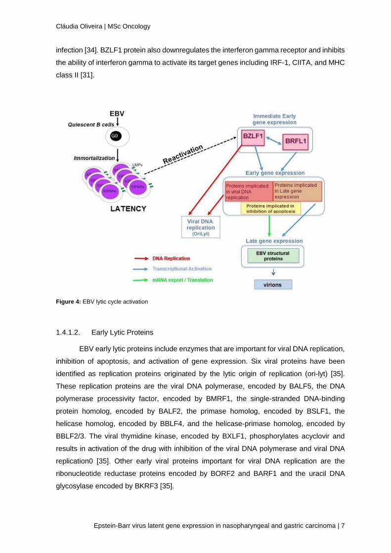
Cláudia Oliveira | MSc Oncology
Epstein-Barr virus latent gene expression in nasopharyngeal and gastric carcinoma | 7
infection [34]. BZLF1 protein also downregulates the interferon gamma receptor and inhibits
the ability of interferon gamma to activate its target genes including IRF-1, CIITA, and MHC
class II [31].
Figure 4: EBV lytic cycle activation
1.4.1.2. Early Lytic Proteins
EBV early lytic proteins include enzymes that are important for viral DNA replication,
inhibition of apoptosis, and activation of gene expression. Six viral proteins have been
identified as replication proteins originated by the lytic origin of replication (ori-lyt) [35].
These replication proteins are the viral DNA polymerase, encoded by BALF5, the DNA
polymerase processivity factor, encoded by BMRF1, the single-stranded DNA-binding
protein homolog, encoded by BALF2, the primase homolog, encoded by BSLF1, the
helicase homolog, encoded by BBLF4, and the helicase-primase homolog, encoded by
BBLF2/3. The viral thymidine kinase, encoded by BXLF1, phosphorylates acyclovir and
results in activation of the drug with inhibition of the viral DNA polymerase and viral DNA
replication0 [35]. Other early viral proteins important for viral DNA replication are the
ribonucleotide reductase proteins encoded by BORF2 and BARF1 and the uracil DNA
glycosylase encoded by BKRF3 [35].
Cláudia Oliveira | MSc Oncology
8 | Epstein-Barr virus latent gene expression in nasopharyngeal and gastric carcinoma
1.4.1.3. Late Lytic Proteins
Late genes encode structural proteins of the virion: glycoproteins, nucleocapsid
proteins, and a viral cytokine [36]. Most of the viral capsid antigen (VCA) is comprised of
the major nucleocapsid protein, which is encoded by BcLF1 [36].
EBV encodes several glycoproteins including gp350, gp110, gp85, gp42, and gp25 [37].
The gp350, encoded by BLLF1, is the major viral envelope protein and it is able to bind
CD21 contributing to the virus entry into the host B-cells. Deletion of gp350 from the virus
markedly reduces the infectivity of the virus, and therefore purified recombinant gp350 is
being studied as a vaccine candidate [37]. EBV gp110, encoded by BALF4, is the homolog
of herpes simplex virus (HSV) glycoprotein B, which is required for HSV entry into cells. The
three remaining EBV glycoproteins, gp85, gp42, and gp25, form a trimolecular complex,
which is responsible for virion penetration of the B-cell membrane [38]. EBV gp85, encoded
by BXLF2, is the homolog of HSV glycoprotein H (gH), which is essential for the fusion of
the virus to B-cells and absorption to epithelial cells [39, 40]. EBV gp25, the product of
BKRF2, acts as a viral chaperone to transport gp85 to the cell membrane [41], and gp42,
encoded by BZLF2, binds to MHC class II molecules [42] and functions as a co-receptor for
virus entry in B-cells [43]. EBV also encodes homologs of HSV glycoprotein N (gN),
encoded by BLRF1, and glycoprotein M (gM), encoded by BBRF3, that are important for
egress of virus from the cell [44].
BCRF1 protein, also termed viral IL-10, shares over 80% amino acid identity with human
IL-10 [45]. This viral IL-10 inhibits interferon gamma secretion by peripheral blood
mononuclear cells and release of IL-12 from macrophages protecting virus-infected cells
from cytotoxic T-cells. [46]. Viral IL-10 also stimulates growth of B-cells and inhibits the
activity of dendritic cells [47, 48].
1.4.2. Latent genes
From all viral proteins coded by EBV genome, only a restricted group may be expressed
in different combinations during latency. [49]. Therefore, latency results from a tight interplay
between viral and host transcription factors, leading to the distinct use of three viral
promoters (Cp, Wp and Qp) which regulate the transcription of different viral genes [50].
The viral genes expressed during latency include: six EBV Nuclear Antigens (EBNA1, 2,
3A, 3B, 3C and LP); three Latent Membrane Proteins (LMP1, LMP2A. and LMP2B); two
small nonpolyadenylated Epstein-Barr Virus-encoded RNAs (EBER1 and 2) and highly
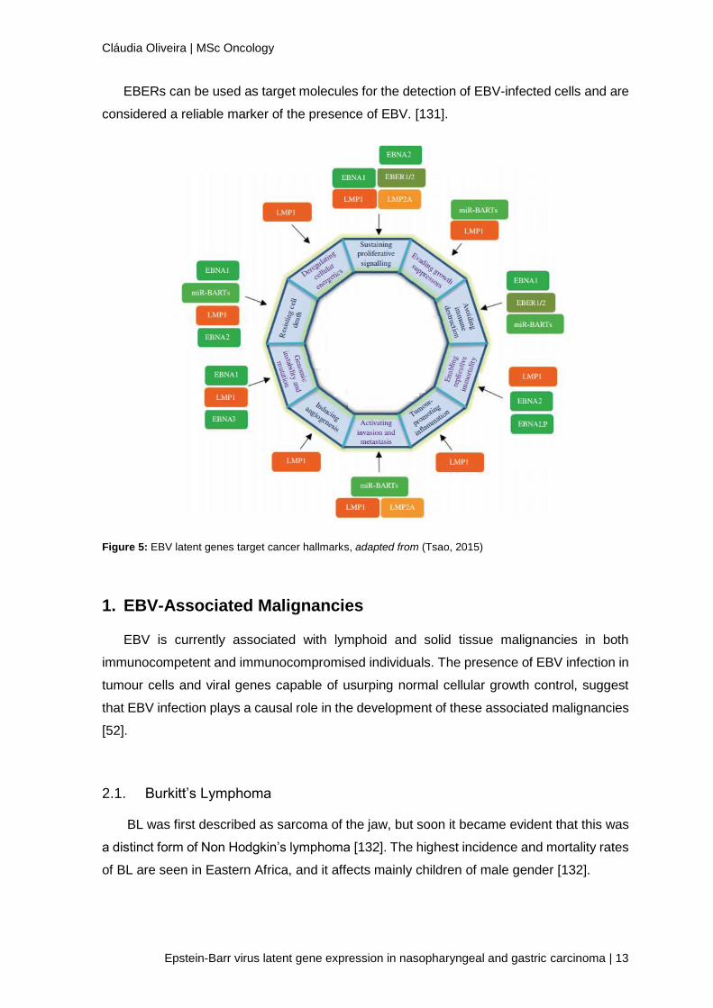
spliced BamHI A rightward transcripts (BARTs) (Figure 5) [13, 51, 52].
Cláudia Oliveira | MSc Oncology
Epstein-Barr virus latent gene expression in nasopharyngeal and gastric carcinoma | 9
1.4.2.1 EBV-nuclear antigens
EBNA1
EBNA1 is the only nuclear EBV antigen expressed in both latent and lytic modes
and it is essential for efficient EBV genome replication, persistence and transcription in
dividing cells. [53, 54]. This protein is expressed in all virus-associated tumours [55, 56].
Literature have referred several functions to EBNA1, such as binding to viral DNA elements
and cellular promoters, activation EBV viral Cp and Wp promoters, inhibition of Qp
promoters, upregulation signal transducers and activation of transcription 1 (STAT1),
downregulation of tumour growth factor-β signaling pathways, reduction of SMAD2,
upregulation of CCL20 [57], inhibition the canonical NF-KB pathway, and enhancing activity
of the AP-1 transcription factor (TF) [56-63].
EBNA1 contributes to the latent infection as it suppresses spontaneous lytic reactivation
and induces a family of microRNAs (miRNAs), which in turn decrease the level of cellular
protein Dicer, inhibiting reactivation of latent EBV [64, 65]. EBNA1 has also been recognized
as inducing the loss of Promyelocytic Leukemia gene (PML) nuclear bodies, and decreased
p53 activation and apoptosis in response to DNA damage in NPC and EBV-associated
gastric cancer (EBVaGC) [66].
E B N A 2
EBNA2 exists in two allelic forms, which differ in the amino acid sequence of the
central region of the molecule [67]. Interestingly, each EBV subtype has a different EBNA2
allelic form. This may probably account for the differences observed in the growth transformation
abilities of each EBV subtype. [68].
EBNA2 expression results from the same pre-mRNA as EBNA-LP, after an
alternative splicing. These two viral proteins are the first EBV proteins expressed during EBV
infection and B cells transformation, nevertheless, only EBNA2 and LMP1 essential for.EBV-
induced immortalization [69, 70]. EBNA2 by itself is responsible for the expression of
several proteins such as LMP1, LMP2A, LMP2B and also the ones arising from the EBNA's
Cp promoter [71-74]. EBNA2 activation of gene expression does not occur though direct
binding of EBNA2 to DNA, but rather through the interaction with the cellular CBF1 (RBP-
J11) protein complex. One important target of EBNA2 is the cellular oncogene c-Myc, this
link may be relevant for EBV induced proliferation and immortalization of B lymphocytes
[75].
Cláudia Oliveira | MSc Oncology
10 | Epstein-Barr virus latent gene expression in nasopharyngeal and gastric carcinoma
EBNA3 FAMILY
There are three EBNA3 proteins, EBNA3A, EBNA3B, and EBNA3C. Studies have
demonstrated that, in contrast with EBNA3B, EBNA3A and EBNA3C are required for B-cell
transformation [76, 77]. The EBNA3 proteins are responsible for increased expression of
both cellular and viral genes. EBNA3C increases the expression of CD21 and co-activates
the LMP1 promoter in conjunction with EBNA2, it interacts with the human metastatic
suppressor protein Nm23-HI and inhibits the ability of the latter to suppress the migration of
BL cells [78]. EBNA3C has also been shown to mediate the degradation of the
retinoblastoma protein (pRb), with the assistance of the SCFSKP2 complex, in transiently or
stably transfected cells [79, 80]. On the other hand, EBNA3B seems to upregulate CD40
and bcl-2 expression [81-83]. These funtions reforce the ability of EBNA3C family in the
maintenance of EBV latency and transformation [81-83].
EBNA-LP
Even though the role of EBNA-LP in EBV-induced B-cell transformation is still uncertain,
it is thought that it plays an important role in the establishment of B-cell immortalization,
once mutant viruses lacking the unique carboxy-terminal domain, are much less efficient at
immortalizing B cells than wild-type viruses [84, 85]. EBNALP coactivates EBNA2
enhancing the expression of the major viral oncoprotein LMP1 [85, 86]. While EBNA-LP has
been shown to bind to p53 and the retinoblastoma protein, it is unclear what significance
these interactions have for the role of EBNALP in B-cell transformation [87, 88]. In addition,
EBNA-LP binds to a number of other cellular proteins including heat shock protein 70, DNA
protein kinase catalytic subunit, HA95 a nuclear protein that may be involved in mitosis, and
in a and b tubulin [87, 89].
In newly infected B cells, a range of EBNA-LP isoforms are expressed, but over time,
the number of expressed isoforms decreases [85]. In addition, EBNA-LP localizes diffusely
throughout the nucleus within the hours post-infection, but with time, it associates with PML
nuclear bodies [90].
1.4.2.1. Latent membrane proteins
LMP1
LMP1 develops a key role in the immortalization of B cells and is considered a major
EBV oncoprotein [91, 92]. It is a membrane protein composed of six hydrophobic clusters
forming three membrane spanning domains connected to a short N-terminal cytoplasmic
domain and also to a long C-terminal cytoplasmic tail of 200 amino acids [93, 94]. It is
Cláudia Oliveira | MSc Oncology
Epstein-Barr virus latent gene expression in nasopharyngeal and gastric carcinoma | 11
relatively highly expressed in most EBV associated tumours, but its expression is rare in
EBV infected healthy individuals [18, 95].
Several studies have demonstrated the LMP1 is involved in proliferation, apoptosis,
angiogenesis, invasion and modulation of immune response, leading to dysregulation of
various cellular pathways in tumour cells, and also affecting the tumour microenvironment [95-
97]. Thus, LMP1 is highly regulated and presents various functions in different cellular
processes [94, 95, 97, 98]. Although it is a latent protein, its transcription is also observed
during lytic infection [99, 100].
L M P 2
LMP2A and LMP2B are LMP2 isoforms transcribed from 2 different promoters and
differing only in the first exon [101, 102]. Both LMP2A and LMP2B are membrane proteins
responsible for the maintenance of EBV latency and pathogenicity [101, 103].
LMP2A is known to regulate several signaling pathways, like proliferation and survival
of B cells even in the absence of normal BCR [104, 105]. LMP2A has been shown to
interfere with switch from latency into lytic EBV infection, either by blocking the activation of
protein tyrosine kinases usually associated with BCR or by providing surrogate BCR
receptor signaling [103]. Moreover, it has been shown that LMP2A expression increases
the signaling capacity of LMP1 in epithelial cells. [106, 107].
Although the LMP2A isoform has been well studied, the LMP2B function in EBV infection
has not been yet uncovered, mainly due to technical limitations associated with inability to
produce antibodies against this protein [101, 103]. Nevertheless, some in vitro studies
indicate a co-localization of this protein with LMP2A at the cellular membrane. It seems
that LMP2B negatively negatively regulates the function of LMP2A in preventing the switch
from latent to lytic EBV replication, , resulting in increased susceptibility to induction of lytic
EBV infection through modulation of BCR and downstream signaling [103].
1.4.3. Other transcripts
BARTs
BARTs, are a family of multispliced rightward transcripts from the BamH1 A region of
the EBV genome [108]. It has been suggested that they may play a special role in epithelial
malignances, once they are expressed at high levels in EBV-infected epithelial cancers, but
not in EBV-transformed lymphocytes [109, 110].
Cláudia Oliveira | MSc Oncology
12 | Epstein-Barr virus latent gene expression in nasopharyngeal and gastric carcinoma
EBV encodes a number of microRNAs (miR-BARTs), that are transcribed from the same
BART transcript [111]. These miR-BARTs are believed to play a key role in tumourigenesis
by targeting multiple viral and cellular genes, and prevention of apoptosis is its major
function in epithelial cancers. In addition miR-BARTs are able to protect EBV-infected
malignant epithelial cells by weakening the host immune response. [108, 111].
Three BART cluster-1 miRNAs, miR-BART1-5p, -16 and -17-5p, can affect the growth-
promoting and pro-apoptotic actions of LMP1 by downregulation of its expression [112].
miR-BART3, on the other hand, targets a nuclear importer receptor, importin 7 (IPO7), for
immune evasion [113]. Recently studies showed that miR-BART22 suppressed expression
of LMP2A in order to protect NPC cells from immunological attack [114]. miR-BART3-5p
promotes cellular growth by targeting the DICE1 tumour-suppressor gene and miR-BART9
targets E-cadherin to enhance invasiveness and metastatic capacity in NPC cells [115,
116]. Moreover, the miR-BARTs facilitate EBV latency by limiting the expression of multiple
lytic genes like BZLF1, BRLF1 and BALF5 in infected epithelial cells [117, 118].
BARF1
BARF1, encoded in the BamH1 A region, is a homologue of CSF1R, and blocks CSF-1
mediated signaling, a pathway of innate immunity [119]. It is considered a major viral
oncogene in epithelial cells, and is highly expressed in NPC and EBV associated gastric
carcinoma (EBVaGC) [120, 121].
BARF1 may drive carcinogenesis by transforming and mortalizingin epithelial cells, and
enable cell survival, by upregulating anti-apoptotic Bcl-2 [122]. Also, secreted hexameric
BARF1 inhibits macrophage colony-stimulating factor (M-CSF), manipulating this way
myeloid cell growth and functions [123]. In B cells and lymphomas, BARF1 expression is
restrained to the viral lytic replication cycle [123].
EBERs
EBV encodes two small RNAs, EBER1 and EBER2, which are non-polyadenylated,
noncoding and expressed abundantly in all forms of cells latently infected with EBV [124].
Despite its abundance and well characterized structure, the function and mechanism of
action is poorly understood. EBERs are reported to be involved in several cellular activities
such as inhibition of apoptosis, increase cell proliferation and induction of tumour formation
[125-127]. Literature suggests also, that EBER-1, which is the most abundant and stable
of the two [128, 129], is excreted from cells as an RNA-protein complex and is able to induce
pro-inflammatory cytokines such as IL-12 via Toll-like receptor 3 (TLR3) [128-130].
Cláudia Oliveira | MSc Oncology
Epstein-Barr virus latent gene expression in nasopharyngeal and gastric carcinoma | 13
EBERs can be used as target molecules for the detection of EBV-infected cells and are
considered a reliable marker of the presence of EBV. [131].
Figure 5: EBV latent genes target cancer hallmarks, adapted from (Tsao, 2015)
1. EBV-Associated Malignancies
EBV is currently associated with lymphoid and solid tissue malignancies in both
immunocompetent and immunocompromised individuals. The presence of EBV infection in
tumour cells and viral genes capable of usurping normal cellular growth control, suggest
that EBV infection plays a causal role in the development of these associated malignancies
[52].
2.1. Burkitt’s Lymphoma
BL was first described as sarcoma of the jaw, but soon it became evident that this was
a distinct form of Non Hodgkin’s lymphoma [132]. The highest incidence and mortality rates
of BL are seen in Eastern Africa, and it affects mainly children of male gender [132].
Cláudia Oliveira | MSc Oncology
14 | Epstein-Barr virus latent gene expression in nasopharyngeal and gastric carcinoma
There are several forms of BL according to its geographic distribution, incidence
magnitude and risk factors (Table 1). BL is a B-cell lymphoma genetically characterized by
a chromosomal translocation that results in deregulation of the c-MYC oncogene [133].
Characteristics Endemic BL Sporadic BL HIV associated BL
Epidemiology
Equatorial Median age 7 yrs Associated with malaria/Climate
Median age 30yrs Children (30%)
Older adults (1%) Low Socio Economical Status
HIV risk groups Median age 10-19
yrs
Clinical Presentation Facial skeleton (50%),
Central Nervous System (33%), other organs
Abdominal, ileo-coecal (80%) Bone marrow (20%)
Other organs also affected
Organ and nodal presentation
Pathology/Morphology Germinal centre B-cell
Chromosomal translocations
Monomorphic medium sized B cells with basophilic cytoplasm and multiple mitotic figures
EBV association 95-100% 30% 30-50%
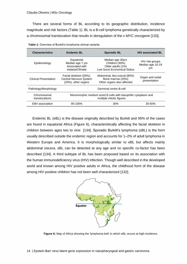
Endemic BL (eBL) is the disease originally described by Burkitt and 95% of the cases
are found in equatorial Africa (Figure 6), characteristically affecting the facial skeleton in
children between ages two to nine [134]. Sporadic Burkitt’s lymphoma (sBL) is the form
usually described outside the endemic region and accounts for 1–2% of adult lymphoma in
Western Europe and America. It is morphologically similar to eBL but affects mainly
abdominal viscera. sBL can be detected at any age and no specific co-factor has been
described [134]. A third subtype of BL has been proposed based on its association with
the human immunodeficiency virus (HIV) infection. Though well described in the developed
world and known among HIV positive adults in Africa, the childhood form of the disease
among HIV positive children has not been well characterized [132].
Table 1: Overview of Burkitt’s lymphoma clinical variants.
Figure 6: Map of Africa showing the 'lymphoma belt' in which eBL occurs at high incidence.
Cláudia Oliveira | MSc Oncology
Epstein-Barr virus latent gene expression in nasopharyngeal and gastric carcinoma | 15
BL has a very close association with EBV, with approximately 95% of eBL showing the
presence of the EBV genome in their tumour cells, while the other form are associated with
EBV in 30-50% of all cases [135].
EBV plays a role in the pathogenesis of BL by deregulation of c-MYC activity and clonal
expansion, direct mutagenesis and immune inactivation. Indeed, EBV is known to transform
resting B cells into latently infected lymphoblastoid cells [132]. The majority of BL’s, show
EBV latency I pattern. This cells carry a wild-type EBV genome and express only EBNA1
from the latent promoter Qp [136]. However, around 15% of endemic tumours, carry an
EBNA2 gene-deleted genome and express EBNA1, -3A, -3B, and -3C from the Wp latent
promoter [137].
2.2. Hodgkin’s Lymphoma
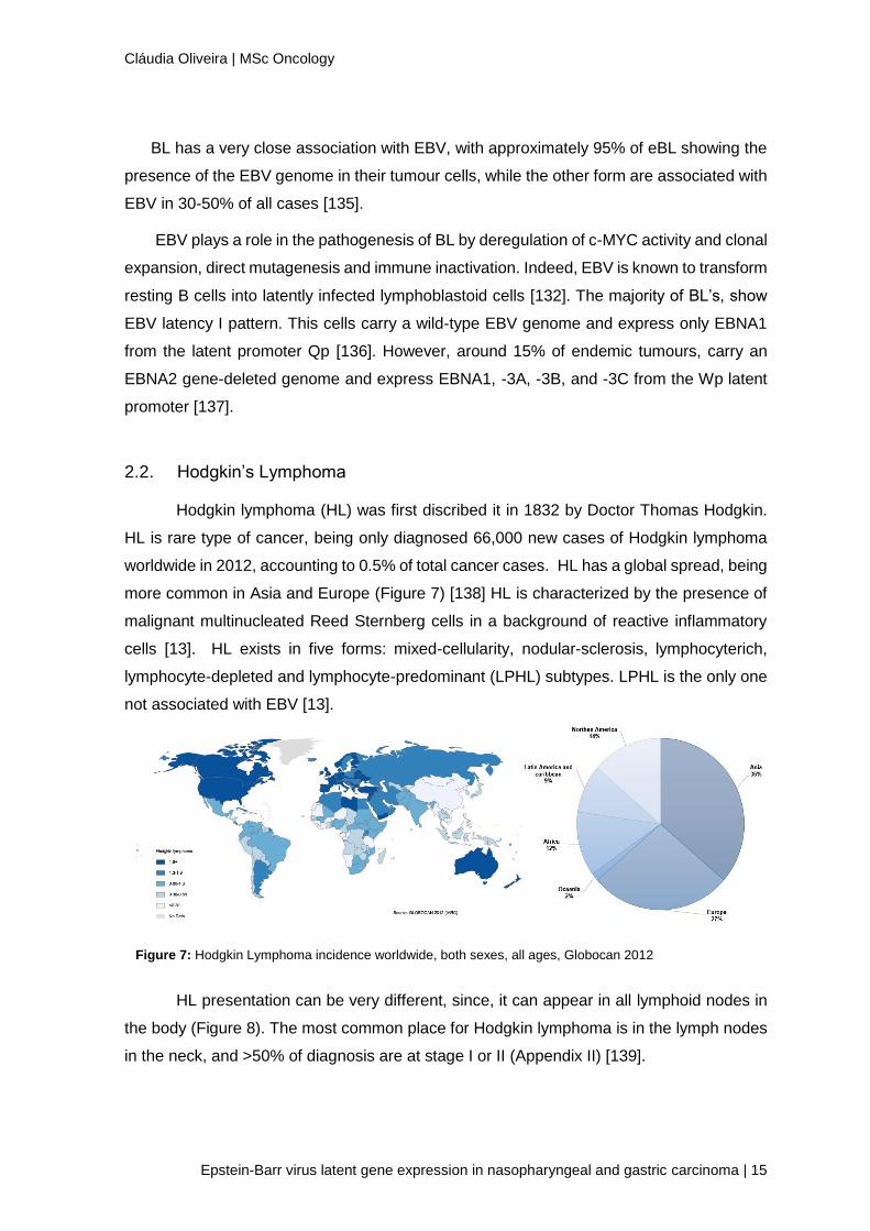
Hodgkin lymphoma (HL) was first discribed it in 1832 by Doctor Thomas Hodgkin.
HL is rare type of cancer, being only diagnosed 66,000 new cases of Hodgkin lymphoma
worldwide in 2012, accounting to 0.5% of total cancer cases. HL has a global spread, being
more common in Asia and Europe (Figure 7) [138] HL is characterized by the presence of
malignant multinucleated Reed Sternberg cells in a background of reactive inflammatory
cells [13]. HL exists in five forms: mixed-cellularity, nodular-sclerosis, lymphocyterich,
lymphocyte-depleted and lymphocyte-predominant (LPHL) subtypes. LPHL is the only one
not associated with EBV [13].
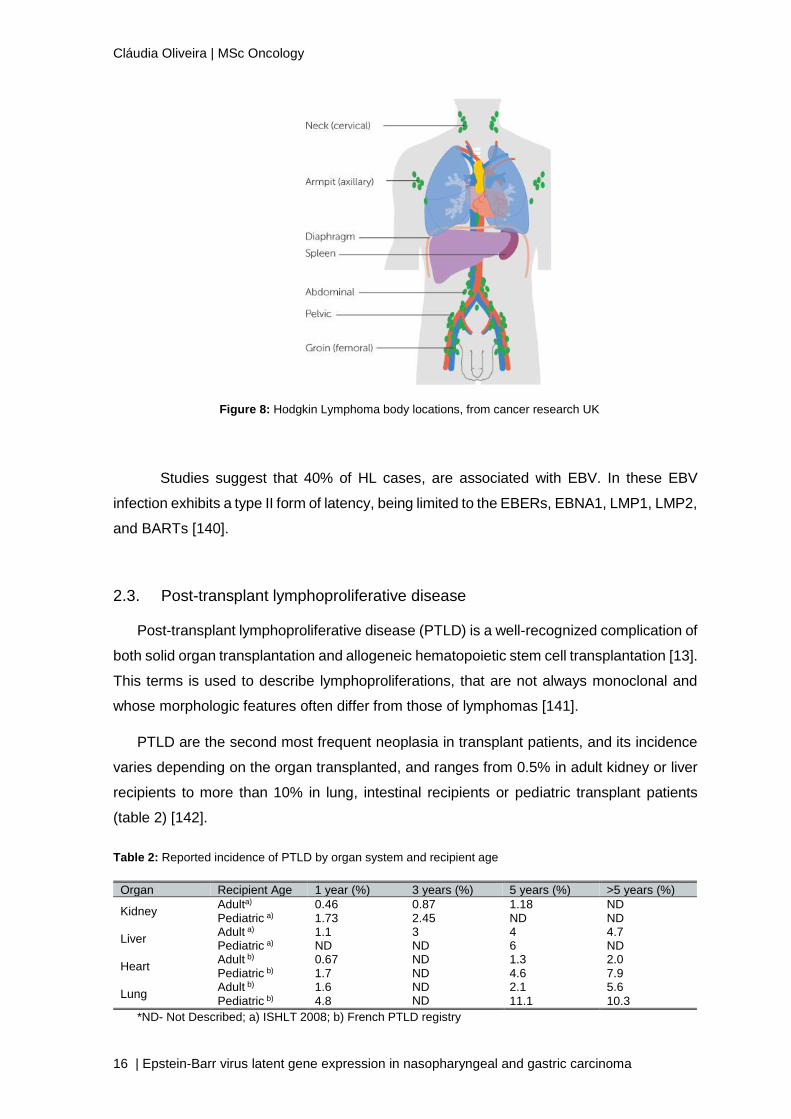
HL presentation can be very different, since, it can appear in all lymphoid nodes in
the body (Figure 8). The most common place for Hodgkin lymphoma is in the lymph nodes
in the neck, and >50% of diagnosis are at stage I or II (Appendix II) [139].
Figure 7: Hodgkin Lymphoma incidence worldwide, both sexes, all ages, Globocan 2012
Cláudia Oliveira | MSc Oncology
16 | Epstein-Barr virus latent gene expression in nasopharyngeal and gastric carcinoma
Studies suggest that 40% of HL cases, are associated with EBV. In these EBV
infection exhibits a type II form of latency, being limited to the EBERs, EBNA1, LMP1, LMP2,
and BARTs [140].
2.3. Post-transplant lymphoproliferative disease
Post-transplant lymphoproliferative disease (PTLD) is a well-recognized complication of
both solid organ transplantation and allogeneic hematopoietic stem cell transplantation [13].
This terms is used to describe lymphoproliferations, that are not always monoclonal and
whose morphologic features often differ from those of lymphomas [141].
PTLD are the second most frequent neoplasia in transplant patients, and its incidence
varies depending on the organ transplanted, and ranges from 0.5% in adult kidney or liver
recipients to more than 10% in lung, intestinal recipients or pediatric transplant patients
(table 2) [142].
Organ Recipient Age 1 year (%) 3 years (%) 5 years (%) >5 years (%)
Kidney Adulta) 0.46 0.87 1.18 ND Pediatric a) 1.73 2.45 ND ND
Liver Adult a) 1.1 3 4 4.7 Pediatric a) ND ND 6 ND
Heart Adult b) 0.67 ND 1.3 2.0 Pediatric b) 1.7 ND 4.6 7.9
Lung Adult b) 1.6 ND 2.1 5.6
Pediatric b) 4.8 ND 11.1 10.3
*ND- Not Described; a) ISHLT 2008; b) French PTLD registry
Table 2: Reported incidence of PTLD by organ system and recipient age
Figure 8: Hodgkin Lymphoma body locations, from cancer research UK
Cláudia Oliveira | MSc Oncology
Epstein-Barr virus latent gene expression in nasopharyngeal and gastric carcinoma | 17
Literature suggest that EBV infection has a major pathogenic role in PTLDs, infecting
60%-80% of PTLD patients, including 100% of early-onset PTLD patients [28]. Up to 2/3 of
PTLD cases are associated with EBV infection of B cells, either because of reactivation of
the virus or from primary EBV infection [13, 102]. Recipients who are EBV seronegative
have a higher risk of developing EBV-induced lymphoma, particularly pediatric patients
[143, 144]. Literature suggest that EBV infection has a major pathogenic role in PTLDs,
infecting 60%-80% of PTLD patients, including 100% of early-onset PTLD patients [28].
In most cases of PTLD, tumour cells express the latency III pattern, with some cells
undergoing lytic replication [145]. However, there is variability between individuals once
latency type I and II patterns have also been detected in PTLD biopsies. In these cases
additional genetic or epigenetic changes are probably required for tumour outgrowth [146,
147].
2.4. Nasopharyngeal Carcinoma
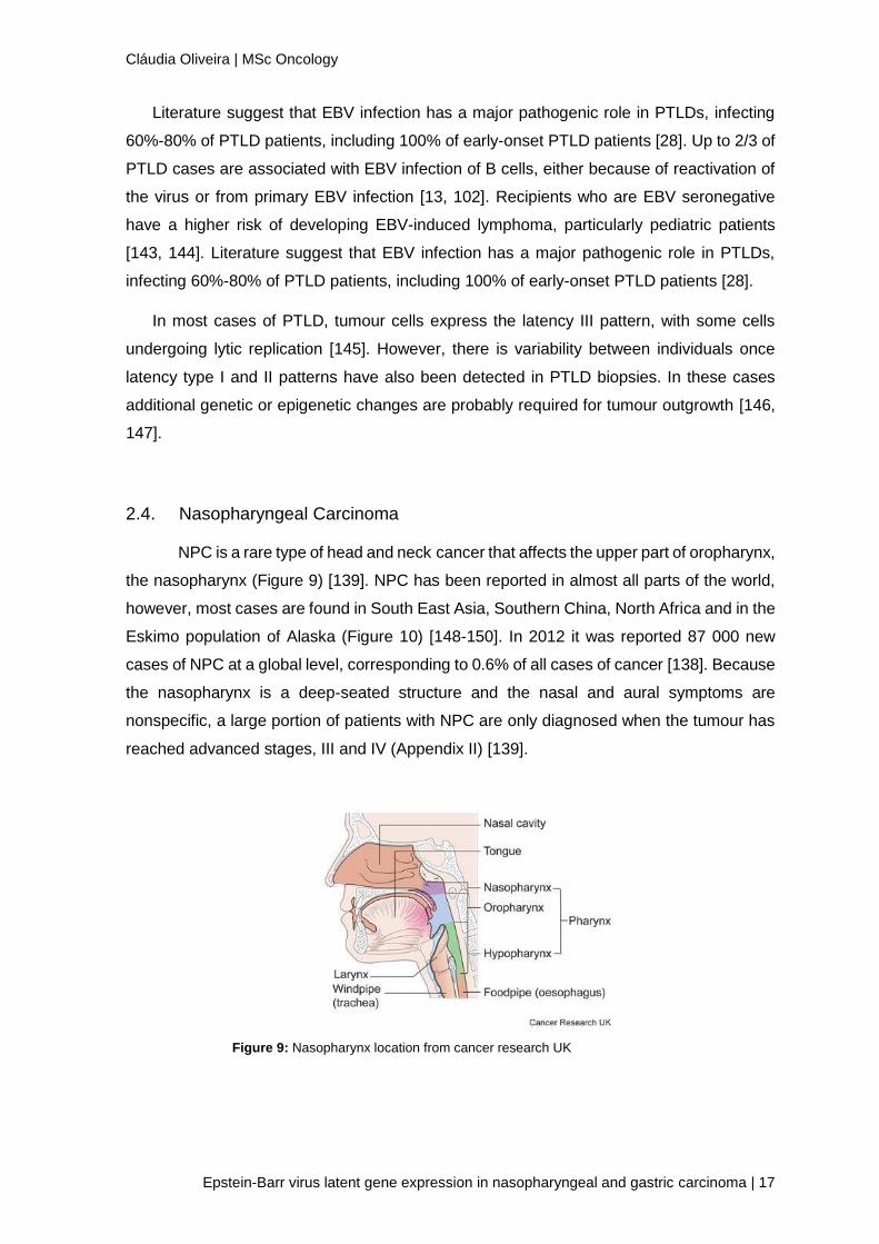
NPC is a rare type of head and neck cancer that affects the upper part of oropharynx,
the nasopharynx (Figure 9) [139]. NPC has been reported in almost all parts of the world,
however, most cases are found in South East Asia, Southern China, North Africa and in the
Eskimo population of Alaska (Figure 10) [148-150]. In 2012 it was reported 87 000 new
cases of NPC at a global level, corresponding to 0.6% of all cases of cancer [138]. Because
the nasopharynx is a deep-seated structure and the nasal and aural symptoms are
nonspecific, a large portion of patients with NPC are only diagnosed when the tumour has
reached advanced stages, III and IV (Appendix II) [139].
Figure 9: Nasopharynx location from cancer research UK
Cláudia Oliveira | MSc Oncology
18 | Epstein-Barr virus latent gene expression in nasopharyngeal and gastric carcinoma
Taking into account 2003 WHO classification NPC can be divided in three main
types: keratinizing squamous cell carcinoma, non-keratinizing carcinoma, which can be
divided diferentiated and undifferentiated, finally in basaloid squamous cell carcinoma
respectively [151, 152]. NPC as several risk factors, such as EBV infection and classic head
and neck aetiological factors including alcohol and tobacco [153].
Even though here is no doubt that NPC has a definite association with EBV, it is still
not clear the specific pathogenic mechanism by which EBV causes NPC. A gap in the
explanation of the EBV pathogenesis in the tumour is the fact that mature nasopharyngeal
cells are not usually infected with EBV, though tumours have been shown to be infected
before transformation [154]. It has been shown that the immature epithelial cells carry CD21
and can be infected by the virus. It is therefore postulated that EBV infects nasopharyngeal
cells that have been stimulated by other environmental factors [155-157].
It appears that latency gene expression in NPC is intermediate between what is seen
in latency I and latency II. The expression of EBNA1 and the EBERs are present in all EBV-
positive NPC cases [158, 159]. It also appears that LMP2A can be detected in about 50%
of NPC [160, 161]. In other hand, LMP1 is identified readily in only 35% of cases. LMP1 has
been identified in all pre invasive lesions, suggesting that its expression is necessary in
early lesions but may not be as essential in established carcinomas [162].
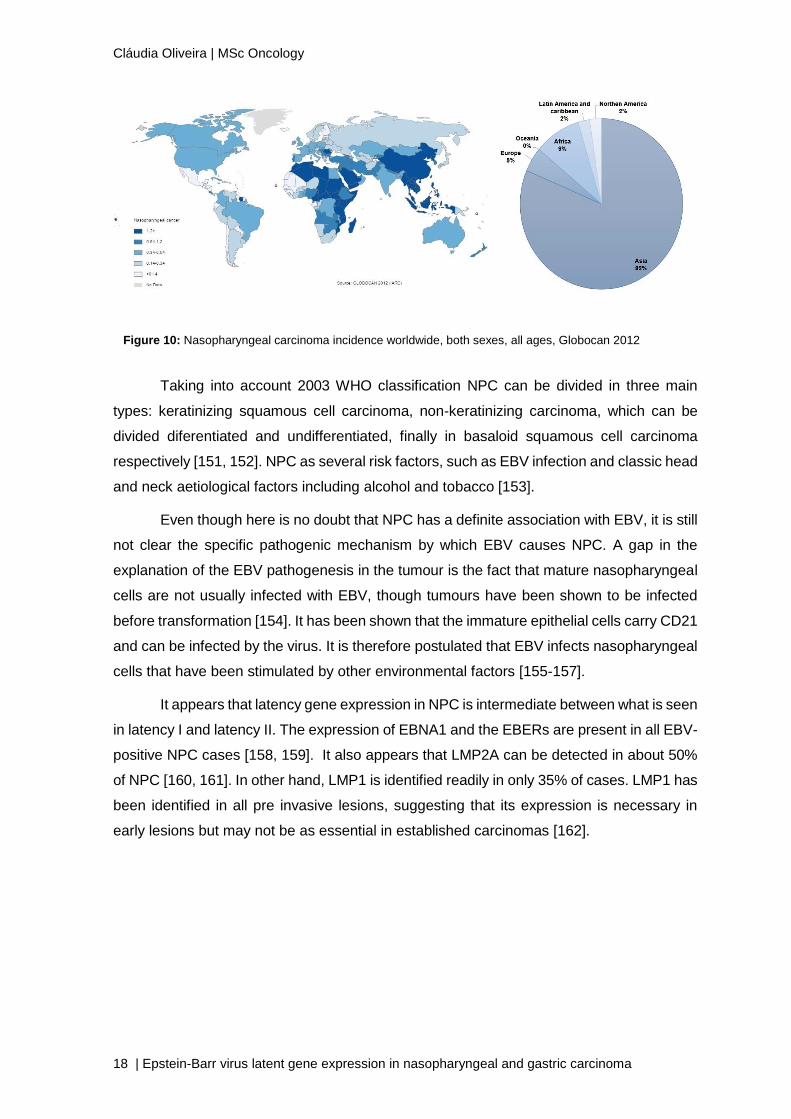
Figure 10: Nasopharyngeal carcinoma incidence worldwide, both sexes, all ages, Globocan 2012
Cláudia Oliveira | MSc Oncology
Epstein-Barr virus latent gene expression in nasopharyngeal and gastric carcinoma | 19
2.5. Gastric Carcinoma
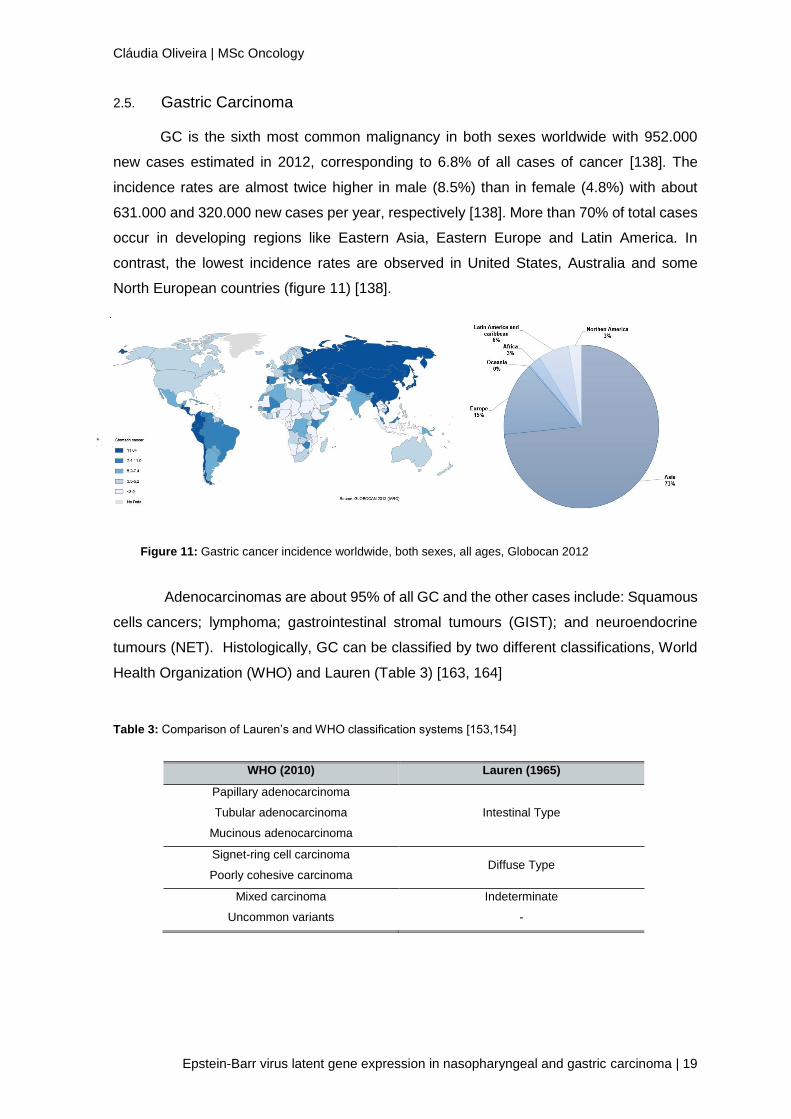
GC is the sixth most common malignancy in both sexes worldwide with 952.000
new cases estimated in 2012, corresponding to 6.8% of all cases of cancer [138]. The
incidence rates are almost twice higher in male (8.5%) than in female (4.8%) with about
631.000 and 320.000 new cases per year, respectively [138]. More than 70% of total cases
occur in developing regions like Eastern Asia, Eastern Europe and Latin America. In
contrast, the lowest incidence rates are observed in United States, Australia and some
North European countries (figure 11) [138].
Adenocarcinomas are about 95% of all GC and the other cases include: Squamous
cells cancers; lymphoma; gastrointestinal stromal tumours (GIST); and neuroendocrine
tumours (NET). Histologically, GC can be classified by two different classifications, World
Health Organization (WHO) and Lauren (Table 3) [163, 164]
Table 3: Comparison of Lauren’s and WHO classification systems [153,154]
WHO (2010) Lauren (1965)
Papillary adenocarcinoma
Intestinal Type Tubular adenocarcinoma
Mucinous adenocarcinoma
Signet-ring cell carcinoma Diffuse Type
Poorly cohesive carcinoma
Mixed carcinoma Indeterminate
Uncommon variants -
Figure 11: Gastric cancer incidence worldwide, both sexes, all ages, Globocan 2012
Cláudia Oliveira | MSc Oncology
20 | Epstein-Barr virus latent gene expression in nasopharyngeal and gastric carcinoma
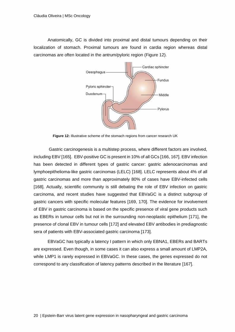
Anatomically, GC is divided into proximal and distal tumours depending on their
localization of stomach. Proximal tumours are found in cardia region whereas distal
carcinomas are often located in the antrum/pyloric region (Figure 12).
Gastric carcinogenesis is a multistep process, where different factors are involved,
including EBV [165]. EBV-positive GC is present in 10% of all GCs [166, 167]. EBV infection
has been detected in different types of gastric cancer: gastric adenocarcinomas and
lymphoepithelioma-like gastric carcinomas (LELC) [168]. LELC represents about 4% of all
gastric carcinomas and more than approximately 80% of cases have EBV-infected cells
[168]. Actually, scientific community is still debating the role of EBV infection on gastric
carcinoma, and recent studies have suggested that EBVaGC is a distinct subgroup of
gastric cancers with specific molecular features [169, 170]. The evidence for involvement
of EBV in gastric carcinoma is based on the specific presence of viral gene products such
as EBERs in tumour cells but not in the surrounding non-neoplastic epithelium [171], the
presence of clonal EBV in tumour cells [172] and elevated EBV antibodies in prediagnostic
sera of patients with EBV-associated gastric carcinoma [173].
EBVaGC has typically a latency I pattern in which only EBNA1, EBERs and BARTs
are expressed. Even though, in some cases it can also express a small amount of LMP2A,
while LMP1 is rarely expressed in EBVaGC. In these cases, the genes expressed do not
correspond to any classification of latency patterns described in the literature [167].
Figure 12: Illustrative scheme of the stomach regions from cancer research UK
Cláudia Oliveira | MSc Oncology
Epstein-Barr virus latent gene expression in nasopharyngeal and gastric carcinoma | 21
3. EBV Latency
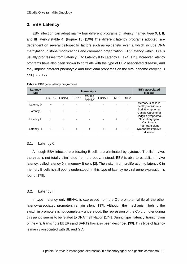
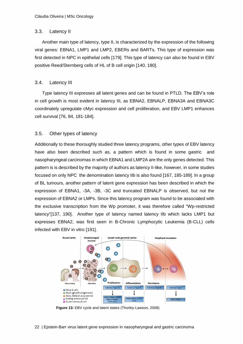
EBV infection can adopt mainly four different programs of latency, named type 0, I, II,
and III latency (table 4) (Figure 13) [106]. The different latency programs adopted, are
dependent on several cell-specific factors such as epigenetic events, which include DNA
methylation, histone modifications and chromatin organization. EBV latency within B cells
usually progresses from Latency III to Latency II to Latency I. [174, 175]. Moreover, latency
programs have also been shown to correlate with the type of EBV associated disease, and
they impose different phenotypic and functional properties on the viral genome carrying B
cell [176, 177].
Table 4: EBV gene latency programmes
Latency type
Transcripts EBV-associated
disease
EBERS EBNA1 EBNA2 EBNA3 FAMILY
EBNALP LMP1 LMP2
Latency 0 + - - - - - - Memory B cells in healthy individuals
Latency I + + - - - - - Burkitt lymphoma, Gastric Carcinoma
Latency II + + - - - + + Hodgkin lymphoma,
Nasopharyngeal Carcinoma
Latency III + + + + + + + Post-transplant
lymphoproliferative disease
3.1. Latency 0
Although EBV-infected proliferating B cells are eliminated by cytotoxic T cells in vivo,
the virus is not totally eliminated from the body. Instead, EBV is able to establish in vivo
latency, called latency 0 in memory B cells [2]. The switch from proliferation to latency 0 in
memory B cells is still poorly understood. In this type of latency no viral gene expression is
found [178].
3.2. Latency I
In type I latency only EBNA1 is expressed from the Qp promoter, while all the other
latency-associated promoters remain silent [137]. Although the mechanism behind the
switch in promoters is not completely understood, the repression of the Cp promoter during
this period seems to be related to DNA methylation [174]. During type I latency, transcription
of the viral transcripts EBERs and BARTs has also been described [30]. This type of latency
is mainly associated with BL and GC.
Cláudia Oliveira | MSc Oncology
22 | Epstein-Barr virus latent gene expression in nasopharyngeal and gastric carcinoma
3.3. Latency II
Another main type of latency, type II, is characterized by the expression of the following
viral genes: EBNA1, LMP1 and LMP2, EBERs and BARTs. This type of expression was
first detected in NPC in epithelial cells [179]. This type of latency can also be found in EBV
positive Reed/Sternberg cells of HL of B cell origin [140, 180].
3.4. Latency III
Type latency III expresses all latent genes and can be found in PTLD. The EBV’s role
in cell growth is most evident in latency III, as EBNA2, EBNALP, EBNA3A and EBNA3C
coordinately upregulate cMyc expression and cell proliferation, and EBV LMP1 enhances
cell survival [76, 84, 181-184].
3.5. Other types of latency
Additionally to these thoroughly studied three latency programs, other types of EBV latency
have also been described such as, a pattern which is found in some gastric and
nasopharyngeal carcinomas in which EBNA1 and LMP2A are the only genes detected. This
pattern is is described by the majority of authors as latency II-like, however, in some studies
focused on only NPC the denomination latency IIb is also found [167, 185-189]. In a group
of BL tumours, another pattern of latent gene expression has been described in which the
expression of EBNA1, -3A, -3B, -3C and truncated EBNALP is observed, but not the
expression of EBNA2 or LMPs. Since this latency program was found to be associated with
the exclusive transcription from the Wp promoter, it was therefore called "Wp-restricted
latency"[137, 190]. Another type of latency named latency IIb which lacks LMP1 but
expresses EBNA2, was first seen in B-Chronic Lymphocytic Leukemia (B-CLL) cells
infected with EBV in vitro [191].
Figure 13: EBV cycle and latent states (Thorley-Lawson, 2008)

II. AIMS OF THE STUDY

Cláudia Oliveira | MSc Oncology
Epstein-Barr virus latent gene expression in nasopharyngeal and gastric carcinoma |25
Although there are many studies in this field, it is still not clear the latency pattern
present in several malignancies, especially in gastric carcinoma. Therefore, it is necessary
to clarify and understand which EBV proteins are expressed, in order to serve as a starting
point for carcinogenesis studies.
The aim of this study is to characterize EBV latency in NPC and GC evaluating the
expression of EBV proteins in different tumors’ and try to establish a clinical correlation
between the viral latency and the malignances.


III. MATERIAL AND
METHODS

Cláudia Oliveira | MSc Oncology
Epstein-Barr virus latent gene expression in nasopharyngeal and gastric carcinoma | 29
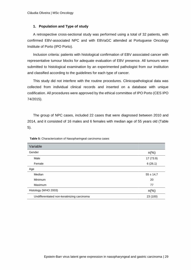
1. Population and Type of study
A retrospective cross-sectional study was performed using a total of 32 patients, with
confirmed EBV-associated NPC and with EBVaGC attended at Portuguese Oncology
Institute of Porto (IPO Porto).
Inclusion criteria: patients with histological confirmation of EBV associated cancer with
representative tumour blocks for adequate evaluation of EBV presence. All tumours were
submitted to histological examination by an experimented pathologist from our institution
and classified according to the guidelines for each type of cancer.
This study did not interfere with the routine procedures. Clinicopathological data was
collected from individual clinical records and inserted on a database with unique
codification. All procedures were approved by the ethical committee of IPO Porto (CES IPO
74/2015).
The group of NPC cases, included 22 cases that were diagnosed between 2010 and
2014, and it consisted of 16 males and 6 females with median age of 55 years old (Table
5).
Variable
Gender n(%)
Male 17 (73.9)
Female 6 (26.1)
Age
Median 55 ± 14,7
Minimum 20
Maximum 77
Histology (WHO 2003) n(%)
Undifferentiated non-keratinizing carcinoma 23 (100)
Table 5: Characterization of Nasopharingeal carcinoma cases
Cláudia Oliveira | MSc Oncology
30 | Epstein-Barr virus latent gene expression in nasopharyngeal and gastric carcinoma
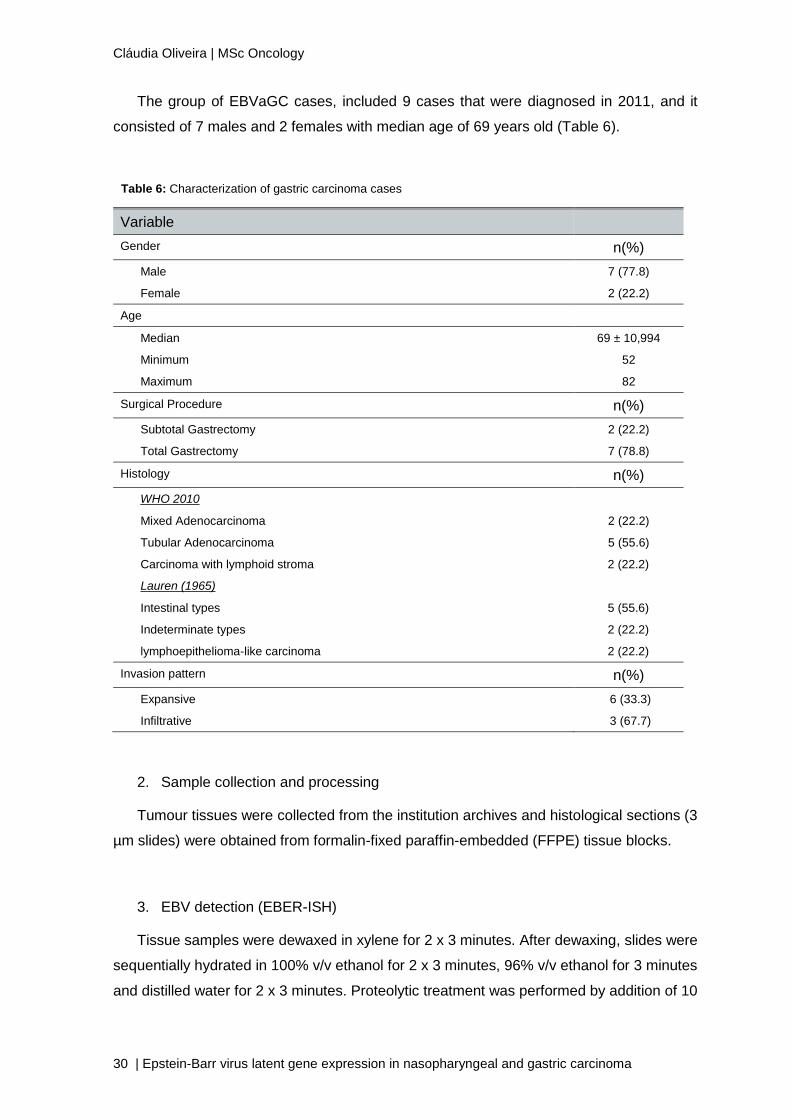
The group of EBVaGC cases, included 9 cases that were diagnosed in 2011, and it
consisted of 7 males and 2 females with median age of 69 years old (Table 6).
2. Sample collection and processing
Tumour tissues were collected from the institution archives and histological sections (3
µm slides) were obtained from formalin-fixed paraffin-embedded (FFPE) tissue blocks.
3. EBV detection (EBER-ISH)
Tissue samples were dewaxed in xylene for 2 x 3 minutes. After dewaxing, slides were
sequentially hydrated in 100% v/v ethanol for 2 x 3 minutes, 96% v/v ethanol for 3 minutes
and distilled water for 2 x 3 minutes. Proteolytic treatment was performed by addition of 10
Variable
Gender n(%)
Male 7 (77.8)
Female 2 (22.2)
Age
Median 69 ± 10,994
Minimum 52
Maximum 82
Surgical Procedure n(%)
Subtotal Gastrectomy 2 (22.2)
Total Gastrectomy 7 (78.8)
Histology n(%)
WHO 2010
Mixed Adenocarcinoma 2 (22.2)
Tubular Adenocarcinoma 5 (55.6)
Carcinoma with lymphoid stroma 2 (22.2)
Lauren (1965)
Intestinal types 5 (55.6)
Indeterminate types 2 (22.2)
lymphoepithelioma-like carcinoma 2 (22.2)
Invasion pattern n(%)
Expansive 6 (33.3)
Infiltrative 3 (67.7)
Table 6: Characterization of gastric carcinoma cases
Cláudia Oliveira | MSc Oncology
Epstein-Barr virus latent gene expression in nasopharyngeal and gastric carcinoma | 31
mM proteinase K and incubation at 37ºC during 30 minutes. Finished the incubation time,
endogenous peroxidase activity was blocked by incubating the slides in 3% hydrogen
peroxide (H2O2) for 10 minutes at room temperature, then solution the slides were immersed
in distilled water for 2 x 3 minutes and then dehydrated in 96% v/v ethanol followed 100%
v/v ethanol for 3 minutes to facilitate air drying.
Epstein-Barr virus was identified by in situ hybridization (ISH) for detection of EBV-
encoded small RNA (EBER). Hybridization results in duplex formation of sequence present
in EBV infected cells (EBERs) and specific probe. The BondTM Ready-to-use ISH EBER
Probe (Leica, Newcastle upon Tyne, UK) was used with a volume of 20 µl for each slide.
Slides were covered with coverslip, and then incubated at 37ºC for 2 hours. Nonspecific
antibody binding was block using UltraVision Large Volume Detection System Anti-
Polyvalent, HRP (THERMO SCIENTIFIC, Fremont, USA). It was incubated for 10 minutes
at room temperature and washing was performed with TBS, 0.1% v/v Trinton X-100 (TBS-
T) 2x 5 minutes.
EBERs detection was performed with BondTM Anti-Fluorescein Antibody (Leica,
Newcastle upon Tyne, UK) diluted 1:150 in TBS, 3% m/v BSA, 0.1% v/v Trinton X-100 with
incubation at room temperature for 30 minutes. After washing 2 x 3 minutes with TBS, the
revelation of hybrids was performed with the UltraVision Large Volume Detection System
Anti-Polyvalent, HRP (THERMO SCIENTIFIC, Fremont, USA). Briefly, the Biotinylated Goat
Anti-Polyvalent Antibody (THERMO SCIENTIFIC, Fremont, USA) was added at room
temperature for 10 minutes, washed with TBS-T 2 x 5 minutes followed by the addition of
Streptavidin Peroxidase (THERMO SCIENTIFIC, Fremont, USA) with incubation for 10
minutes at room temperature. Streptavidin shows high affinity with several secondary
antibody-conjugated biotin molecules providing a good revelation signal. Detection of
hybrids is achieved by enzymatic reaction using a specific substrate to peroxidase.
ImmPACTTM DAB, Peroxidase Substrate (VECTOR, Burlingame, CA USA) was used
during 4 minutes at room temperature and diluted 3:100. The final washing was performed
with distillated water 2 x 5 minutes.
Mayer’s hemalum solution (Millipore, Darmstadt, Germany) was used as counterstain
for 10-20 seconds, depending of dye’s use. After coloration, slides were washed in running
water for 5 minutes and the following step was sequential dehydration in 70% v/v ethanol
for 2 x 4 minutes, 96% v/v ethanol for 2 x 4 minutes, 100% v/v ethanol for 2 x 4 minutes and
xylene for 2 x 4 minutes. Mounting was performed with Microscopy Entellan (MERCK,
Darmstadt, Germany).
Cláudia Oliveira | MSc Oncology
32 | Epstein-Barr virus latent gene expression in nasopharyngeal and gastric carcinoma
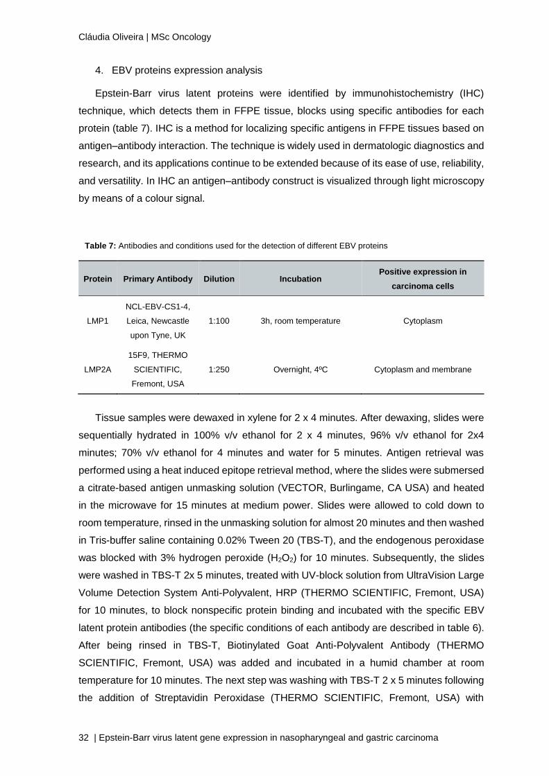
4. EBV proteins expression analysis
Epstein-Barr virus latent proteins were identified by immunohistochemistry (IHC)
technique, which detects them in FFPE tissue, blocks using specific antibodies for each
protein (table 7). IHC is a method for localizing specific antigens in FFPE tissues based on
antigen–antibody interaction. The technique is widely used in dermatologic diagnostics and
research, and its applications continue to be extended because of its ease of use, reliability,
and versatility. In IHC an antigen–antibody construct is visualized through light microscopy
by means of a colour signal.
Protein Primary Antibody Dilution Incubation Positive expression in
carcinoma cells
LMP1
NCL-EBV-CS1-4,
Leica, Newcastle
upon Tyne, UK
1:100 3h, room temperature Cytoplasm
LMP2A
15F9, THERMO
SCIENTIFIC,
Fremont, USA
1:250 Overnight, 4ºC Cytoplasm and membrane
Tissue samples were dewaxed in xylene for 2 x 4 minutes. After dewaxing, slides were
sequentially hydrated in 100% v/v ethanol for 2 x 4 minutes, 96% v/v ethanol for 2x4
minutes; 70% v/v ethanol for 4 minutes and water for 5 minutes. Antigen retrieval was
performed using a heat induced epitope retrieval method, where the slides were submersed
a citrate-based antigen unmasking solution (VECTOR, Burlingame, CA USA) and heated
in the microwave for 15 minutes at medium power. Slides were allowed to cold down to
room temperature, rinsed in the unmasking solution for almost 20 minutes and then washed
in Tris-buffer saline containing 0.02% Tween 20 (TBS-T), and the endogenous peroxidase
was blocked with 3% hydrogen peroxide (H2O2) for 10 minutes. Subsequently, the slides
were washed in TBS-T 2x 5 minutes, treated with UV-block solution from UltraVision Large
Volume Detection System Anti-Polyvalent, HRP (THERMO SCIENTIFIC, Fremont, USA)
for 10 minutes, to block nonspecific protein binding and incubated with the specific EBV
latent protein antibodies (the specific conditions of each antibody are described in table 6).
After being rinsed in TBS-T, Biotinylated Goat Anti-Polyvalent Antibody (THERMO
SCIENTIFIC, Fremont, USA) was added and incubated in a humid chamber at room
temperature for 10 minutes. The next step was washing with TBS-T 2 x 5 minutes following
the addition of Streptavidin Peroxidase (THERMO SCIENTIFIC, Fremont, USA) with
Table 7: Antibodies and conditions used for the detection of different EBV proteins
Cláudia Oliveira | MSc Oncology
Epstein-Barr virus latent gene expression in nasopharyngeal and gastric carcinoma | 33
incubation for 10 minutes at room temperature. Detection of hybrids is achieved by
enzymatic reaction using a specific substrate to peroxidase, 3, 3'-diaminobenzidine (DAB)
ImmPACTTM DAB (VECTOR, Burlingame, CA USA) was used during 4 minutes at room
temperature and diluted 3:100. The final washing was performed with water 2 x 5 minutes.
Mayer’s hemalum solution (Millipore, Darmstadt, Germany) was used as counterstain
for 10-30 seconds, depending of dye’s use. After coloration, slides were washed in running
water for 5 minutes and the following step was sequential dehydration in 70% v/v ethanol
for 4 minutes, 96% v/v ethanol for 2 x 4 minutes, 100% v/v ethanol for 2 x 4 minutes and
xylene for 2 x 4 minutes. Mounting was performed with Microscopy Entellan (MERCK,
Darmstadt, Germany).
5. Quality control
Positive and negative tissues controls were used in order to ensure the quality of the
protocol. As a positive control for EBV proteins, PTLD FFPE tissue samples were used,
once this malignancy expresses all EBV proteins. As for negative control, it was used GC
or NPC FFPE tissue samples that lacked the specific antigen.
6. Data analysis
The clinicopathological characteristics of the tumour were compared to the expression
of EBV latency proteins and consequently to the EBV latency pattern in GC and NPC
pacients.


IV. RESULTS

Cláudia Oliveira | MSc Oncology
Epstein-Barr virus latent gene expression in nasopharyngeal and gastric carcinoma | 37
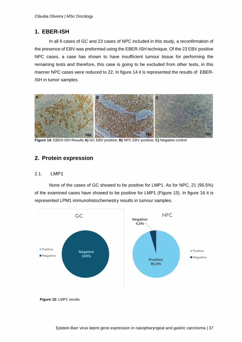
1. EBER-ISH
In all 9 cases of GC and 23 cases of NPC included in this study, a reconfirmation of
the presence of EBV was preformed using the EBER-ISH technique. Of the 23 EBV positive
NPC cases, a case has shown to have insufficient tumour tissue for performing the
remaining tests and therefore, this case is going to be excluded from other tests, in this
manner NPC cases were reduced to 22. In figure 14 it is represented the results of EBER-
ISH in tumor samples.
Figure 14: EBER-ISH Results A) GC EBV positive; B) NPC EBV positive; C) Negative control
2. Protein expression
2.1. LMP1
None of the cases of GC showed to be positive for LMP1. As for NPC, 21 (95.5%)
of the examined cases have showed to be positive for LMP1 (Figure 15). In figure 16 it is
represented LPM1 immunohistochemestry results in tumour samples.
Negative100%
GC
Positive
Negative
Positive95,5%
Negative4,5%
NPC
Positive
Negative
Figure 15: LMP1 results
Cláudia Oliveira | MSc Oncology
38 | Epstein-Barr virus latent gene expression in nasopharyngeal and gastric carcinoma
Figure 16: IHC LMP1 results A) GC case negative for LMP1; B) NPC case positive for LMP1; C) NPC case
negative for LMP1; D) Positive Control; E) Negative control
2.2. LMP2A
Of all cases of GC, only 4 proved to be positive for LMP2a. As for NPC, all 22 cases
examined proved to be positive for LMP2a (Figure 17). In figure 18 it is represented LPM1
immunohistochemestry results in tumour samples.
Positive44,4%Negative
55,6%
GC
Positive
NegativePositive
100%
NPC
Positive
Negative
Figure 17: LMP2a results
Cláudia Oliveira | MSc Oncology
Epstein-Barr virus latent gene expression in nasopharyngeal and gastric carcinoma | 39
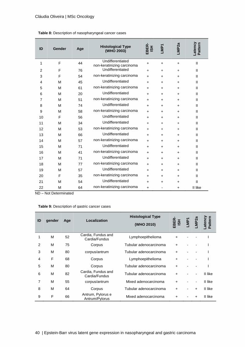
Figure 18: IHC LMP2a results A) GC case positive for LMP2a; B) GC case negative for LMP2a; C) NPC case
positive for LMP2a; D) Positive control; E) Negative control
3. EBV latency
Assuming that EBNA1 is expressed in all EBV associated tumours, it was given to
each of the cases used in this study an EBV latency pattern based on the expression of
LMP1 and LMP2a.
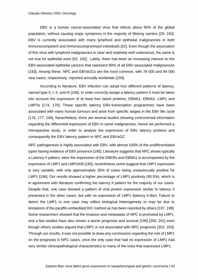
3.1. NPC
Table 8 describes the principal baseline characteristics of each NPC case used in
this study, including sex, age, histological type and expressed proteins. The majority of
cases, 21 (95.5%), had the expression of both LMP1 and LMP2a, which corresponds to a
latency II pattern. Only 1 case (4.5%) expressed a distinct latency pattern similar to latency
II, but without the expression of LMP1. This pattern is going to be referred as “latency II-
like”.
3.2. EBVaGC
Table 9 describes the principal baseline characteristics of each EBVaGC case used
in this study, including sex, age, tumour location, histological type and expressed proteins.
From all 9 cases of GC, 5 (55.6%) had no expression of LMP1 and LMP2a. In these, a
latency I pattern is observed. The remaining 4 cases, had the expression of LMP2a, but not
LMP1. These cases presented a “latency II-like” pattern.
Cláudia Oliveira | MSc Oncology
40 | Epstein-Barr virus latent gene expression in nasopharyngeal and gastric carcinoma
ND – Not Determinated
ID Gender Age Histological Type
(WHO 2003)
EB
ER
-
ISH
LM
P1
LM
P2
a
La
ten
cy
Pa
tte
rn
1 F 44 Undifferentiated
non-keratinizing carcinoma + + + II
2 F 76 Undifferentiated + + + II
3 F 54 non-keratinizing carcinoma + + + II
4 M 45 Undifferentiated + + + II
5 M 61 non-keratinizing carcinoma + + + II
6 M 20 Undifferentiated + + + II
7 M 51 non-keratinizing carcinoma + + + II
8 M 74 Undifferentiated + + + II
9 M 58 non-keratinizing carcinoma + + + II
10 F 56 Undifferentiated + + + II
11 M 34 Undifferentiated + + + II
12 M 53 non-keratinizing carcinoma + + + II
13 M 66 Undifferentiated + + + II
14 M 57 non-keratinizing carcinoma + + + II
15 M 71 Undifferentiated + + + II
16 M 41 non-keratinizing carcinoma + + + II
17 M 71 Undifferentiated + + + II
18 M 77 non-keratinizing carcinoma + + + II
19 M 57 Undifferentiated + + + II
20 F 35 non-keratinizing carcinoma + + + II
21 M 54 Undifferentiated + + + II
22 M 64 non-keratinizing carcinoma + - + II like
ID gender Age Localization Histological Type
(WHO 2010)
EB
ER
-
ISH
LM
P1
LM
P2
a
La
ten
cy
Pa
tte
rn
1 M 52 Cardia, Fundus and
Cardia/Fundus Lymphoepithelioma + - - I
2 M 75 Corpus Tubular adenocarcinoma + - - I
3 M 80 corpus/antrum Tubular adenocarcinoma + - - I
4 F 68 Corpus Lymphoepithelioma + - - I
5 M 80 Corpus Tubular adenocarcinoma + - - I
6 M 82 Cardia, Fundus and
Cardia/Fundus Tubular adenocarcinoma + - - II like
7 M 55 corpus/antrum Mixed adenocarcinoma + - - II like
8 M 64 Corpus Tubular adenocarcinoma + - + II like
9 F 66 Antrum, Pylorus e
Antrum/Pylorus Mixed adenocarcinoma + - + II like
Table 9: Description of gastric cancer cases
Table 8: Description of nasopharyngeal cancer cases

V. DISCUSSION

Cláudia Oliveira | MSc Oncology
Epstein-Barr virus latent gene expression in nasopharyngeal and gastric carcinoma | 43
EBV is a human cancer-associated virus that infects about 90% of the global
population, without causing major symptoms in the majority of lifelong carriers [29, 192].
EBV is currently associated with many lymphoid and epithelial malignancies in both
immunocompetent and immunocompromised individuals [52]. Even though the association
of this virus with lymphoid malignancies is clear and relatively well understood, the same is
not true for epithelial ones [52, 192]. Lately, there has been an increasing interest on the
EBV-associated epithelial cancers that represent 80% of all EBV-associated malignancies
[193]. Among these, NPC and EBVaGCs are the most common, with 78 000 and 84 000
new cases, respectively, reported annually worldwide [193].
According to literature, EBV infection can adopt four different patterns of latency,
named type 0, I, II, and III [106]. In order correctly assign a latency pattern it must be taken
into account the expression of at least four latent proteins, EBNA1, EBNA2, LMP1 and
LMP2a [174, 175]. These specific latency EBV-transcription programmes have been
associated with many human tumours and arise from specific stages in the EBV life cycle
[176, 177, 194]. Nevertheless, there are several studies showing controversial information
regarding the differential expression of EBV in some malignancies, hence we performed a
retrospective study, in order to analyze the expression of EBV latency proteins and
consequently the EBV latency pattern in NPC and EBVaGC.
NPC pathogenesis is highly associated with EBV, with almost 100% of the undifferentiated
types having evidence of EBV presence [195]. Literature suggests that NPC shows typically
a Latency II pattern, were the expression of the EBERs and EBNA1 is accompanied by the
expression of LMP1 and LMP2A/B [195], nevertheless some suggest that LMP1 expression
is very variable, with only approximately 35% of cases being unequivocally positive for
LMP1 [196]. Our results showed a higher percentage of LMP1 positivity (95.5%), which is
in agreement with literature confirming the latency II pattern for the majority of our cases.
Despite that, one case showed a pattern of viral protein expression similar to latency II
presented in the other cases, but with no expression of LMP1 (latency II-like). Failure to
detect the LMP1 in one case may reflect biological heterogeneity or may be due to
limitations of the paraffin-embedded IHC method as has been reported by others [197, 198].
Some researchers showed that the invasion and metastasis of NPC is promoted by LMP1,
and a few studies have also shown a worse prognosis and survival [199] [200, 201] even
though others studies argued that LMP1 is not associated with NPC prognosis [202, 203].
Through our results, it was not possible to draw any conclusions regarding the role of LMP1
on the prognosis in NPC cases, once the only case that had no expression of LMP1 had
very similar clinicopathological characteristics to many of the ones that expressed LMP1.
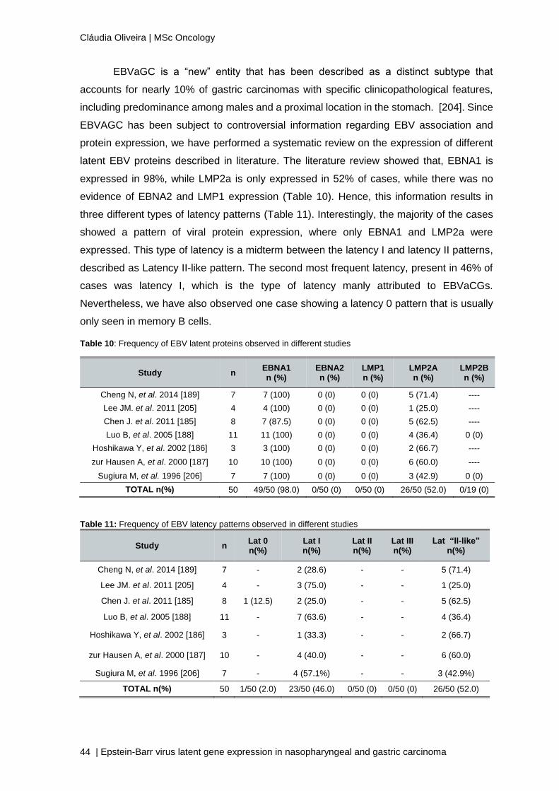
Cláudia Oliveira | MSc Oncology
44 | Epstein-Barr virus latent gene expression in nasopharyngeal and gastric carcinoma
EBVaGC is a “new” entity that has been described as a distinct subtype that
accounts for nearly 10% of gastric carcinomas with specific clinicopathological features,
including predominance among males and a proximal location in the stomach. [204]. Since
EBVAGC has been subject to controversial information regarding EBV association and
protein expression, we have performed a systematic review on the expression of different
latent EBV proteins described in literature. The literature review showed that, EBNA1 is
expressed in 98%, while LMP2a is only expressed in 52% of cases, while there was no
evidence of EBNA2 and LMP1 expression (Table 10). Hence, this information results in
three different types of latency patterns (Table 11). Interestingly, the majority of the cases
showed a pattern of viral protein expression, where only EBNA1 and LMP2a were
expressed. This type of latency is a midterm between the latency I and latency II patterns,
described as Latency II-like pattern. The second most frequent latency, present in 46% of
cases was latency I, which is the type of latency manly attributed to EBVaCGs.
Nevertheless, we have also observed one case showing a latency 0 pattern that is usually
only seen in memory B cells.
Table 10: Frequency of EBV latent proteins observed in different studies
Study n EBNA1 n (%)
EBNA2 n (%)
LMP1 n (%)
LMP2A n (%)
LMP2B n (%)
Cheng N, et al. 2014 [189] 7 7 (100) 0 (0) 0 (0) 5 (71.4) ----
Lee JM. et al. 2011 [205] 4 4 (100) 0 (0) 0 (0) 1 (25.0) ----
Chen J. et al. 2011 [185] 8 7 (87.5) 0 (0) 0 (0) 5 (62.5) ----
Luo B, et al. 2005 [188] 11 11 (100) 0 (0) 0 (0) 4 (36.4) 0 (0)
Hoshikawa Y, et al. 2002 [186] 3 3 (100) 0 (0) 0 (0) 2 (66.7) ----
zur Hausen A, et al. 2000 [187] 10 10 (100) 0 (0) 0 (0) 6 (60.0) ----
Sugiura M, et al. 1996 [206] 7 7 (100) 0 (0) 0 (0) 3 (42.9) 0 (0)
TOTAL n(%) 50 49/50 (98.0) 0/50 (0) 0/50 (0) 26/50 (52.0) 0/19 (0)
Table 11: Frequency of EBV latency patterns observed in different studies
Study n Lat 0 n(%)
Lat I n(%)
Lat II n(%)
Lat III n(%)
Lat “II-like” n(%)
Cheng N, et al. 2014 [189] 7 - 2 (28.6) - - 5 (71.4)
Lee JM. et al. 2011 [205] 4 - 3 (75.0) - - 1 (25.0)
Chen J. et al. 2011 [185] 8 1 (12.5) 2 (25.0) - - 5 (62.5)
Luo B, et al. 2005 [188] 11 - 7 (63.6) - - 4 (36.4)
Hoshikawa Y, et al. 2002 [186] 3 - 1 (33.3) - - 2 (66.7)
zur Hausen A, et al. 2000 [187] 10 - 4 (40.0) - - 6 (60.0)
Sugiura M, et al. 1996 [206] 7 - 4 (57.1%) - - 3 (42.9%)
TOTAL n(%) 50 1/50 (2.0) 23/50 (46.0) 0/50 (0) 0/50 (0) 26/50 (52.0)
Cláudia Oliveira | MSc Oncology
Epstein-Barr virus latent gene expression in nasopharyngeal and gastric carcinoma | 45
In our study, the results were congruent to those described from this literature
review, since both of the most frequent latency patterns were also observed in our study.
The only difference was that latency I, was the most frequent with 55.6% of cases, while
latency II-like was observed in 44.4% of the cases. It is suggested by literature that, the lack
of LMP1 expression in EBVaGC, indicates that LMP1 may not be necessary for maintaining
the tumour malignant state, but it might participate in an earlier stage of the tumour
development, and is down-regulated afterwards [206]. Alternatively, the lack of LMP1 may
result of a clonal selection of LMP1-negative tumour cells by immunological pressure, once
EBV-specific cytotoxic T cells are potentially directed against the viral latent proteins other
than EBNA1 [206]. Recently, it was also demonstrated that LMP2A up-regulates surviving
gene expression through the NF-kB pathway and that it up-regulates cellular DNA
methyltransferase 1 through the phosphorylation of STAT3, causing promoter
hypermethylation of a tumour suppressor gene, PTEN, in EBVaGC [185]. Since LMP2A has
demonstrated to play important roles in the oncogenic processes of EBVaGC, and it is
expressed in about half of the cases, it is crucial to not be restricted to three types of latency
patterns that are described and accepted by literature, and thereby new ones, with different
tyes of protein expression, should be taken into consideration.
Even though our results are important and this is the first study to describe latency
patterns in our population, it would be important to complement this work with some extra
information. For example, compare GC llymphoepithelioma with NPC undifferentiated non-
keratinizing carcinoma, once they have similar histology, but it seems that they have
different latency patter, for this it would be necessary to increase the number of cases of
GC. It would be also important to complement our results with information from other
proteins, such as EBNA1, that in this study is assumed to be expressed in very tumour, but
it should be done in order to eliminate possible doubts of the existence of latency 0; EBNA2;
and finally BARF1, once it is suggested by literature that it might act as a viral oncogene in
the development of EBVaGC [187].
It is very important correctly identify all latency patterns and which carcinogenic
processes are taking place in each of them, because once we know it, it is much easier to
have an accurate prognosis and by consequence a proper treatment for each individual.


VI. CONCLUSION

Cláudia Oliveira | MSc Oncology
Epstein-Barr virus latent gene expression in nasopharyngeal and gastric carcinoma | 49
EBV infection can adopt different patterns of latency, and these have a close
correlation with the different types of EBV associated diseases. With this study it was possible
to confirm that in our population, NPC is characterized by a latency II pattern, while EBVaGC
shows a latency I pattern. We were also able to identify a novel, and yet not accepted,
latency pattern associated mainly associated with EBVaGC, but also described in one case
of NPC. We described that latency pattern as latency II-like, and it is characterized by the
expression of EBNA1 and LMP2a.
This is the first study regarding latency patterns of NPC and EBVaGC in Portugal
and further studies are required to confirm these evidences, especially with more cases and
ideally from different regions of Portugal. It also important to increase the panel of proteins
studied so we can correctly describe the different latency patterns, and in order to better
understand the role of each protein in tumour carcinogenesis, functional assays would also
be important.


VII. REFERENCES

Cláudia Oliveira | MSc Oncology
Epstein-Barr virus latent gene expression in nasopharyngeal and gastric carcinoma | 53
1. Burkitt, D., Determining the climatic limitations of a children's cancer common in Africa. Br Med J, 1962. 2(5311): p. 1019-23.
2. Gromminger, S., J. Mautner, and G.W. Bornkamm, Burkitt lymphoma: the role of Epstein-Barr virus revisited. Br J Haematol, 2012. 156(6): p. 719-29.
3. Epstein, M.A., B.G. Achong, and Y.M. Barr, Virus Particles in Cultured Lymphoblasts from Burkitt's Lymphoma. Lancet, 1964. 1(7335): p. 702-3.
4. Henle, G. and W. Henle, Immunofluorescence in cells derived from Burkitt's lymphoma. J Bacteriol, 1966. 91(3): p. 1248-56.
5. Henle, G., W. Henle, and V. Diehl, Relation of Burkitt's tumor-associated herpes-ytpe virus to infectious mononucleosis. Proc Natl Acad Sci U S A, 1968. 59(1): p. 94-101.
6. zur Hausen, H., et al., EBV DNA in biopsies of Burkitt tumours and anaplastic carcinomas of the nasopharynx. Nature, 1970. 228(5276): p. 1056-8.
7. Roizman, B., Family Herpesviridae: A Brief Introduction. 5th ed. Field Virology, ed. D.H. Knipe, PM. 2007, Philadelphia: Lippincott-Williams & Wilkins Publishers.
8. Johannsen, E., et al., Proteins of purified Epstein-Barr virus. Proc Natl Acad Sci U S A, 2004. 101(46): p. 16286-91.
9. Kieff, E.R., AB., Epstein-Barr Virus and Its Replication. Fields Virology, ed. H.P. Knipe DM. 2007, Philadelphia.
10. Sanchez-Pinel, A., et al., Identification of a novel EBV-induced membrane glycoprotein of 43 kDa with H667 MAb. Virology, 1991. 180(1): p. 31-40.
11. Zone, V. http://viralzone.expasy.org/viralzone/all_by_species/185.html. 2015. 12. Baer, R., et al., DNA sequence and expression of the B95-8 Epstein-Barr virus genome.
Nature, 1984. 310(5974): p. 207-11. 13. Kutok, J.L. and F. Wang, Spectrum of Epstein-Barr virus-associated diseases. Annu Rev
Pathol, 2006. 1: p. 375-404. 14. Hurley, E.A. and D.A. Thorley-Lawson, B cell activation and the establishment of Epstein-
Barr virus latency. J Exp Med, 1988. 168(6): p. 2059-75. 15. Straus, S.E., et al., Epstein-Barr Virus Infections: Biology, Pathogenesis, and Management.
Annals of Internal Medicine, 1993. 118(1): p. 45-58. 16. Lucchesi, W., et al., Differential gene regulation by Epstein-Barr virus type 1 and type 2
EBNA2. J Virol, 2008. 82(15): p. 7456-66. 17. Khanim, F., et al., BHRF1, a viral homologue of the Bcl-2 oncogene, is conserved at both
the sequence and functional level in different Epstein-Barr virus isolates. J Gen Virol, 1997. 78 ( Pt 11): p. 2987-99.
18. Middeldorp, J.M., et al., Pathogenic roles for Epstein-Barr virus (EBV) gene products in EBV-associated proliferative disorders. Crit Rev Oncol Hematol, 2003. 45(1): p. 1-36.
19. Neves, M., et al., Characterization of Epstein-Barr virus strains and LMP1-deletion variants in Portugal. J Med Virol, 2015. 87(8): p. 1382-8.
20. Peh, S.-C., et al., Epstein-Barr Virus (EBV) Subtypes and Variants in Malignant Tissue from Malaysian Patients. Journal of clinical and experimental hematopathology, 2003. 43(2): p. 61-69.
21. Borisch, B., et al., Distribution and localization of Epstein-Barr virus subtypes A and B in AIDS-related lymphomas and lymphatic tissue of HIV-positive patients. J Pathol, 1992. 168(2): p. 229-36.
22. Levitsky, V. and M.G. Masucci, Manipulation of immune responses by Epstein-Barr virus. Virus Res, 2002. 88(1-2): p. 71-86.
23. Ohga, S., et al., Immunological aspects of Epstein-Barr virus infection. Crit Rev Oncol Hematol, 2002. 44(3): p. 203-15.
24. Kuppers, R., B cells under influence: transformation of B cells by Epstein-Barr virus. Nat Rev Immunol, 2003. 3(10): p. 801-12.
25. Lopes, V., L.S. Young, and P.G. Murray, Epstein-Barr virus-associated cancers: aetiology and treatment. Herpes, 2003. 10(3): p. 78-82.
Cláudia Oliveira | MSc Oncology
54 |Epstein-Barr virus latent gene expression in nasopharyngeal and gastric carcinoma
26. Young, L.S. and P.G. Murray, Epstein-Barr virus and oncogenesis: from latent genes to tumours. Oncogene, 2003. 22(33): p. 5108-21.
27. Linde, A., Diagnosis of Epstein-Barr virus-related diseases. Scand J Infect Dis Suppl, 1996. 100: p. 83-8.
28. Thompson, M.P. and R. Kurzrock, Epstein-Barr virus and cancer. Clin Cancer Res, 2004. 10(3): p. 803-21.
29. Young, L.S. and A.B. Rickinson, Epstein-Barr virus: 40 years on. Nat Rev Cancer, 2004. 4(10): p. 757-68.
30. Fiorini, S. and T. Ooka, Secretion of Epstein-Barr virus-encoded BARF1 oncoprotein from latently infected B cells. Virol J, 2008. 5: p. 70.
31. Morrison, T.E., et al., Inhibition of IFN-gamma signaling by an Epstein-Barr virus immediate-early protein. Immunity, 2001. 15(5): p. 787-99.
32. Chevallier-Greco, A., et al., Both Epstein-Barr virus (EBV)-encoded trans-acting factors, EB1 and EB2, are required to activate transcription from an EBV early promoter. Embo j, 1986. 5(12): p. 3243-9.
33. Feederle, R., et al., The Epstein-Barr virus lytic program is controlled by the co-operative functions of two transactivators. Embo j, 2000. 19(12): p. 3080-9.
34. Sinclair, A.J., M. Brimmell, and P.J. Farrell, Reciprocal antagonism of steroid hormones and BZLF1 in switch between Epstein-Barr virus latent and productive cycle gene expression. J Virol, 1992. 66(1): p. 70-7.
35. Fixman, E.D., G.S. Hayward, and S.D. Hayward, trans-acting requirements for replication of Epstein-Barr virus ori-Lyt. J Virol, 1992. 66(8): p. 5030-9.
36. Janz, A., et al., Infectious Epstein-Barr virus lacking major glycoprotein BLLF1 (gp350/220) demonstrates the existence of additional viral ligands. J Virol, 2000. 74(21): p. 10142-52.
37. Jackman, W.T., et al., Expression of Epstein-Barr virus gp350 as a single chain glycoprotein for an EBV subunit vaccine. Vaccine, 1999. 17(7-8): p. 660-8.
38. Miller, N. and L.M. Hutt-Fletcher, A monoclonal antibody to glycoprotein gp85 inhibits fusion but not attachment of Epstein-Barr virus. J Virol, 1988. 62(7): p. 2366-72.
39. Molesworth, S.J., et al., Epstein-Barr virus gH is essential for penetration of B cells but also plays a role in attachment of virus to epithelial cells. J Virol, 2000. 74(14): p. 6324-32.
40. Urquiza, M., et al., Identifying gp85-regions involved in Epstein-Barr virus binding to B-lymphocytes. Biochem Biophys Res Commun, 2004. 319(1): p. 221-9.
41. Yaswen, L.R., et al., Epstein-Barr virus glycoprotein gp85 associates with the BKRF2 gene product and is incompletely processed as a recombinant protein. Virology, 1993. 195(2): p. 387-96.
42. Spriggs, M.K., et al., The extracellular domain of the Epstein-Barr virus BZLF2 protein binds the HLA-DR beta chain and inhibits antigen presentation. J Virol, 1996. 70(8): p. 5557-63.
43. Wang, X., et al., Epstein-Barr virus uses different complexes of glycoproteins gH and gL to infect B lymphocytes and epithelial cells. J Virol, 1998. 72(7): p. 5552-8.
44. Lake, C.M. and L.M. Hutt-Fletcher, Epstein-Barr virus that lacks glycoprotein gN is impaired in assembly and infection. J Virol, 2000. 74(23): p. 11162-72.
45. Moore, K.W., et al., Pillars article: homology of cytokine synthesis inhibitory factor (IL-10) to the Epstein-Barr virus gene BCRFI. Science. 1990. 248: 1230-1234. J Immunol, 2012. 189(5): p. 2072-6.
46. Hsu, D.H., et al., Expression of interleukin-10 activity by Epstein-Barr virus protein BCRF1. Science, 1990. 250(4982): p. 830-2.
47. Miyazaki, I., R.K. Cheung, and H.M. Dosch, Viral interleukin 10 is critical for the induction of B cell growth transformation by Epstein-Barr virus. J Exp Med, 1993. 178(2): p. 439-47.
48. Takayama, T., et al., Retroviral delivery of viral interleukin-10 into myeloid dendritic cells markedly inhibits their allostimulatory activity and promotes the induction of T-cell hyporesponsiveness. Transplantation, 1998. 66(12): p. 1567-74.
Cláudia Oliveira | MSc Oncology
Epstein-Barr virus latent gene expression in nasopharyngeal and gastric carcinoma | 55
49. Werner, M., et al., Epstein-Barr virus latency switch in human B-cells: a physico-chemical model. BMC Syst Biol, 2007. 1: p. 40.
50. Murray, P.G. and L.S. Young, Epstein-Barr virus infection: basis of malignancy and potential for therapy. Expert Rev Mol Med, 2001. 3(28): p. 1-20.
51. Carbone, A., A. Gloghini, and G. Dotti, EBV-associated lymphoproliferative disorders: classification and treatment. Oncologist, 2008. 13(5): p. 577-85.
52. Tsao, S.W., et al., The role of Epstein-Barr virus in epithelial malignancies. J Pathol, 2015. 235(2): p. 323-33.
53. Malik-Soni, N. and L. Frappier, Nucleophosmin contributes to the transcriptional activation function of the Epstein-Barr virus EBNA1 protein. J Virol, 2014. 88(4): p. 2323-6.
54. Liu, C.D., et al., The nuclear chaperone nucleophosmin escorts an Epstein-Barr Virus nuclear antigen to establish transcriptional cascades for latent infection in human B cells. PLoS Pathog, 2012. 8(12): p. e1003084.
55. Cheng, T.C., et al., Expression of Epstein-Barr nuclear antigen 1 in gastric carcinoma cells is associated with enhanced tumorigenicity and reduced cisplatin sensitivity. Int J Oncol, 2010. 36(1): p. 151-60.
56. O'Neil, J.D., et al., Epstein-Barr virus-encoded EBNA1 modulates the AP-1 transcription factor pathway in nasopharyngeal carcinoma cells and enhances angiogenesis in vitro. J Gen Virol, 2008. 89(Pt 11): p. 2833-42.
57. Baumforth, K.R., et al., Expression of the Epstein-Barr virus-encoded Epstein-Barr virus nuclear antigen 1 in Hodgkin's lymphoma cells mediates Up-regulation of CCL20 and the migration of regulatory T cells. Am J Pathol, 2008. 173(1): p. 195-204.
58. Canaan, A., et al., EBNA1 regulates cellular gene expression by binding cellular promoters. Proc Natl Acad Sci U S A, 2009. 106(52): p. 22421-6.
59. Dresang, L.R., D.T. Vereide, and B. Sugden, Identifying sites bound by Epstein-Barr virus nuclear antigen 1 (EBNA1) in the human genome: defining a position-weighted matrix to predict sites bound by EBNA1 in viral genomes. J Virol, 2009. 83(7): p. 2930-40.
60. Sugden, B. and N. Warren, A promoter of Epstein-Barr virus that can function during latent infection can be transactivated by EBNA-1, a viral protein required for viral DNA replication during latent infection. J Virol, 1989. 63(6): p. 2644-9.
61. Wood, V.H., et al., Epstein-Barr virus-encoded EBNA1 regulates cellular gene transcription and modulates the STAT1 and TGFbeta signaling pathways. Oncogene, 2007. 26(28): p. 4135-47.
62. Flavell, J.R., et al., Down-regulation of the TGF-beta target gene, PTPRK, by the Epstein-Barr virus encoded EBNA1 contributes to the growth and survival of Hodgkin lymphoma cells. Blood, 2008. 111(1): p. 292-301.
63. Valentine, R., et al., Epstein-Barr virus-encoded EBNA1 inhibits the canonical NF-kappaB pathway in carcinoma cells by inhibiting IKK phosphorylation. Mol Cancer, 2010. 9: p. 1.
64. Sivachandran, N., X. Wang, and L. Frappier, Functions of the Epstein-Barr virus EBNA1 protein in viral reactivation and lytic infection. J Virol, 2012. 86(11): p. 6146-58.
65. Mansouri, S., et al., Epstein-Barr virus EBNA1 protein regulates viral latency through effects on let-7 microRNA and dicer. J Virol, 2014. 88(19): p. 11166-77.
66. Sivachandran, N., F. Sarkari, and L. Frappier, Epstein-Barr nuclear antigen 1 contributes to nasopharyngeal carcinoma through disruption of PML nuclear bodies. PLoS Pathog, 2008. 4(10): p. e1000170.
67. Wu, D.Y., et al., Epstein-Barr virus nuclear protein 2 (EBNA2) binds to a component of the human SNF-SWI complex, hSNF5/Ini1. J Virol, 1996. 70(9): p. 6020-8.
68. Tsui, S. and W.H. Schubach, Epstein-Barr virus nuclear protein 2A forms oligomers in vitro and in vivo through a region required for B-cell transformation. J Virol, 1994. 68(7): p. 4287-94.
69. Alfieri, C., M. Birkenbach, and E. Kieff, Early events in Epstein-Barr virus infection of human B lymphocytes. Virology, 1991. 181(2): p. 595-608.
Cláudia Oliveira | MSc Oncology
56 |Epstein-Barr virus latent gene expression in nasopharyngeal and gastric carcinoma
70. Peng, R., et al., The Epstein-Barr virus EBNA-LP protein preferentially coactivates EBNA2-mediated stimulation of latent membrane proteins expressed from the viral divergent promoter. J Virol, 2005. 79(7): p. 4492-505.
71. Kempkes, B., et al., Epstein-Barr virus nuclear antigen 2-estrogen receptor fusion proteins transactivate viral and cellular genes and interact with RBP-J kappa in a conditional fashion. Virology, 1995. 214(2): p. 675-9.
72. Le Clorennec, C., et al., EBV latency III immortalization program sensitizes B cells to induction of CD95-mediated apoptosis via LMP1: role of NF-kappaB, STAT1, and p53. Blood, 2006. 107(5): p. 2070-8.
73. Sjoblom, A., et al., Domains of the Epstein-Barr virus nuclear antigen 2 (EBNA2) involved in the transactivation of the latent membrane protein 1 and the EBNA Cp promoters. J Gen Virol, 1995. 76 ( Pt 11): p. 2669-78.
74. Polack, A., et al., c-myc activation renders proliferation of Epstein-Barr virus (EBV)-transformed cells independent of EBV nuclear antigen 2 and latent membrane protein 1. Proc Natl Acad Sci U S A, 1996. 93(19): p. 10411-6.
75. Zhao, B., et al., RNAs induced by Epstein-Barr virus nuclear antigen 2 in lymphoblastoid cell lines. Proc Natl Acad Sci U S A, 2006. 103(6): p. 1900-5.
76. Tomkinson, B., E. Robertson, and E. Kieff, Epstein-Barr virus nuclear proteins EBNA-3A and EBNA-3C are essential for B-lymphocyte growth transformation. J Virol, 1993. 67(4): p. 2014-25.
77. Tomkinson, B. and E. Kieff, Use of second-site homologous recombination to demonstrate that Epstein-Barr virus nuclear protein 3B is not important for lymphocyte infection or growth transformation in vitro. J Virol, 1992. 66(5): p. 2893-903.
78. Subramanian, C., M.A. Cotter, 2nd, and E.S. Robertson, Epstein-Barr virus nuclear protein EBNA-3C interacts with the human metastatic suppressor Nm23-H1: a molecular link to cancer metastasis. Nat Med, 2001. 7(3): p. 350-5.
79. Knight, J.S., N. Sharma, and E.S. Robertson, Epstein-Barr virus latent antigen 3C can mediate the degradation of the retinoblastoma protein through an SCF cellular ubiquitin ligase. Proc Natl Acad Sci U S A, 2005. 102(51): p. 18562-6.
80. Jha, H.C., et al., EBNA3C-mediated regulation of aurora kinase B contributes to Epstein-Barr virus-induced B-cell proliferation through modulation of the activities of the retinoblastoma protein and apoptotic caspases. J Virol, 2013. 87(22): p. 12121-38.
81. Wang, F., et al., Epstein-Barr virus latent membrane protein (LMP1) and nuclear proteins 2 and 3C are effectors of phenotypic changes in B lymphocytes: EBNA-2 and LMP1 cooperatively induce CD23. J Virol, 1990. 64(5): p. 2309-18.
82. Allday, M.J., D.H. Crawford, and J.A. Thomas, Epstein-Barr virus (EBV) nuclear antigen 6 induces expression of the EBV latent membrane protein and an activated phenotype in Raji cells. J Gen Virol, 1993. 74 ( Pt 3): p. 361-9.
83. Silins, S.L. and T.B. Sculley, Modulation of vimentin, the CD40 activation antigen and Burkitt's lymphoma antigen (CD77) by the Epstein-Barr virus nuclear antigen EBNA-4. Virology, 1994. 202(1): p. 16-24.
84. Mannick, J.B., et al., The Epstein-Barr virus nuclear protein encoded by the leader of the EBNA RNAs is important in B-lymphocyte transformation. J Virol, 1991. 65(12): p. 6826-37.
85. Ling, P.D., J. Tan, and R. Peng, Nuclear-cytoplasmic shuttling is not required for the Epstein-Barr virus EBNA-LP transcriptional coactivation function. J Virol, 2009. 83(14): p. 7109-16.
86. Sinclair, A.J., et al., EBNA-2 and EBNA-LP cooperate to cause G0 to G1 transition during immortalization of resting human B lymphocytes by Epstein-Barr virus. Embo j, 1994. 13(14): p. 3321-8.
87. Szekely, L., et al., EBNA-5, an Epstein-Barr virus-encoded nuclear antigen, binds to the retinoblastoma and p53 proteins. Proc Natl Acad Sci U S A, 1993. 90(12): p. 5455-9.
Cláudia Oliveira | MSc Oncology
Epstein-Barr virus latent gene expression in nasopharyngeal and gastric carcinoma | 57
88. Portal, D., et al., EBV nuclear antigen EBNALP dismisses transcription repressors NCoR and RBPJ from enhancers and EBNA2 increases NCoR-deficient RBPJ DNA binding. Proc Natl Acad Sci U S A, 2011. 108(19): p. 7808-13.
89. Han, I., et al., EBNA-LP associates with cellular proteins including DNA-PK and HA95. J Virol, 2001. 75(5): p. 2475-81.
90. Ling, P.D., et al., Mediation of Epstein-Barr virus EBNA-LP transcriptional coactivation by Sp100. Embo j, 2005. 24(20): p. 3565-75.
91. Jansson, A., et al., Activity of the LMP1 gene promoter in Epstein-Barr virus-transformed cell lines is modulated by sequence variations in the promoter-proximal CRE site. J Gen Virol, 2007. 88(Pt 7): p. 1887-94.
92. Kilger, E., et al., Epstein-Barr virus-mediated B-cell proliferation is dependent upon latent membrane protein 1, which simulates an activated CD40 receptor. Embo j, 1998. 17(6): p. 1700-9.
93. Ahsan, N., et al., Epstein-Barr virus transforming protein LMP1 plays a critical role in virus production. J Virol, 2005. 79(7): p. 4415-24.
94. Middeldorp, J.M. and D.M. Pegtel, Multiple roles of LMP1 in Epstein-Barr virus induced immune escape. Semin Cancer Biol, 2008. 18(6): p. 388-96.
95. Zhao, Y., et al., LMP1 stimulates the transcription of eIF4E to promote the proliferation, migration and invasion of human nasopharyngeal carcinoma. Febs j, 2014. 281(13): p. 3004-18.
96. Eliopoulos, A.G. and L.S. Young, LMP1 structure and signal transduction. Semin Cancer Biol, 2001. 11(6): p. 435-44.
97. Satoh, T., M. Fukuda, and T. Sairenji, Distinct patterns of mitogen-activated protein kinase phosphorylation and Epstein-Barr virus gene expression in Burkitt's lymphoma cell lines versus B lymphoblastoid cell lines. Virus Genes, 2002. 25(1): p. 15-21.
98. Tworkoski, K. and N. Raab-Traub, LMP1 promotes expression of insulin-like growth factor 1 (IGF1) to selectively activate IGF1 receptor and drive cell proliferation. J Virol, 2015. 89(5): p. 2590-602.
99. Boos, H., et al., Enhancement of Epstein-Barr virus membrane protein (LMP) expression by serum, TPA, or n-butyrate in latently infected Raji cells. Virology, 1987. 159(1): p. 161-5.
100. Prince, S., et al., Latent membrane protein 1 inhibits Epstein-Barr virus lytic cycle induction and progress via different mechanisms. J Virol, 2003. 77(8): p. 5000-7.
101. Wasil, L.R., et al., The effect of Epstein-Barr virus Latent Membrane Protein 2 expression on the kinetics of early B cell infection. PLoS One, 2013. 8(1): p. e54010.
102. Ok, C.Y., L. Li, and K.H. Young, EBV-driven B-cell lymphoproliferative disorders: from biology, classification and differential diagnosis to clinical management. Exp Mol Med, 2015. 47: p. e132.
103. Rechsteiner, M.P., et al., Latent membrane protein 2B regulates susceptibility to induction of lytic Epstein-Barr virus infection. J Virol, 2008. 82(4): p. 1739-47.
104. Mori, T. and T. Sairenji, Functional role of phosphatidylinositol 3-kinase/Akt pathway on cell growth and lytic cycle of Epstein-Barr virus in the Burkitt's lymphoma cell line, P3HR-1. Virus Genes, 2006. 32(3): p. 327-34.
105. Rovedo, M. and R. Longnecker, Epstein-barr virus latent membrane protein 2B (LMP2B) modulates LMP2A activity. J Virol, 2007. 81(1): p. 84-94.
106. Young, L.S., C.W. Dawson, and A.G. Eliopoulos, The expression and function of Epstein-Barr virus encoded latent genes. Mol Pathol, 2000. 53(5): p. 238-47.
107. Portis, T. and R. Longnecker, Epstein-Barr virus (EBV) LMP2A mediates B-lymphocyte survival through constitutive activation of the Ras/PI3K/Akt pathway. Oncogene, 2004. 23(53): p. 8619-28.
108. Marquitz, A.R. and N. Raab-Traub, The role of miRNAs and EBV BARTs in NPC. Semin Cancer Biol, 2012. 22(2): p. 166-72.
Cláudia Oliveira | MSc Oncology
58 |Epstein-Barr virus latent gene expression in nasopharyngeal and gastric carcinoma
109. Strong, M.J., et al., Differences in gastric carcinoma microenvironment stratify according to EBV infection intensity: implications for possible immune adjuvant therapy. PLoS Pathog, 2013. 9(5): p. e1003341.
110. Cui, J., et al., Comprehensive characterization of the genomic alterations in human gastric cancer. Int J Cancer, 2015. 137(1): p. 86-95.
111. Lung, R.W., J.H. Tong, and K.F. To, Emerging roles of small Epstein-Barr virus derived non-coding RNAs in epithelial malignancy. Int J Mol Sci, 2013. 14(9): p. 17378-409.
112. Lo, A.K., et al., Modulation of LMP1 protein expression by EBV-encoded microRNAs. Proc Natl Acad Sci U S A, 2007. 104(41): p. 16164-9.
113. Dolken, L., et al., Systematic analysis of viral and cellular microRNA targets in cells latently infected with human gamma-herpesviruses by RISC immunoprecipitation assay. Cell Host Microbe, 2010. 7(4): p. 324-34.
114. Lung, R.W., et al., Modulation of LMP2A expression by a newly identified Epstein-Barr virus-encoded microRNA miR-BART22. Neoplasia, 2009. 11(11): p. 1174-84.
115. Lei, T., et al., Targeting of DICE1 tumor suppressor by Epstein-Barr virus-encoded miR-BART3* microRNA in nasopharyngeal carcinoma. Int J Cancer, 2013. 133(1): p. 79-87.
116. Hsu, C.Y., et al., The Epstein-Barr virus-encoded microRNA MiR-BART9 promotes tumor metastasis by targeting E-cadherin in nasopharyngeal carcinoma. PLoS Pathog, 2014. 10(2): p. e1003974.
117. Jung, Y.J., et al., MicroRNA miR-BART20-5p stabilizes Epstein-Barr virus latency by directly targeting BZLF1 and BRLF1. J Virol, 2014. 88(16): p. 9027-37.
118. Barth, S., et al., Epstein-Barr virus-encoded microRNA miR-BART2 down-regulates the viral DNA polymerase BALF5. Nucleic Acids Res, 2008. 36(2): p. 666-75.
119. Ohashi, M., et al., An Epstein-Barr virus encoded inhibitor of Colony Stimulating Factor-1 signaling is an important determinant for acute and persistent EBV infection. PLoS Pathog, 2012. 8(12): p. e1003095.
120. Seto, E., et al., Epstein-Barr virus (EBV)-encoded BARF1 gene is expressed in nasopharyngeal carcinoma and EBV-associated gastric carcinoma tissues in the absence of lytic gene expression. J Med Virol, 2005. 76(1): p. 82-8.
121. Stevens, S.J., et al., Noninvasive diagnosis of nasopharyngeal carcinoma: nasopharyngeal brushings reveal high Epstein-Barr virus DNA load and carcinoma-specific viral BARF1 mRNA. Int J Cancer, 2006. 119(3): p. 608-14.
122. Wang, Q., et al., Anti-apoptotic role of BARF1 in gastric cancer cells. Cancer Lett, 2006. 238(1): p. 90-103.
123. Hoebe, E.K., et al., Epstein-Barr virus transcription activator R upregulates BARF1 expression by direct binding to its promoter, independent of methylation. J Virol, 2012. 86(20): p. 11322-32.
124. Nanbo, A., et al., Epstein-Barr virus RNA confers resistance to interferon-alpha-induced apoptosis in Burkitt's lymphoma. Embo j, 2002. 21(5): p. 954-65.
125. Wong, H.L., et al., Stable expression of EBERs in immortalized nasopharyngeal epithelial cells confers resistance to apoptotic stress. Mol Carcinog, 2005. 44(2): p. 92-101.
126. Yang, L., et al., Epstein-Barr virus (EBV)-encoded RNA promotes growth of EBV-infected T cells through interleukin-9 induction. Cancer Res, 2004. 64(15): p. 5332-7.
127. Repellin, C.E., et al., Lymphoid hyperplasia and lymphoma in transgenic mice expressing the small non-coding RNA, EBER1 of Epstein-Barr virus. PLoS One, 2010. 5(2): p. e9092.
128. Martin, C., et al., Prevalence of Epstein-Barr virus associated with nasal lymphoma in patients attending the regional hospital of Valdivia, Chile, between 1987 and 2005. J Med Virol, 2010. 82(5): p. 825-8.
129. Clarke, P.A., N.A. Sharp, and M.J. Clemens, Expression of genes for the Epstein-Barr virus small RNAs EBER-1 and EBER-2 in Daudi Burkitt's lymphoma cells: effects of interferon treatment. J Gen Virol, 1992. 73 ( Pt 12): p. 3169-75.
Cláudia Oliveira | MSc Oncology
Epstein-Barr virus latent gene expression in nasopharyngeal and gastric carcinoma | 59
130. Iwakiri, D., et al., Epstein-Barr virus (EBV)-encoded small RNA is released from EBV-infected cells and activates signaling from Toll-like receptor 3. J Exp Med, 2009. 206(10): p. 2091-9.
131. Chang, K.L., et al., Description of an in situ hybridization methodology for detection of Epstein-Barr virus RNA in paraffin-embedded tissues, with a survey of normal and neoplastic tissues. Diagn Mol Pathol, 1992. 1(4): p. 246-55.
132. Orem, J., et al., Burkitt's lymphoma in Africa, a review of the epidemiology and etiology. Afr Health Sci, 2007. 7(3): p. 166-75.
133. van den Bosch, C.A., Is endemic Burkitt's lymphoma an alliance between three infections and a tumour promoter? Lancet Oncol, 2004. 5(12): p. 738-46.
134. Ferry, J.A., Burkitt's lymphoma: clinicopathologic features and differential diagnosis. Oncologist, 2006. 11(4): p. 375-83.
135. Stefan, D.C. and R. Lutchman, Burkitt lymphoma: epidemiological features and survival in a South African centre. Infect Agent Cancer, 2014. 9: p. 19.
136. Tao, Q., et al., Epstein-Barr virus (EBV) in endemic Burkitt's lymphoma: molecular analysis of primary tumor tissue. Blood, 1998. 91(4): p. 1373-81.
137. Kelly, G.L., et al., Epstein-Barr virus nuclear antigen 2 (EBNA2) gene deletion is consistently linked with EBNA3A, -3B, and -3C expression in Burkitt's lymphoma cells and with increased resistance to apoptosis. J Virol, 2005. 79(16): p. 10709-17.
138. Ferlay, J., et al., Cancer incidence and mortality patterns in Europe: estimates for 40 countries in 2012. Eur J Cancer, 2013. 49(6): p. 1374-403.
139. UK, C.R., http://www.cancerresearchuk.org/. 2013. 140. Deacon, E.M., et al., Epstein-Barr virus and Hodgkin's disease: transcriptional analysis of
virus latency in the malignant cells. J Exp Med, 1993. 177(2): p. 339-49. 141. Caillard, S., et al., Epidemiology of posttransplant lymphoproliferative disorders in adult
kidney and kidney pancreas recipients: report of the French registry and analysis of subgroups of lymphomas. Am J Transplant, 2012. 12(3): p. 682-93.
142. Opelz, G. and B. Dohler, Lymphomas after solid organ transplantation: a collaborative transplant study report. Am J Transplant, 2004. 4(2): p. 222-30.
143. Ho, M., et al., Epstein-Barr virus infections and DNA hybridization studies in posttransplantation lymphoma and lymphoproliferative lesions: the role of primary infection. J Infect Dis, 1985. 152(5): p. 876-86.
144. Holmes, R.D. and R.J. Sokol, Epstein-Barr virus and post-transplant lymphoproliferative disease. Pediatr Transplant, 2002. 6(6): p. 456-64.
145. Rea, D., et al., Patterns of Epstein-Barr virus latent and replicative gene expression in Epstein-Barr virus B cell lymphoproliferative disorders after organ transplantation. Transplantation, 1994. 58(3): p. 317-24.
146. Tanner, J.E. and C. Alfieri, The Epstein-Barr virus and post-transplant lymphoproliferative disease: interplay of immunosuppression, EBV, and the immune system in disease pathogenesis. Transpl Infect Dis, 2001. 3(2): p. 60-9.
147. Hunt, B.J., et al., Epstein-Barr virus associated Burkitt lymphoma in a heart transplant recipient. Transplantation, 1996. 62(6): p. 869-72.
148. Gullo, C., W.K. Low, and G. Teoh, Association of Epstein-Barr virus with nasopharyngeal carcinoma and current status of development of cancer-derived cell lines. Ann Acad Med Singapore, 2008. 37(9): p. 769-77.
149. Huang, E.S., et al., Detection of low level of human papilloma virus type 16 DNA sequences in cancer cell lines derived from two well-differentiated nasopharyngeal cancers. J Med Virol, 1993. 40(3): p. 244-50.
150. Breda, E., et al., Epstein-Barr virus detection in nasopharyngeal carcinoma: implications in a low-risk area. Braz J Otorhinolaryngol, 2010. 76(3): p. 310-5.
151. Niedobitek, G., Epstein-Barr virus infection in the pathogenesis of nasopharyngeal carcinoma. Mol Pathol, 2000. 53(5): p. 248-54.
Cláudia Oliveira | MSc Oncology
60 |Epstein-Barr virus latent gene expression in nasopharyngeal and gastric carcinoma
152. Jarrett, A.F., A.A. Armstrong, and E. Alexander, Epidemiology of EBV and Hodgkin's lymphoma. Ann Oncol, 1996. 7 Suppl 4: p. 5-10.
153. Chan, A.T., Nasopharyngeal carcinoma. Ann Oncol, 2010. 21 Suppl 7: p. vii308-12. 154. Weinreb, M., et al., The role of Epstein-Barr virus in Hodgkin's disease from different
geographical areas. Arch Dis Child, 1996. 74(1): p. 27-31. 155. Yu, M.C., Diet and nasopharyngeal carcinoma. FEMS Microbiol Immunol, 1990. 2(4): p.
235-42. 156. Yu, M.C., Nasopharyngeal carcinoma: epidemiology and dietary factors. IARC Sci Publ,
1991(105): p. 39-47. 157. Wu, Y.T., H.L. Luo, and D.R. Johnson, Effect of nickel sulfate on cellular proliferation and
Epstein-Barr virus antigen expression in lymphoblastoid cell lines. Cancer Lett, 1986. 32(2): p. 171-9.
158. Niedobitek, G., et al., The Epstein-Barr virus encoded membrane protein (LMP) induces phenotypic changes in epithelial cells. Virchows Arch B Cell Pathol Incl Mol Pathol, 1992. 62(1): p. 55-9.
159. Young, L.S., et al., Epstein-Barr virus gene expression in nasopharyngeal carcinoma. J Gen Virol, 1988. 69 ( Pt 5): p. 1051-65.
160. Morrison, J.A. and N. Raab-Traub, Roles of the ITAM and PY motifs of Epstein-Barr virus latent membrane protein 2A in the inhibition of epithelial cell differentiation and activation of {beta}-catenin signaling. J Virol, 2005. 79(4): p. 2375-82.
161. Heussinger, N., et al., Expression of the Epstein-Barr virus (EBV)-encoded latent membrane protein 2A (LMP2A) in EBV-associated nasopharyngeal carcinoma. J Pathol, 2004. 203(2): p. 696-9.
162. Pathmanathan, R., et al., Clonal proliferations of cells infected with Epstein-Barr virus in preinvasive lesions related to nasopharyngeal carcinoma. N Engl J Med, 1995. 333(11): p. 693-8.
163. Lauren, P., THE TWO HISTOLOGICAL MAIN TYPES OF GASTRIC CARCINOMA: DIFFUSE AND SO-CALLED INTESTINAL-TYPE CARCINOMA. AN ATTEMPT AT A HISTO-CLINICAL CLASSIFICATION. Acta Pathol Microbiol Scand, 1965. 64: p. 31-49.
164. Cancer, I.A.f.R.o. http://www.iarc.fr/. 2010. 165. Murphy, G., et al., Meta-analysis shows that prevalence of Epstein-Barr virus-positive
gastric cancer differs based on sex and anatomic location. Gastroenterology, 2009. 137(3): p. 824-33.
166. Sousa, H., et al., Epstein-Barr virus is associated with gastric carcinoma: the question is what is the significance? World J Gastroenterol, 2008. 14(27): p. 4347-51.
167. Uemura, Y., et al., A unique morphology of Epstein-Barr virus-related early gastric carcinoma. Cancer Epidemiol Biomarkers Prev, 1994. 3(7): p. 607-11.
168. Herath, C.H. and R. Chetty, Epstein-Barr virus-associated lymphoepithelioma-like gastric carcinoma. Arch Pathol Lab Med, 2008. 132(4): p. 706-9.
169. Comprehensive molecular characterization of gastric adenocarcinoma. Nature, 2014. 513(7517): p. 202-9.
170. Zhao, J., et al., Genome-wide identification of Epstein-Barr virus-driven promoter methylation profiles of human genes in gastric cancer cells. Cancer, 2013. 119(2): p. 304-12.
171. Shibata, D. and L.M. Weiss, Epstein-Barr virus-associated gastric adenocarcinoma. Am J Pathol, 1992. 140(4): p. 769-74.
172. Imai, S., et al., Gastric carcinoma: monoclonal epithelial malignant cells expressing Epstein-Barr virus latent infection protein. Proc Natl Acad Sci U S A, 1994. 91(19): p. 9131-5.
173. Levine, P.H., et al., Elevated antibody titers to Epstein-Barr virus prior to the diagnosis of Epstein-Barr-virus-associated gastric adenocarcinoma. Int J Cancer, 1995. 60(5): p. 642-4.
Cláudia Oliveira | MSc Oncology
Epstein-Barr virus latent gene expression in nasopharyngeal and gastric carcinoma | 61
174. Day, L., et al., Chromatin profiling of Epstein-Barr virus latency control region. J Virol, 2007. 81(12): p. 6389-401.
175. Yoshioka, M., et al., Unique Epstein-Barr virus (EBV) latent gene expression, EBNA promoter usage and EBNA promoter methylation status in chronic active EBV infection. J Gen Virol, 2003. 84(Pt 5): p. 1133-40.
176. Thorley-Lawson, D.A. and A. Gross, Persistence of the Epstein-Barr virus and the origins of associated lymphomas. N Engl J Med, 2004. 350(13): p. 1328-37.
177. Thorley-Lawson, D.A. and M.J. Allday, The curious case of the tumour virus: 50 years of Burkitt's lymphoma. Nat Rev Microbiol, 2008. 6(12): p. 913-24.
178. Miyashita, E.M., et al., A novel form of Epstein-Barr virus latency in normal B cells in vivo. Cell, 1995. 80(4): p. 593-601.
179. Fahraeus, R., et al., Expression of Epstein-Barr virus-encoded proteins in nasopharyngeal carcinoma. Int J Cancer, 1988. 42(3): p. 329-38.
180. Pallesen, G., et al., Expression of Epstein-Barr virus latent gene products in tumour cells of Hodgkin's disease. Lancet, 1991. 337(8737): p. 320-2.
181. Longnecker, R., et al., Deletion of DNA encoding the first five transmembrane domains of Epstein-Barr virus latent membrane proteins 2A and 2B. J Virol, 1993. 67(8): p. 5068-74.
182. Robertson, E.S., et al., Epstein-Barr virus nuclear protein 3C modulates transcription through interaction with the sequence-specific DNA-binding protein J kappa. J Virol, 1995. 69(5): p. 3108-16.
183. Kaye, K.M., K.M. Izumi, and E. Kieff, Epstein-Barr virus latent membrane protein 1 is essential for B-lymphocyte growth transformation. Proc Natl Acad Sci U S A, 1993. 90(19): p. 9150-4.
184. Bollard, C.M., C.M. Rooney, and H.E. Heslop, T-cell therapy in the treatment of post-transplant lymphoproliferative disease. Nat Rev Clin Oncol, 2012. 9(9): p. 510-9.
185. Chen, J.N., et al., Epstein-Barr virus genome polymorphisms of Epstein-Barr virus-associated gastric carcinoma in gastric remnant carcinoma in Guangzhou, southern China, an endemic area of nasopharyngeal carcinoma. Virus Res, 2011. 160(1-2): p. 191-9.
186. Hoshikawa, Y., et al., Evidence of lytic infection of Epstein-Barr virus (EBV) in EBV-positive gastric carcinoma. J Med Virol, 2002. 66(3): p. 351-9.
187. zur Hausen, A., et al., Unique transcription pattern of Epstein-Barr virus (EBV) in EBV-carrying gastric adenocarcinomas: expression of the transforming BARF1 gene. Cancer Res, 2000. 60(10): p. 2745-8.
188. Luo, B., et al., Expression of Epstein-Barr virus genes in EBV-associated gastric carcinomas. World J Gastroenterol, 2005. 11(5): p. 629-33.
189. Cheng, N., et al., Is gastric lymphoepithelioma-like carcinoma a special subtype of EBV-associated gastric carcinoma? New insight based on clinicopathological features and EBV genome polymorphisms. Gastric Cancer, 2015. 18(2): p. 246-55.
190. Kelly, G.L. and A.B. Rickinson, Burkitt lymphoma: revisiting the pathogenesis of a virus-associated malignancy. Hematology Am Soc Hematol Educ Program, 2007: p. 277-84.
191. Klein, E., N. Nagy, and A.E. Rasul, EBV genome carrying B lymphocytes that express the nuclear protein EBNA-2 but not LMP-1: Type IIb latency. Oncoimmunology, 2013. 2(2): p. e23035.
192. Vockerodt, M., et al., The Epstein-Barr virus and the pathogenesis of lymphoma. J Pathol, 2015. 235(2): p. 312-22.
193. Cohen, J.I., et al., Epstein-Barr virus: an important vaccine target for cancer prevention. Sci Transl Med, 2011. 3(107): p. 107fs7.
194. Thorley-Lawson, D.A., EBV the prototypical human tumor virus--just how bad is it? J Allergy Clin Immunol, 2005. 116(2): p. 251-61; quiz 262.
195. Young, L.S. and C.W. Dawson, Epstein-Barr virus and nasopharyngeal carcinoma. Chin J Cancer, 2014. 33(12): p. 581-90.
Cláudia Oliveira | MSc Oncology
62 |Epstein-Barr virus latent gene expression in nasopharyngeal and gastric carcinoma
196. Tao, Q., et al., Epstein-Barr virus (EBV) and its associated human cancers--genetics, epigenetics, pathobiology and novel therapeutics. Front Biosci, 2006. 11: p. 2672-713.
197. Qi, Z.L., et al., Comparison of three methods for the detection of Epstein-Barr virus in Hodgkin's lymphoma in paraffin-embedded tissues. Mol Med Rep, 2013. 7(1): p. 89-92.
198. Lauritzen, A.F., U. Hording, and H.W. Nielsen, Epstein-Barr virus and Hodgkin's disease: a comparative immunological, in situ hybridization, and polymerase chain reaction study. Apmis, 1994. 102(7): p. 495-500.
199. Li, X., et al., Recombinant adeno-associated virus mediated RNA interference inhibits metastasis of nasopharyngeal cancer cells in vivo and in vitro by suppression of Epstein-Barr virus encoded LMP-1. Int J Oncol, 2006. 29(3): p. 595-603.
200. Hariwiyanto, B., et al., LMP1 and LMP2 may be prognostic factors for outcome of therapy in nasopharyngeal cancers in Indonesia. Asian Pac J Cancer Prev, 2010. 11(3): p. 763-6.
201. Kondo, S., et al., Epstein-Barr virus latent membrane protein 1 induces the matrix metalloproteinase-1 promoter via an Ets binding site formed by a single nucleotide polymorphism: enhanced susceptibility to nasopharyngeal carcinoma. Int J Cancer, 2005. 115(3): p. 368-76.
202. Jeon, Y.K., et al., Molecular characterization of Epstein-Barr virus and oncoprotein expression in nasopharyngeal carcinoma in Korea. Head Neck, 2004. 26(7): p. 573-83.
203. Hu, L.F., et al., Differences in the growth pattern and clinical course of EBV-LMP1 expressing and non-expressing nasopharyngeal carcinomas. Eur J Cancer, 1995. 31a(5): p. 658-60.
204. Shinozaki-Ushiku, A., A. Kunita, and M. Fukayama, Update on Epstein-Barr virus and gastric cancer (review). Int J Oncol, 2015. 46(4): p. 1421-34.
205. Lee, J.M., et al., Expression of Epstein-Barr Virus Gene and Clonality of Infiltrated T Lymphocytes in Epstein-Barr Virus-associated Gastric Carcinoma. Immune Netw, 2011. 11(1): p. 50-8.
206. Sugiura, M., et al., Transcriptional analysis of Epstein-Barr virus gene expression in EBV-positive gastric carcinoma: unique viral latency in the tumour cells. Br J Cancer, 1996. 74(4): p. 625-31.

VIII. APPENDIX

Cláudia Oliveira | MSc Oncology
Epstein-Barr virus latent gene expression in nasopharyngeal and gastric carcinoma | 65
Appendix I

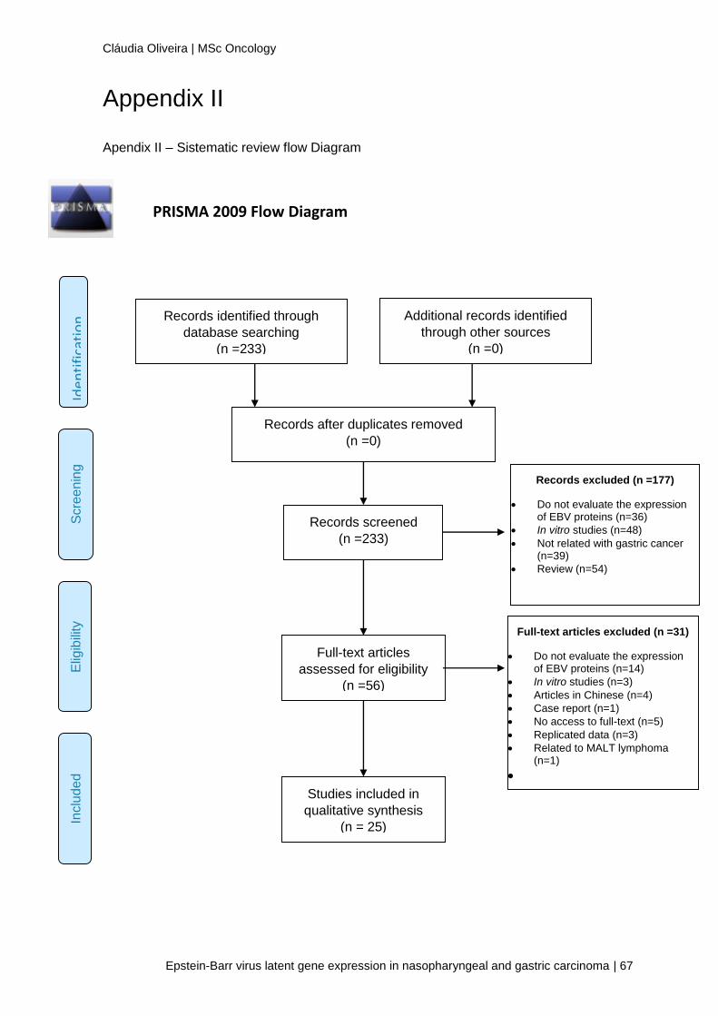
Cláudia Oliveira | MSc Oncology
Epstein-Barr virus latent gene expression in nasopharyngeal and gastric carcinoma | 67
Appendix II Apendix II – Sistematic review flow Diagram
PRISMA 2009 Flow Diagram
Records identified through
database searching
(n =233)
Scre
en
ing
Inclu
de
d
Elig
ibili
ty
Iden
tifi
cati
on
Additional records identified
through other sources
(n =0)
Records after duplicates removed
(n =0)
Records screened
(n =233)
Full-text articles
assessed for eligibility
(n =56)
Full-text articles excluded (n =31)
Do not evaluate the expression of EBV proteins (n=14)
In vitro studies (n=3)
Articles in Chinese (n=4)
Case report (n=1)
No access to full-text (n=5)
Replicated data (n=3)
Related to MALT lymphoma (n=1)
Studies included in
qualitative synthesis
(n = 25)
Records excluded (n =177)
Do not evaluate the expression of EBV proteins (n=36)
In vitro studies (n=48)
Not related with gastric cancer (n=39)
Review (n=54)