Embed Size (px)
Citation preview
British Journal of Ophthalmology, 1978, 62, 543-546
Foveo-macular retinitis, solar retinopathy, and traumaR. H. B. GREYFrom the Department of Clinical Ophthalmology, Institiute of Ophthalmology, and Moorfields EyeHospital, London
SUMMARY Three patients are described with foveal lesions resembling minute holes followingtrauma. The similarity of the lesions to foveomacular retinitis and solar retinopathy suggests thatall these conditions produce a similar, localised neuroretinal lesion with sparing of the pigmentepithelium. Loss of the photoreceptors at the fovea would be expected to produce a lesionresembling a small retinal hole.
Cordes (1944) described a series of patients whopresented with blurred vision and a circular greyishlesion at the macula. After 1 to 2 weeks the lesionresolved, leaving a minute red foveal hole withvariable visual recovery. He called the conditionfoveomacular retinitis. Subsequent authors con-sidered this disorder to be central serous retinopathy,but Kerr and Little (1966) described a further seriesof patients with foveomacular retinitis and empha-sised the difference between these two conditions.On account of the vast preponderance of young, fit,male military recruits in all the reported series offoveomacular retinitis and because of the similarityof the lesions to solar retinopathy, Ewald andRitchey (1970) looked specifically for evidence ofsun-gazing. Of their 47 patients 690% admitted tosun-gazing with or without the use of drugs, andthey concluded that foveomacular retinitis was aself-inflicted condition largely to avoid active militaryservice. Marlor et al. (1973) examined 147 Marinerecruits with foveomacular retinitis in an attempt toelucidate the aetiology of this condition; they wereunable to confirm sun-gazing as the cause.The purpose of this report is to describe 3 patients
with the appearance of foveomacular retinitis inwhom the cause is known to be blunt trauma andto compare the fundus lesion with solar retinopathy.
Case histories
CASE 1A 19-year-old male presented to the casualtydepartment with blurred vision in the left eye afterbeing hit by a football the previous day. The leftvisual acuity was 6/9 and there was a mild anterior
Address for reprints: R. H. B. Grey, FRCS, MoorfieldsEye Hospital, City Road, London EC1V 2PD
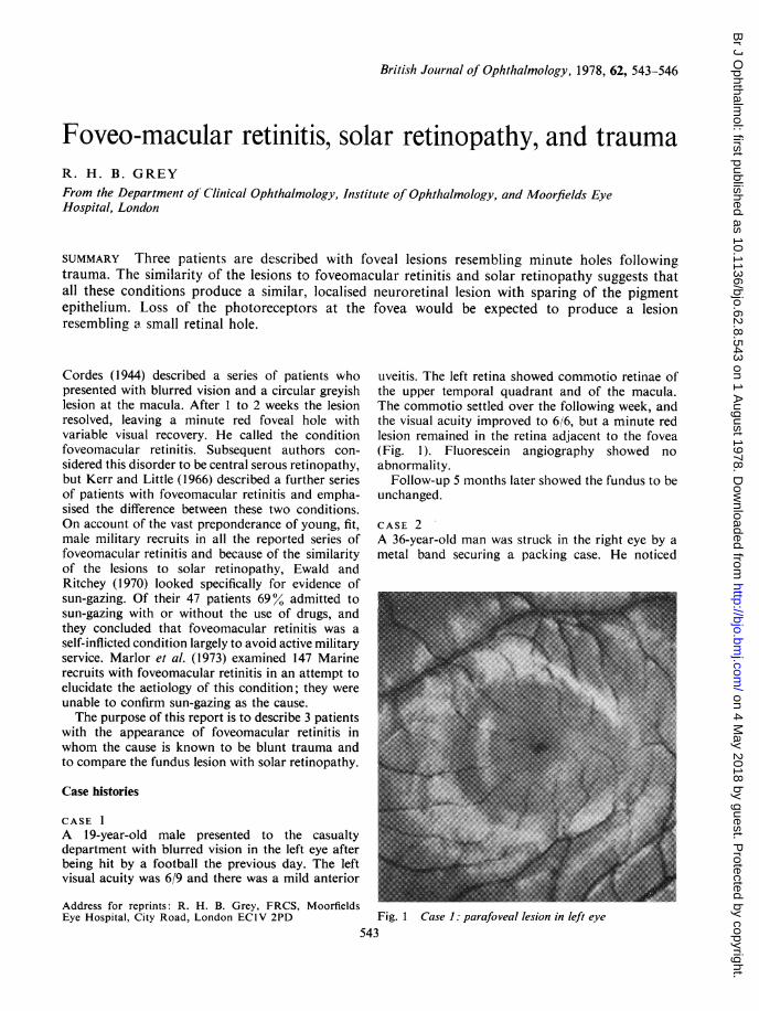
uveitis. The left retina showed commotio retinae ofthe upper temporal quadrant and of the macula.The commotio settled over the following week, andthe visual acuity improved to 6/6, but a minute redlesion remained in the retina adjacent to the fovea(Fig. 1). Fluorescein angiography showed noabnormality.
Follow-up 5 months later showed the fundus to beunchanged.
CASE 2A 36-year-old man was struck inmetal band securing a packing
the right eye by acase. He noticed
Fig. 1 Case 1: parafoveal lesion in left eye
543
on 4 May 2018 by guest. P
rotected by copyright.http://bjo.bm
j.com/
Br J O
phthalmol: first published as 10.1136/bjo.62.8.543 on 1 A
ugust 1978. Dow
nloaded from
R. H. B. Grey
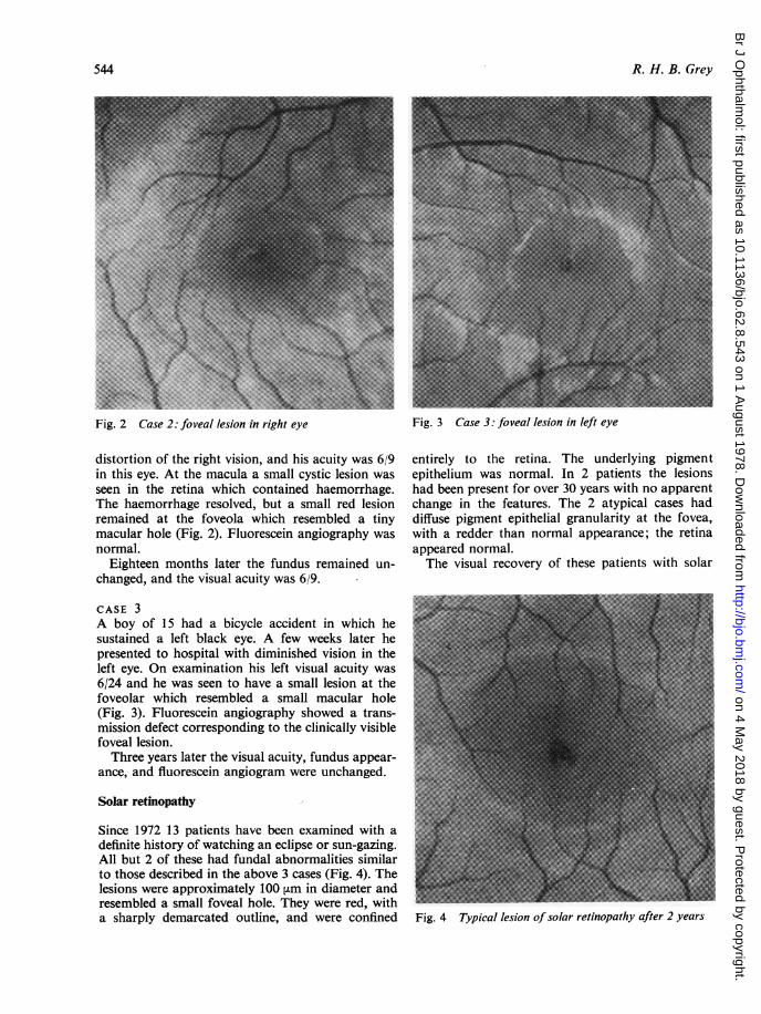
Fig. 2 Case 2: foveal lesion in right eye
distortion of the right vision, and his acuity was 6/9in this eye. At the macula a small cystic lesion wasseen in the retina which contained haemorrhage.The haemorrhage resolved, but a small red lesionremained at the foveola which resembled a tinymacular hole (Fig. 2). Fluorescein angiography wasnormal.
Eighteen months later the fundus remained un-changed, and the visual acuity was 6/9.
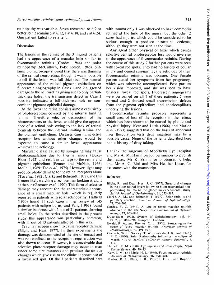
CASE 3A boy of 15 had a bicycle accident in which hesustained a left black eye. A few weeks later hepresented to hospital with diminished vision in theleft eye. On examination his left visual acuity was6/24 and he was seen to have a small lesion at thefoveolar which resembled a small macular hole(Fig. 3). Fluorescein angiography showed a trans-mission defect corresponding to the clinically visiblefoveal lesion.
Three years later the visual acuity, fundus appear-ance, and fluorescein angiogram were unchanged.
Solar retinopathy
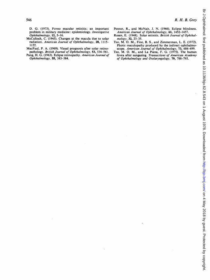
Since 1972 13 patients have been examined with adefinite history of watching an eclipse or sun-gazing.All but 2 of these had fundal abnormalities similarto those described in the above 3 cases (Fig. 4). Thelesions were approximately 100 ,um in diameter andresembled a small foveal hole. They were red, witha sharply demarcated outline, and were confined
Fig. 3 Case 3: foveal lesion in left eye
entirely to the retina. The underlying pigmentepithelium was normal. In 2 patients the lesionshad been present for over 30 years with no apparentchange in the features. The 2 atypical cases haddiffuse pigment epithelial granularity at the fovea,with a redder than normal appearance; the retinaappeared normal.The visual recovery of these patients with solar
Fig. 4 Typical lesion of solar retinopathy after 2 years
544
on 4 May 2018 by guest. P
rotected by copyright.http://bjo.bm
j.com/
Br J O
phthalmol: first published as 10.1136/bjo.62.8.543 on 1 A
ugust 1978. Dow
nloaded from
Foveo-macu/lar retinitis, solar Eretilnopatliy, ci,idtrauima5
retinopathy was variable. Seven recovered to 6 9 orbetter, but 2 remained at 6 12, 1 at 6 18, and 2 at 6 24.One patient failed to re-attend.
Discussion
The lesions in the retinae of the 3 injured patientshad the appearance of a macular hole similar tofoveomacular retinitis (Cordes, 1944) and solarretinopathy (McCulloch, 1945; Rosen, 1948). Slit-lamp biomicroscopy showed that there was thinningot the central neuroretina, though it was impossibleto tell if the lesion was full thickness. The normalappearance of the retinal pigment epithelium onfluorescein angiography in Cases 1 and 2 suggesteddamage to the neuroretina giving rise to only partial-thickness holes; the transmission defect in Case 3possibly indicated a full-thickness hole or con-comitant pigment epithelial damage.
At the fovea the retina consists almost exclusivelyof photoreceptors covered by the internal limitinglamina. Therefore selective destruction of thephotoreceptors at the fovea would give the appear-ance of a retinal hole owing to the lack of retinalelements between the internal limiting lamina andthe pigment epithelium. Diseases causing selectivereceptor loss without other changes would beexpected to cause a similar foveal appearancewhatever the aetiology.
Macular disease caused by sun-gazing may causephotocoagulation due to thermal injury (Duke-Elder, 1972) and result in damage to the retina andpigment epithelium (Penner and McNair, 1966;McFaul, 1969; Tso et al., 1975). However, light mayproduce photic damage to the retinal receptors alone(Tso et al., 1972; Clarke and Behrendt, 1972), and thisis more likely watching an eclipse than looking straightat the sun (Geeraets et al., 1970). This form of selectivedamage may account for the characteristic appear-ance of a small macular hole, which is regularlyreported in patients with solar retinopathy. Hatfield(1970) found 11 such cases in her review of 145patients with eclipse burns, and Pang (1963) founda similar incidence with 2 out of 21 patients showingsmall holes. In the series described in the presentstudy this appearance was particularly common,with 11 out of 13 patients affected in this way.Trauma has been shown to cause receptor damage
(Blight and Hart, 1977). In their experiments thedamage was demonstrated at the site of impact andwas not confined to the receptors; regeneration wasalso shown to occur. However, it is conceivable thatselective photoreceptor damage may occur in manunder some circumstances and produce the uniquechanges which give rise to the clinical appearance ofa foveal red spot. Of the 3 patients described here
with trauma only I was observed to hiave commotioretinae at the time of the injury, but the other 2cases had injuries which could be considered to beserious enough to produce intraretinal oedema,although they were not seen at the time.Any agent either physical or toxic which causes
selective central photoreceptor loss would give riseto the appearance of foveomacular retinitis. Duringthe course of this study 7 further patients were seenwith foveal red spots. They had no history of ocularinjury and denied sun-gazing; the aetiology of theirfoveomacular retinitis was obscure. One femalepatient dated her symptoms from her pregnancy,which was otherwise uncomplicated. Post partumher vision improved, and she was seen to havebilateral foveal red spots. Fluorescein angiogramswere performed on all 7 of these patients; 5 werenormal and 2 showed small transmission defectsfrom the pigment epithelium and choriocapillarisunderlying the lesions.Foveomacular retinitis probably represents a
small area of loss of the receptors in the retina,which has been shown to be caused by photic andphysical injury. Kerr and Little (1966) and Marloret al. (1973) suggested that on the basis of abnormalliver flocculation tests drug ingestion may be apossible cause. None of the patients described herehad a history of drug taking.
I thank the surgeons of Moorfields Eye Hospitaland Mr A. M. Hamilton for permission to publishtheir cases, Mr K. Sehmi for photographic help,and Mr A. C. Bird and Miss Heather Lucas forassistance with the manuscript.
References
Blight, R., and Dean Hart, J. C. (1977). Structural chanigesin the outer retinal layers following blunt mechanical non-perforating trauma to the globe: an experimental study.British Journal of Opthalmtiology, 61, 573-587.
Clarke, A. M., and Behrendt, T. (1972). Solar retinitis andpupillary reaction. Amnerican Journal of Ophthalclmology.73, 700-703.
Cordes, F. C. (1944). A type of foveo macular retinitisobserved in the US Navy. Almerican, Jouirnial of Ophthal-tmology,, 27, 803-816.
Duke-Elder (1972). System of Ophthalmology, vol. 14,Pt. 2, pp. 885-896. Kimpton: London.
Ewald, R. A., and Ritchey, C. L. (1970). Sungazing as thecause of foveo macular retiniitis. Amierican Jouarital ofOphthalmology, 70, 491-497.
Geeraets, W. J., Nooney, T. W., Svoboda, J. R., and Ching,F. C. (1970). Solar Retinopathy following the eclipse ofMarch 7 1970. Medical College of Virgimiia Quarterly, 6,3-7.
Hatfield, E. M. (1970). Eye injuries and solar eclipse. SightSavinig Review, 40, 79-85.
Kerr, L. M., and Little, H. L. (1966). Foveo macular retinitis.Archives of Ophthalmology, 76, 498-504.
Marlor, R. L., Blais, B. R., Preston, F. R., and Boydon,
545
on 4 May 2018 by guest. P
rotected by copyright.http://bjo.bm
j.com/
Br J O
phthalmol: first published as 10.1136/bjo.62.8.543 on 1 A
ugust 1978. Dow
nloaded from
R. H. B. Grey
D. G. (1973). Foveo macular retinitis; an importantproblem in military medicine: epidemiology. InvestigativeOphthalmology, 12, 5-16.
McCulloch, C. (1945). Changes at the macula due to solarradiation. American Journal of Ophthalmology, 28, 1115-1122.
MacFaul, P. A. (1969). Visual prognosis after solar retino-pathology. British Journal of Ophthalmology, 53, 534-541.
Pang, H. G. (1963). Eclipse retinopathy. American Journal ofOphthalmology, 55, 383-384.
Penner, R., and McNair, J. N. (1966). Eclipse blindness.American Journal of Ophthalmology, 61, 1452-1457.
Rosen, E. (1948). Solar retinitis. British Journal of Ophthal-mology, 32, 23-35.
Tso, M. 0. M., Fine, B. S., and Zimmerman, L. E. (1972).Photic maculopathy produced by the indirect ophthalmo-scope. American Journal of Ophthalmology, 73, 686-699.
Tso, M. 0. M., and La Piana, F. G. (1975). The humanfovea after sungazing. Transactions of American Academyof Ophthalmology and Otolaryngology, 79, 788-795.
546
on 4 May 2018 by guest. P
rotected by copyright.http://bjo.bm
j.com/
Br J O
phthalmol: first published as 10.1136/bjo.62.8.543 on 1 A
ugust 1978. Dow
nloaded from