Embed Size (px)
Citation preview
358 Clinical Advances in Hematology & Oncology Volume 12, Issue 6 June 2014
Malignancies Associated With Epstein-Barr Virus: Pathobiology, Clinical Features, and Evolving Treatments Natalia Neparidze, MD, and Jill Lacy, MD
Abstract: Epstein-Barr virus (EBV) is associated with a wide
variety of B-cell–derived lymphoid neoplasms, including Burkitt
lymphoma, lymphomas arising in immunocompromised patients
(post-transplant and HIV-associated lymphomas), and Hodgkin
lymphoma. In addition, EBV has been linked to some T-cell
lymphomas (angioimmunoblastic T-cell lymphoma, extranodal
nasal-type natural killer/T-cell lymphoma, and other rare histo-
types), nasopharyngeal cancer, and a subset of gastric cancers.
Advances in our understanding of the pathobiology of EBV onco-
genesis, including the transforming and immunogenic properties
of the virus and the role of immune dysregulation, have provided
the rationale for new treatment strategies. Emerging EBV-specific
therapeutic approaches include activation of lytic viral infec-
tion combined with antiviral drugs, inhibition of EBV-induced
oncogenic cellular signaling pathways, adoptive EBV-specific
T-cell therapies, and EBV vaccines. This review summarizes the
pathobiology, clinical features, and treatment of EBV-associated
malignancies, including new and evolving therapies focused on
exploiting the pathobiology of EBV.
Introduction
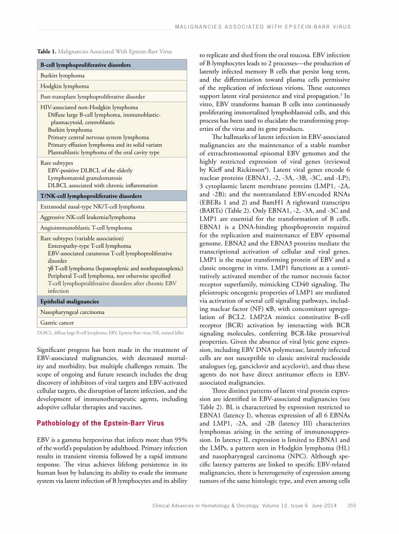
Epstein-Barr virus (EBV), a ubiquitous B-lymphotropic herpesvi-rus, was the first virus directly linked to cancer in humans. Since its discovery and association with Burkitt lymphoma (BL) 50 years ago, EBV has been associated with a heterogeneous group of lymphomas and epithelial tumors (Table 1). It infects B and T lym-phocytes, follicular dendritic cells, smooth muscle cells, squamous epithelium of the oropharynx and nasopharynx, and glandular epithelium of the thyroid, stomach, and salivary glands. B lympho-cytes are the major cellular reservoir for EBV persistence, and the majority of EBV-associated malignancies derive from EBV-infected B cells.1 EBV encodes an array of products that mimic or activate antiapoptotic molecules, cytokines, and signal transducers, thereby promoting EBV infection, immortalization, and transformation.2
Dr Neparidze is an assistant professor of medicine and Dr Lacy is an associate professor of medicine in the Section of Medical Oncology at the Yale University School of Medicine in West Haven, Connecticut.
Address correspondence to:Natalia Neparidze, MDAssistant Professor of MedicineSection of Medical Oncology Yale University School of Medicine VA Connecticut Healthcare Center 950 Campbell Avenue (III-d) West Haven, CT 06516 Phone: 203-937-3421 Fax: 203-937-3803E-mail: [email protected]
KeywordsBurkitt lymphoma, Epstein-Barr virus, gastric cancer, Hodgkin lymphoma, nasopharyngeal, post-transplant lymphoproliferative disorder
Clinical Advances in Hematology & Oncology Volume 12, Issue 6 June 2014 359
M A L I G N A N C I E S A S S O C I AT E D W I T H E P S T E I N - B A R R V I R U S
Significant progress has been made in the treatment of EBV-associated malignancies, with decreased mortal-ity and morbidity, but multiple challenges remain. The scope of ongoing and future research includes the drug discovery of inhibitors of viral targets and EBV-activated cellular targets, the disruption of latent infection, and the development of immunotherapeutic agents, including adoptive cellular therapies and vaccines.
Pathobiology of the Epstein-Barr Virus
EBV is a gamma herpesvirus that infects more than 95% of the world’s population by adulthood. Primary infection results in transient viremia followed by a rapid immune response. The virus achieves lifelong persistence in its human host by balancing its ability to evade the immune system via latent infection of B lymphocytes and its ability
to replicate and shed from the oral mucosa. EBV infection of B lymphocytes leads to 2 processes—the production of latently infected memory B cells that persist long term, and the differentiation toward plasma cells permissive of the replication of infectious virions. These outcomes support latent viral persistence and viral propagation.3 In vitro, EBV transforms human B cells into continuously proliferating immortalized lymphoblastoid cells, and this process has been used to elucidate the transforming prop-erties of the virus and its gene products.
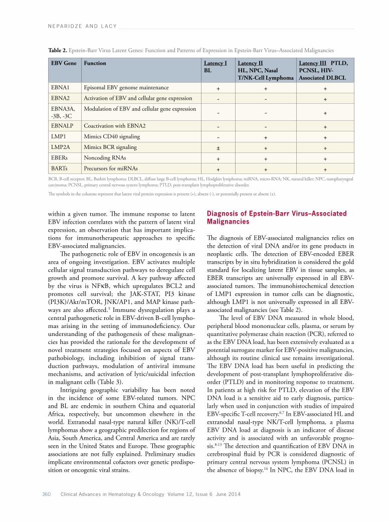
The hallmarks of latent infection in EBV-associated malignancies are the maintenance of a stable number of extrachromosomal episomal EBV genomes and the highly restricted expression of viral genes (reviewed by Kieff and Rickinson4). Latent viral genes encode 6 nuclear proteins (EBNA1, -2, -3A, -3B, -3C, and -LP); 3 cytoplasmic latent membrane proteins (LMP1, -2A, and -2B); and the nontranslated EBV-encoded RNAs (EBERs 1 and 2) and BamH1 A rightward transcripts (BARTs) (Table 2). Only EBNA1, -2, -3A, and -3C and LMP1 are essential for the transformation of B cells. EBNA1 is a DNA-binding phosphoprotein required for the replication and maintenance of EBV episomal genome. EBNA2 and the EBNA3 proteins mediate the transcriptional activation of cellular and viral genes. LMP1 is the major transforming protein of EBV and a classic oncogene in vitro. LMP1 functions as a consti-tutively activated member of the tumor necrosis factor receptor superfamily, mimicking CD40 signaling. The pleiotropic oncogenic properties of LMP1 are mediated via activation of several cell signaling pathways, includ-ing nuclear factor (NF) κB, with concomitant upregu-lation of BCL2. LMP2A mimics constitutive B-cell receptor (BCR) activation by interacting with BCR signaling molecules, conferring BCR-like prosurvival properties. Given the absence of viral lytic gene expres-sion, including EBV DNA polymerase, latently infected cells are not susceptible to classic antiviral nucleoside analogues (eg, ganciclovir and acyclovir), and thus these agents do not have direct antitumor effects in EBV-associated malignancies.
Three distinct patterns of latent viral protein expres-sion are identified in EBV-associated malignancies (see Table 2). BL is characterized by expression restricted to EBNA1 (latency I), whereas expression of all 6 EBNAs and LMP1, -2A, and -2B (latency III) characterizes lymphomas arising in the setting of immunosuppres-sion. In latency II, expression is limited to EBNA1 and the LMPs, a pattern seen in Hodgkin lymphoma (HL) and nasopharyngeal carcinoma (NPC). Although spe-cific latency patterns are linked to specific EBV-related malignancies, there is heterogeneity of expression among tumors of the same histologic type, and even among cells
Table 1. Malignancies Associated With Epstein-Barr Virus
B-cell lymphoproliferative disorders
Burkitt lymphoma
Hodgkin lymphoma
Post-transplant lymphoproliferative disorder
HIV-associated non-Hodgkin lymphoma Diffuse large B-cell lymphoma, immunoblastic- plasmacytoid, centroblastic Burkitt lymphoma Primary central nervous system lymphoma Primary effusion lymphoma and its solid variant Plasmablastic lymphoma of the oral cavity type
Rare subtypes EBV-positive DLBCL of the elderly Lymphomatoid granulomatosis DLBCL associated with chronic inflammation
T/NK-cell lymphoproliferative disorders
Extranodal nasal-type NK/T-cell lymphoma
Aggressive NK-cell leukemia/lymphoma
Angioimmunoblastic T-cell lymphoma
Rare subtypes (variable association) Enteropathy-type T-cell lymphoma EBV-associated cutaneous T-cell lymphoproliferative
disorder γδ T-cell lymphoma (hepatosplenic and nonhepatosplenic) Peripheral T-cell lymphoma, not otherwise specified T-cell lymphoproliferative disorders after chronic EBV
infection
Epithelial malignancies
Nasopharyngeal carcinoma
Gastric cancerDLBCL, diffuse large B-cell lymphoma; EBV, Epstein-Barr virus; NK, natural killer.
360 Clinical Advances in Hematology & Oncology Volume 12, Issue 6 June 2014
N E PA R I D Z E A N D L A C Y
within a given tumor. The immune response to latent EBV infection correlates with the pattern of latent viral expression, an observation that has important implica-tions for immunotherapeutic approaches to specific EBV-associated malignancies.
The pathogenetic role of EBV in oncogenesis is an area of ongoing investigation. EBV activates multiple cellular signal transduction pathways to deregulate cell growth and promote survival. A key pathway affected by the virus is NFκB, which upregulates BCL2 and promotes cell survival; the JAK-STAT, PI3 kinase (PI3K)/Akt/mTOR, JNK/AP1, and MAP kinase path-ways are also affected.5 Immune dysregulation plays a central pathogenetic role in EBV-driven B-cell lympho-mas arising in the setting of immunodeficiency. Our understanding of the pathogenesis of these malignan-cies has provided the rationale for the development of novel treatment strategies focused on aspects of EBV pathobiology, including inhibition of signal trans-duction pathways, modulation of antiviral immune mechanisms, and activation of lytic/suicidal infection in malignant cells (Table 3).
Intriguing geographic variability has been noted in the incidence of some EBV-related tumors. NPC and BL are endemic in southern China and equatorial Africa, respectively, but uncommon elsewhere in the world. Extranodal nasal-type natural killer (NK)/T-cell lymphomas show a geographic predilection for regions of Asia, South America, and Central America and are rarely seen in the United States and Europe. These geographic associations are not fully explained. Preliminary studies implicate environmental cofactors over genetic predispo-sition or oncogenic viral strains.
Diagnosis of Epstein-Barr Virus–Associated Malignancies
The diagnosis of EBV-associated malignancies relies on the detection of viral DNA and/or its gene products in neoplastic cells. The detection of EBV-encoded EBER transcripts by in situ hybridization is considered the gold standard for localizing latent EBV in tissue samples, as EBER transcripts are universally expressed in all EBV-associated tumors. The immunohistochemical detection of LMP1 expression in tumor cells can be diagnostic, although LMP1 is not universally expressed in all EBV-associated malignancies (see Table 2).
The level of EBV DNA measured in whole blood, peripheral blood mononuclear cells, plasma, or serum by quantitative polymerase chain reaction (PCR), referred to as the EBV DNA load, has been extensively evaluated as a potential surrogate marker for EBV-positive malignancies, although its routine clinical use remains investigational. The EBV DNA load has been useful in predicting the development of post-transplant lymphoproliferative dis-order (PTLD) and in monitoring response to treatment. In patients at high risk for PTLD, elevation of the EBV DNA load is a sensitive aid to early diagnosis, particu-larly when used in conjunction with studies of impaired EBV-specific T-cell recovery.6,7 In EBV-associated HL and extranodal nasal-type NK/T-cell lymphoma, a plasma EBV DNA load at diagnosis is an indicator of disease activity and is associated with an unfavorable progno-sis.8-13 The detection and quantification of EBV DNA in cerebrospinal fluid by PCR is considered diagnostic of primary central nervous system lymphoma (PCNSL) in the absence of biopsy.14 In NPC, the EBV DNA load in
Table 2. Epstein-Barr Virus Latent Genes: Function and Patterns of Expression in Epstein-Barr Virus–Associated Malignancies
EBV Gene Function Latency IBL
Latency II HL, NPC, Nasal T/NK-Cell Lymphoma
Latency III PTLD, PCNSL, HIV-Associated DLBCL
EBNA1 Episomal EBV genome maintenance + + +EBNA2 Activation of EBV and cellular gene expression - - +EBNA3A, -3B, -3C
Modulation of EBV and cellular gene expression - - +
EBNALP Coactivation with EBNA2 - - +LMP1 Mimics CD40 signaling - + +LMP2A Mimics BCR signaling ± + +EBERs Noncoding RNAs + + +BARTs Precursors for miRNAs + + +
BCR, B-cell receptor; BL, Burkitt lymphoma; DLBCL, diffuse large B-cell lymphoma; HL, Hodgkin lymphoma; miRNA, micro-RNA; NK, natural killer; NPC, nasopharyngeal carcinoma; PCNSL, primary central nervous system lymphoma; PTLD, post-transplant lymphoproliferative disorder.
The symbols in the columns represent that latent viral protein expression is present (+), absent (-), or potentially present or absent (±).
Clinical Advances in Hematology & Oncology Volume 12, Issue 6 June 2014 361
M A L I G N A N C I E S A S S O C I AT E D W I T H E P S T E I N - B A R R V I R U S
plasma is useful as a diagnostic and prognostic tool and in monitoring response to treatment.15,16
Epstein-Barr Virus–Associated Lymphoproliferative Disorders
The EBV-associated B- and T-cell lymphoproliferative disorders are a heterogeneous group of malignancies but share the feature of harboring latent EBV within tumor cells. Immunodeficiency states, such as HIV infection, congenital immunodeficiencies, post-transplant immu-nosuppression, and chronic active EBV infection, increase the risk for EBV-associated lymphoma.
These lymphomas display different patterns of latent viral gene expression, with the potential for distinct novel treatment approaches, such as T-cell immunotherapy target-ing EBV.17 However, at present, the standard treatment of EBV-associated lymphomas is usually identical to that of their EBV-negative counterparts, with the exception of PTLD.
Burkitt LymphomaThe isolation of virus particles from BL cell lines in 1964 led to the discovery of EBV and its association with BL. BL is a highly aggressive non-Hodgkin lymphoma (NHL) characterized by a diffuse infiltrate of monomorphic, medium-size B cells in a “starry sky” pattern, imparted by
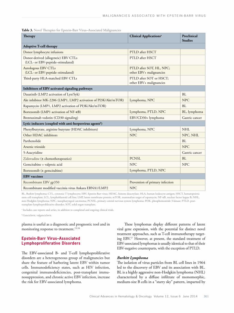
Table 3. Novel Therapies for Epstein-Barr Virus–Associated Malignancies
Therapy Clinical Applicationsa
Preclinical Studies
Adoptive T-cell therapy
Donor lymphocyte infusions PTLD after HSCT
Donor-derived (allogeneic) EBV CTLs (LCL- or EBV-peptide–stimulated)
PTLD after HSCT
Autologous EBV CTLs (LCL- or EBV-peptide–stimulated)
PTLD after SOT, HL, NPC; other EBV+ malignancies
Third-party HLA-matched EBV CTLs PTLD after SOT or HSCT; other EBV+ malignancies
Inhibitors of EBV-activated signaling pathways
Dasatinib (LMP2 activation of Lyn/Syk) BL
Akt inhibitor MK-2206 (LMP1, LMP2 activation of PI3K/Akt/mTOR) Lymphoma, NPC NPC
Rapamycin (LMP1, LMP2 activation of PI3K/Akt/mTOR) BL
Bortezomib (LMP1 activation of NF-κB) Lymphoma, PTLD, NPC BL, lymphoma
Brentuximab vedotin (CD30 signaling) EBV/CD30+ lymphoma Gastric cancer
Lytic inducers (coupled with anti-herpesvirus agentsb)
Phenylbutyrate, arginine butyrate (HDAC inhibitors) Lymphoma, NPC NHL
Other HDAC inhibitors NPC NPC, NHL
Parthenolide BL
Arsenic trioxide NPC
5-Azacytidine Gastric cancer
Zidovudine (± chemotherapeutics) PCNSL BL
Gemcitabine + valproic acid NPC NPC
Bortezomib (± gemcitabine) Lymphoma, PTLD, NPC
EBV vaccines
Recombinant EBV gp350 Prevention of primary infection
Recombinant modified vaccinia virus Ankara EBNA1/LMP2 NPCBL, Burkitt lymphoma; CTL, cytotoxic T lymphocyte; EBV, Epstein-Barr virus; HDAC, histone deacetylase; HLA, human leukocyte antigen; HSCT, hematopoietic stem cell transplant; LCL, lymphoblastoid cell line; LMP, latent membrane protein; mTOR, mammalian target of rapamycin; NF-κB, nuclear factor kappa B; NHL, non-Hodgkin lymphoma; NPC, nasopharyngeal carcinoma; PCNSL, primary central nervous system lymphoma; PI3K, phosphoinositide 3-kinase; PTLD, post-transplant lymphoproliferative disorder; SOT, solid organ transplant.
a Includes case reports and series, in addition to completed and ongoing clinical trials.
b Ganciclovir, valganciclovir.
362 Clinical Advances in Hematology & Oncology Volume 12, Issue 6 June 2014
N E PA R I D Z E A N D L A C Y
numerous benign macrophages, and by an extremely high proliferative index, with a Ki-67 approaching 100%. BL is divided into 3 subtypes: endemic (eBL), sporadic (sBL), and HIV-associated. eBL presents as tumors affecting the jaw and facial bones in young children in equatorial Africa, whereas sBL occurs worldwide and involves the gut, upper respiratory tract, or Waldeyer ring.18 HIV-associated BL characteristically involves lymph nodes and bone marrow. BL is not universally associated with EBV.19 Although more than 90% of cases of eBL are EBV-positive, only 5% to 20% of cases of sBL and 40% of cases of HIV-associated BL are EBV-positive.20-22
EBV gene expression in BL cells from primary tissue is highly restricted, with expression limited to EBNA1 (latency I). The absence of consistent expression of the immunogenic EBNA and LMP proteins facilitates eva-sion from cytotoxic T-lymphocyte (CTL)–mediated immunosurveillance and contributes to BL pathogenesis. Moreover, the use of adoptive cellular therapies in BL is limited by the insensitivity of EBNA1-restricted BL cells to CTL-mediated cytotoxicity.
Because EBV is not essential in the pathogenesis of BL, the mechanisms by which EBV contributes to the development of BL remain uncertain.23 In eBL, it is believed that hyperstimulation of B cells and suppres-sion of T-cell activity by chronic malarial infection is permissive for the reactivation of EBV in infected B cells, leading to a dramatic expansion of EBV-infected B-cell populations. In both EBV-positive and EBV-negative BL, constitutive activation of the c-MYC oncogene through its translocation into one of the immunoglobulin loci is the critical oncogenic event.22,24,25 Whether there is a causal relationship between EBV and the translocation of MYC is unknown.
The optimal treatment of BL has yet to be defined, and patients with BL should be enrolled in clinical tri-als whenever possible. Outside a clinical trial, treatment consists of intense combination chemotherapy regimens with CNS prophylaxis, such as fractionated cyclophos-phamide, vincristine, doxorubicin, and dexamethasone (hyper-CVAD); cyclophosphamide, vincristine, doxo-rubicin, and high-dose methotrexate (CODOX-M) with ifosfamide, etoposide, and high-dose cytarabine (IVAC); or the Cancer and Leukemia Group B (CALGB) 9251 protocol, usually in combination with rituximab (Rituxan, Genentech/Biogen Idec).26-30 The less intensive regimens used in other types of NHL, such as cyclo-phosphamide, doxorubicin, vincristine, and prednisone (CHOP), are not adequate therapy and result in frequent relapses.31,32 At present, EBV-negative and EBV-positive BLs are treated in an identical manner.
Novel agents focused on exploiting aspects of EBV biology are under investigation for the treatment of
EBV-positive BL in preclinical studies. Although viral gene expression is generally restricted to EBNA1 (type I latency), LMP2A expression has been demonstrated in some primary BL biopsies when assessed by PCR and Western blotting.33,34 This finding provides the rationale for the development of inhibitors of LMP2A-induced cel-lular targets, such as the PI3K/Akt/mTOR pathway. In a model of EBV-associated BL in Tg6/λ-MYC transgenic mice, the mTOR inhibitor rapamycin reversed spleno-megaly and decreased tumor growth and metastasis in bone marrow.35 An alternative approach is the induction of lytic EBV infection, leading to cell lysis, coupled with anti-EBV agents. Thus, bortezomib (Velcade, Millennium Pharmaceuticals) may activate EBV lytic gene expression in BL cell lines in the context of endoplasmic reticulum stress, with C/EBPβ playing a role in this process,36 and lytic cytotoxicity induced by lactone parthenolide in combination with ganciclovir has shown promise as a virus-targeted therapy in BL in studies in vitro.37 Recently, high-throughput screen technologies have identified small molecular inhibitors of EBNA1, and further development of EBNA1 inhibitors may provide a treatment specific for EBV latent infection.38
Hodgkin LymphomaThe identification of EBV DNA, EBER RNA, and LMP1 in Reed-Sternberg (RS) cells in a subset of HL has confirmed the link between EBV and HL.39-41 Fur-thermore, epidemiologic and case-control studies have shown an increased risk for EBV-positive but not EBV-negative HL in individuals with a history of infectious mononucleosis or an altered serologic response to EBV latent antigens, supporting a causal association between EBV and HL.39,42-44 The prevalence of EBV varies widely among the pathologic subtypes of HL. EBV is present in 70% of cases of mixed-cellularity HL, 95% of cases of lymphocyte-depleted HL, and 10% to 40% of cases of nodular sclerosing HL, whereas nodular lymphocyte-predominant HL is generally EBV-negative.45 HL arising in the setting of immunodeficiency (eg, HIV infection, iatrogenic immunodeficiency) is usually EBV-positive.
EBV gene expression in RS cells is restricted to EBNA1, LMP1, LMP2A and -2B, and EBERs (type II latency).46 The EBV genomes found in RS cells are clonal, indicating that EBV infection precedes clonal expansion and implicating an etiologic role of the virus. However, the precise role of EBV in HL pathogenesis is uncertain.47 Expression of LMP1 and LMP2A may prevent apoptosis through the induction of antiapoptotic proteins. RS cells (both EBV-positive and EBV-negative) produce immuno-suppressive cytokines such as interleukin 10, interleukin 13, and transforming growth factor-β.48,49 EBV infection of primary RS cells and RS cell–derived cell lines has been
Clinical Advances in Hematology & Oncology Volume 12, Issue 6 June 2014 363
M A L I G N A N C I E S A S S O C I AT E D W I T H E P S T E I N - B A R R V I R U S
shown to increase expression of the CCL20 chemokines, which in turn increases the migration of CD4+/FOXp3+ regulatory T cells (Tregs). This observation identifies a mechanism by which EBV-infected RS cells can recruit Tregs to the HL microenvironment and prevent immune responses against the virus-infected RS cells.50,51
Standard treatment of EBV-positive HL is not dif-ferent from that for EBV-negative HL of the same stage, histology, and prognosis52 and is guided by clinical stage and risk stratification. Systemic chemotherapy with doxorubicin, bleomycin, vinblastine, and dacarbazine (ABVD), followed by involved-field radiotherapy when indicated, is considered the gold standard. The regimen of bleomycin, etoposide, doxorubicin, cyclophosphamide, vincristine, procarbazine, and prednisone (BEACOPP) is an alternative option for patients with high-risk, advanced-stage disease.
The success of adoptive immunotherapy with ex vivo expanded allogeneic or autologous EBV-specific CTLs in PTLD (described below) has led to the application of this strategy in HL. Limited experience with EBV-specific CTLs in patients with recurrent, refractory, EBV-positive HL has shown promise.53-55 However, this strategy has not been widely adopted because of the complexity of the technique. In addition, the immune microenvironment of the tumor might impede the efficacy of CTLs in HL.
The incidence of classic HL is increased in settings of impaired immunity, including after transplant. Post-transplant HL is invariably EBV-positive and should fulfill the diagnostic criteria for classic HL. The majority of patients are men, and all have received post-transplant immunosuppression.56-59 The time from transplant to the onset of the disease ranges from a few months to several years and is generally longer than that for non-Hodgkin PTLDs.59,60 In 50% of cases, the disease presents as extra-nodal masses in liver or lung, and other extranodal sites can be involved.
The optimal treatment of post-transplant HL is not well defined. The clinical course is aggressive, and the outcome is poor. The majority of patients are initially managed by reduction or withdrawal of immunosuppres-sion. The use of chemotherapy may be limited because of comorbidities, and the response rate is lower than in classic HL. Rituximab is highly effective in non-Hodgkin PTLD,61 and some patients with post-transplant HL respond to rituximab.
Post-transplant Lymphoproliferative DisorderIt is widely recognized that the incidence of lymphopro-liferative disorders is increased in transplant recipients of both solid organ and hematopoietic stem cell allografts. The vast majority of these PTLDs are associated with EBV. The process likely begins with dysregulated EBV-
driven B-cell proliferation due to impaired EBV-specific T-cell–mediated immune surveillance of infected recipi-ent or donor B cells. This leads to a dramatic expansion of the EBV-infected B-cell population, the acquisition of mutations, and ultimately, malignant transformation.62
Approximately 95% of all PTLDs are associated with EBV, as shown by EBER expression in tissue-infiltrating lymphocytes and/or immunoblasts. There are 4 major World Health Organization (WHO) categories of PTLD: early lesions, polymorphic PTLD, monomorphic PTLD, and classic HL-type PTLD. In practice, a clear separation between the WHO categories of PTLD is not always pos-sible. Early lesions, polymorphic PTLD, and monomor-phic PTLD probably represent a pathologic spectrum. Early lesions are polyclonal/oligoclonal, whereas poly-morphic PTLD and monomorphic PTLD are usually monoclonal by immunoglobulin gene rearrangement and EBV episomal testing, although the latter is not routinely performed in clinical practice.63 The diagnosis of PTLD is based upon an evaluation of histologic, immunophe-notypic, virologic, and genetic studies interpreted in the context of the clinical scenario.63-65
PTLD-like tumors occasionally occur in patients without transplants who are immunosuppressed for other reasons, such as patients with rheumatoid arthritis who are on methotrexate therapy. As in PTLD, these tumors are often EBV-positive and respond favorably to immune reconstitution.
The incidence of PTLD is greatest within the first year after transplant. Major factors related to the risk for its development are the degree of T-cell–specific immuno-suppression and the EBV seronegative status of the recipi-ent. Specific risk factors for PTLD after hematopoietic stem cell transplant (HSCT) include T-cell depletion of the allograft, T-cell–depleting conditioning regimens, the use of antithymocyte globulin, acute and chronic graft-versus-host disease (GVHD), second allogeneic HSCT, and age older than 50 years.66
Management strategies that may reduce the inci-dence of overt PTLD include limiting exposure to aggres-sive immunosuppressive regimens by judicious tapering to maintenance target levels and the use of anti-EBV prophylaxis (eg, ganciclovir) to prevent EBV reactiva-tion.67-69 Preemptive treatment of the reactivation of EBV infection, as determined by monitoring the EBV load in peripheral blood, with rituximab or with reduced immu-nosuppression can prevent PTLD and clear EBV from the peripheral blood.7 Thus, many transplant centers routinely incorporate post-transplant surveillance of the EBV DNA load and preemptive treatment strategies into their transplant protocols.
PTLD is a life-threatening complication of alloge-neic transplantation. Early in its course, PTLD may cause

364 Clinical Advances in Hematology & Oncology Volume 12, Issue 6 June 2014
N E PA R I D Z E A N D L A C Y
minimal or no symptoms. When it is symptomatic, the manifestations are variable and include constitutional symptoms (eg, fever, weight loss, fatigue), lymphade-nopathy, and dysfunction of affected organs (eg, severe hepatitis, pneumonia, colitis, nephritis). Involvement of extranodal sites is common, including the CNS. In 25% of patients, the allograft itself is infiltrated with PTLD, which can cause allograft failure.70,71 Laboratory studies often demonstrate an elevated lactate dehydrogenase level and monoclonal protein in serum or urine.
Treatment for PTLD depends on the subtype of PTLD, type of allograft, need for rapid cytoreduction, and treatment-associated toxicities. Treatment entails a reduction of immunosuppression to permit restoration of the EBV-specific CTL response, unless graft rejec-tion precludes this intervention. Other options include rituximab, cytotoxic chemotherapy, and radiation. Single-agent rituximab is highly effective and is considered the first-line treatment at many transplant centers.61 Because of the heterogeneity of PTLD and the unique features of each case, approaches to both initial treatment and salvage therapy must be individualized. In general, in the absence of fulminant disease, treatment proceeds in a stepwise fashion, with the most intensive therapies reserved for patients with pathologically and clinically aggressive or recurrent disease. Notably, EBV-negative PTLD occurs later (usually 2 years) after transplant and does not respond as well to the withdrawal of immunosuppression.72
CNS involvement is a particularly poor prognostic feature of PTLD.73,74 Treatment strategies include the use of antiviral agents, immunotherapy, radiation therapy, and chemotherapy, but outcomes remain dismal. Although the use of intense chemotherapy poses unique risks to transplant recipients, high-dose methotrexate can be efficacious and tolerable in patients with CNS PTLD.75-78
Adoptive transfer of EBV-specific CTLs is a highly effective investigational approach for the prevention or treatment of PTLD. PTLDs are characterized by the expression of all of the immunodominant EBV latency proteins (latency III), and thus, in contrast to BL (latency I), they are amenable to T-cell–based cellular therapies. This strategy typically uses EBV-infected lymphoblastic cell lines to repetitively stimulate donor-derived T cells (or autologous T cells in the setting of a solid organ transplant), followed by ex vivo expansion over several weeks and finally transfer to the affected patient.79-81 In contrast to unmanipulated donor lymphocyte infusions, EBV-specific CTLs can reconstitute an in vivo immune response without inducing GVHD. Prophylactic infu-sions of EBV-specific CTLs prevent PTLD in virtually all patients, and the clinical outcomes of patients with overt PTLD are favorable, with the majority of patients achiev-ing durable remissions.79,82,83 However, the widespread use
of adoptive cellular therapy for PTLD and other EBV-associated malignancies is limited by the need for special-ized facilities and the length of time required to prepare the EBV-specific CTLs (8-12 weeks). Recent efforts have focused on developing technologies that will decrease the time required to produce EBV-specific CTLs and thus broaden the applicability of this approach, such as rapid ex vivo culture, rapid isolation of EBV peptide–selected CTLs, and the use of banked, “third party” human leuko-cyte antigen–typed EBV-specific T-cell lines.84-87
HIV-Associated Non-Hodgkin LymphomasHIV infection is associated with a dramatic increase in the risk of developing NHL; 40% of these cases are associated with EBV. The risk of HIV-associated NHL, also known as AIDS-related NHL, is related to the degree of immune dysfunction and is greatest in patients with low CD4-positive cell counts (<100/μL) and high HIV loads. Although the incidence of HIV-associated NHL has decreased with the widespread use of highly active combination antiretroviral therapy (ART), these diseases continue to make up a substantial portion of NHLs in the United States (6% of diffuse large cell B-cell lymphomas [DLBCLs], 20% of BLs, and 27% of PCNSLs).
NHLs arising in the setting of HIV are generally dif-fuse aggressive or highly aggressive subtypes. In contrast to PTLD, which includes polyclonal lesions, HIV-associated NHLs are always monoclonal. The most common forms are DLBCL with immunoblastic or centroblastic histology, BL, and PCNSL. Less common lymphomas, encountered almost exclusively in HIV-infected patients, include plas-mablastic lymphoma of the oral cavity type and human herpesvirus 8/Kaposi sarcoma herpesvirus–positive primary effusion lymphoma (PEL) and its solid variant. Although 40% of HIV-associated lymphomas are EBV-positive, the incidence varies with the histologic subtype and site of disease. EBV is present in nearly 100% of PCNSLs, PELs, and plasmablastic lymphomas; in 70% of DLBCLs (100% of immunoblastic and 40% of centroblastic DLBCLs); and in just 30% of BLs. EBV-positive HIV-associated NHLs typically exhibit plasmacytoid-plasmablastic differentiation as a unifying histopathologic feature.88
EBV-associated lymphomagenesis in HIV infec-tion is attributed to the transforming properties of EBV in conjunction with impaired immunosurveillance of EBV. In contrast to PTLD, EBV-positive HIV-associated NHLs are always monoclonal, implicating an important pathogenetic role of superimposed genetic events. Altered EBV antibody patterns and decreased EBV-specific T-cell responses are shown to precede the onset of EBV-positive HIV-associated NHL,89-91 although the EBV DNA load in peripheral blood mononuclear cells is not predictive of lymphoma occurrence in patients with HIV infection.92 In
Clinical Advances in Hematology & Oncology Volume 12, Issue 6 June 2014 365
M A L I G N A N C I E S A S S O C I AT E D W I T H E P S T E I N - B A R R V I R U S
healthy individuals, CD27-positive memory B cells are the main carriers of EBV infection. Despite the loss of memory B cells in HIV, elevated EBV loads are observed, suggesting that populations of B cells other than memory B cells are implicated in EBV persistence and lymphomagenesis.93
As with NHL in HIV-negative patients, the choice of chemotherapy regimen, need for CNS prophylaxis, and role of radiotherapy are dictated by the pathologic subtype, stage, and institutional preference. The inclusion of ART in management improves the response rate and survival and decreases opportunistic infections in patients with HIV-associated NHL undergoing chemotherapy. In contrast to PTLD, HIV-associated NHLs are inad-equately treated with immune reconstitution alone (ie, the initiation of ART). Special considerations in treat-ment include the increased risk for infection. Patients should receive growth factor support and Pneumocystis prophylaxis with consideration of enteric antibiotic, anti-herpetic, and/or antifungal prophylaxis. The inclusion of rituximab improves remission rates in CD20-positive HIV-associated NHLs, although it may be associated with an increased risk for infectious deaths in patients who have severe lymphopenia (CD4-positive cell count <50/μL).94 Pooled analysis from 19 prospective trials revealed that the inclusion of rituximab, the use of a dose-intense regimen such as doxorubicin, cyclophosphamide, vindesine, bleomycin, and prednisone (ACVBP) or an infusional regimen such as etoposide, prednisone, vincris-tine, cyclophosphamide, and doxorubicin (EPOCH), and concurrent ART are all associated with improved overall survival in HIV-associated NHL.95 Recent evidence sug-gests that incorporating high-dose methotrexate into initial therapy results in lower rates of CNS relapse in patients with high-risk DLBCL.96
The presence of EBV has been exploited to develop novel therapies for HIV-associated NHLs. The strategy of pharmacologic induction of lytic infection is thought to induce cytotoxicity and render tumor cells susceptible to antiherpes nucleoside analogues (eg, ganciclovir), which require phosphorylation by the lytic-specific EBV thymi-dine kinase.97,98 Several agents, including short-chain fatty acids and other histone deacetylase inhibitors, bortezomib, and chemotherapeutic agents, disrupt EBV latency and sensitize EBV-transformed B cells to nucleoside antiviral agents in vitro.36,97,99,100 This strategy has shown promise in a pilot study of arginine butyrate in combination with ganciclovir in patients with refractory EBV-positive lymphoid malignancies.100 Zidovudine (AZT), alone or with chemotherapy (eg, hydroxyurea), induces apopto-sis in EBV-positive BL cell lines, possibly by inhibition of NFκB and activation of the lytic cycle.101 Thus, the combination of AZT with methotrexate or hydroxyurea, both of which penetrate the blood-brain barrier, may be
of particular benefit in PCNSL, and responses in patients with PCNSL have been reported anecdotally with these combinations.102
Other novel therapeutic approaches exploit the acti-vation of signal transduction pathways by EBV, including NFκB and PI3K/Akt/mTOR. Bortezomib and rapamy-cin have shown promise in preclinical and limited clinical studies for the treatment of PEL, which responds poorly to traditional chemotherapy.103-105 Other novel agents have shown promise in PEL cell lines or murine xenograft PEL models, including interferon alfa combined with arsenic trioxide, the anti-CD30 drug conjugate brentuximab vedotin (Adcetris, Seattle Genetics), and histone deacety-lase inhibitors.105-108 There have been isolated reports of success with intracavitary cidofovir and with systemic cidofovir combined with interferon alfa and ART.109,110
Rare EBV-Associated B-Cell Lymphoproliferative DisordersEBV-Positive Diffuse Large B-Cell Lymphoma of the Elderly (“Senile Type”). Although DLBCL in immuno-competent patients is rarely EBV-positive, the uncommon EBV-positive DLBCL of the elderly (“senile type”) is an EBV1-positive variant of DLBCL that occurs in patients older than 50 years without any known immunodefi-ciency or prior lymphoma. It is postulated that chronic inflammation and the immune senescence of aging may be cofactors in the development of this subtype. Although rare in Western countries, DLBCL of the elderly accounts for 8% to 10% of cases of DLBCL in Asia.111
The histopathology is characterized by RS-like cells and polymorphic features. Expression of CD30, EBER, and LMP1 is detectable in more than 90% of cases, and both latency II and latency III patterns of EBV gene expression have been described. Clonality of the immunoglobulin genes and EBV genome can usually be detected by molecular techniques. DLBCL of the elderly is characterized by prominent NFκB pathway activation, likely mediated by EBV.112,113 Treatment consists of the rituximab (R)-CHOP regimen, although in significant numbers of patients the disease is refractory to chemo-therapy. The clinical course is aggressive, and patients have a median survival of about 2 years.114
Lymphomatoid Granulomatosis. Lymphomatoid granu-lomatosis (LYG) is a rare EBV-associated lymphoprolif-erative disease. Although most patients with LYG are not overtly immunocompromised, the disease is encountered in patients with genetic and iatrogenic immunodeficiency syndromes. LYG invariably involves the lungs in a nodular pattern, but it may also affect the CNS, skin, liver, and kidneys.115 Pathologically, it is characterized by an angio-centric and angiodestructive infiltrate made up of a small
366 Clinical Advances in Hematology & Oncology Volume 12, Issue 6 June 2014
N E PA R I D Z E A N D L A C Y
number of atypical clonal EBV-positive B cells in a poly-morphous inflammatory background of T cells, plasma cells, and histiocytes. The EBV-positive B cells of LYG are CD20- and CD30-positive and are seen in increasing numbers with increasing grade. Low-grade LYG occasion-ally undergoes spontaneous remission and is managed with strategies designed to enhance the host’s immune system (eg, withdrawal of immunosuppressive agent, interferon). High-grade LYG has inferior outcomes and is best man-aged with chemotherapy; regimens like R-CHOP and high-dose cytarabine may result in long-term remission.116 LYG can lead to progressive organ failure or transform into overt EBV-positive lymphoma without appropriate recog-nition and management. Advances in our understanding of the biology of LYG, particularly the role of EBV in patho-genesis, offer promise for the development of improved management strategies.
DLCBL Associated With Chronic Inflammation. DLBCL associated with chronic inflammation, most often with chronic pyothorax, is a rare form of large B-cell NHL, often developing after 20 to 40 years in patients with pyothorax or tuberculous pleuritis.117 EBV is consistently detectable in the neoplastic cells in this entity, and latent EBV gene expression is type III in most cases. DLBCL associated with chronic inflammation affects older individuals, and the majority of cases have been reported from Japan. Patients present with fever, chest pain, pleural effusion, and tumor masses in the thoracic cavity. Laboratory studies show leukocytosis and inflam-matory reactive changes. The histopathology is character-ized by a diffuse proliferation of lymphoid cells consisting of atypical large cells positive for CD45 and CD20 and small lymphoid cells.118-120 Cell lines and animal models are needed to better understand this rare lymphoma.121 The use of R-CHOP may lead to complete remission.122
T-Cell Lymphomas The majority of EBV-associated malignancies are of B-cell origin, likely reflecting the transforming potential of EBV in B cells and the large reservoir of latently infected B cells. However, a number of uncommon T-cell lympho-proliferative diseases have been associated with EBV, especially in Asia and Latin America. Most EBV-asso-ciated T-cell lymphomas exhibit a cytotoxic phenotype, with type II latency (EBNA1, LMP1, LMP2A and -2B, EBERs). Because T cells are refractory to EBV infection in vitro, the mechanism by which EBV infects the T cells is unknown. Some EBV-associated T-cell lymphomas arise in the backdrop of chronic active EBV infection or immunosuppression, and the presence of EBV often confers a worse prognosis. The pathogenetic role of EBV in T-cell lymphomas is largely unknown.
The most common EBV-positive T-cell lympho-proliferative diseases are angioimmunoblastic T-cell lym-phoma and extranodal nasal-type NK/T-cell lymphoma. Rare entities reported to be EBV-positive include a subset of peripheral T-cell lymphomas, enteropathy-type T-cell lymphoma, γδ T-cell lymphomas (hepatosplenic and nonhepatosplenic), T-cell lymphoproliferative disorders after chronic EBV infection, EBV-associated cutaneous T-cell lymphoproliferative disorders (especially in Asia), and aggressive NK-cell leukemia/lymphoma.
Angioimmunoblastic T-Cell Lymphoma. Angioimmuno-blastic T-cell lymphoma (AITL) is a CD4-positive peripheral T-cell lymphoma; the malignant cell of AITL corresponds to a subset of follicular helper T cells.123,124 EBV is detected in up to 100% of lymph nodes involved by AITL.125 However, in situ hybridization techniques have demonstrated EBV only in infiltrating immunoblastic B cells, which may be clonal.126 Thus, AITL is actually an EBV-negative T-cell malignancy that is tightly linked to infiltrating EBV-positive B cells. The pathogenetic relationship of EBV to AITL remains uncer-tain, and interestingly, EBV-positive B-cell lymphomas have complicated the treatment of AITL.
Clinically, AITLs are characterized by generalized lymphadenopathy, hepatosplenomegaly, systemic symp-toms, and an aggressive course with a poor response to therapy. Polyclonal hypergammaglobulinemia, positive Coombs test, autoimmune phenomena, cryoglobulins, and cold agglutinins may be seen.127,128 Treatment gener-ally consists of CHOP-like chemotherapy, although there is no consensus regarding the optimal treatment. The sur-vival rates appear higher with autologous HSCT in first complete remission, and therefore consolidative auto-HSCT should be considered in appropriate patients.129
Extranodal Nasal-Type T/NK-Cell Lymphoma. Nasal-type T/NK-cell lymphoma is a rare tumor that is invari-ably associated with EBV. It is an angiodestructive tumor of the nasal cavity and has a geographic predilection for Asia (especially China) and Central America.130 Distinc-tive genotypic and phenotypic features of these lympho-mas include absence of T-cell antigens, expression of the NK-cell marker CD56, and absence of T-cell receptor gene rearrangement. The nasal cavity is the most frequent site of involvement, but the tumor may involve extranodal sites, such as the skin, testis, kidney, upper gastrointestinal tract, and orbit.131 Some nasal NK/T-cell lymphomas have been associated with chronic active EBV infection.132,133 An elevated EBV DNA load in plasma before treatment is associated with a poor prognosis in early-stage disease and an inferior response to chemotherapy.11,13
The extent of the disease dictates the choice of treat-ment. Localized disease is managed by radiotherapy, often
Clinical Advances in Hematology & Oncology Volume 12, Issue 6 June 2014 367
M A L I G N A N C I E S A S S O C I AT E D W I T H E P S T E I N - B A R R V I R U S
with concurrent chemotherapy; disseminated disease is treated with chemotherapy (eg, CHOP, CHOP-Bleo, l-asparaginase–containing regimens).134,135 Large random-ized trials comparing treatments for this disease entity are lacking. The prognosis of patients with disseminated disease is poor.
Epstein-Barr Virus–Associated Epithelial Malignancies
EBV is an etiologic factor in nonkeratinizing NPC and a subset of gastric carcinoma. The pathogenetic role of EBV in epithelial carcinogenesis is not completely understood. As in EBV-associated lymphomas, infection in these epi-thelial malignancies is latent, with expression of EBNA1, LMP2A, and LMP2B and variable expression of LMP1, as well as the nontranslated EBERs and BARTs. The LMP oncoproteins are believed to play a direct oncogenic role in EBV-associated epithelial malignancies, given their profound effects on cellular gene expression and cell growth and survival. The recent discovery of EBV-encoded micro-RNAs showed that these micro-RNAs function as post-transcriptional gene regulators and may play a role in carcinogenesis.136-138
EBV infection alone is insufficient to transform epithelial cells, and multiple genetic and epigenetic abnor-malities have been described in EBV-associated NPC and gastric cancers. In NPC, recurring chromosomal aberra-tions (eg, loss of 3p, 9p, and 14q; gain of 12p and 3q) and widespread hypermethylation of the genome result in inac-tivation of key tumor suppressor genes (eg, p16/CDKN2A, RASSF1A, E-cadherin/CDH1) and activation of key oncogenes (eg, PI3K/PIK3CA).139 Several lines of evidence in NPC suggest that preexisting genetic alterations in pre-cursor dysplastic lesions are important in susceptibility to EBV infection and maintenance of latency, and that EBV promotes additional epigenetic change.
Nasopharyngeal CarcinomaThe geographic incidence of undifferentiated nonke-ratinizing NPC (type III) varies widely. The incidence is highest in southern China, where NPC is the fourth most common cancer diagnosis. In contrast to keratin-izing squamous cell NPC, endemic NPC is generally associated with EBV.140,141 Clonal EBV genomes have been detected in precursor dysplastic lesions of the nasopharynx and in invasive NPC, supporting a possible causal link between EBV and NPC.142,143 EBV serologies in NPC are aberrant, characterized by the presence of immunoglobulin A (IgA) antibodies directed against EBV viral capsid antigen (VCA IgA) and early antigen (EA IgA). The plasma EBV load correlates with the diagnosis and the pre- and post-treatment prognosis of
NPC, and it is useful in response assessment and post-treatment surveillance.144-147 EBV serologies and EBV DNA load have been useful in screening for NPC in areas where NPC is endemic.148,149
Currently, the standard treatment of NPC is radio-therapy for stage I disease and concurrent chemoradio-therapy with cisplatin for locally advanced disease in stages II through IVB (reviewed by Ma and colleagues150). Although current treatments for NPC have improved the 5-year survival rate to 50%, metastatic disease for which there is no curative therapy eventually develops in 20% to 25% of patients.
As in EBV-associated lymphomas, the presence of EBV has been exploited in the development of novel treatments for recurrent and metastatic NPC. These approaches include targeting EBV-activated signal trans-duction pathways, lytic cycle induction, and immuno-modulation with autologous EBV-specific CTLs or EBV-specific vaccines.151-157 Chemotherapy in combination with targeted therapy or immunotherapy may further improve treatment results in the future.
Epstein-Barr Virus–Associated Gastric CancerThe role of EBV in gastric cancer was first suggested in studies of patients from Asia.158,159 EBV genomes were identified within the gastric carcinoma and adjacent dysplastic epithelium but were absent in surrounding lymphocytes, stromal cells, intestinal metaplasia, and normal mucosa. In addition, the detection of monoclonal EBV episomes in EBV-associated gastric cancer strongly suggested that EBV plays an etiologic role in gastric carci-nogenesis. EBV-associated gastric cancer is characterized by global and nonrandom CpG island methylation in the promoter region of many cancer-related genes with asso-ciated silencing, including PTEN, p16, and E-cadherin (CpG island methylation phenotype, or CIMP).160,161 The pattern of latent EBV gene expression is more restricted than in NPC in that LMP1 is not expressed in EBV-associated gastric cancer.162 Histopathologically, there are 2 subtypes of EBV-associated gastric cancer—lym-phoepithelioma-like carcinoma and conventional gastric adenocarcinoma—and they make up nearly 10% of all cases of gastric cancer.
There have been conflicting data in the literature regarding the clinicopathologic characteristics and prognosis of EBV-associated gastric cancer. Overall, it is believed that the clinical and molecular characteristics of EBV-associated gastric cancer may be quite different from those of conventional gastric adenocarcinoma.163 Further studies are needed to determine the effect of EBV on the clinical course and survival of patients with gastric cancer, and whether there are any treatment implications related to the presence of EBV.
368 Clinical Advances in Hematology & Oncology Volume 12, Issue 6 June 2014
N E PA R I D Z E A N D L A C Y
Summary
EBV possesses potent growth-transforming properties, and its role in the pathogenesis of a range of lymphoid and epithelial malignancies is well established. Over the past 2 decades, an evolving understanding of the diversity and pathobiology of EBV-related malignancies has paralleled the evolution of EBV-based therapeutic approaches. Although EBV-specific therapies remain largely investigational, several strategies exploiting aspects of EBV pathobiology have shown promise. These strate-gies include inhibition of viral targets and EBV-activated cellular targets, disruption of latent infection coupled with antiviral drugs, and adoptive cellular therapies. It is anticipated that ongoing studies of the pathobiology of specific EBV-associated malignancies will lead to novel therapies appropriate for each disease.
DisclosuresThe authors have no relevant conflicts of interest to disclose.
References
1. Saha A, Robertson ES. Epstein-Barr virus-associated B-cell lymphomas: patho-genesis and clinical outcomes. Clin Cancer Res. 2011;17(10):3056-3063. 2. Young LS, Murray PG. Epstein-Barr virus and oncogenesis: from latent genes to tumours. Oncogene. 2003;22(33):5108-5121. 3. Thorley-Lawson DA, Hawkins JB, Tracy SI, Shapiro M. The pathogenesis of Epstein-Barr virus persistent infection. Curr Opin Virol. 2013;3(3):227-232. 4. Kieff E, Rickinson AB. Epstein-Barr virus and its replication. In: Knipe DN, Howley PM, eds. Fields Virology. 5th ed. Philadelphia, PA: Lippincott Williams & Wilkins; 2007.5. Ersing I, Bernhardt K, Gewurz BE. NF-κB and IRF7 pathway activation by Epstein-Barr virus Latent Membrane Protein 1. Viruses. 2013;5(6):1587-1606. 6. Meij P, van Esser JW, Niesters HG, et al. Impaired recovery of Epstein-Barr virus (EBV)–specific CD8+ T lymphocytes after partially T-depleted allogeneic stem cell transplantation may identify patients at very high risk for progressive EBV reacti-vation and lymphoproliferative disease. Blood. 2003;101(11):4290-4297. 7. Reddy N, Rezvani K, Barrett AJ, Savani BN. Strategies to prevent EBV reacti-vation and posttransplant lymphoproliferative disorders (PTLD) after allogeneic stem cell transplantation in high-risk patients. Biol Blood Marrow Transplant. 2011;17(5):591-597. 8. Kwong YL, Pang AW, Leung AY, Chim CS, Tse E. Quantification of circulat-ing Epstein-Barr virus DNA in NK/T-cell lymphoma treated with the SMILE protocol: diagnostic and prognostic significance. Leukemia. 2014;28(4):865-870.9. Kanakry JA, Li H, Gellert LL, et al. Plasma Epstein-Barr virus DNA predicts outcome in advanced Hodgkin lymphoma: correlative analysis from a large North American cooperative group trial. Blood. 2013;121(18):3547-3553. 10. Hohaus S, Santangelo R, Giachelia M, et al. The viral load of Epstein-Barr virus (EBV) DNA in peripheral blood predicts for biological and clinical character-istics in Hodgkin lymphoma. Clin Cancer Res. 2011;17(9):2885-2892. 11. Wang ZY, Liu QF, Wang H, et al. Clinical implications of plasma Epstein-Barr virus DNA in early-stage extranodal nasal-type NK/T-cell lymphoma patients receiving primary radiotherapy. Blood. 2012;120(10):2003-2010. 12. Jarrett RF. Risk factors for Hodgkin’s lymphoma by EBV status and signifi-cance of detection of EBV genomes in serum of patients with EBV-associated Hodgkin’s lymphoma. Leuk Lymphoma. 2003;44(S3)(suppl 3):S27-S32. 13. Ito Y, Kimura H, Maeda Y, et al. Pretreatment EBV-DNA copy number is pre-dictive of response and toxicities to SMILE chemotherapy for extranodal NK/T-cell lymphoma, nasal type. Clin Cancer Res. 2012;18(15):4183-4190. 14. Cinque P, Cingolani A, Bossolasco S, Antinori A. Positive predictive value of Epstein-Barr virus DNA detection in HIV-related primary central nervous system lymphoma. Clin Infect Dis. 2004;39(9):1396-1397, author reply 1397-1398.
15. Leung SF, Chan KC, Ma BB, et al. Plasma Epstein-Barr viral DNA load at mid-point of radiotherapy course predicts outcome in advanced-stage nasopharyngeal carcinoma [published online March 17, 2014]. Ann Oncol. 2014. doi:10.1093/annonc/mdu117.16. Chan KC, Lo YM. Circulating EBV DNA as a tumor marker for nasopharyn-geal carcinoma. Semin Cancer Biol. 2002;12(6):489-496. 17. Bollard CM, Gottschalk S, Torrano V, et al. Sustained complete responses in patients with lymphoma receiving autologous cytotoxic T lymphocytes targeting Epstein-Barr virus latent membrane proteins. J Clin Oncol. 2014;32(8):798-808.18. Yustein JT, Dang CV. Biology and treatment of Burkitt’s lymphoma. Curr Opin Hematol. 2007;14(4):375-381. 19. de Leval L, Hasserjian RP. Diffuse large B-cell lymphomas and Burkitt lym-phoma. Hematol Oncol Clin North Am. 2009;23(4):791-827. 20. van den Bosch CA. Is endemic Burkitt’s lymphoma an alliance between three infections and a tumour promoter? Lancet Oncol. 2004;5(12):738-746. 21. Wright DH. What is Burkitt’s lymphoma and when is it endemic? Blood. 1999;93(2):758.22. Young LS, Rickinson AB. Epstein-Barr virus: 40 years on. Nat Rev Cancer. 2004;4(10):757-768. 23. Mutalima N, Molyneux E, Jaffe H, et al. Associations between Burkitt lym-phoma among children in Malawi and infection with HIV, EBV and malaria: results from a case-control study. PLoS ONE. 2008;3(6):e2505. 24. Allday MJ. How does Epstein-Barr virus (EBV) complement the activa-tion of Myc in the pathogenesis of Burkitt’s lymphoma? Semin Cancer Biol. 2009;19(6):366-376. 25. Brady G, Macarthur GJ, Farrell PJ. Epstein-Barr virus and Burkitt lymphoma. Postgrad Med J. 2008;84(993):372-377. 26. Magrath I, Adde M, Shad A, et al. Adults and children with small non-cleaved-cell lymphoma have a similar excellent outcome when treated with the same che-motherapy regimen. J Clin Oncol. 1996;14(3):925-934.27. Mead GM, Sydes MR, Walewski J, et al; UKLG LY06 collaborators. An inter-national evaluation of CODOX-M and CODOX-M alternating with IVAC in adult Burkitt’s lymphoma: results of United Kingdom Lymphoma Group LY06 study. Ann Oncol. 2002;13(8):1264-1274. 28. Mead GM, Barrans SL, Qian W, et al; UK National Cancer Research Insti-tute Lymphoma Clinical Studies Group; Australasian Leukaemia and Lymphoma Group. A prospective clinicopathologic study of dose-modified CODOX-M/IVAC in patients with sporadic Burkitt lymphoma defined using cytogenetic and immu-nophenotypic criteria (MRC/NCRI LY10 trial). Blood. 2008;112(6):2248-2260. 29. Rizzieri DA, Johnson JL, Niedzwiecki D, et al. Intensive chemotherapy with and without cranial radiation for Burkitt leukemia and lymphoma: final results of Cancer and Leukemia Group B Study 9251. Cancer. 2004;100(7):1438-1448. 30. Thomas DA, Cortes J, O’Brien S, et al. Hyper-CVAD program in Burkitt’s-type adult acute lymphoblastic leukemia. J Clin Oncol. 1999;17(8):2461-2470.31. Smeland S, Blystad AK, Kvaløy SO, et al. Treatment of Burkitt’s/Burkitt-like lymphoma in adolescents and adults: a 20-year experience from the Nor-wegian Radium Hospital with the use of three successive regimens. Ann Oncol. 2004;15(7):1072-1078. 32. Bishop PC, Rao VK, Wilson WH. Burkitt’s lymphoma: molecular pathogen-esis and treatment. Cancer Invest. 2000;18(6):574-583. 33. Bell AI, Groves K, Kelly GL, et al. Analysis of Epstein-Barr virus latent gene expres-sion in endemic Burkitt’s lymphoma and nasopharyngeal carcinoma tumour cells by using quantitative real-time PCR assays. J Gen Virol. 2006;87(pt 10):2885-2890. 34. Xue SA, Labrecque LG, Lu QL, et al. Promiscuous expression of Epstein-Barr virus genes in Burkitt’s lymphoma from the central African country Malawi. Int J Cancer. 2002;99(5):635-643. 35. Cen O, Longnecker R. Rapamycin reverses splenomegaly and inhibits tumor development in a transgenic model of Epstein-Barr virus-related Burkitt’s lym-phoma. Mol Cancer Ther. 2011;10(4):679-686. 36. Shirley CM, Chen J, Shamay M, et al. Bortezomib induction of C/EBPβ mediates Epstein-Barr virus lytic activation in Burkitt lymphoma. Blood. 2011;117(23):6297-6303. 37. Li Y, Zhang Y, Fu M, et al. Parthenolide induces apoptosis and lytic cytotoxicity in Epstein-Barr virus-positive Burkitt lymphoma. Mol Med Rep. 2012;6(3):477-482.38. Thompson S, Messick T, Schultz DC, Reichman M, Lieberman PM. Develop-ment of a high-throughput screen for inhibitors of Epstein-Barr virus EBNA1. J Biomol Screen. 2010;15(9):1107-1115. 39. Weiss LM, Strickler JG, Warnke RA, Purtilo DT, Sklar J. Epstein-Barr viral DNA in tissues of Hodgkin’s disease. Am J Pathol. 1987;129(1):86-91.40. Wu TC, Mann RB, Charache P, et al. Detection of EBV gene expression in Reed-Sternberg cells of Hodgkin’s disease. Int J Cancer. 1990;46(5):801-804.
Clinical Advances in Hematology & Oncology Volume 12, Issue 6 June 2014 369
M A L I G N A N C I E S A S S O C I AT E D W I T H E P S T E I N - B A R R V I R U S
41. Kapatai G, Murray P. Contribution of the Epstein Barr virus to the molecular pathogenesis of Hodgkin lymphoma. J Clin Pathol. 2007;60(12):1342-1349. 42. Alexander FE, Jarrett RF, Lawrence D, et al. Risk factors for Hodgkin’s disease by Epstein-Barr virus (EBV) status: prior infection by EBV and other agents. Br J Cancer. 2000;82(5):1117-1121.43. Levin LI, Chang ET, Ambinder RF, et al. Atypical prediagnosis Epstein-Barr virus serology restricted to EBV-positive Hodgkin lymphoma. Blood. 2012;120(18):3750-3755. 44. Hjalgrim H, Askling J, Rostgaard K, et al. Characteristics of Hodgkin’s lym-phoma after infectious mononucleosis. N Engl J Med. 2003;349(14):1324-1332. 45. Chapman AL, Rickinson AB. Epstein-Barr virus in Hodgkin’s disease. Ann Oncol. 1998;9(suppl 5):S5-S16. 46. Deacon EM, Pallesen G, Niedobitek G, et al. Epstein-Barr virus and Hodgkin’s disease: transcriptional analysis of virus latency in the malignant cells. J Exp Med. 1993;177(2):339-349. 47. Massini G, Siemer D, Hohaus S. EBV in Hodgkin lymphoma. Mediterr J Hematol Infect Dis. 2009;1(2):e2009013.48. Kapp U, Yeh WC, Patterson B, et al. Interleukin 13 is secreted by and stimulates the growth of Hodgkin and Reed-Sternberg cells. J Exp Med. 1999;189(12):1939-1946. 49. Küppers R. B cells under influence: transformation of B cells by Epstein-Barr virus. Nat Rev Immunol. 2003;3(10):801-812. 50. Baumforth KR, Birgersdotter A, Reynolds GM, et al. Expression of the Epstein-Barr virus-encoded Epstein-Barr virus nuclear antigen 1 in Hodgkin’s lymphoma cells mediates up-regulation of CCL20 and the migration of regulatory T cells. Am J Pathol. 2008;173(1):195-204. 51. Marshall NA, Culligan DJ, Tighe J, Johnston PW, Barker RN, Vickers MA. The relationships between Epstein-Barr virus latent membrane protein 1 and regu-latory T cells in Hodgkin’s lymphoma. Exp Hematol. 2007;35(4):596-604. 52. Ansell SM. Hodgkin lymphoma: 2012 update on diagnosis, risk-stratification, and management. Am J Hematol. 2012;87(12):1096-1103. 53. Bollard CM, Aguilar L, Straathof KC, et al. Cytotoxic T lymphocyte therapy for Epstein-Barr virus+ Hodgkin’s disease. J Exp Med. 2004;200(12):1623-1633. 54. Lucas KG, Salzman D, Garcia A, Sun Q. Adoptive immunotherapy with allo-geneic Epstein-Barr virus (EBV)-specific cytotoxic T-lymphocytes for recurrent, EBV-positive Hodgkin disease. Cancer. 2004;100(9):1892-1901. 55. Sing AP, Ambinder RF, Hong DJ, et al. Isolation of Epstein-Barr virus (EBV)-specific cytotoxic T lymphocytes that lyse Reed-Sternberg cells: impli-cations for immune-mediated therapy of EBV+ Hodgkin’s disease. Blood. 1997;89(6):1978-1986.56. Bierman PJ, Vose JM, Langnas AN, et al. Hodgkin’s disease following solid organ transplantation. Ann Oncol. 1996;7(3):265-270. 57. Dharnidharka VR, Douglas VK, Hunger SP, Fennell RS. Hodgkin’s lymphoma after post-transplant lymphoproliferative disease in a renal transplant recipient. Pediatr Transplant. 2004;8(1):87-90. 58. Goyal RK, McEvoy L, Wilson DB. Hodgkin disease after renal transplantation in childhood. J Pediatr Hematol Oncol. 1996;18(4):392-395. 59. Garnier JL, Lebranchu Y, Dantal J, et al. Hodgkin’s disease after transplanta-tion. Transplantation. 1996;61(1):71-76. 60. Carbone A, Spina M, Gloghini A, Tirelli U. Classical Hodgkin’s lymphoma arising in different host’s conditions: pathobiology parameters, therapeutic options, and outcome. Am J Hematol. 2011;86(2):170-179. 61. Choquet S, Leblond V, Herbrecht R, et al. Efficacy and safety of rituximab in B-cell post-transplantation lymphoproliferative disorders: results of a prospective multicenter phase 2 study. Blood. 2006;107(8):3053-3057. 62. Chiusolo P, Metafuni E, Cattani P, et al. Prospective evaluation of Epstein-Barr virus reactivation after stem cell transplantation: association with monoclonal gammopathy. J Clin Immunol. 2010;30(6):894-902. 63. Parker A, Bowles K, Bradley JA, et al; Haemato-oncology Task Force of the Brit-ish Committee for Standards in Haematology and British Transplantation Society. Diagnosis of post-transplant lymphoproliferative disorder in solid organ transplant recipients—BCSH and BTS Guidelines. Br J Haematol. 2010;149(5):675-692. 64. Campo E, Swerdlow SH, Harris NL, Pileri S, Stein H, Jaffe ES. The 2008 WHO classification of lymphoid neoplasms and beyond: evolving concepts and practical applications. Blood. 2011;117(19):5019-5032. 65. Gupta S, Fricker FJ, González-Peralta RP, Slayton WB, Schuler PM, Dharnid-harka VR. Post-transplant lymphoproliferative disorder in children: recent outcomes and response to dual rituximab/low-dose chemotherapy combination. Pediatr Transplant. 2010;14(7):896-902. 66. Landgren O, Gilbert ES, Rizzo JD, et al. Risk factors for lymphoprolif-erative disorders after allogeneic hematopoietic cell transplantation. Blood. 2009;113(20):4992-5001.
67. Cesaro S, Pegoraro A, Tridello G, et al. A prospective study on modulation of immunosuppression for Epstein-Barr virus reactivation in pediatric patients who underwent unrelated hematopoietic stem-cell transplantation. Transplantation. 2010;89(12):1533-1540. 68. Höcker B, Böhm S, Fickenscher H, et al. (Val-)Ganciclovir prophylaxis reduces Epstein-Barr virus primary infection in pediatric renal transplantation. Transpl Int. 2012;25(7):723-731. 69. Shapiro R, Scantlebury VP, Jordan ML, et al. Pediatric renal transplantation under tacrolimus-based immunosuppression. Transplantation. 1999;67(2):299-303. 70. Hou HA, Yao M, Tang JL, et al. Poor outcome in post transplant lymphoprolif-erative disorder with pulmonary involvement after allogeneic hematopoietic SCT: 13 years’ experience in a single institute. Bone Marrow Transplant. 2009;43(4):315-321. 71. Rasche L, Kapp M, Einsele H, Mielke S. EBV-induced post transplant lym-phoproliferative disorders: a persisting challenge in allogeneic hematopoetic SCT. Bone Marrow Transplant. 2014;49(2):163-167.72. Nelson BP, Nalesnik MA, Bahler DW, Locker J, Fung JJ, Swerdlow SH. Epstein-Barr virus-negative post-transplant lymphoproliferative disorders: a dis-tinct entity? Am J Surg Pathol. 2000;24(3):375-385. 73. Buell JF, Gross TG, Hanaway MJ, et al. Posttransplant lymphoproliferative disorder: significance of central nervous system involvement. Transplant Proc. 2005;37(2):954-955. 74. Maecker B, Jack T, Zimmermann M, et al. CNS or bone marrow involvement as risk factors for poor survival in post-transplantation lymphoproliferative disorders in children after solid organ transplantation. J Clin Oncol. 2007;25(31):4902-4908. 75. Xu QS, Ye S, Zhou YQ, Sheng JF, Ye K, Zheng SS. Posttransplantation lym-phoproliferative disorder involving the central nervous system in liver transplant recipients. Hepatobiliary Pancreat Dis Int. 2008;7(5):551-554.76. Twist CJ, Castillo RO. Treatment of recurrent posttransplant lymphoprolifera-tive disorder of the central nervous system with high-dose methotrexate [published online August 1, 2013]. Case Rep Transplant. 2013. doi:1155/2013/765230.77. Traum AZ, Rodig NM, Pilichowska ME, Somers MJ. Central nervous system lymphoproliferative disorder in pediatric kidney transplant recipients. Pediatr Transplant. 2006;10(4):505-512. 78. Taj MM, Messahel B, Mycroft J, et al. Efficacy and tolerability of high-dose methotrexate in central nervous system positive or relapsed lymphoproliferative disease following liver transplant in children. Br J Haematol. 2008;140(2):191-196. 79. Heslop HE, Slobod KS, Pule MA, et al. Long-term outcome of EBV-specific T-cell infusions to prevent or treat EBV-related lymphoproliferative disease in transplant recipients. Blood. 2010;115(5):925-935. 80. Comoli P, Labirio M, Basso S, et al. Infusion of autologous Epstein-Barr virus (EBV)-specific cytotoxic T cells for prevention of EBV-related lymphoproliferative disorder in solid organ transplant recipients with evidence of active virus replica-tion. Blood. 2002;99(7):2592-2598. 81. Savoldo B, Goss JA, Hammer MM, et al. Treatment of solid organ transplant recipients with autologous Epstein Barr virus-specific cytotoxic T lymphocytes (CTLs). Blood. 2006;108(9):2942-2949. 82. Bollard CM, Rooney CM, Heslop HE. T-cell therapy in the treatment of post-transplant lymphoproliferative disease. Nat Rev Clin Oncol. 2012;9(9):510-519. 83. Doubrovina E, Oflaz-Sozmen B, Prockop SE, et al. Adoptive immunotherapy with unselected or EBV-specific T cells for biopsy-proven EBV+ lymphomas after allogeneic hematopoietic cell transplantation. Blood. 2012;119(11):2644-2656. 84. Ricciardelli I, Brewin J, Lugthart G, Albon SJ, Pule M, Amrolia PJ. Rapid gen-eration of EBV-specific cytotoxic T lymphocytes resistant to calcineurin inhibitors for adoptive immunotherapy. Am J Transplant. 2013;13(12):3244-3252. 85. Barker JN, Doubrovina E, Sauter C, et al. Successful treatment of EBV-associ-ated posttransplantation lymphoma after cord blood transplantation using third-party EBV-specific cytotoxic T lymphocytes. Blood. 2010;116(23):5045-5049. 86. Icheva V, Kayser S, Wolff D, et al. Adoptive transfer of Epstein-Barr virus (EBV) nuclear antigen 1-specific t cells as treatment for EBV reactivation and lymphoprolifera-tive disorders after allogeneic stem-cell transplantation. J Clin Oncol. 2013;31(1):39-48. 87. Moosmann A, Bigalke I, Tischer J, et al. Effective and long-term control of EBV PTLD after transfer of peptide-selected T cells. Blood. 2010;115(14):2960-2970. 88. Gloghini A, Dolcetti R, Carbone A. Lymphomas occurring specifically in HIV-infected patients: from pathogenesis to pathology. Semin Cancer Biol. 2013;23(6):457-467. 89. Mueller N, Mohar A, Evans A, et al. Epstein-Barr virus antibody patterns preced-ing the diagnosis of non-Hodgkin’s lymphoma. Int J Cancer. 1991;49(3):387-393. 90. van Baarle D, Hovenkamp E, Callan MF, et al. Dysfunctional Epstein-Barr virus (EBV)-specific CD8(+) T lymphocytes and increased EBV load in HIV-1 infected individuals progressing to AIDS-related non-Hodgkin lymphoma. Blood. 2001;98(1):146-155.
370 Clinical Advances in Hematology & Oncology Volume 12, Issue 6 June 2014
N E PA R I D Z E A N D L A C Y
91. Epeldegui M, Widney DP, Martínez-Maza O. Pathogenesis of AIDS lym-phoma: role of oncogenic viruses and B cell activation-associated molecular lesions. Curr Opin Oncol. 2006;18(5):444-448. 92. Piriou ER, van Dort K, Nanlohy NM, Miedema F, van Oers MH, van Baarle D. Altered EBV viral load setpoint after HIV seroconversion is in accordance with lack of predictive value of EBV load for the occurrence of AIDS-related non-Hodgkin lymphoma. J Immunol. 2004;172(11):6931-6937. 93. Richard Y, Amiel C, Jeantils V, et al. Changes in blood B cell phenotypes and Epstein-Barr virus load in chronically human immunodeficiency virus–infected patients before and after antiretroviral therapy. J Infect Dis. 2010;202(9):1424-1434. 94. Kaplan LD, Lee JY, Ambinder RF, et al. Rituximab does not improve clinical outcome in a randomized phase 3 trial of CHOP with or without rituximab in patients with HIV-associated non-Hodgkin lymphoma: AIDS-Malignancies Con-sortium Trial 010. Blood. 2005;106(5):1538-1543. 95. Barta SK, Xue X, Wang D, et al. Treatment factors affecting outcomes in HIV-associated non-Hodgkin lymphomas: a pooled analysis of 1546 patients. Blood. 2013;122(19):3251-3262. 96. Cheah C. Incorporating high-dose IV methotrexate into initial therapy results in lower rates of central nervous system (CNS) relapse in patients with high-risk diffuse large B-cell lymphoma (DLBCL). Paper presented at: 55th ASH Annual Meeting and Exposition; December 7-10, 2013; New Orleans, LA. Abstract 4353.97. Mentzer SJ, Fingeroth J, Reilly JJ, Perrine SP, Faller DV. Arginine butyrate-induced susceptibility to ganciclovir in an Epstein-Barr-virus-associated lym-phoma. Blood Cells Mol Dis. 1998;24(2):114-123. 98. Feng WH, Hong G, Delecluse HJ, Kenney SC. Lytic induction therapy for Epstein-Barr virus-positive B-cell lymphomas. J Virol. 2004;78(4):1893-1902. 99. Ghosh SK, Perrine SP, Williams RM, Faller DV. Histone deacetylase inhibitors are potent inducers of gene expression in latent EBV and sensitize lymphoma cells to nucleoside antiviral agents. Blood. 2012;119(4):1008-1017. 100. Perrine SP, Hermine O, Small T, et al. A phase 1/2 trial of arginine butyrate and ganciclovir in patients with Epstein-Barr virus-associated lymphoid malignan-cies. Blood. 2007;109(6):2571-2578. 101. Kurokawa M, Ghosh SK, Ramos JC, et al. Azidothymidine inhibits NF-kappaB and induces Epstein-Barr virus gene expression in Burkitt lymphoma. Blood. 2005;106(1):235-240. 102. Bayraktar UD, Diaz LA, Ashlock B, et al. Zidovudine-based lytic-inducing chemotherapy for Epstein-Barr virus-related lymphomas. Leuk Lymphoma. 2014;55(4):786-794.103. Boulanger E, Meignin V, Oksenhendler E. Bortezomib (PS-341) in patients with human herpesvirus 8-associated primary effusion lymphoma. Br J Haematol. 2008;141(4):559-561. 104. Granato M, Santarelli R, Lotti LV, et al. JNK and macroautophagy activation by bortezomib has a pro-survival effect in primary effusion lymphoma cells. PLoS ONE. 2013;8(9):e75965. 105. Bhatt S, Ashlock BM, Toomey NL, et al. Efficacious proteasome/HDAC inhibitor combination therapy for primary effusion lymphoma. J Clin Invest. 2013;123(6):2616-2628. 106. Sin SH, Roy D, Wang L, et al. Rapamycin is efficacious against primary effu-sion lymphoma (PEL) cell lines in vivo by inhibiting autocrine signaling. Blood. 2007;109(5):2165-2173. 107. Abou-Merhi R, Khoriaty R, Arnoult D, et al. PS-341 or a combination of arsenic trioxide and interferon-alpha inhibit growth and induce caspase-dependent apoptosis in KSHV/HHV-8-infected primary effusion lymphoma cells. Leukemia. 2007;21(8):1792-1801. 108. Bhatt S, Ashlock BM, Natkunam Y, et al. CD30 targeting with brentuximab vedotin: a novel therapeutic approach to primary effusion lymphoma. Blood. 2013;122(7):1233-1242. 109. Stingaciu S, Ticchioni M, Sudaka I, Haudebourg J, Mounier N. Intracavitary cidofovir for human herpes virus-8-associated primary effusion lymphoma in an HIV-negative patient. Clin Adv Hematol Oncol. 2010;8(5):367-374.110. Halfdanarson TR, Markovic SN, Kalokhe U, Luppi M. A non-chemotherapy treatment of a primary effusion lymphoma: durable remission after intra-cavitary cidofovir in HIV negative PEL refractory to chemotherapy. Ann Oncol. 2006;17(12):1849-1850. 111. Oyama T, Ichimura K, Suzuki R, et al. Senile EBV+ B-cell lymphopro-liferative disorders: a clinicopathologic study of 22 patients. Am J Surg Pathol. 2003;27(1):16-26. 112. Ok CY, Papathomas TG, Medeiros LJ, Young KH. EBV-positive diffuse large B-cell lymphoma of the elderly. Blood. 2013;122(3):328-340. 113. Montes-Moreno S, Odqvist L, Diaz-Perez JA, et al. EBV-positive diffuse large B-cell lymphoma of the elderly is an aggressive post-germinal center B-cell
neoplasm characterized by prominent nuclear factor-kB activation. Mod Pathol. 2012;25(7):968-982. 114. Oyama T, Yamamoto K, Asano N, et al. Age-related EBV-associated B-cell lymphoproliferative disorders constitute a distinct clinicopathologic group: a study of 96 patients. Clin Cancer Res. 2007;13(17):5124-5132. 115. Dunleavy K, Roschewski M, Wilson WH. Lymphomatoid granulomatosis and other Epstein-Barr virus associated lymphoproliferative processes. Curr Hema-tol Malig Rep. 2012;7(3):208-215. 116. Aoki T, Harada Y, Matsubara E, et al. Long-term remission after multiple relapses in an elderly patient with lymphomatoid granulomatosis after rituximab and high-dose cytarabine chemotherapy without stem-cell transplantation. J Clin Oncol. 2013;31(22):e390-e393. 117. Aozasa K. Pyothorax-associated lymphoma. J Clin Exp Hematop. 2006;46(1):5-10. 118. Nakatsuka S, Yao M, Hoshida Y, Yamamoto S, Iuchi K, Aozasa K. Pyothorax-associated lymphoma: a review of 106 cases. J Clin Oncol. 2002;20(20):4255-4260. 119. Aozasa K, Takakuwa T, Nakatsuka S. Pyothorax-associated lymphoma: a lym-phoma developing in chronic inflammation. Adv Anat Pathol. 2005;12(6):324-331. 120. Narimatsu H, Ota Y, Kami M, et al. Clinicopathological features of pyotho-rax-associated lymphoma; a retrospective survey involving 98 patients. Ann Oncol. 2007;18(1):122-128. 121. Taniguchi A, Hashida Y, Nemoto Y, Taguchi T, Iwahara Y, Daibata M. Pyothorax-associated lymphoma (PAL) with biclonal Epstein-Barr virus infec-tion: characterization of a novel PAL cell line with unique features. Leuk Res. 2013;37(11):1545-1550. 122. Nakagawa T, Nakano T, Masuda R, et al. Pyothorax-associated lymphoma: complete remission achieved by chemotherapy alone [published online March 29, 2013]. Gen Thorac Cardiovasc Surg. 2013.123. Krenacs L, Schaerli P, Kis G, Bagdi E. Phenotype of neoplastic cells in angio-immunoblastic T-cell lymphoma is consistent with activated follicular B helper T cells. Blood. 2006;108(3):1110-1111. 124. de Leval L, Rickman DS, Thielen C, et al. The gene expression profile of nodal peripheral T-cell lymphoma demonstrates a molecular link between angio-immunoblastic T-cell lymphoma (AITL) and follicular helper T (TFH) cells. Blood. 2007;109(11):4952-4963. 125. Weiss LM, Jaffe ES, Liu XF, Chen YY, Shibata D, Medeiros LJ. Detection and localization of Epstein-Barr viral genomes in angioimmunoblastic lymph-adenopathy and angioimmunoblastic lymphadenopathy-like lymphoma. Blood. 1992;79(7):1789-1795.126. Bräuninger A, Spieker T, Willenbrock K, et al. Survival and clonal expansion of mutating “forbidden” (immunoglobulin receptor-deficient) epstein-barr virus-infected b cells in angioimmunoblastic t cell lymphoma. J Exp Med. 2001;194(7):927-940. 127. Sallah S, Gagnon GA. Angioimmunoblastic lymphadenopathy with dysprotein-emia: emphasis on pathogenesis and treatment. Acta Haematol. 1998;99(2):57-64. 128. Federico M, Rudiger T, Bellei M, et al. Clinicopathologic characteristics of angioimmunoblastic T-cell lymphoma: analysis of the international peripheral T-cell lymphoma project. J Clin Oncol. 2013;31(2):240-246. 129. Kyriakou C, Canals C, Goldstone A, et al; Outcome-Lymphoma Working Party of the European Group for Blood and Marrow Transplantation. High-dose therapy and autologous stem-cell transplantation in angioimmunoblastic lymphoma: complete remission at transplantation is the major determinant of Outcome-Lymphoma Working Party of the European Group for Blood and Mar-row Transplantation. J Clin Oncol. 2008;26(2):218-224.130. Jaffe ES, Chan JK, Su IJ, et al. Report of the Workshop on Nasal and Related Extranodal Angiocentric T/Natural Killer Cell Lymphomas. Definitions, differen-tial diagnosis, and epidemiology. Am J Surg Pathol. 1996;20(1):103-111. 131. Rizvi MA, Evens AM, Tallman MS, Nelson BP, Rosen ST. T-cell non-Hodg-kin lymphoma. Blood. 2006;107(4):1255-1264. 132. Kimura H, Ito Y, Kawabe S, et al. EBV-associated T/NK-cell lymphopro-liferative diseases in nonimmunocompromised hosts: prospective analysis of 108 cases. Blood. 2012;119(3):673-686. 133. Kato S, Miyata T, Takata K, et al. Epstein-Barr virus-positive cytotoxic T-cell lymphoma followed by chronic active Epstein-Barr virus infection-associated T/NK-cell lymphoproliferative disorder: a case report. Hum Pathol. 2013;44(12):2849-2852. 134. Li YX, Fang H, Liu QF, et al. Clinical features and treatment outcome of nasal-type NK/T-cell lymphoma of Waldeyer ring. Blood. 2008;112(8):3057-3064. 135. Yamaguchi M, Kwong YL, Kim WS, et al. Phase II study of SMILE chemo-therapy for newly diagnosed stage IV, relapsed, or refractory extranodal natural killer (NK)/T-cell lymphoma, nasal type: the NK-Cell Tumor Study Group study. J Clin Oncol. 2011;29(33):4410-4416. 136. Lung RW, Tong JH, To KF. Emerging roles of small Epstein-Barr virus derived non-coding RNAs in epithelial malignancy. Int J Mol Sci. 2013;14(9):17378-17409.
Clinical Advances in Hematology & Oncology Volume 12, Issue 6 June 2014 371
M A L I G N A N C I E S A S S O C I AT E D W I T H E P S T E I N - B A R R V I R U S
137. Dawson CW, Port RJ, Young LS. The role of the EBV-encoded latent mem-brane proteins LMP1 and LMP2 in the pathogenesis of nasopharyngeal carcinoma (NPC). Semin Cancer Biol. 2012;22(2):144-153. 138. Marquitz AR, Raab-Traub N. The role of miRNAs and EBV BARTs in NPC. Semin Cancer Biol. 2012;22(2):166-172. 139. Lo KW, Chung GT, To KF. Deciphering the molecular genetic basis of NPC through molecular, cytogenetic, and epigenetic approaches. Semin Cancer Biol. 2012;22(2):79-86. 140. Chang ET, Adami HO. The enigmatic epidemiology of nasopharyngeal carci-noma. Cancer Epidemiol Biomarkers Prev. 2006;15(10):1765-1777. 141. Maxwell JH, Kumar B, Feng FY, et al. HPV-positive/p16-positive/EBV-negative nasopharyngeal carcinoma in white North Americans. Head Neck. 2010;32(5):562-567.142. Yeung WM, Zong YS, Chiu CT, et al. Epstein-Barr virus carriage by nasopha-ryngeal carcinoma in situ. Int J Cancer. 1993;53(5):746-750. 143. Pathmanathan R, Prasad U, Sadler R, Flynn K, Raab-Traub N. Clonal pro-liferations of cells infected with Epstein-Barr virus in preinvasive lesions related to nasopharyngeal carcinoma. N Engl J Med. 1995;333(11):693-698. 144. Leung SF, Chan AT, Zee B, et al. Pretherapy quantitative measurement of circulating Epstein-Barr virus DNA is predictive of posttherapy distant failure in patients with early-stage nasopharyngeal carcinoma of undifferentiated type. Cancer. 2003;98(2):288-291. 145. Leung SF, Zee B, Ma BB, et al. Plasma Epstein-Barr viral deoxyribonucleic acid quantitation complements tumor-node-metastasis staging prognostication in nasopharyngeal carcinoma. J Clin Oncol. 2006;24(34):5414-5418. 146. Chan AT, Lo YM, Zee B, et al. Plasma Epstein-Barr virus DNA and residual disease after radiotherapy for undifferentiated nasopharyngeal carcinoma. J Natl Cancer Inst. 2002;94(21):1614-1619. 147. Lin JC, Wang WY, Chen KY, et al. Quantification of plasma Epstein-Barr virus DNA in patients with advanced nasopharyngeal carcinoma. N Engl J Med. 2004;350(24):2461-2470. 148. Adham M, Greijer AE, Verkuijlen SA, et al. Epstein-Barr virus DNA load in nasopharyngeal brushings and whole blood in nasopharyngeal carcinoma patients before and after treatment. Clin Cancer Res. 2013;19(8):2175-2186. 149. Chan KC, Hung EC, Woo JK, et al. Early detection of nasopharyngeal car-cinoma by plasma Epstein-Barr virus DNA analysis in a surveillance program. Cancer. 2013;119(10):1838-1844.
150. Ma BB, Hui EP, Chan AT. Systemic approach to improving treatment out-come in nasopharyngeal carcinoma: current and future directions. Cancer Sci. 2008;99(7):1311-1318. 151. Chan AT, Hsu MM, Goh BC, et al. Multicenter, phase II study of cetuximab in combination with carboplatin in patients with recurrent or metastatic nasopha-ryngeal carcinoma. J Clin Oncol. 2005;23(15):3568-3576. 152. Lee NY, Zhang Q, Pfister DG, et al. Addition of bevacizumab to standard chemoradiation for locoregionally advanced nasopharyngeal carcinoma (RTOG 0615): a phase 2 multi-institutional trial. Lancet Oncol. 2012;13(2):172-180. 153. Straathof KC, Bollard CM, Popat U, et al. Treatment of nasopharyngeal carci-noma with Epstein-Barr virus–specific T lymphocytes. Blood. 2005;105(5):1898-1904. 154. Wong CH, Loong HH, Hui CW, et al. Preclinical evaluation of the PI3K-mTOR dual inhibitor PF-04691502 as a novel therapeutic drug in nasopharyngeal carcinoma. Invest New Drugs. 2013;31(6):1399-1408. 155. Comoli P, Pedrazzoli P, Maccario R, et al. Cell therapy of stage IV naso-pharyngeal carcinoma with autologous Epstein-Barr virus-targeted cytotoxic T lymphocytes. J Clin Oncol. 2005;23(35):8942-8949. 156. Hui EP, Taylor GS, Jia H, et al. Phase I trial of recombinant modified vaccinia ankara encoding Epstein-Barr viral tumor antigens in nasopharyngeal carcinoma patients. Cancer Res. 2013;73(6):1676-1688. 157. Wildeman MA, Novalic Z, Verkuijlen SA, et al. Cytolytic virus activation therapy for Epstein-Barr virus-driven tumors. Clin Cancer Res. 2012;18(18):5061-5070. 158. Shibata D, Weiss LM. Epstein-Barr virus-associated gastric adenocarcinoma. Am J Pathol. 1992;140(4):769-774.159. Shin WS, Kang MW, Kang JH, et al. Epstein-Barr virus-associated gastric adenocarcinomas among Koreans. Am J Clin Pathol. 1996;105(2):174-181.160. Chang MS, Uozaki H, Chong JM, et al. CpG island methylation status in gastric carcinoma with and without infection of Epstein-Barr virus. Clin Cancer Res. 2006;12(10):2995-3002. 161. Hino R, Uozaki H, Murakami N, et al. Activation of DNA methyltransferase 1 by EBV latent membrane protein 2A leads to promoter hypermethylation of PTEN gene in gastric carcinoma. Cancer Res. 2009;69(7):2766-2774. 162. Fukayama M. Epstein-Barr virus and gastric carcinoma. Pathol Int. 2010;60(5):337-350. 163. Lee JH, Kim SH, Han SH, An JS, Lee ES, Kim YS. Clinicopathological and molecular characteristics of Epstein-Barr virus-associated gastric carcinoma: a meta-analysis. J Gastroenterol Hepatol. 2009;24(3):354-365.