Embed Size (px)
Citation preview

Epstein-Barr Virus
Juan Flores
Jasmine Ibarra
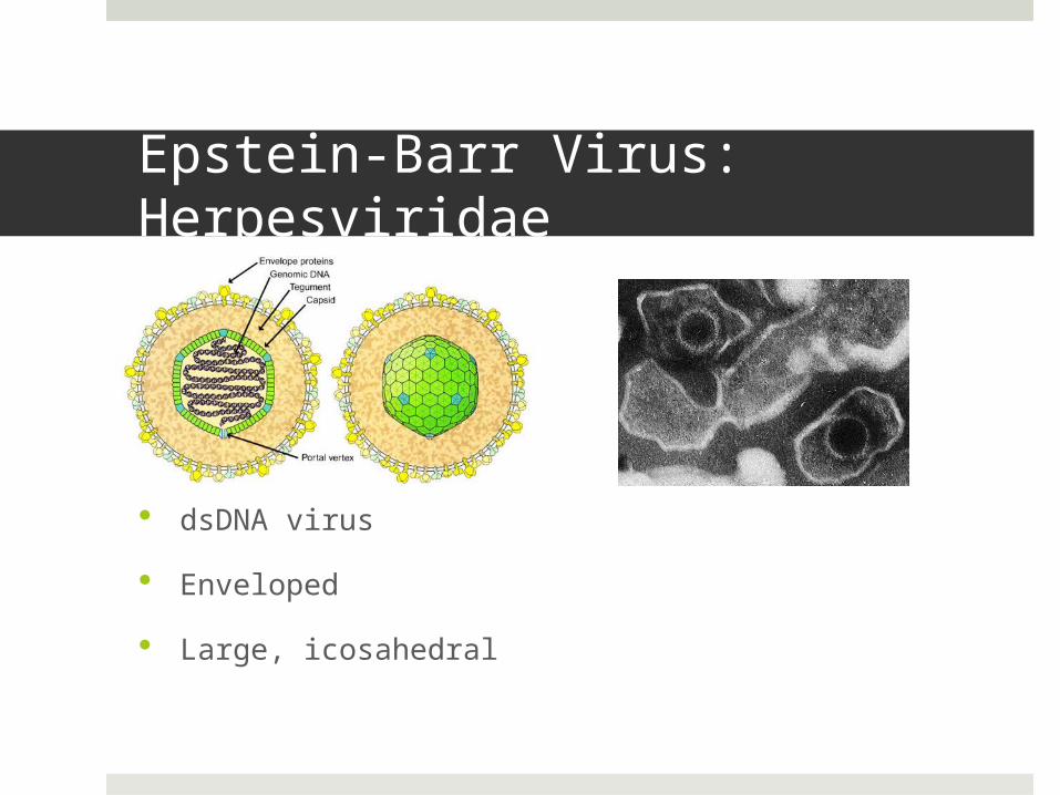
Epstein-Barr Virus: Herpesviridae
dsDNA virus
Enveloped
Large, icosahedral

Classification: Group I
Structure
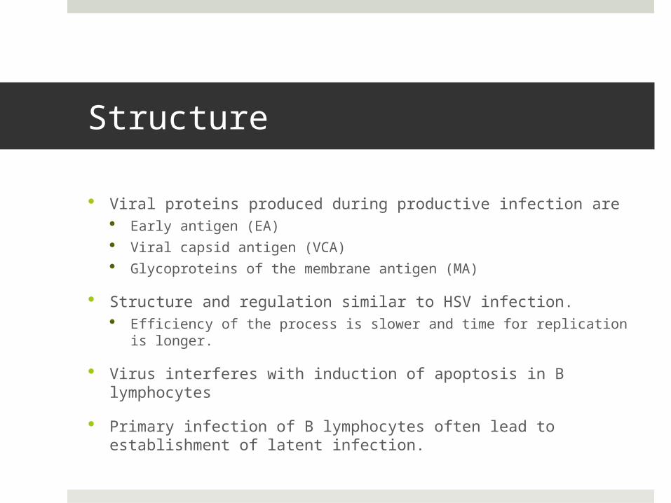
Viral proteins produced during productive infection are Early antigen (EA) Viral capsid antigen (VCA) Glycoproteins of the membrane antigen (MA)
Structure and regulation similar to HSV infection. Efficiency of the process is slower and time for replication is
longer.
Virus interferes with induction of apoptosis in B lymphocytes
Primary infection of B lymphocytes often lead to establishment of latent infection.
Transmission
Saliva
Close oral contact
Sharing of toothbrush

Pathogenesis of EBV
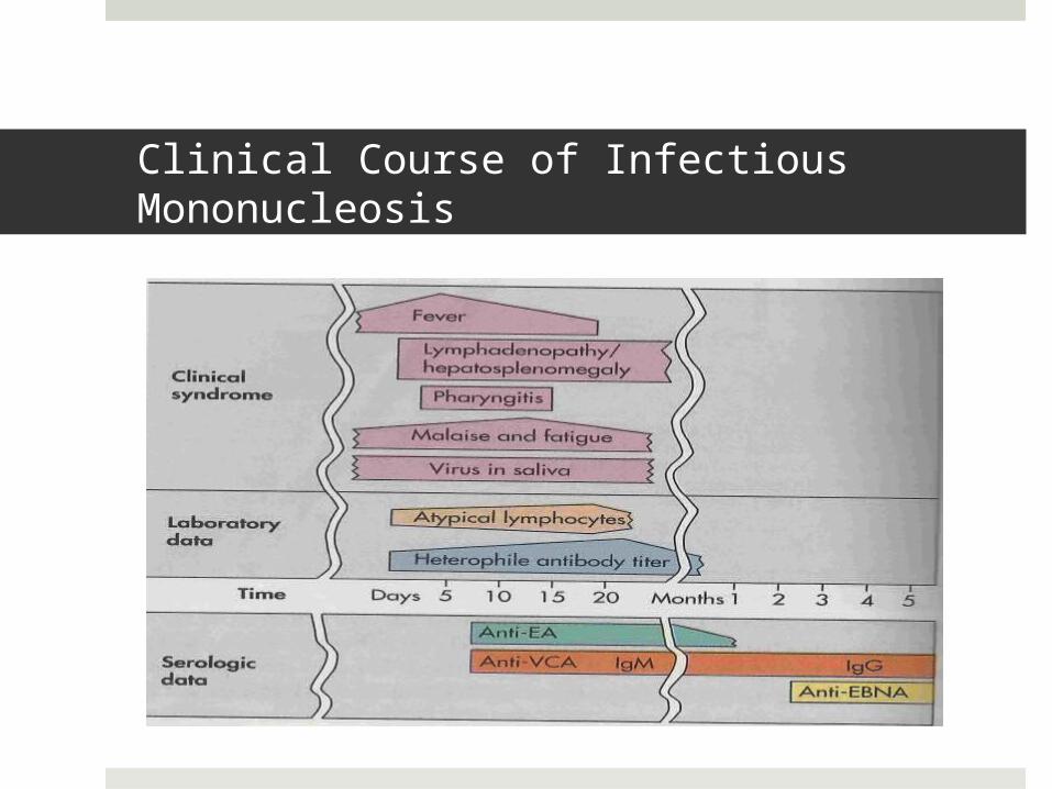
Clinical Course of Infectious Mononucleosis
Epstein-Barr Virus Case Study
A 17-year-old high school student has had low-grade fever and malaise for several days, followed by sore throat, swollen cervical lymph nodes, and increasing fatigue. The patient also notes some discomfort in the left upper quadrant of the abdomen. The sore throat, lymphadenopathy, and fever gradually resolve over the next 2 weeks, but the patient’s full energy level does not return for another 6 weeks.
1. What lab tests would confirm the diagnosis of EBV-induced mononucleosis and distinguish it from CMV infection?
The clinical diagnosis of infectious mononucleosis is suggested on the basis of the symptoms of fever, sore throat, swollen lymph glands, and the age of the patient.
Heterophile antibody test (“Monospot” test) Rapid test for mononucleosis due to EBV Sensitive for heterophile antibodies produced by the
immune system in response to infection.
1. What lab tests would confirm the diagnosis of EBV-induced mononucleosis and distinguish it from CMV infection?
White blood cell count 40%-70% of patients have elevated White Blood Cell
(WBC) count of 10,000-20,000 cells/mL 10% of patients have a WBC count greater than 25,000
cells/mL of blood by the second week of illness. Approximately 80-90% of patients have lymphocytosis
with more than 50% lymphocytes. 20%-40% of the lymphocytes in patients are atypical.
WBC appear larger, have a lower nuclear-to-cytoplasmic ratio, and have a nucleus that is less dense than that of normal lymphocytes.
1. What lab tests would confirm the diagnosis of EBV-induced mononucleosis and distinguish it from CMV infection?
EBV serology antigens would confirm for EBV Viral capsid antigens Early antigens EBV nuclear antigen (EBNA) Differentiation if the immunoglobulin G and M of the
capsid

2. To what characteristic diagnostic feature of the disease does mononucleosis refer?
Mononucleosis Results of antigen-independent proliferation of infected B cells
that crowd out RBC and cause anemia.
Results from the expansion in numbers of T cells upon stimulation by the EBV-infected B cells.
Mononuclear cells are large, with ragged edges and have an abundance of cytoplasm.
Mononucleosis-like syndromes accompany other infections of lymphocytes including CMV and HIV. The excess of atypical lymphocytes present in the patients’ blood smear is indicative of mononucleosis.
3. What causes the swollen glands and fatigue?
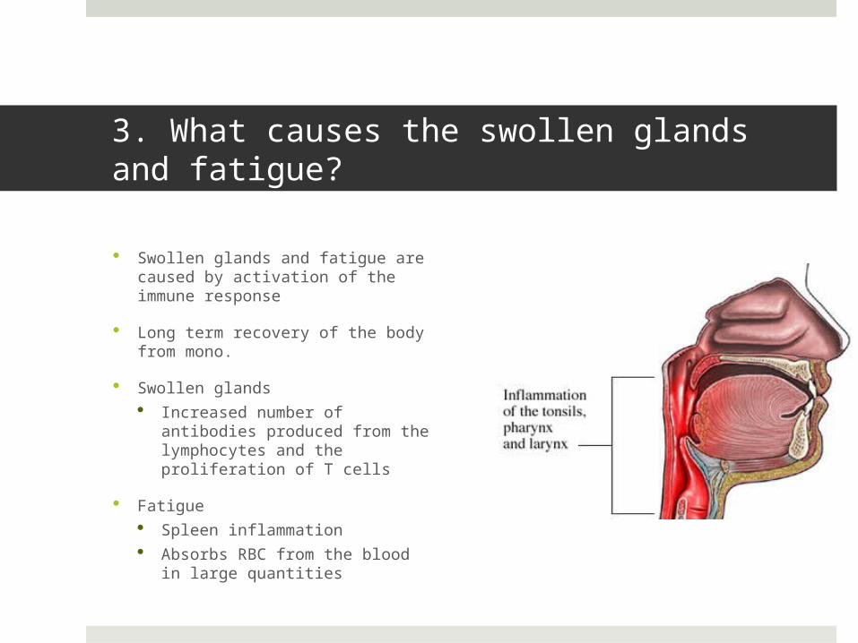
Swollen glands and fatigue are caused by activation of the immune response
Long term recovery of the body from mono.
Swollen glands Increased number of
antibodies produced from the lymphocytes and the proliferation of T cells
Fatigue Spleen inflammation Absorbs RBC from the blood in
large quantities
4. Who is at greatest risk for a serious outcome of an EBV infection? What is the outcome? Why?
Children Asymptomatic disease or mild symptoms due to their less
active immune system
Adolescents & adults At risk for infectious mononucleosis
Immunocompromised individuals At highest risk for life-threatening neoplastic disease
References
Bennett N. “Pediatric mononucleosis and Epstein-Barr Virus Infection” Medscape. (2012) Retrieved November 2012.
Murray, P.R. et al. (2005). Medical Microbiology 5th edition. Philadelphia, PA: Elsevier-Mosby
Wagner, E.K. et al. (2006). Basic Virology (3rd ed.). Malden, MA: Blackwell