Embed Size (px)
Citation preview
REGIONAL ENTERITIS INVOLVING THE DUODENUM
Report of Two Cases
CHARLES H. BROWN, M.D. and JAMES R. SIMS, Jr., M.D.* Department of Gastroenterology
RE G I O N A L enteritis is a disease with diverse and protean manifestations that
>• may simulate those of many other gastrointestinal conditions. Although the terms "terminal ileitis" and "regional ileitis" have been used as synonyms for "regional enteritis," they are misleading for the disease is not limited to the terminal i leum but may involve any segment of the small intestine and portions of the colon. T h e disease frequently involves the small intestine with "skip areas" of inflammation with normal intestinal mucosa between the involved segments. Comfort and associates1 have suggested that the condition be termed "nonspecif ic granulomatous gastroenteritis," but this vague term merely emphasizes the fact that little has been added to our knowledge of regional enteritis since Crohn's2 original description of its etiology and pathogenesis in 1932. "Reg ional enteritis" remains the most widely accepted name for this condition.
Regional enteritis involves the terminal ileum in 75 per cent of the cases. T h e frequency of involvement of a specific segment is directly related to its prox-imity to the ileocecal valve; however, isolated cases of enteritis occurring high in the j e junum have been reported. Involvement of the duodenum, a relatively short segment, is unusual; there are only 13 reports1»3-7 of such cases in the literature. Although some of the reported cases are not described histopatho-logically, all of them must be considered as nonspecific, granulomatous, cicatrizing inflammation of the uppermost intestinal tract (duodenum), because of the clinical characteristics and the surgical descriptions of the pathology.
Comfort 1 reported a typical clinical picture presented by patients having regional enteritis involving the duodenum. H e found that symptoms could be classified into four categories: (1) Continuous and intermittent upper abdominal distress intensified by food, loss of weight and strength, nausea and occasional vomiting; (2) Diarrhea, steatorrheal and episodic in type; (3) Gastric retention, denoted by succussion splash and by morning aspiration of greater than normal amounts of gastric contents (and, if we may add, the vomiting of food particles eaten as much as eight hours previously); and (4) Evidence of deficient absorp-tion. M a n y of these symptoms are due to a high partial obstruction and are not specific for regional enteritis. W e have seen all of them, including diarrhea, in patients with mechanical obstruction of the upper je junum. Yet, cases of regional
* Former Fellow in the Department of General Surgery; now in the United States Army.
9 5
other uses require permission. on March 21, 2022. For personal use only. Allwww.ccjm.orgDownloaded from
BROWN AND SIMS
enteritis involving the duodenum should not go unrecognized and they should never be erroneously labeled as functional disease.
T w o case reports of patients having regional enteritis involving the duo-denum are presented because of the rarity of the condition, the importance of recognizing it, and the peculiar symptoms that the disease produced.
CASE REPORTS
Case 1. A 34 year old woman was first seen in May 1951 at which time she complained of nausea and vomiting of one to two quarts of liquid two to four hours after the evening meal. The symptoms had been present for 12 months. Occasionally the sensation of fullness that preceded the vomiting had awakened her during the night and she had obtained relief by vomiting. There had been no pain, hematemesis, jaundice or diarrhea, and no loss in weight despite the emesis. Five months before admission a roentgen examination made elsewhere had shown a "stricture of the duodenum" and "gastric retention."
Physical examination was essentially normal: she was 62 inches tall and weighed 165 pounds with no evidence of loss of weight. Laboratory examinations, including blood count, urinalysis, stool examination, blood sugar, calcium, urea, albumin-globulin ratio, prothrombin time, serology and serum amylase, were all normal. Gastric analysis yielded 1500 cc. of fluid with a free HC1 of 20 and a total acidity of 33 units.
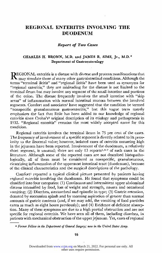
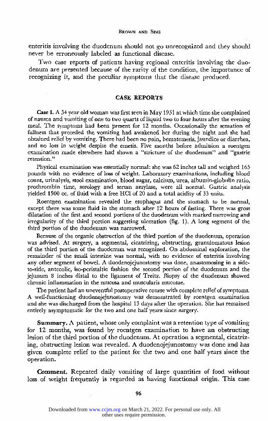
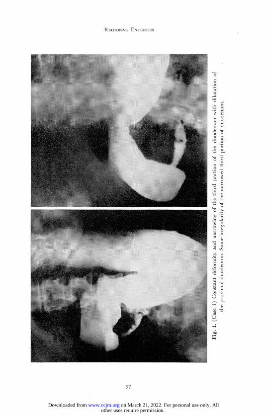
Roentgen examination revealed the esophagus and the stomach to be normal, except there was some fluid in the stomach after 12 hours of fasting. There was gross dilatation of the first and second portions of the duodenum with marked narrowing and irregularity of the third portion suggesting ulceration (fig. 1). A long segment of the third portion of the duodenum was narrowed.
Because of the organic obstruction of the third portion of the duodenum, operation was advised. At surgery, a segmental, cicatrizing, obstructing, granulomatous lesion of the third portion of the duodenum was recognized. On abdominal exploration, the remainder of the small intestine was normal, with no evidence of enteritis involving any other segment of bowel. A duodenojejunostomy was done, anastomosing in a side-to-side, antecolic, iso-peristaltic fashion the second portion of the duodenum and the jejunum 8 inches distal to the ligament of Treitz. Biopsy of the duodenum showed chronic inflammation in the mucosa and muscularis mucosae.
The patient had an uneventful postoperative course with complete relief of symptoms. A well-functioning duodenojejunostomy was demonstrated by roentgen examination and she was discharged from the hospital 13 days after the operation. She has remained entirely asymptomatic for the two and one half years since surgery.
Summary. A patient, whose only complaint was a retention type of vomiting for 12 months, was found by roentgen examination to have an obstructing lesion of the third portion of the duodenum. At operation a segmental, cicatriz-ing, obstructing lesion was revealed. A duodenojejunostomy was done and has given complete relief to the patient for the two and one half years since the operation.
Comment. Repeated daily vomiting of large quantities of food without loss of weight frequently is regarded as having functional origin. This case
9 6
other uses require permission. on March 21, 2022. For personal use only. Allwww.ccjm.orgDownloaded from
REGIONAL ENTERITIS
other uses require permission. on March 21, 2022. For personal use only. Allwww.ccjm.orgDownloaded from
BROWN AND SIMS
demonstrates that such vomiting due to an organic lesion may occur without causing any loss of weight.
In connection with obstructive lesions of the duodenum and upper je junum distal to the mid-second portion of the duodenum, there is one point concerning the choice of operation about which we could find little discussion in the literature. In cases such as case 1, it would seem that a duodenojejunostomy is indicated rather than a gastrojejunostomy. If there is a complete duodenal obstruction distal to the ampulla of Vater, and a gastrojejunostomy has been done, the alkaline pancreatic juices and bile can gain access to the small intestine only by regurgitation through the stomach. If instead, a duodeno-jejunostomy has been done, the alkaline bile and pancreatic juices can gain ready access to the small intestine through the anastomosis. Thus, if the cica-trizing process approaches complete stenosis, the proximal portion of the small bowel receiving the acid gastric juice is not denied a direct and free supply of alkaline bile and pancreatic secretions when a duodenojejunostomy is done. In such cases, a duodenojejunostomy may result in fewer sequelae of the opera-tion, such as distressing regurgitation of bile into the stomach and a lower incidence of marginal ulcer, than would a gastrojejunostomy.
Case 2. A 24 year old woman in October 1948 first complained of a sharp epigastric pain with radiation to both right and left upper quadrants. In addition she had com-plaints referable to every other system in the body characteristic of a conversion neurosis. The epigastric pain occurred once or twice a week, was of 10 to 15 minutes' duration, and was associated with nausea. Complete studies were negative cxccpt for some question-able deformity of the duodenal bulb. She was given antispasmodics and was asked to return in one month for a recheck roentgenographic examination of the stomach.
She was next seen three years later (August 2, 1951) at which time she complained of nausea and vomiting. A 30-pound loss of weight had occurred during the preceding two and one half months. For one month prior to the appearance of these symptoms she had had watery, bloody, mucous diarrhea and had experienced lower abdominal cramps. She had been married one month prior to the onset of the present illness; this was her second marriage and already it was fraught with emotional problems. She referred to her husband in uncomplimentary terms, and stated her belief that he was responsible for her illness. She was admitted to the hospital with a tentative diagnosis of anorexia nervosa.
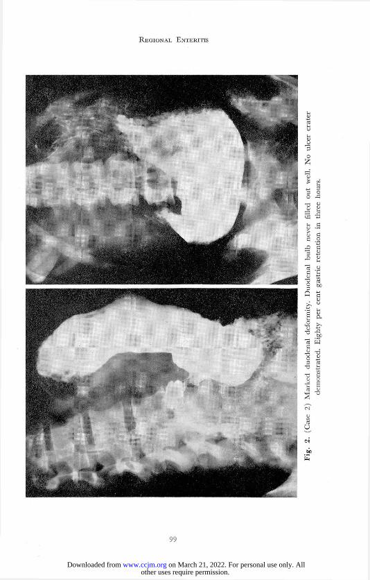
Intravenous fluids to correct a marked electrolytic imbalance, gastric tube feedings, and finally frequent small oral feedings were given. Psychiatric consultation confirmed the diagnosis of a conversion neurosis. Bccause of continued vomiting despite therapy, gastroenterologic consultation was requested two weeks after admission to the hospital. An upper gastrointestinal x-ray examination showed marked gastric retention, presumably due to pyloric obstruction. Barium enema examination showed narrowing and distortion of the mucosal pattern of the terminal ileum (fig. 2).
At operation, the stomach was thick walled, edematous and dilated. The duodenum was greatly thickened throughout its first portion and rubbery firm to palpation. Its serosal surface was injected with many telangiectatic vessels and showed a loss of its normal glistening surface. The induration in the duodenal wall was diffuse, and no ulcer crater could be palpated. The induration began just distal to the pylorus and extended to the distal extremity of the first portion of the duodenum. The terminal ileum was found to be involved in a similar process and was typical of those found in cases of
9 8
other uses require permission. on March 21, 2022. For personal use only. Allwww.ccjm.orgDownloaded from
REGIONAL ENTERITIS
other uses require permission. on March 21, 2022. For personal use only. Allwww.ccjm.orgDownloaded from
BROWN AND SIMS
regional enteritis. A vagotomy and gastroenterostomy was done, and since the lesion in the terminal ileum was not severe enough to cause obstruction, no resection was attempted.
The patient had an uneventful postoperative course. She gained 25 pounds in weight and felt well during the first two months after operation except for mild abdominal cramps following emotional upsets. A year after operation she requested psychiatric consultation because of extreme marital discord. Two years after operation she occasion-ally had loose stools two to three times a day and lower abdominal cramps. Roentgen examination of the small bowel showed no change in the terminal ileum, the distal ileum being narrowed and irregular for 4 to 5 inches.
Summary. A patient with severe emotional problems was treated for two weeks for anorexia nervosa before it was discovered that she had almost complete gastric obstruction and terminal ileitis. She improved following vagotomy and gastroenterostomy and, except for occasional mild episodes of diarrhea and lower abdominal cramps, remained relatively symptom-free for two years following surgery. Her emotional problems continued to disturb her.
Comment. This patient demonstrates quite well the fact that patients with functional disease may have organic disease as well. Before a diagnosis of anorexia nervosa is made, it is essential to rule out any possible organic gastrointestinal disease that could cause the patient's symptoms. A diagnosis of anorexia nervosa must be made not only by positive evidence of a severe psychoneurosis, as in this patient, but also by excluding any possibility of the existence of organic disease.
DISCUSSION
Regional enteritis is a disease with manifestations simulating many other pathologic gastrointestinal conditions including functional abdominal disorders. The diagnosis is frequently difficult to determine until after the disease has existed for some time and has resulted in obstruction of the small intestine. The two patients whose case reports arc presented, exhibited symptoms of gastric obstruction due to regional enteritis involving the duodenum. Other symptoms more frequently present are those of diarrhea, lower abdominal pain, symptoms due to partial obstruction, and fistulae formation. Fever may be a predominant or the single symptom for a long time. W e saw a third patient who had a cholecystectomy for a low-grade fever and a nonfunctioning gallbladder. She continued to have a fever after the cholecystectomy. It was not until 18 months after the cholecystectomy that she developed diarrhea, and a diagnosis of regional enteritis was established by roentgen examination and at operation.
Diarrhea is frequently present but there can be extensive regional enteritis without diarrhea. In the third patient mentioned in the preceding paragraph, the disease was present and caused a low-grade fever for at least 18 months before diarrhea developed. In cases 1 and 2 in which there was involvement of the duodenum, only one patient had diarrhea and that for only one month
1 0 0
other uses require permission. on March 21, 2022. For personal use only. Allwww.ccjm.orgDownloaded from
REGIONAL ENTERITIS
before the operation. If diarrhea is a necessary symptom before one becomes suspicious of this condition, many cases will be diagnosed late in the course of the disease or never will be correctly diagnosed.
In some patients anemia may be the chief symptom and may be present for a long period before other manifestations of regional enteritis appear. This disease should be considered in the differential diagnosis of unexplained anemia. Because of the marked tendency to fistulae formation in cases of regional enteritis, we believe that the possibility of the presence of this disease should be considered in any patient with a fistula, including a perianal fistula.
Regional enteritis involves the terminal ileum in 75 per cent of the cases. Frequently the terminal ileum will be filled on barium enema examination, and it is possible to exclude many cases by this examination alone. In suspected cases of regional enteritis, we believe that a barium enema examination should be done before any x-ray studies of the small intestine. In some cases, roentgen studies of the small intestine will not be necessary. In others, barium enema examination may suggest disease of the terminal ileum which may or may not be substantiated by roentgen examinations of the small intestine.
It is frequently difficult to demonstrate disease in the small intestine by x-ray examination until that disease has become quite extensive and has caused at least partial obstruction. Of all the possible gastrointestinal x-ray studies, that of the small intestine is the least satisfactory. Therefore, in cases in which regional enteritis or other small-intestinal diseases are suspected, it may be necessary to repeat the roentgen study of the small bowel later to establish the true condition. If regional enteritis is suspected clinically despite a "normal" small bowel on x-ray examination, it is always advisable to repeat the roentgen examination after several months.
SUMMARY
T w o cases of regional enteritis involving the duodenum are reported. Such involvement is rare; only 13 such cases having been previously recorded. Both patients presented symptoms of gastric or duodenal obstruction. Both were relieved by side-tracking operations, a duodenojejunostomy in one and a gastrojejunostomy in the other. The rationale of doing a duodenojejunostomy rather than a gastrojejunostomy for obstructive lesions of the duodenum and jejunum distal to the mid-second portion of the duodenum is discussed. The possibility of mistakenly considering patients with regional enteritis as having functional disease (one of the patients having been treated for anorexia nervosa for two weeks) is discussed, and the various manifestations of regional enteritis are presented. Regional enteritis should be considered in the differential diagnosis of obstructing lesions of the duodenum.
101
other uses require permission. on March 21, 2022. For personal use only. Allwww.ccjm.orgDownloaded from
BROWN AND SIMS
References
1. Comfort, M. W., Weber, H. M., Baggenstoss, A. H. and Kiely, W. F.: Nonspecific granu-lomatous inflammation of stomach and duodenum: its relation to regional enteritis. Am. J. M. Sc. 220:616-632 (Dec.) 1950.
2. Crohn, B. B., Ginzburg, L. and Oppenheimer, G. D.: Regional ileitis; pathologic and clinical entity. J.A.M.A. 99:1323-1329 (Oct. 15) 1932.,
3. Carlisle, J. C. and Judd, E. S., Jr.: Regional enteritis involving the duodenum: report of case. Proc. Staff Meet., Mayo Clin. 27:569-574 (Dec. 31) 1952.
4. Shapiro, R.: Regional ileitis; summary of literature. Am. J. M. Sc. 198:269-292 (Aug.) 1939.
5. Janus, W. L.: Regional jejunitis; 3 cases. Radiology 50:532-535 (April) 1948.
6. Hawthorne, H. R. and Frobese, A. S.: Chronic stenosing regional enteritis—surgical pathology and experience in surgical treatment. Ann. Surg. 130:233-241 (Aug.) 1949.
7. Case Records of the Massachusetts General Hospital; No. 35171, Regional enteritis. New England J. Med. 240:692-694 (April 28) 1949.
1 0 2
other uses require permission. on March 21, 2022. For personal use only. Allwww.ccjm.orgDownloaded from