Embed Size (px)
Citation preview
Instructions for use
Title Molecular cloning and characterization of the corticoid receptors from the American alligator
Author(s) Oka, Kaori; Kohno, Satomi; Urushitani, Hiroshi; Guillette, Louis J., Jr.; Ohta, Yasuhiko; Iguchi, Taisen; Katsu,Yoshinao
Citation Molecular and Cellular Endocrinology, 365(2): 153-161
Issue Date 2013-01-30
Doc URL http://hdl.handle.net/2115/52257
Type article (author version)
Additional Information There are other files related to this item in HUSCAP. Check the above URL.
File Information MCE365-2_153-161.pdf
Hokkaido University Collection of Scholarly and Academic Papers : HUSCAP
1
Molecular cloning and characterization of the corticoid receptors from the
American alligator
Short title: alligator glucocorticoid and mineralocorticoid receptors
Kaori Oka 1
, Satomi Kohno 2
, Hiroshi Urushitani3, 4, a
, Louis J. Guillette Jr. 2, Yasuhiko Ohta
5,
Taisen Iguchi 3, 4, #
and Yoshinao Katsu 1, 3, 6, #
1Graduate School of Life Science, Hokkaido University, Sapporo 060-0810, Japan;
2Department of Obstetrics and Gynecology, Medical University of South Carolina and
Hollings Marine Laboratory, 221 Ft. Johnson Rd., Charleston, SC 29412, USA; 3Okazaki
Institute for Integrative Bioscience, National Institutes of Natural Sciences, Okazaki
444-8787, Japan ; 4
National Institute for Basic Biology, Okazaki 444-8787, Japan ;
5Department of Veterinary Medicine, Faculty of Agriculture, Tottori University, Tottori
680-8553, Japan; 6Department of Biological Sciences, Hokkaido University, Sapporo
060-0810, Japan
#Address all correspondence to: Taisen Iguchi or Yoshinao Katsu, Okazaki Institute for
Integrative Bioscience, National Institutes of Natural Sciences, Okazaki 444-8787, Japan
(T.I.), or Department of Biological Sciences, Hokkaido University, Kita-10, Nishi-8, Kita-ku,
Sapporo 060-0810, Japan (Y.K.).
E-mail:[email protected] (Taisen Iguchi); or [email protected] (Yoshinao Katsu)
aPresent address: Environmental Quality Measurement Section, Research Center for
Environmental Risk, National Institute for Environmental Studies, 16-2 Onogawa, Tsukuba,
Ibaraki 305-8506, Japan
Disclosure statement: All authors have nothing to disclose.
Key words: alligator, glucocorticoid receptor, mineralocorticoid receptor, cloning,
transactivation
Abbreviations: GR, glucocorticoid receptor; MR, mineralocorticoid receptor; RACE, rapid
amplification cDNA ends; MMTV, murine mammary tumor virus; DMSO, dimethylsulfoxide
2
Abstract
Steroid hormones are essential for health in vertebrates. Corticosteroids, for
example, have a regulatory role in many physiological functions, such as osmoregulation,
respiration, immune responses, stress responses, reproduction, growth, and metabolism.
Although extensively studied in mammals and some non-mammalian species, the molecular
mechanisms of corticosteroid hormone (glucocorticoids and mineralocorticoids) action are
poorly understood in reptiles. Here, we have evaluated hormone receptor-ligand interactions in
the American alligator (Alligator mississippiensis), following the isolation of cDNAs encoding
a glucocorticoid receptor (GR) and a mineralocorticoid receptor (MR). The full-length
alligator GR (aGR) and aMR cDNAs were obtained using 5’ and 3’ rapid amplification cDNA
ends (RACE). The deduced amino acid sequences exhibited high identity to the chicken
orthologs (aGR: 83%; aMR: 90%). Using transient transfection assays of mammalian cells,
both aGR and aMR proteins displayed corticosteroid-dependent activation of transcription
from keto-steroid hormone responsive, murine mammary tumor virus promoters. We further
compared the ligand-specifity of human, chicken, Xenopus, and zebrafish GR and MR. We
found that the alligator and chicken GR/MR have very similar amino acid sequences, and this
translates to very similar ligand specificity. This is the first report of the full-coding regions
of a reptilian GR and MR, and the examination of their transactivation by steroid hormones.
3
1. Introduction
Steroid hormones are essential for health in all vertebrates, including reptiles, with
many reported actions. These actions are mediated, at least in part, by specific receptors
localized in or near the nucleus of target cells. Two distinct types of corticosteroid receptor,
a glucocorticoid receptor (GR) and a mineralocorticoid receptor (MR) have been isolated in
vertebrates to date. Corticosteroid hormones, the glucocorticoids and mineralocorticoids,
were originally named for their distinct physiological functions in mammals.
Glucocorticoids, including cortisol and corticosterone, were identified for their role in liver
glycogen deposition, whereas the mineralocorticoid, predominantly aldosterone, regulated
mineral balance principally by controlling sodium retention in the kidney (Norman and
Litwack, 1997).
Thornton (2001) proposed that the ancestral condition for the jawed vertebrates
(Gnathostomata) was the presence of two forms of corticosteroid receptor. Both forms have
been found in chondrichthyan and osteichthyan fishes, amphibians, birds and mammals. To
data, however, cDNAs encoding full length GR and MR have not been reported for a reptile
nor has their function been compared with other vertebrate classes. These receptors are
important as they hold a basal position in the evolution of vertebrate steroid receptors
(Thornton, 2001) and the reptilian forms would add to our understanding of the evolution and
function of these receptors in phylogenetically related avian and mammalian species. A
full-coding region of a reptilian (green anole, Anolis carolonensis) GR-like sequence has
been registered in GenBank. However, as this sequence is predicted from genomic
information (A. carolinesis chromosome 2 genomic scaffold, AnCar2.0, whole genome
shotgun sequence, NW_003338615), the functional analysis of this receptor was not reported.
Here, we report the isolation of cDNA clones encoding American alligator (Alligator
mississippiensis) homologs of the glucocorticoid and mineralocorticoid receptors. We
analyzed their phylogenic relationship with other known vertebrate steroid receptors. The
transactivation functions of GR and MR were determined by expressing these two receptors
separately in transiently transfected cultured cell lines, using a general reporter gene assay.
Further, we compared the ligand-specificity of GR and MR from the alligator to other
vertebrates (human, chicken, Xenopus, and zebrafish) .
2. Materials and Methods
2.1. Animals
All experiments in this study involving alligator were carried out under the guidelines
specified by the Institutional Animal Care and use Committee at the University of Florida and
National Institutes of Natural Sciences. All fieldwork was conducted under permits from
4
the Florida Fish and Wildlife Conservation Commission and the U.S. Fish and Wildlife
Service. Juvenile American alligators (A. mississippiensis) were collected by hand from
airboats from the Lake Woodruff National Wildlife Refuge (NWR), Volusia County, Florida,
USA. Animals were killed with a lethal dose of Nembutal, and tissues were isolated and
stored at -80˚C until analysis.
Alligator eggs were collected from Lake Woodruff NWR, and transported to the
University of Florida (Gainesville, FL, USA), where they were incubated in damp sphagnum
moss at male-producing (33.5˚C) or female-producing (30.0˚C) temperatures. Embryonic
developmental stages were determined primarily according to the criteria described by
Ferguson (1985), with some further resolution based on our previous experience working
with this species (Kohno and Guillette, 2012). Embryos were dissected at stages 19, 20, 21,
23 and 24. The gonad-adrenal-mesonephric complex (GAM) was isolated under sterile
conditions and stored at -80˚C until analyzed. Neonates (< 48 hours old) and one-month-old
animals were killed with a lethal dose of Nembutal, and GAM tissues were isolated and
stored at -80˚C in RNAlater (Ambion, Austin, TX) until analyzed. GAMs isolated from
one-month-old animal, and fixed in RNAlater, were carefully dissected into three portions;
the gonad, adrenal and mesonephros (Kohno et al., 2010).
An adult female frog (Xenopus laevis) and zebrafish (Danio rerio) were purchased
from a local supplier. Animals were overdosed with MS-222 and tissues obtained by sterile
necropsy.
2.2. Chemical reagents
We obtained chemicals from Sigma-Aldrich Corp.; aldosterone,
deoxycorticosterone, corticosterone, cortisol, cortisone, pregnenolone, progesterone,
androstenedione, 5-dihydrotestosterone, 17-estradiol, and diethylstilbestrol. All
chemicals were dissolved in dimethylsulfoxide (DMSO). The concentration of DMSO in
the culture medium did not exceed 0.1%.
2.3. Molecular cloning of steroid hormone receptors
For the MR, two conserved amino acid regions (FMDDKDY and QVVKWAK) of
vertebrate MRs were selected and degenerate oligonucleotides were used as primers for
polymerase chain reaction (PCR). As a template for PCR, the first-strand cDNA was
synthesized from 2 g of total RNA isolated from the alligator liver. After amplification, an
additional primer set, QSFHYRI and IVYAGYD, was used for the second PCR. For the GR,
two conserved amino acid regions, QVKTEKE/D and QNWQRFY of vertebrate GRs were
selected and degenerate oligonucleotides were used as primers for PCR. First-strand cDNA
5
was synthesized from 2 g of total RNA isolated from the liver of alligator after amplification,
and an additional primer set, GQMYHYD and NMLGGRQ, was used for the second PCR.
The amplified DNA fragments were subcloned with the TA-cloning plasmid
pCR2.1 vector (Invitrogen, Carlsbad, CA), sequenced using a BigDye terminator Cycle
Sequencing-kit (Applied Biosystems, Foster City, CA) with T7 and SP6 primers, and
analyzed on an Applied Biosystems 3130 Genetic Analyzer (Applied Biosystems). The 5’-
and 3’-ends of the MR and GR cDNAs were amplified by rapid amplification of the cDNA
end (RACE) using a SMART RACE cDNA Amplification kit (BD Biosciences Clontech,
Palo Alto, CA). All sequences generated were searched for similarity using Blastn and
Blastp on the web servers of the National Center of Biotechnology Information.
2.4. Construction of plasmid vectors
The full-coding regions of the alligator GR, human GR (accession number;
NM_000901), chicken GR (accession number; NM_001159345), Xenopus GR (accession
number; NM_001090605), zebrafish GR (accession number; NM_001100403), alligator MR,
human MR (accession number; NM_000176,), chicken MR (accession number;
NM_001037826), Xenopus MR (accession number; NM_001088062), and zebrafish MR
(accession number; NM_001100403) were amplified by PCR with KOD DNA polymerase
(TOYOBO Biochemicals, Osaka, Japan). The PCR products were gel-purified and ligated
into the pcDNA3.1 vector (Invitrogen). The mouse mammary tumor virus-long terminal
repeat (MMTV-LTR) was amplified from the pMSG vector (Pharmacia) by PCR, and
inserted into the pGL3-basic vector that contained the Photinus pyralis lucifease gene
(Promega).
2.5. RNA isolation and RT-PCR
Total RNA was prepared from tissues of both female and male alligators using
RNeasy. For RT-PCR, 2 g of total RNA was reverse-transcribed using SuperScript III
transcriptase (Invitrogen) and oligo (dT) primer. The following primer sets were used for
RT-PCR: 5’-CACGCCATCCTGCGTCTGGACTTG-3’ and
5’-ATTCTGTCGGCGATGCCAGGGTAC-3’ for -actin,
5’-CGTTCTGGTAAGAAGCTGGA-3’ and 5’-TGACACCAACAGCAACAGTC-3’ for
EF1, 5’-ATAGTTCCTGCATCACTGCCACAG-3’ and
5’-TCAGGCCCTCCTTAGGAATTGTAG-3’ for GR,
5’-AACCAGACACAGCAGAGTACCTGC-3’ and
5’-TTCCACCAGCATGGCTGGAAACTC-3’ for MR. Twenty-eight cycles (for -actin and
EF1or thirty cycles (for GR and MR) of amplification were carried out under the
6
following conditions: denaturing at 94◦C for 30 sec, annealing at 58
◦C for 30 sec, and
extension at 72◦C for 1 min. At completion of the PCR, DNA fragments were resolved on
1.2% agarose gels.
2.6. Transactivation assays
Transcription assays were performed in human embryonic kidney 293 (HEK293)
cell. HEK293 cells were seeded in 24-well plates at 5x10⁴ cells/well in phenol-red free
Dulbecco’s modified Eagle’s medium (Sigma-Aldrich Corp.) with 10%
charcoal/dextran-treated fetal bovine serum (Hyclone, South Logan, UT). After 24 h, the
cells were transfected with DNAs using Fugene 6 transfection reagent (Roche Diagnostics,
Basel, Switzerland) according to the manufacturer’s instructions. After 20 h of incubation,
steroid hormones were introduced to the media. After an additional 24 h, the cells were
collected, and the luciferase activity of the cells was measured by a chemiluminescence assay
employing the Dual-Luciferase Reporter Assay System (Promega). Luminescence was
measured using a Turner Designs Luminometer TD-20/20 (Promega). Promoter activity
was calculated as firefly (Photinus pyralis)-luciferase activity/sea pansy (Renilla
reniformis)-luciferase activity. All transfections were preformed in triplicate.
2.7. Phylogenetic analysis and databases
All sequences generated were searched for similarity using PSI-Blast with
American alligator GR or MR at web servers of the National Center of Biotechnology
Information. The searches were repeated until any new sequence would not be obtained
within 500 sequences in the Protein Reference Sequences database. After brief analysis of
the phylogeny with selected species, the data revealed AR and PR were the closest out-group
of GR and MR as described before in Bridgham et al. (2009). Thus GR and MR were
analyzed with AR and PR. Phylogenic tree was estimated by phylogeny.fr (Dereeper et al.,
2008) with T-Coffee, Gblocks and phyML. MEGA5 were used to edit the phylogenic tree
(Tamura et al., 2011). The NCBI gene ID of sequences in the phylogeny are: 324021675,
324021673, 324021671, 321117150, 66528642, 58535455, 21322252, 4504133, 158508572,
112363098, 121247453, 144227212, 327269138, 327265250, 327285073, 226817306,
45383982, 301612214, 301607929, 62858859, 154240734 and 261599016.
2.8. Statistical methods
Results are presented as mean ± SEM. Comparisons between two groups were
performed using Student’s t-test, and all multi-group comparisons were performed by
ANOVA followed by Bonferroni post hoc analyses using GraphPad Prism (version 4.0c;
7
GraphPad Software, Inc., San Diego, CA). P < 0.05 was considered statistically significant.
3. Results
3.1. Alligator GR and MR
Using standard PCR techniques, partial DNA fragments were amplified from
alligator liver RNA. Two different DNA fragments were obtained showing similarity to the
GR and MR, respectively. A full-length alligator GR cDNA (GenBank Accession No.
AB701407) and MR cDNA (GenBank Accession No. AB701406) were cloned using RACE.
The cDNA for alligator GR (aGR) is composed of a predicted 780 amino acids with a
calculated molecular mass of 85.2 kDa, whereas the MR (aMR) is composed of a predicted
985 amino acids with a calculated molecular mass of 107.4 kDa (Fig. 1). Using the
nomenclature of Krust et al. (1986), the aGR and aMR sequences can be divided into four
domains (A/B, C, D and E(F) domains) based on its sequence homology with other steroid
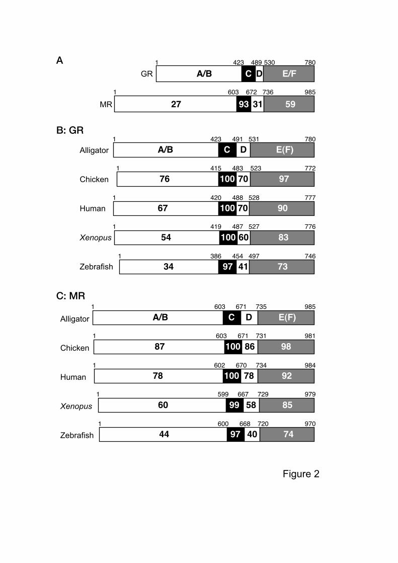
hormone receptors. aGR and aMR shared 27% identity in the A/B domain, 93% identity in
the C domain, 31% identity in the D domain, and 59% identity in the E(F) domain (Fig. 2A).
Thus, the C domain (DNA-binding domain) is highly conserved, with the E(F) domain
(ligand-binding domain) exhibiting conservation as well between the aGR and aMR.
Amino acid sequences of the aGR and aMR with the other vertebrates examined, show an
over-all homology of 39%. The over-all homologies of aGR with human, chicken, Xenopus
and alpha form zebrafish (Schaaf et al., 2008) GRs were 76, 83, 67 and 52%, respectively.
The over-all homologies of the aMR with human, chicken, Xenopus or zebrafish MRs were
82, 90, 68 and 54%, respectively. When examined in greater detail, the aGR shared 76-34%,
100-97%, 70-41%, and 97-73% identities in the A/B, C, D, and E(F) domains with the other
species examined, respectively (Fig. 2B). A similar analyses for the aMR indicated 87-44%,
100-97%, 86-40%, and 98-74% identities in the A/B, C, D, and E(F) domains, respectively
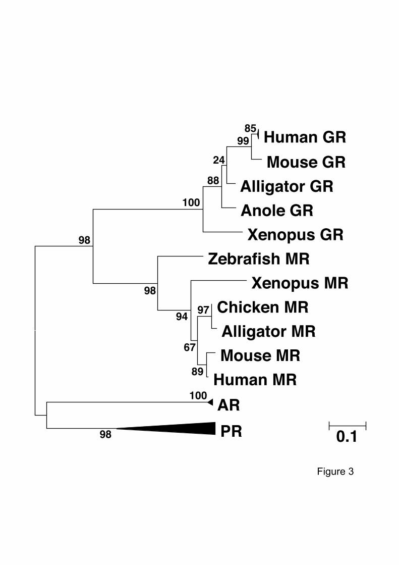
(Fig. 2C). As would be predicted from previous studies using various forms of genetic
information, a phylogenetic analysis indicated that the alligator corticoid receptors were more
closely related to those of birds rather than those of mammals and amphibians (Fig. 3).
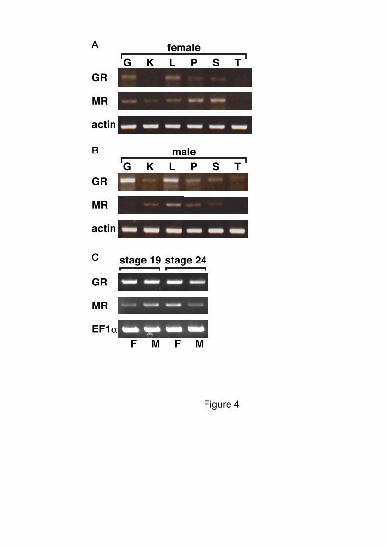
The mRNA expression levels of both corticoid receptors in various tissues, from
both female and male juvenile alligators, were measured by RT-PCR. Expression of aGR in
the female kidney, and female and male thyroid appeared less, based on the band intensity on
the gel, compared with other tissues. Similarly, expression of the aMR in female and male
thyroid, and the male gonad was lower compared with other tissues. We also analyzed the
expression level of these receptors in the GAM during embryonic development. Both aGR
and aMR mRNAs were detected at stages 19 and 24, stages just prior to and the last stage of
the thermo-sensitive period for sex determination, but using gel band intensity alone, the
8
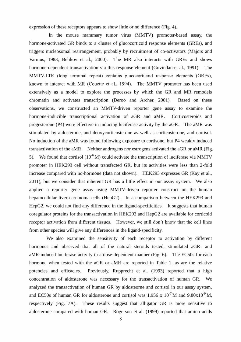
expression of these receptors appears to show little or no difference (Fig. 4).
In the mouse mammary tumor virus (MMTV) promoter-based assay, the
hormone-activated GR binds to a cluster of glucocorticoid response elements (GREs), and
triggers nucleosomal rearrangement, probably by recruitment of co-activators (Majors and
Varmus, 1983; Belikov et al., 2000). The MR also interacts with GREs and shows
hormone-dependent transactivation via this response element (Govindan et al., 1991). The
MMTV-LTR (long terminal repeat) contains glucocorticoid response elements (GREs),
known to interact with MR (Couette et al., 1994). The MMTV promoter has been used
extensively as a model to explore the processes by which the GR and MR remodels
chromatin and activates transcription (Deroo and Archer, 2001). Based on these
observations, we constructed an MMTV-driven reporter gene assay to examine the
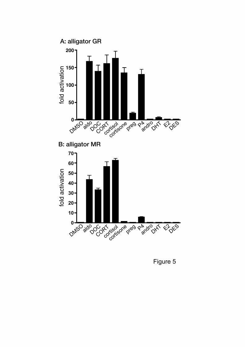
hormone-inducible transcriptional activation of aGR and aMR. Corticosteroids and
progesterone (P4) were effective in inducing luciferase activity by the aGR. The aMR was
stimulated by aldosterone, and deoxycorticosterone as well as corticosterone, and cortisol.
No induction of the aMR was found following exposure to cortisone, but P4 weakly induced
transactivation of the aMR. Neither androgens nor estrogens activated the aGR or aMR (Fig.
5). We found that cortisol (10-6
M) could activate the transcription of luciferase via MMTV
promoter in HEK293 cell without transfected GR, but its activities were less than 2-fold
increase compared with no-hormone (data not shown). HEK293 expresses GR (Kay et al.,
2011), but we consider that inherent GR has a little effect in our assay system. We also
applied a reporter gene assay using MMTV-driven reporter construct on the human
hepatocellular liver carcinoma cells (HepG2). In a comparison between the HEK293 and
HepG2, we could not find any difference in the ligand-specificities. It suggests that human
coregulator proteins for the transactivation in HEK293 and HepG2 are available for corticoid
receptor activation from different tissues. However, we still don’t know that the cell lines
from other species will give any differences in the ligand-specificity.
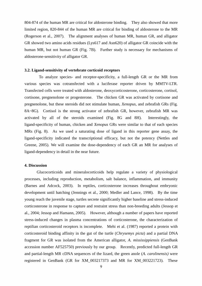
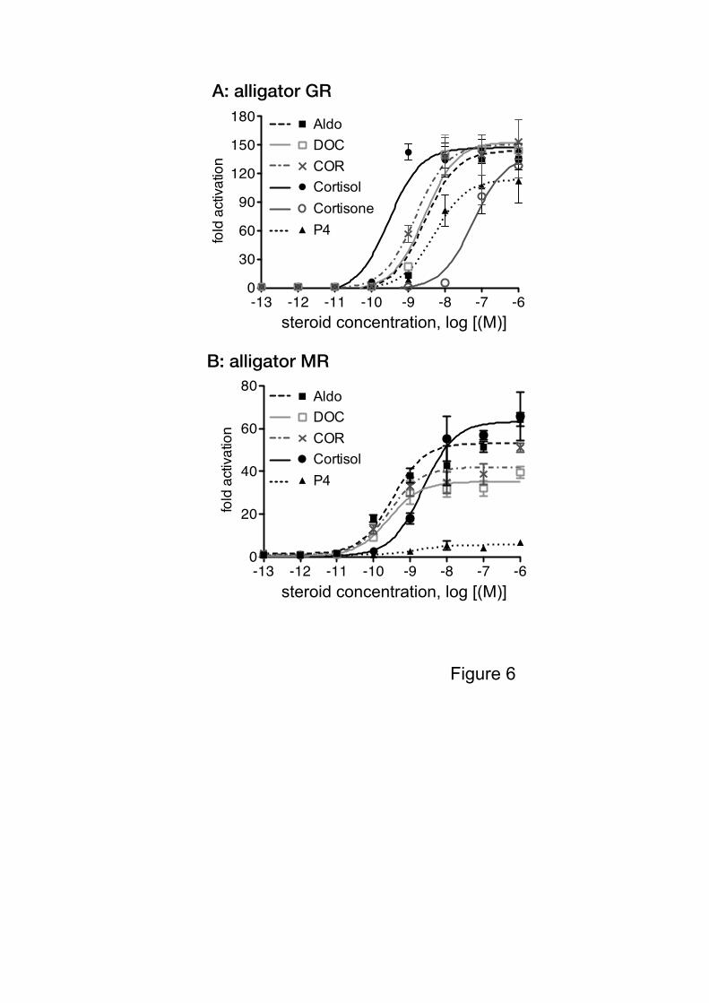
We also examined the sensitivity of each receptor to activation by different
hormones and observed that all of the natural steroids tested, stimulated aGR- and
aMR-induced luciferase activity in a dose-dependent manner (Fig. 6). The EC50s for each
hormone when tested with the aGR or aMR are reported in Table 1, as are the relative
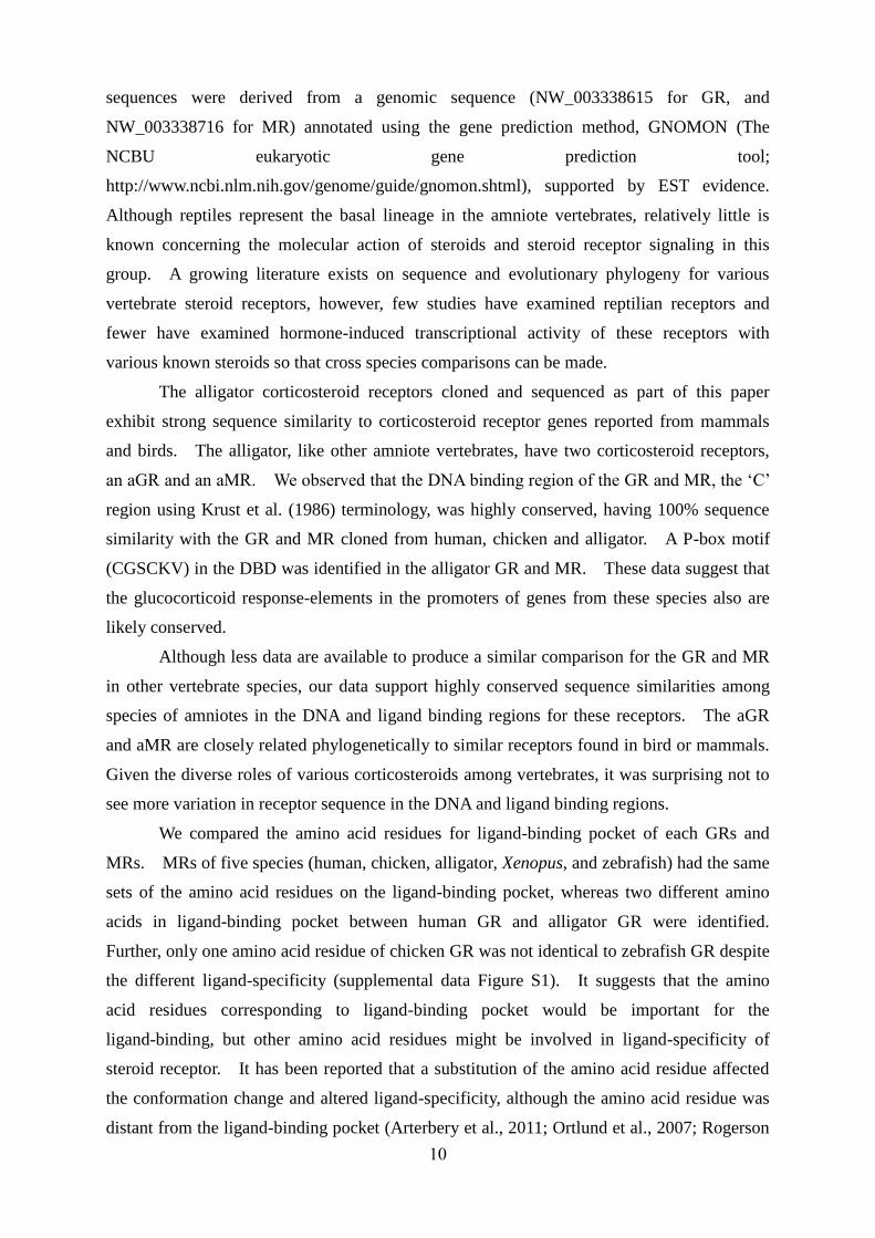
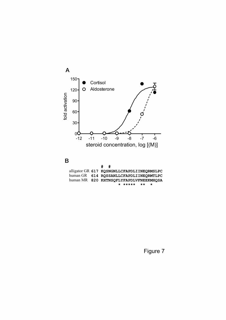
potencies and efficacies. Previously, Rupprecht et al. (1993) reported that a high
concentration of aldosterone was necessary for the transactivation of human GR. We
analyzed the transactivation of human GR by aldosteorne and cortisol in our assay system,
and EC50s of human GR for aldosterone and cortisol was 1.956 x 10⁻⁷
M and 9.80x10⁻⁹
M,
respectively (Fig. 7A). These results suggest that alligator GR is more sensitive to
aldosterone compared with human GR. Rogerson et al. (1999) reported that amino acids
9
804-874 of the human MR are critical for aldosterone binding. They also showed that more
limited region, 820-844 of the human MR are critical for binding of aldosterone to the MR
(Rogerson et al., 2007). The alignment analyses of human MR, human GR, and alligator
GR showed two amino acids residues (Lys617 and Asn620) of alligator GR coincide with the
human MR, but not human GR (Fig. 7B). Further study is necessary for mechanisms of
aldosterone-sensitivity of alligator GR.
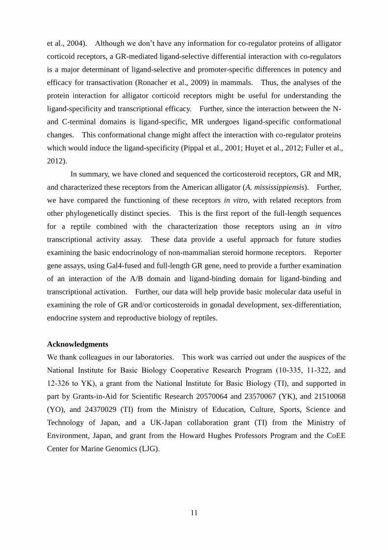
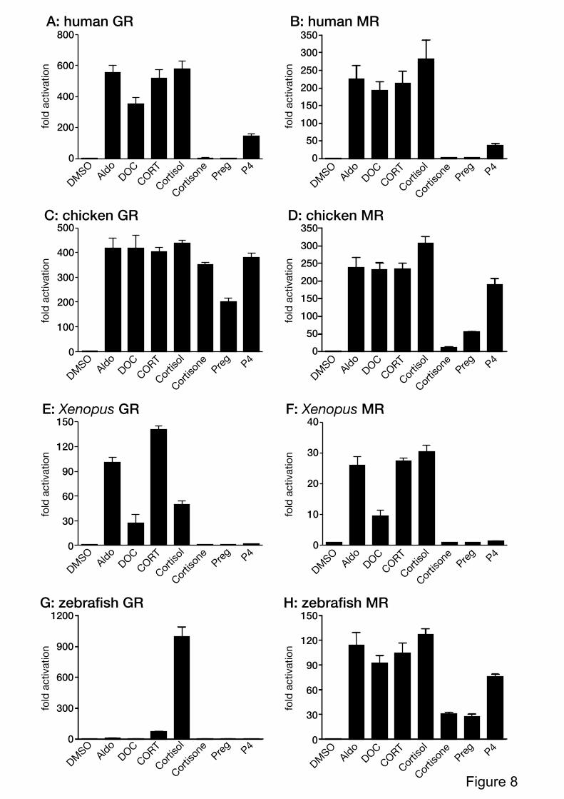
3.2. Ligand-sensitivity of vertebrate corticoid receptors
To analyze species- and receptor-specificity, a full-length GR or the MR from
various species was cotransfected with a luciferase reporter driven by MMTV-LTR.
Transfected cells were treated with aldosterone, deoxycorticosterone, corticosterone, cortisol,
cortisone, pregnenolone or progesterone. The chicken GR was activated by cortisone and
pregnenolone, but these steroids did not stimulate human, Xenopus, and zebrafish GRs (Fig.
8A~8G). Cortisol is the strong activator of zebrafish GR, however, zebrafish MR was
activated by all of the steroids examined (Fig. 8G and 8H). Interestingly, the
ligand-specificity of human, chicken and Xenopus GRs were similar to that of each species
MRs (Fig. 8). As we used a saturating dose of ligand in this reporter gene assay, the
ligand-specificity indicated the transcriptional efficacy, but not the potency (Nettles and
Greene, 2005). We will examine the dose-dependency of each GR an MR for analyses of
ligand-dependency in detail in the near future.
4. Discussion
Glucocorticoids and mineralocorticoids help regulate a variety of physiological
processes, including reproduction, metabolism, salt balance, inflammation, and immunity
(Barnes and Adcock, 2003). In reptiles, corticosterone increases throughout embryonic
development until hatching (Jennings et al., 2000; Medler and Lance, 1998). By the time
young reach the juvenile stage, turtles secrete significantly higher baseline and stress-induced
corticosterone in response to capture and restraint stress than non-breeding adults (Jessop et
al., 2004; Jessop and Hamann, 2005). However, although a number of papers have reported
stress-induced changes in plasma concentrations of corticosterone, the characterization of
reptilian corticosteroid receptors is incomplete. Mehi et al. (1987) reported a protein with
corticosteroid binding affinity in the gut of the turtle (Chrysemys picta) and a partial DNA
fragment for GR was isolated from the American alligator, A. mississippiensis (GenBank
accession number AF525750) previously by our group. Recently, predicted full-length GR
and partial-length MR cDNA sequences of the lizard, the green anole (A. carolinensis) were
registered in GenBank (GR for XM_003217373 and MR for XM_003221723). These
10
sequences were derived from a genomic sequence (NW_003338615 for GR, and
NW_003338716 for MR) annotated using the gene prediction method, GNOMON (The
NCBU eukaryotic gene prediction tool;
http://www.ncbi.nlm.nih.gov/genome/guide/gnomon.shtml), supported by EST evidence.
Although reptiles represent the basal lineage in the amniote vertebrates, relatively little is
known concerning the molecular action of steroids and steroid receptor signaling in this
group. A growing literature exists on sequence and evolutionary phylogeny for various
vertebrate steroid receptors, however, few studies have examined reptilian receptors and
fewer have examined hormone-induced transcriptional activity of these receptors with
various known steroids so that cross species comparisons can be made.
The alligator corticosteroid receptors cloned and sequenced as part of this paper
exhibit strong sequence similarity to corticosteroid receptor genes reported from mammals
and birds. The alligator, like other amniote vertebrates, have two corticosteroid receptors,
an aGR and an aMR. We observed that the DNA binding region of the GR and MR, the ‘C’
region using Krust et al. (1986) terminology, was highly conserved, having 100% sequence
similarity with the GR and MR cloned from human, chicken and alligator. A P-box motif
(CGSCKV) in the DBD was identified in the alligator GR and MR. These data suggest that
the glucocorticoid response-elements in the promoters of genes from these species also are
likely conserved.
Although less data are available to produce a similar comparison for the GR and MR
in other vertebrate species, our data support highly conserved sequence similarities among
species of amniotes in the DNA and ligand binding regions for these receptors. The aGR
and aMR are closely related phylogenetically to similar receptors found in bird or mammals.
Given the diverse roles of various corticosteroids among vertebrates, it was surprising not to
see more variation in receptor sequence in the DNA and ligand binding regions.
We compared the amino acid residues for ligand-binding pocket of each GRs and
MRs. MRs of five species (human, chicken, alligator, Xenopus, and zebrafish) had the same
sets of the amino acid residues on the ligand-binding pocket, whereas two different amino
acids in ligand-binding pocket between human GR and alligator GR were identified.
Further, only one amino acid residue of chicken GR was not identical to zebrafish GR despite
the different ligand-specificity (supplemental data Figure S1). It suggests that the amino
acid residues corresponding to ligand-binding pocket would be important for the
ligand-binding, but other amino acid residues might be involved in ligand-specificity of
steroid receptor. It has been reported that a substitution of the amino acid residue affected
the conformation change and altered ligand-specificity, although the amino acid residue was
distant from the ligand-binding pocket (Arterbery et al., 2011; Ortlund et al., 2007; Rogerson
11
et al., 2004). Although we don’t have any information for co-regulator proteins of alligator
corticoid receptors, a GR-mediated ligand-selective differential interaction with co-regulators
is a major determinant of ligand-selective and promoter-specific differences in potency and
efficacy for transactivation (Ronacher et al., 2009) in mammals. Thus, the analyses of the
protein interaction for alligator corticoid receptors might be useful for understanding the
ligand-specificity and transcriptional efficacy. Further, since the interaction between the N-
and C-terminal domains is ligand-specific, MR undergoes ligand-specific conformational
changes. This conformational change might affect the interaction with co-regulator proteins
which would induce the ligand-specificity (Pippal et al., 2001; Huyet et al., 2012; Fuller et al.,
2012).
In summary, we have cloned and sequenced the corticosteroid receptors, GR and MR,
and characterized these receptors from the American alligator (A. mississippiensis). Further,
we have compared the functioning of these receptors in vitro, with related receptors from
other phylogenetically distinct species. This is the first report of the full-length sequences
for a reptile combined with the characterization those receptors using an in vitro
transcriptional activity assay. These data provide a useful approach for future studies
examining the basic endocrinology of non-mammalian steroid hormone receptors. Reporter
gene assays, using Gal4-fused and full-length GR gene, need to provide a further examination
of an interaction of the A/B domain and ligand-binding domain for ligand-binding and
transcriptional activation. Further, our data will help provide basic molecular data useful in
examining the role of GR and/or corticosteroids in gonadal development, sex-differentiation,
endocrine system and reproductive biology of reptiles.
Acknowledgments
We thank colleagues in our laboratories. This work was carried out under the auspices of the
National Institute for Basic Biology Cooperative Research Program (10-335, 11-322, and
12-326 to YK), a grant from the National Institute for Basic Biology (TI), and supported in
part by Grants-in-Aid for Scientific Research 20570064 and 23570067 (YK), and 21510068
(YO), and 24370029 (TI) from the Ministry of Education, Culture, Sports, Science and
Technology of Japan, and a UK-Japan collaboration grant (TI) from the Ministry of
Environment, Japan, and grant from the Howard Hughes Professors Program and the CoEE
Center for Marine Genomics (LJG).
12
References
Arterbery, A.S., Fergus, D.J., Fogarty, E.A., Mayberry, J., Deitcher, D.L., Lee Kraus, W.,
Bass, A.H., 2011. Evolution of ligand specificity in vertebrate corticosteroid
receptors. BMC Evo.l Biol. 14;11:14.
Barnes, P.J., Adcock, I.M., 2003. How do corticosteroids work in asthma? Ann. Intern.
Med. 139, 359-370.
Belikov, S., Gelius, B., Almouzni, G., Wrange, O., 2000. Hormone activation induces
nucleosome positioning in vivo. EMBO J. 19, 1023-1033.
Bridgham, J.T., Ortlund, E.A., Thornton, J.W., 2009. An epistatic ratchet constrains the
direction of glucocorticoid receptor evolution. Nature 461, 515-519.
Couette, B., Le Ricousse, S., Fortin, D., Rafestin-Oblin, M.E., Richard-Foy, H., 1994. The
establishment of the long terminal repeat of the mouse mammary tumor virus into
CV-1 cells allows a functional analysis of steroid receptors. Biochim. Biophys. Acta.
1219, 607-612.
Dereeper, A., Guignon, V., Blanc, G., Audic, S., Buffet, S., Chevenet, F., Dufayard, J.F.,
Guindon, S., Lefort, V., Lescot, M., Claverie, J.M., Gascuel, O., 2008. Phylogeny.fr:
robust phylogenetic analysis for the non-specialist. Nucleic Acids Res. 36, W465-469.
Deroo, B.J., Archer, T.K., 2001. Glucocorticoid receptor-mediated chromatin remodeling
in vivo. Oncogene 20, 3039-3046.
Ferguson, M.W.J., 1985. Reproductive biology and embryology of the crocodilians. In:
Gans C. Maderson P, Billete F (eds): Biology of the Reptilia, vol 14, pp329-491 (Wiley
and Sons, Yew York).
Fuller, P., Yao, Y., Yang, J., Young, M.J., 2012. Mechanisms of ligand specificity of the
mineralocorticoid receptor. J. Endocrinol. 213, 15-24.
Govindan, M.V., Leclerc, S., Roy, R., Rathanaswami, P., Xie, B.X., 1991. Differential
regulation of mouse mammary tumor virus-bacterial chloramphenicol
13
acetyltransferase chimeric gene by human mineralocorticoid hormone-receptor
complexes. J. Steroid Biochem. Mol. Biol. 39, 91-103.
Huyet, J., Pinon, G.M., Fay, M.R., Rafestin-Oblin, M.E., Fagart, J., 2012. Structural
determinants of ligand binding to the mineralocorticoid receptor. Mol. Cell.
Endocrinol. 350, 187-195.
Jennings, D.H., Weiss, S.L. Moore, M.C., 2000. Ontogenic changes in embryonic yolk
steroid content in tree lizards: transfer of hormones from the developing embryo to
the york? Am. Zool. 40, 1075-1076.
Jessop, T.S., Sumner, J.M., Limpus, C.J., Whittier, J.M., 2004. Interplay between plasma
hormone profiles, sex and body condition in immature hawksbill turtles
(Eretmochelys imbricata) subjected to a capture stress protocol. Comp. Biochem.
Physiol. A 137, 197-204.
Jessop, T.S., Hamann, M., 2005. Interplay between age class, sex and stress response in
green turtle (Chelonia mydas). Aust. J. Zool. 53, 131-136.
Kay, P., Schlossmacher, G., Matthews, L., Sommer, P., Singh, D., White, A., Ray, D. 2011.
Loss of glucocorticoid receptor expression by DNA methylation prevents
glucocorticoid induced apoptosis in human small cell lung cancer cells. PLoS One
6(10):e24839.
Kohno, S., Katsu, Y., Urushitani, H., Ohta, Y., Iguchi, T., Guillette, L.J., 2010. Potential
contributions of heat shock proteins to temperature-dependent sex determination in
the American alligator. Sex. Dev. 4, 73-87.
Kohno, S., Guillette, L.J., 2012. Endocrine disruption and reptiles: Using the unique
attributes of temperature-dependent sex determination to assess impacts. In:
Endocrine Disrupter Risk Assessment: Testing and Prediction Methods (Matthiessen, P.
ed.), J. Wiley and sons, New York
Krust, A., Green, S., Argos, P., Kumar, V., Walter, P., Bornert, J.M., Chambon, P., 1986.
The chicken oestrogen receptor sequence: homology with v-erbA and the human
oestrogen and glucocorticoid receptors. EMBO J. 5, 891-897.
14
Majors, J., Varmus, H.E., 1983. A small region of the mouse mammary tumor virus long
terminal repeat confers glucocorticoid hormone regulation on a linked heterologous
gene. Proc. Natl. Acad. Sci. U.S.A. 80, 5866-5870.
Medler, K.F., Lance, V.A., 1998. Sex differences in plasma corticosterone levels in
alligator (Alligator mississippiensis) embryo. J. Exp. Zool. 280, 238-244.
Mehi, A.Z., DiBattusta, J.A., Sandor, T., 1987. Characterization of steroid receptors in the
gut and kidney of the frog (Rana catesbeiana) and in the gut of the turtle (Chrysemys
picta). J. Steroid Biochem. 26, 627-639.
Nettles, K.W., Greene, G.L., 2005. Ligand control of coregulator recruitment to nuclear
receptors. Annu. Rev. Physiol. 67, 309-333.
Norman, A.W., Litwack, G., 1997. Hormones. 2nd ed. An Diego: Academic Press
Ortlund, E.A., Bridgham, J.T., Redinbo, M.R., Thornton, J,W., 2007. Crystal structure of
an ancient protein: Evolution by conformational epistasis. Science 317, 1544-1548.
Pippal, J.B., Cheung, C.M.I., Yao, Y-Z., Brennan, F.E., Fuller, P.J., 2011. Characterization
of the zebrafish (Danio rerio) mineralocorticoid receptor. Mol. Cell. Endocrinol. 332,
58-66.
Rogerson, F.M., Dimopoulos, N., Sluka, P., Chu, S., Curtis, A.J., Fuller, P.J., 1999.
Structural determinants of aldosterone binding selectivity in the mineralocorticoid
receptor. J. Biol. Chem. 274, 36305-36311.
Rogerson, F.M., Yao, Y.Z., Smith, B.J., Fuller, P.J., 2004. Differences in the determinants
of eplerenone, spironolactone and aldosterone binding to the mineralocorticoid
receptor. Clin. Exp. Pharmacol. Physiol. 31, 704-709.
Rogerson, F.M., Yao, Y.Z., Elsass, R.E., Dimopoulos, N., Smith, B.J., Fuller, P.J., 2007. A
critical region in the mineralocorticoid receptor for aldosterone binding and
activation by cortisol: evidence for a common mechanism governing ligand binding
specificity in steroid hormone receptors. Mol. Endocrinol. 21, 817-828.
15
Ronacher, K., Hadley, K., Avenant, C., Stubsrud, E., Simons, S.S. Jr, Louw, A., Hapgood,
J.P., 2009. Ligand-selective transactivation and transrepression via the
glucocorticoid receptor: role of cofactor interaction. Mol. Cell. Endocrinol. 299,
219-231.
Rupprecht, R., Arriza, J.L., Spengler, D., Reul, J.M., Evans, R.M., Holsboer, F., Damm, K.,
1993. Transactivation and synergistic properties of the mineralocorticoid receptor:
relationship to the glucocorticoid receptor. Mol. Endocrinol. 7, 597-603.
Schaaf, M.J., Champagne, D., van Laanen, I.H., van Wijk, D.C., Meijer, A.H., Meijer, O.C.,
Spaink, H.P., Richardson, M.K., 2008. Discovery of a functional glucocorticoid
receptor beta-isoform in zebrafish. Endocrinology 149, 1591-1599.
Tamura, K., Peterson, D., Peterson, N., Stecher, G., Nei, M., Kumar, S., 2011. MEGA5:
molecular evolutionary genetics analysis using maximum likelihood, evolutionary
distance, and maximum parsimony methods. Mol. Biol. Evol. 28, 2731-2739.
Thornton, J.W., 2001. Evolution of vertebrate steroid receptors from an ancestral
estrogen receptor by ligand exploitation and serial genome expansions. Proc. Natl.
Acad. Sci. U.S.A. 98, 5671-5676.
Figure Legends
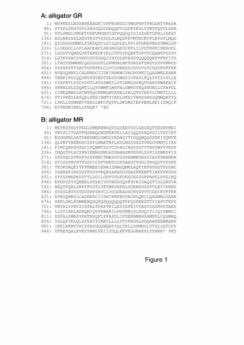
Figure 1. Alligator glucocorticoid receptor (GR) and mineralocorticoid receptor (MR).
(A) The deduced amino acid sequence of alligator GR. Numbers on the side represent the
position of amino acid residues in sequence. (B) The deduced amino acid sequence of
alligator MR. Numbers on the side represent the position of amino acid residues in
sequence.
Figure 2. Domain structure of alligator GR and MR and homology with GR/MR from other
species.
(A) Comparison of alligator GR with alligator MR. The functional A/B to E/F domains are
schematically represented with the numbers of amino acid residues indicated. (B) Domain
structure of GR in alligator, and identity with chicken, human, Xenopus, and zebrafish GRs.
(C) Domain structure of MR in alligator, and identity with chicken, human, Xenopus, and
zebrafish MRs. The functional A/B to E/F domains are schematically represented with the
16
numbers of amino acid residues indicated. The numbers within each box indicated the
percent identity of the domain compared to alligator GR or GR. GenBank accession: human
GR; NM_000901, chicken GR; NM_001159345, Xenopus GR; NM_001090605, zebrafish
GR; NM_001100403). Numbers (%) shows the over-all identity. (C) Comparison of
alligator MR with MRs of several species (human, chicken, Xenopus, and zebrafish;
GenBank accession: human MR; NM_000176, chicken MR; NM_001037826, Xenopus MR;
NM_001088062, zebrafish GR; NM_001100403).
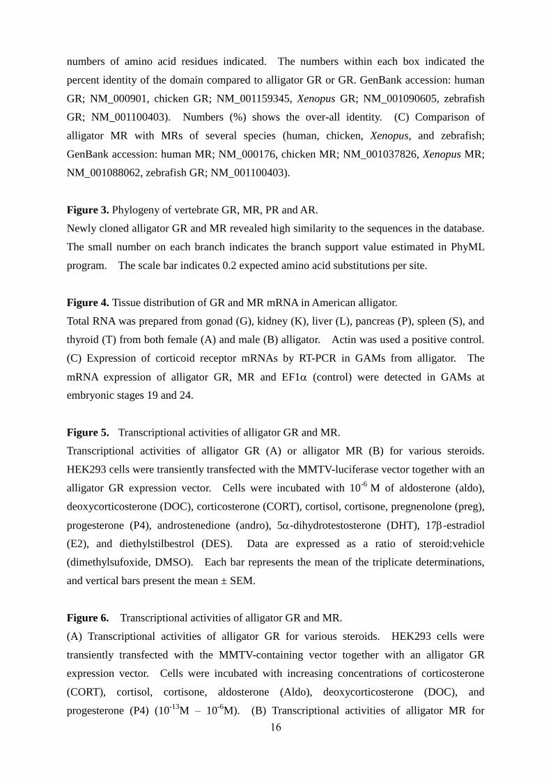
Figure 3. Phylogeny of vertebrate GR, MR, PR and AR.
Newly cloned alligator GR and MR revealed high similarity to the sequences in the database.
The small number on each branch indicates the branch support value estimated in PhyML
program. The scale bar indicates 0.2 expected amino acid substitutions per site.
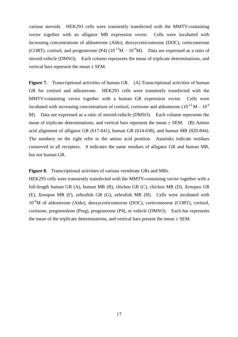
Figure 4. Tissue distribution of GR and MR mRNA in American alligator.
Total RNA was prepared from gonad (G), kidney (K), liver (L), pancreas (P), spleen (S), and
thyroid (T) from both female (A) and male (B) alligator. Actin was used a positive control.
(C) Expression of corticoid receptor mRNAs by RT-PCR in GAMs from alligator. The
mRNA expression of alligator GR, MR and EF1 (control) were detected in GAMs at
embryonic stages 19 and 24.
Figure 5. Transcriptional activities of alligator GR and MR.
Transcriptional activities of alligator GR (A) or alligator MR (B) for various steroids.
HEK293 cells were transiently transfected with the MMTV-luciferase vector together with an
alligator GR expression vector. Cells were incubated with 10-6
M of aldosterone (aldo),
deoxycorticosterone (DOC), corticosterone (CORT), cortisol, cortisone, pregnenolone (preg),
progesterone (P4), androstenedione (andro), 5-dihydrotestosterone (DHT), 17-estradiol
(E2), and diethylstilbestrol (DES). Data are expressed as a ratio of steroid:vehicle
(dimethylsufoxide, DMSO). Each bar represents the mean of the triplicate determinations,
and vertical bars present the mean ± SEM.
Figure 6. Transcriptional activities of alligator GR and MR.
(A) Transcriptional activities of alligator GR for various steroids. HEK293 cells were
transiently transfected with the MMTV-containing vector together with an alligator GR
expression vector. Cells were incubated with increasing concentrations of corticosterone
(CORT), cortisol, cortisone, aldosterone (Aldo), deoxycorticosterone (DOC), and
progesterone (P4) (10-13
M – 10-6
M). (B) Transcriptional activities of alligator MR for
17
various steroids. HEK293 cells were transiently transfected with the MMTV-containing
vector together with an alligator MR expression vector. Cells were incubated with
increasing concentrations of aldosterone (Aldo), deoxycorticosterone (DOC), corticosterone
(CORT), cortisol, and progesterone (P4) (10-13
M – 10-6
M). Data are expressed as a ratio of
steroid:vehicle (DMSO). Each column represents the mean of triplicate determinations, and
vertical bars represent the mean ± SEM.
Figure 7. Transcriptional activities of human GR. (A) Transcriptional activities of human
GR for cortisol and aldosterone. HEK293 cells were transiently transfected with the
MMTV-containing vector together with a human GR expression vector. Cells were
incubated with increasing concentrations of cortisol, cortisone and aldosterone (10-12
M – 10-6
M). Data are expressed as a ratio of steroid:vehicle (DMSO). Each column represents the
mean of triplicate determinations, and vertical bars represent the mean ± SEM. (B) Amino
acid alignment of alligator GR (617-641), human GR (614-638), and human MR (820-844).
The numbers on the right refer to the amino acid position. Astarisks indicate residues
conserved in all receptors. # indicates the same residues of alligator GR and human MR,
but not human GR.
Figure 8. Transcriptional activities of various vertebrate GRs and MRs.
HEK293 cells were transiently transfected with the MMTV-containing vector together with a
full-length human GR (A), human MR (B), chicken GR (C), chicken MR (D), Xenopus GR
(E), Xenopus MR (F), zebrafish GR (G), zebrafish MR (H). Cells were incubated with
10-6
M of aldosterone (Aldo), deoxycorticosteorne (DOC), corticosteorne (CORT), cortisol,
cortisone, pregnenolone (Preg), progesteorne (P4), or vehicle (DMSO). Each bar represents
the mean of the triplicate determinations, and vertical bars present the mean ± SEM.
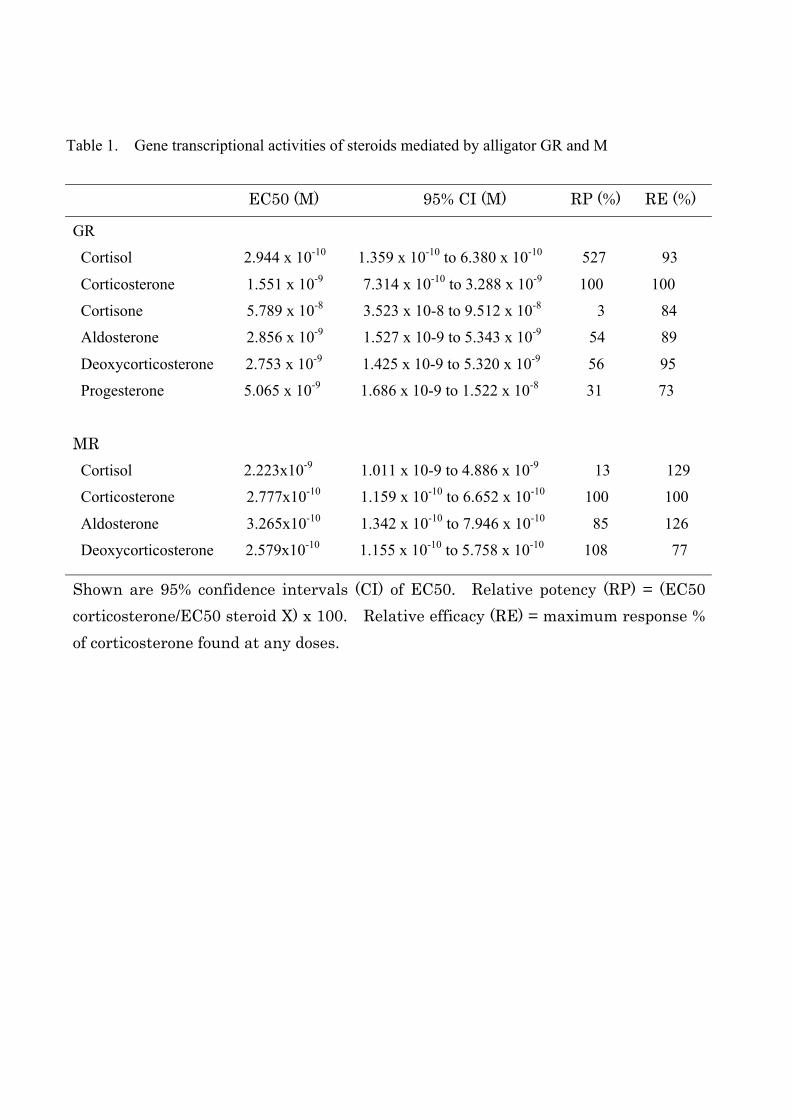
Table 1. Gene transcriptional activities of steroids mediated by alligator GR and M
EC50 (M) 95% CI (M) RP (%) RE (%)
GR
Cortisol 2.944 x 10⁻¹⁰ 1.359 x 10⁻¹⁰ to 6.380 x 10⁻¹⁰ 527 93
Corticosterone 1.551 x 10⁻⁹ 7.314 x 10⁻¹⁰ to 3.288 x 10⁻⁹ 100 100
Cortisone 5.789 x 10⁻⁸ 3.523 x 10⁻⁸ to 9.512 x 10⁻⁸ 3 84
Aldosterone 2.856 x 10⁻⁹ 1.527 x 10⁻⁹ to 5.343 x 10⁻⁹ 54 89
Deoxycorticosterone 2.753 x 10⁻⁹ 1.425 x 10⁻⁹ to 5.320 x 10⁻⁹ 56 95
Progesterone 5.065 x 10⁻⁹ 1.686 x 10⁻⁹ to 1.522 x 10⁻⁸ 31 73
MR Cortisol 2.223x10⁻⁹ 1.011 x 10⁻⁹ to 4.886 x 10⁻⁹ 13 129
Corticosterone 2.777x10⁻¹⁰ 1.159 x 10⁻¹⁰ to 6.652 x 10⁻¹⁰ 100 100
Aldosterone 3.265x10⁻¹⁰ 1.342 x 10⁻¹⁰ to 7.946 x 10⁻¹⁰ 85 126
Deoxycorticosterone 2.579x10⁻¹⁰ 1.155 x 10⁻¹⁰ to 5.758 x 10⁻¹⁰ 108 77
Shown are 95% confidence intervals (CI) of EC50. Relative potency (RP) = (EC50 corticosterone/EC50 steroid X) x 100. Relative efficacy (RE) = maximum response % of corticosterone found at any doses.
1: MDPKESLNSSEREEAGKIVRFNDKGGIVMDFHPTFRGGATVKASA 46: STSPLPASTSPLPASSQSDSKQQPVLGDFSKGLVSNVPQPDLSKA 91: VSLSMGLYMGETDSKVMGNDIGFPQQGQIGISSGETDFRLLEESI136: ASLNKSSGLAEGTKGTVSSGLSLKQDFPVMTNCNVPLEPGTLMQG181: QIGSSGGNMKLFSEDQSTLDILQDLELPPISPGKEPNGSPWRLDP226: LLDEGGLLSPLAADEAFLHEGNSGEDCKPLILPDTKPKINERGDL271: LSSPPVQMPQVKTEKEDFIELCTPGIKQEKTGPVYCQANFPGSSV316: LGTKVSAISVHGVSTSGGQIYHYDLNTASHSQQQDQKPVFNVIPS361: LTAGTENWNRCQGSGDDPLSPMGNLNFSGRSSFSNGYSSPGMRSD406: VSSSPSTTSATTGPPPKLCLVCSDEASGCHYGVLTCGSCKVFFKR451: AVEGQHNYLCAGRNDCIIDKIRRKNCPACRYRKCLQAGMNLEARK496: TKKKIKGIQQSNVSSVRDTPESPGNKSIVPASLPQLTPTLVSLLE541: VIEPEVLYSGYDSTLPDSSWRILSTLNMLGGRQVVAAVKWAKAIP586: GFRNLHLDDQMTLLQYSWMFLMSFALGWRSYKQSNGNLLCFAPDL631: IINEQRMSLPCMYEQCKNMLMVAGELSRLQVSYEEYLCMKTLLLL676: STIPKEGLKSQALFEEIRMTYIKELGKAIVKREGNSSQNWQRFYQ721: LTKLLDSMHEVVENLLNFCFQTFLDKSMSIEFPEMLAEIISNQIP766: KYSNGNIKKLLFHQK* 780
1: METKGYHSYPEGLDMEKRWSQVSQSAEYSSLGAGEQTDESNYMEI 46: VNVSCITGAFPNSNAQGNGKEKPELLACLQQDSNQPGILTSDIKT 91: ESDSKELSATVAESMGLYMDSIRDADYTYDQQNQGSPGKIYQNVE136: QLVKFYKENGHCSSPLNNATRPLRSLMSDSGSSVNGGVMHTIIKS181: PIMCQEKSPSGCSPQNMTSSVCSPAGINSVSSTTTNFGNFVVNSP226: INQGTPLSCSPNIENRGSMLHSPAHASNVGSPLSSPISSMKSPIS271: SPPSHCSVKSPVSSPNNITMRSSVSSPANMNSRSSIASPSNANNR316: STLSSPAVSTVGSSICSPVNNSLGFSAAGTPGGLSRGQDTVPSPE361: TKDKGAQEITFPKMEEIENAISNNGQMNLAQFIKPEPDGTFGSAC406: IGENSKINSDSPFSVPVKQESAKHSCSGASFKRNPTINPFPFSDG451: SYFSFMDDKDYYTLSGILGPPVSSFDGSCEGSGFPNPGLPVGIKQ496: EPDDGSYYQENRLPSSAIVGVNSGGQSFHYRIGAQGTISLSRPVA541: REQTFQHLSAFPPVSTLVETWKSHPDLSSRRNDGYPVLEYIPENV586: SSSSLRSVSTGSSRPSKVCLVCGDEASGCHYGVVTCGSCKVFFKR631: AVEGQHNYLCAGRNDCIIDKIRRKNCPACRLQKCLQAGMNLGARK676: SKKLGKLKGMHEEQSQPQPQQQQQQPPPQSPEEGTTYIAPVTEPS721: VNTALVPHVSISPALTPAPVKILESIEPEIVYAGYDSSKPDTAEY766: LLSTLNRLAGKQMIQVVKWAKILPGFRNLPLEDQITLIQYSWMCL811: SSFALSWRSYKHTNSQFLYFAPDLIFDEERMRHSAMFELCQGMHQ856: ISLQFVRLQLSFEEYTIMKVLLLLSTVPRDGLKSQAAFEEMRANY901: IKELKKMVTKCPSNSGQSWQRFYQLTKLLDSMHDLVTDLLEFCFY946: TFRESQALKVEFPAMLVEIISDQLPKVESGNAKPLYFHRK* 985
Figure 1
B: alligator MR
A: alligator GR
A/B C D E/F
27 93 31 59
1 423 489 530 780
1 603 672 736 985
MR
GRA
C: MR
B: GR
34 97 41 73
54 100 60 83
67 100 70 90
76 100 70 97
A/B C D E(F)1 423 491 531 780
1 415 483 523 772
1 420 488 528 777
1 419 487 527 776
1 386 454 497 746
44 97 40 74
60 99 58 85
78 100 78 92
87 100 86 98
A/B C D E(F)1 603 671 735 985
1 603 671 731 981
1 602 670 734 984
1 599 667 729 979
1 600 668 720 970
Alligator
Chicken
Human
Xenopus
Zebrafish
Figure 2
Alligator
Chicken
Human
Xenopus
Zebrafish
Human GR Mouse GR
Alligator GR Anole GR Xenopus GR
Zebrafish MR Xenopus MR
Chicken MR Alligator MR Mouse MR
Human MR AR PR
8599
24
88
100
97
89
67
94
98
98
98
100
0.1
Figure 3
GR
MR
EF1α
stage 19 stage 24
F M F M
GR
MR
actin
female
male G K L P S T
GR
MR
actin
G K L P S T
Figure 4
A
C
B
200
150
100
50
0
70
50
30
20
0
60
40
10
fold
act
ivat
ion
fold
act
ivat
ion
DMSO aldoDOC
CORTco
rtisol
cortis
onepreg
androDHT E2
DESP4
DMSO aldoDOC
CORTco
rtisol
cortis
onepreg
androDHT E2
DESP4
Figure 5
A: alligator GR
B: alligator MR
-13 -12 -11 -10 -9 -8 -7 -60
30
60
90
120
150
180
P4Cortisone
DOCAldo
CortisolCOR
fold
acti
vatio
n
-13 -12 -11 -10 -9 -8 -7 -60
20
40
60
80
P4CortisolCORDOCAldo
fold
acti
vatio
n
steroid concentration, log [(M)]
steroid concentration, log [(M)]
Figure 6
A: alligator GR
B: alligator MR
steroid concentration, log [(M)]
fold
acti
vatio
n
Figure 7
A
B # #617 KQSNGNLLCFAPDLIINEQRMSLPC614 RQSSANLLCFAPDLIINEQRMTLPC820 KHTNSQFLYFAPDLVFNEEKMHQSA * ***** ** *
alligator GRhuman GRhuman MR
800
600
400
200
0
350
300
250
200
150
100
50
0
350
300
250
200
150
100
50
0
500
400
300
200
100
0
150
120
90
60
30
0
1200
900
600
300
0
150
120
90
60
30
0
40
30
20
10
0
DMSO
AldoDOC
CORT
Cortis
ol
Cortis
one
Preg P4
fold
act
ivat
ion
DMSO
AldoDOC
CORT
Cortis
ol
Cortis
one
Preg P4
DMSO
AldoDOC
CORT
Cortis
ol
Cortis
one
Preg P4
DMSO
AldoDOC
CORT
Cortis
ol
Cortis
one
Preg P4
DMSO
AldoDOC
CORT
Cortis
ol
Cortis
one
Preg P4
DMSO
AldoDOC
CORT
Cortis
ol
Cortis
one
Preg P4
DMSO
AldoDOC
CORT
Cortis
ol
Cortis
one
Preg P4
DMSO
AldoDOC
CORT
Cortis
ol
Cortis
one
Preg P4
fold
act
ivat
ion
fold
act
ivat
ion
fold
act
ivat
ion
fold
act
ivat
ion
fold
act
ivat
ion
fold
act
ivat
ion
fold
act
ivat
ion
Figure 8
A: human GR
E: Xenopus GR
C: chicken GR
G: zebrafish GR
B: human MR
F: Xenopus MR
D: chicken MR
H: zebrafish MR