Embed Size (px)
Citation preview
Unusual Thoracic Manifestations in Filariasis
Due to Loa Loa*
Results of Treatment with Hetrasan and Naphuride Sodium
SAMUEL H. MADELL, M.D. and CLIFFORD L. SPINGARN, M.D.
New York, New York
E NDEMIC only in parts of Africa, filariasis
caused by the nematode Loa loa is rarely observed in this country. We recently
had opportunity to study a patient with this disease and report our findings since an unusual clinical picture was encountered and some interesting results followed treatment with hetrazan@ (1 -diethylcarbamyl-4-methylpipera- zine hydrochloride) and naphuride sodium.@
Human infection with Loa loa occurs chiefly along the west coast of equatorial Africa. The disease is common in Nigeria, the Anglo- Egyptian Sudan, French and Belgian Congo and Ungola. 26 Man is infected with the parasite by the bite of the insect vector, the blood sucking mangrove fly, Chrysops dimidiata. While feed- ing the female flies deposit the larval worms on the skin. The latter quickly penetrate into the subcutaneous tissues where they mature into adults in about four months. The adult females give birth to microfilariae which enter the blood stream and infect the blood sucking insects. In the fly the microfilariae migrate from the stomach through the thoracic muscles to the proboscis.
The worms develop very slowly in the human host. About four years elapse between the time of infection and the appearance of the next generation of microfilariae in the blood. The adults live in and move freely through the loose connective tissues. They are about 1.5 inches in length and resemble small white threads. The microfilariae are about 300 micra in length and are found in the blood in greatest numbers dur- ing the day, in contrast to the nocturnal periodicity of the microfilariae of Wuchereria bancrofti.28
In many patients with the disease the worms can be seen and felt beneath the skin. They often appear beneath the conjunctivae where they
cause severe pain, chemosis and lacrimation. In the subcutaneous tissues the worms may move several inches in a few minutes.12
Allergic reactions to the parasites give rise to recurring subcutaneous and intramuscular swell- ings and nodules called “Calabar swellings,” after a town in Nigeria where loiasis is com- mon.12~14-16 They may occur on any part of the body surface but are most common on the fore- arms. They are usually 1 or 2 inches in diameter and may persist for about five days. As a rule they are attended by itching and burning, rarely by pain. When they occur frequently in the same area chronic cystic tumors may be formed. These are often attached to the tendon sheaths and muscular aponeuroses and contain straw-colored fluid.28
Involvement and obstruction of the lym- phatics by the adult worms occurs but is not as frequent as in infections with Wuchereria ban- crofti. Severe lymphatic edema, lymph varix, hydrocele and varicocele have been noted in the hyperendemic areas. l2 Eosinophilia is a com- mon hematologic finding.
CASE REPORT
A thirty-one year old Negro male was ad- mitted to the Second Medical Service (Dr. I. Snapper) of The Mount Sinai Hospital on June 15, 1951. He complained of chills and fever for two days and a painful mass in the left pectoral region for one week.
In 1946 a painless, slightly raised swelling, about the size of a fist, was noted on his chest below the left clavicle. He was examined at Harlem Hospital in New York City and a four- plus positive serologic reaction for syphilis was found. He was treated for lues for a period of two years with injections of bismuth and
* From the Departments of Medicine and Microbiology of The Mount Sinai Hospital, New York, N. Y.
272 AMERICAN JOURNAL OF MEDICINE
Filariasis Due to Loa Loa-Madell, Sfiingarn
arsenical preparations. He did not receive penicillin. During this interval the swelling on his anterior chest wall fluctuated in size but never caused any discomfort. It disappeared completely for variable periods of time. In 1950 he developed a dry cough. X-rays of his chest revealed a large mass in his mediastinum and he was advised to enter a hospital for observa- tion. However, he declined to do so and con- tinued to work as an elevator operator.
He was born in Nigeria and lived there until 1941. At that time he got a job as a steward abroad a British merchant ship. In 1945 he came to New York City where he has lived since. Aside from several attacks of acute urethritis, his health in Nigeria was good.
On examination the patient was found to be a well nourished and developed Negro male who had a slight cough productive of small amounts of mucoid sputum. The body temperature (rectal) was 101.2’F., the pulse rate was 104, respiratory rate 24 and the blood pressure was 145/100. The trachea deviated to the right and was freely movable. The neck veins were flat. There were several hard, discrete, non- tender almond-sized lymph nodes in each axilla.
The striking physical finding was an elevated, fluctuant, tender swelling of irregular outline covering the left pectoral region. It extended from the lower margin of the pectoral muscles to the anterior border of the trapezius and from the left sternal border to the anterior axillary line. On recumbency the non-pulsatile mass diminished in size and a fullness appeared in the left supraclavicular fossa. At the inferolateral border of the mass there was a hard, fixed, non-tender nodule (2 by 3 cm.). There were similar but smaller nodules at the superolateral and superomedial borders. Dullness and dimin- ished breath sounds were noted at the base of the left lung posteriorly and over the left anterior chest from the second to fourth ribs parasternally. The liver was percussed one fingerbreadth below the costal margin. There were no other palpable abdominal organs or masses.
The hemoglobin concentration of the blood was 13.0 gm. per cent. The white blood count was 16,000 per cu. mm. The differential count was 67 per cent segmented, 16 per cent non- segmented polymorphonuclear leukocytes, 5 per cent lymphocytes and 7 per cent eosinophils. The platelet count was 100,000. Blood Wasser- mann and Kahn tests were negative. The
AUGUST, 1953
erythrocyte sedimentation rate was 109 mm. in one hour (Westergren) . Urinalysis was negative for sugar, albumin and formed elements. Blood culture was negative. A search for Hargrave’s cells in the blood was negative. Blood urea nitrogen was 14 mg. per cent, blood sugar 105 mg. per cent, blood uric acid, 3.6 mg. per cent, serum alkaline phosphatase, 5 K-A units, serum total protein, 6.6 gm. per cent,albumin, 3.6 gm. per cent and globulin 3.0 gm. per cent.
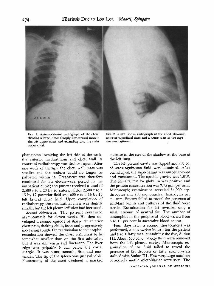
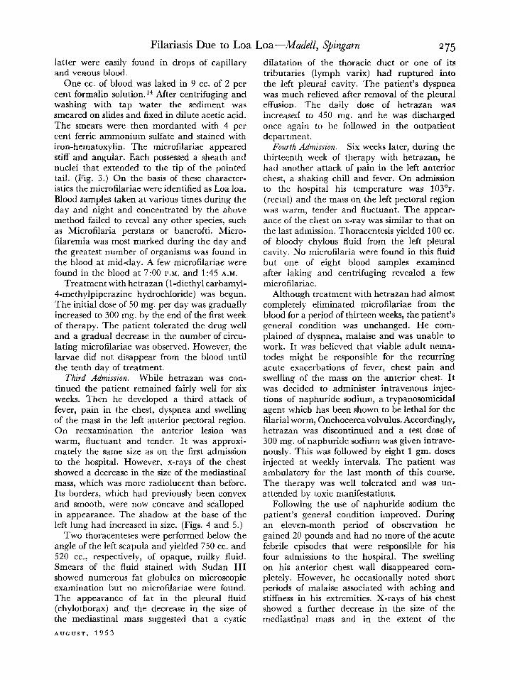
Fluoroscopy and an x-ray of the chest re- vealed a large, dense, homogeneous, sharply demarcated mass, about 8 cm. in diameter, situated in the anterosuperior region of the left chest and extending slightly into the right lung field at the level of the aortic arch. The trachea was deviated to the right and there was a small pleural effusion at the left base. (Figs. 1 and 2.) The mass did not pulsate. There was no delay in the passage of barium through the esophagus.
Bronchoscopy revealed narrowing and distor- tion of the left main bronchus particularly from the anterior aspect. There was no demonstrable intrabronchial abnormality. Biopsy of a left axillary lymph node showed the changes of chronic non-specific inflammation. Examination of the sternal bone marrow showed mild plasmacytosis but the marrow was otherwise normal. An intradermal skin test with Echino. coccus antigen was negative.
A biopsy obtained from the wall of the center of the mass showed huge dilated channels measuring approximately 0.5 cm. in diameter from which a profuse amount of straw-colored fluid drained. Microscopically, the specimen had the appearance of inflamed cystic hygroma.
In an attempt to determine whether the cystic chest wall mass communicated with the deeper lymphatics of the thorax the patient was given a fatty meal containing Sudan III and the superficial swelling was then aspirated. The fluid obtained contained blood and autolysed cells but no dye-stained particles were seen.
Although it was now possible to classify the chest wall mass histologically, the problem of the nature of the mediastinal tumor and its possible relation to the former remained un- solved. Because of the patient’s birth place and the lymphatic nature of the superficial mass, the possibility of filariasis was considered. However, microfilariae were not found in the peripheral blood.
It was thought that the mediastinal tumor was either a lymphoma or a cavernous lym-
Filariasis Due to Loa Loa-Mudell, S@ingarn
FIG. 1. Anteroposterior radiograph of the chest,
showing a large, dense sharply demarcated mass in
the left upper chest and extending into the right
upper chest.
phangioma involving the left side of the neck, the anterior mediastinum and chest wall. A course of radiotherapy was decided upon. After one week of therapy the chest wall mass was smaller and the nodules could no longer be palpated within it. Treatment was therefore continued for an eleven-week period in the outpatient clinic; the patient received a total of 2,500 r to a 20 by 20 anterior field, 2,500 r to a 15 by 17 posterior field and 600 r to a 15 by 10 left lateral chest field. Upon completion of radiotherapy the mediastinal mass was slightly smaller but the left pleural effusion had increased.
Second Admission. The patient remained asymptomatic for eleven weeks. He then de- veloped a second episode of sharp left anterior chest pain, shaking chills, fever and progressively increasing cough. On readmission to the hospital examination showed the chest wall mass to be somewhat smaller than on the first admission but it was still warm and fluctuant. The liver edge was palpable 5 cm. below the costal margin. It was blunt, smooth, firm and non- tender, The tip of the spleen was just palpable. Fluoroscopy of the chest disclosed a marked
FIG. 2. Right lateral radiograph of the chest showing
anterior superficial mass and a dense mass in the supe-
rior mediastinum.
increase in the size of the shadow at the base of the left lung.
The left pleural cavity was tapped and 750 cc. of serosanguineous fluid were obtained. After centrifuging the supernatant was amber colored and translucent. The specific gravity was 1.019. The Rivalta test for globulin was positive and the protein concentration was 9.75 gm. per cent. Microscopic examination revealed 84,000 ery- throcytes and 250 mononuclear leukocytes per cu. mm. Smears failed to reveal the presence of acid-fast bacilli and cultures of the fluid were sterile. Examination for fat revealed only a small amount of neutral fat. The number of eosinophils in the peripheral blood varied from 5 to 10 per cent in successive blood counts.
Four days later a second thoracentesis was performed, about twelve hours after the patient had had a fatty meal containing the dye, Sudan III. About 600 cc. of bloody fluid were removed from the left pleural cavity. Microscopic ex- amination of the fluid failed to reveal the presence of fat droplets or fatty acid crystals stained with Sudan III. However, large numbers of actively motile microfilariae were seen. The
AMERlCAN JOURNAL OF MEDICINE
Filariasis Due to Loa Loa--Made& S’ingarn 275 latter were easily found in drops of capillary and venous blood.
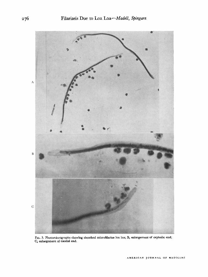
One cc. of blood was laked in 9 cc. of 2 per cent formalin solution.i4 After centrifuging and washing with tap water the sediment was smeared on slides and fixed in dilute acetic acid. The smears were then mordanted with 4 per cent ferric ammonium sulfate and stained with iron-hematoxylin. The microfilariae appeared stiff and angular. Each possessed a sheath and nuclei that extended to the tip of the pointed tail. (Fig. 3.) On the basis of these character- istics the microfilariae were identified as Loa loa. Blood samples taken at various times during the day and night and concentrated by the above method failed to reveal any other species, such as Microfilaria perstans or bancrofti. Micro- filaremia was most marked during the day and the greatest number of organisms was found in the blood at mid-day. A few microfilariae were found in the blood at 7:00 P.M. and 1:45 A.M.
Treatment with hetrazan (l-diethyl carbamyl- 4-methylpiperazine hydrochloride) was begun. The initial dose of 50 mg. per day was gradually increased to 300 mg. by the end of the first week of therapy. The patient tolerated the drug well and a gradual decrease in the number of circu- lating microfilariae was observed. However, the larvae did not disappear from the blood until the tenth day of treatment.
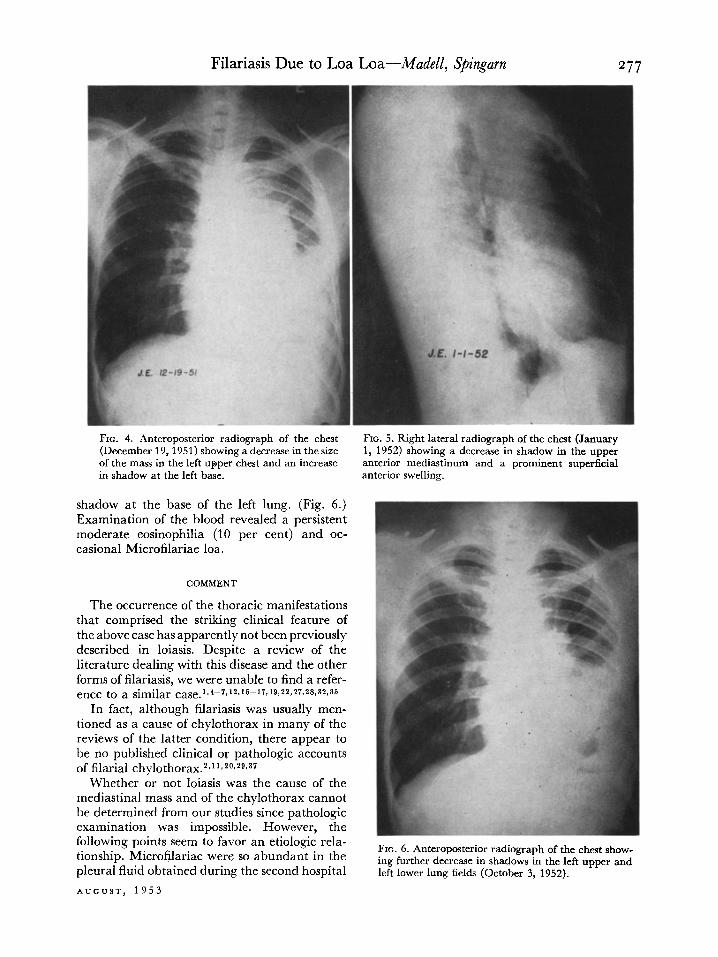
Third Admission. While hetrazan was con- tinued the patient remained fairly well for six weeks. Then he developed a third attack of fever, pain in the chest, dyspnea and swelling of the mass in the left anterior pectoral region. On reexamination the anterior lesion was warm, fluctuant and tender. It was approxi- mately the same size as on the first admission to the hospital. However, x-rays of the chest showed a decrease in the size of the mediastinal mass, which was more radiolucent than before. Its borders, which had previously been convex and smooth, were now concave and scalloped in appearance. The shadow at the base of the left lung had increased in size. (Figs. 4 and 5.)
Two thoracenteses were performed below the angle of the left scapula and yielded 750 cc. and 520 cc., respectively, of opaque, milky fluid. Smears of the fluid stained with Sudan III showed numerous fat globules on microscopic examination but no microfilariae were found. The appearance of fat in the pleural fluid (chylothorax) and the decrease in the size of the mediastinal mass suggested that a cystic
AUDrJST, 1953
dilatation of the thoracic duct or one of its tributaries (lymph varix) had ruptured into the left pleural cavity. The patient’s dyspnea was much relieved after removal of the pleural effusion. The daily dose of hetrazan was increased to 450 mg. and he was discharged once again to be followed in the outpatient department.
Fourth Admission. Six weeks later, during the thirteenth week of therapy with hetrazan, he had another attack of pain in the left anterior chest, a shaking chill and fever. On admission to the hospital his temperature was 103'F.
(rectal) and the mass on the left pectoral region was warm, tender and fluctuant. The appear- ance of the chest on x-ray was similar to that on the last admission. Thoracentesis yielded 100 cc. of bloody chylous fluid from the left pleural cavity. No microfilaria were found in this fluid but one of eight blood samples examined after laking and centrifuging revealed a few microfilariae.
Although treatment with hetrazan had almost completely eliminated microfilariae from the blood for a period of thirteen weeks, the patient’s general condition was unchanged. He com- plained of dyspnea, malaise and was unable to work. It was believed that viable adult nema- todes might be responsible for the recurring acute exacerbations of fever, chest pain and swelling of the mass on the anterior chest. It was decided to administer intravenous injec- tions of naphuride sodium, a trypanosomicidal agent which has been shown to be lethal for the filarial worm, Onchocerca volvulus. Accordingly, hetrazan was discontinued and a test dose of 300 mg. of naphuride sodium was given intrave- nously. This was followed by eight 1 gm. doses injected at weekly intervals. The patient was ambulatory for the last month of this course. The therapy was well tolerated and was un- attended by toxic manifestations.
Following the use of naphuride sodium the patient’s general condition improved. During an eleven-month period of observation he gained 20 pounds and had no more of the acute febrile episodes that were responsible for his four admissions to the hospital. The swelling on his anterior chest wall disappeared com- pletely. However, he occasionally noted short periods of malaise associated with aching and stiffness in his extremities. X-rays of his chest showed a further decrease in the size of the mediastinal mass and in the extent of the
276 Filariasis Due to Loa Loa-Ahdell, Spingarn
FIG. 3. Photomicrographs showing sheathed microfilariae loa loa; B, enlargement of cephalic end; C, enlargement of caudal end.
AMERICAN JOURNAL OF MEDICINE
Filariasis Due to Loa Loa-Ahdell, S’ingam
FIG. 4. Anteroposterior radiograph of the chest (December 19,195l) showing a decrease in the size of the mass in the left upper chest and an increase in shadow at the left base.
shadow at the base of the left lung. (Fig. 6.) Examination of the blood revealed a persistent moderate eosinophilia (10 per cent) and oc- casional Microfilariae loa.
COMMENT
The occurrence of the thoracic manifestations that comprised the striking clinical feature of the above case has apparently not been previously described in loiasis. Despite a review of the literature dealing with this disease and the other forms of filariasis, we were unable to find a refer- ence to a similar ca~e.1~4-7,12~16-17~19~29,27~28~32~35
In fact, although filariasis was usually men- tioned as a cause of chylothorax in many of the reviews of the latter condition, there appear to be no published clinical or pathologic accounts of filarial chylothorax.2~11*20,2g,37
Whether or not loiasis was the cause of the mediastinal mass and of the chylothorax cannot be determined from our studies since pathologic examination was impossible. However, the following points seem to favor an etiologic rela- tionship. Microfilariae were so abundant in the pleural fluid obtained during the second hospital
AUGUST, 1953
FIG. 5. Right lateral radiograph of the chest (January 1, 1952) showing a decrease in shadow in the upper anterior mediastinum and a prominent superficial anterior swelling.
diograph of the chc ing further decrease in shadows in the left upper and left lower lung fields (October 3, 1952).
278 Filariasis Due to Loa
admission that it is reasonable to attribute their presence to the localization of adult worms in the mediastinum. Froes reported the finding of numerous microfilariae in the ascitic fluid in a case of Bancroftian filariasis with ascites2* The appearance of chyle in the pleural fluid and the concomitant shrinkage of the mediastinal mass suggested that the latter was a lymph varix, arising from the thoracic duct, that had rup- tured and spilled its contents into the pleural cavity. It is conceivable that the Loa loa adults could produce obstruction of the thoracic duct or one of its tributaries in the mediastinum. The worms have been known to cause lym- phatic obstruction and lymph varices in the inguinal regions and at autopsy they have been found in every part of the body including the heart and pericardium.12s2s Tissue reactions to the microfilariae have been observed by Klotz who found them within fibrotic nodules in the spleen and other tissues.13
In filariasis due to Wuchereria bancrofti instances of obstruction and extreme dilation of the thoracic duct below the diaphragm have been described in several reports.27 Young found an enormous retroperitoneal lymph varix and a greatly dilated thoracic duct in a case of chyluria. Among nine other cases of filarial chyluria collected from the literature by Stephens and Yorke there were three in which obstruction and dilatation of the retroperitoneal portion of the thoracic duct was observed at postmortem examination.27 Wise reported the finding of a large chylous cyst between the two layers of the mesentery in a case of ascites and also described an abdominal lymphangiovarix containing adult worms in some of the varices.27
In our case the recurrent swelling on the left pectoral region had the characteristics of a cutaneous lymph varix. The periodic acute in- flammatory reactions within it were in all likeli- hood episodes of filarial lymphangitis.lg The disappearance of this mass after the chemo- therapy of the loiasis is in favor of its filarial etiology.
It is possible that this patient had two inde- pendent conditions, namely, loiasis and a cystic lymphangioma of the mediastinum and an- terior chest wall. Loiasis has been known to occur in the absence of clinical manifestations and our patient never experienced some of the common symptoms of the disease, i.e., the movements of the live worms beneath the skin and the occurrence of the typical migratory
Loa--1Madel1, S’ingarn
calabar swellings. A cavernous lymphangioma was found by Neuhof and Swift in a patient with a soft cystic swelling in the. left supra- clavicular region and a large left pleural ef- fusion.2s Chyle was obtained from the mass and from the pleural cavity. Exploration of the left supraclavicular region disclosed a diffuse, fatty mass behind the clavicle. Biopsy revealed this to be a lymphangioma.
Hetrazan$(l-diethylcarbamyl-4-methylpiper- azine hydrochloride) was shown to have a marked filariacidal action in Bancroftian filar- iasis by Santiago-Stevenson et a1.23 Oral admin- istration of the drug caused a rapid disappear- ance of microfilariae from the blood and favor- ably affected the course of the disease. Sub- sequently, encouraging therapeutic results with hetrazan were reported in loiasis and onchocer- ciasis.3s18,24,26,30,32 The drug was effective against the microfilariae volvulus and loa in man in doses comparable to those used in W. bancrofti infections but had no effect on microfilariae perstans or ozzardi.‘O
In loiasis the use of the drug has been attended by prompt cessation of the Calabar swellings and clearing of the blood of microfilariae within a week. Murgatroyd and Woodruff18 treated nine patients with doses of 2 to 6 mg. per kg. for periods of from ten to twenty-one days and observed recurrence of symptoms in only one case during a six-month period of observation. Shookoff and Dwork24 treated five cases with 300 mg. a day for seven to ten days. Four pa- tients were relieved of Calabar swellings for four to ten months. Prior to treatment none of the group had had symptom-free intervals for more than two months. Stefanapoulo and Schneider26 treated twenty patients with 3 to 6 mg. per kg. per day for seven to ten days. In nineteen patients all symptoms disappeared within forty-eight hours. In most of these cases symptoms recurred several weeks after the cessation of therapy but they were less severe.
Reactions, supposedly of an allergic nature, have occurred during the early stages of treat- ment with hetrazan and it has been advised that treatment be begun with small doses. The common untoward reactions have included nausea, vomiting, fever, diarrhea, pruritus, morbilliform and papular skin eruptions. The mode of action of the drug on the adult Loa loa is not known and there is no evidence that the worms are killed by it even when given for prolonged periods. WoodrufPB postulated that
AMERICAN JOURNAL OF MEDICINE
Filariasis Due to Loa Loa-Mudell, Spingarn 279 the drug acts as an opsonin, sensitizing the microfilariae to the phagocytic activity of the cells of the reticula-endothelial system.
In our case, although hetrazan was given continuously for thirteen weeks in doses up to 7 mg. per kg. per day, the blood was not com- pletely cleared of microfilariae and there was little change in the general condition of the patient. The mass on the anterior chest wall persisted and was the site of repeated acute inflammatory flare-ups. This clinical behavior was attributed to the presence of live worms and their toxic products and to repeated secondary infections of the obstructed lymphatics. Beye and his co-workers’ observed that continuous treatment of W. bancrofti infections for more than one year did not reduce the frequency of recurrent attacks of acute filarial lymphangitis.
followed by the cessation of acute exacerbations, disappearance of the chest wall mass and a 20 pound gain in weight during an eleven-month follow-up period.
Acknowledgment: The authors wish to express their indebtedness to Dr. I. Snapper for his advice in the diagnosis and treatment of this case. We also wish to thank Mr. Fred Lazarus for his technical assistance in the demonstration of the morphology of the microfilaria. The naphuride sodium used in this case was supplied generously by the Winthrop Chemical Company.
REFERENCES
The use of naphuride sodium in this case was prompted by the recent report that it had a lethal action on Onchocerca volvulus.3~34 In 1926 Chopra and Rao 31 tried this drug in two cases of W. bancrofti infection and failed to observe an effect on the microfilariae with the small doses that they used. The drug was well tolerated by our patient. The change in the clinical course and the general physical improve- ment that was observed during the eleven- month period following its use suggested that it may have been of value in reducing the number of live nematodes in this case.
1. BEYE, H. K., EDGAR, S. A., MILLE, R., KESSEL, J. F. and BAMBRIDGE, B. Preliminary observations on the prevalence, clinical manifestations and control of filariasis in the Society Islands. Am. J. Trap. Med. & Hyg., 1: 637, 1952.
2. BROWN, R. B. and DUNN, R. G. Lymphogenous cysts of the mediastinum. U. S. Armed Forces Med. .I., 2: 1651, 1951.
3. BURCH, T. A. and ASHBURN, I,. L. Experimental therapy of onchocerciasis with suramin and hetrazan. Am. J. Trap. Med., 31: 617, 1951.
4. CLOT~ER, E. J. K. Filariasis due to Loa loa. Clinics, 2: 875, 1943.
5. FAUST, E. C. Filariasis. h’ew Orleans M. & Surg. J., 97: 115, 1944.
6. GRIEG, E. D. Notes on cases of Calabar swellings with radiologic observations. J. Trap. Med. &? Hy,?., 43: 19, 1940.
Naphuride sodium is a toxic substance and may cause adrenal or renal damage.gT33 It is slowly eliminated from the body and has a cumulative action. The initial dose should not exceed 300 mg. intravenously because of possible idiosyncrasy. One gm. can usually be adminis- tered safely, intravenously, once a week until a total of 10 gm. has been administered. The reported toxic effects include chills, fever, head- ache, nausea, pruritus, conjunctivitis, stomatitis, purpura, hemoglobinuria and agranulocytosis.
SUMMARY
1. A case of loiasis is reported in which a lymph varix of the chest wall, a mediastinal tumor and a chylous pleural effusion were observed.
7. GUY, W. H., COHEN, H. and JACOB, F. M. Infection with Loa loa. Arch. Dermat. C3 Syph., 47: 763, 1943.
8. HAWKIFVG, F. Some recent work on filariasis. Tr. Roy. SOG. Trap. Med. & Hyg., 44: 153, 1951.
9. HUMPHREYS, E. M. and DONALDSON, L. Degenera- tion of the adrenal cortex produced by germanin. Am. J. Path., 17: 767, 1941.
10. KENNEY, M. and HEWITT, R. The treatment of Bancroftian filariasis with hetrazan in British Guiana. Am. J. Trap. Med., 29: 89, 1949.
11. JAHSMAN, W. Chylothorax, brief review of the literature, report of three non-traumatic cases. Ann. Int. Med., 21: 669, 1944.
12. JOHNSTONE, R. D. S. Lo&is. Lancet, 1: 250, 1947. 13. KLOTZ, 0. Nodular fibrosis of the spleen associated
with Loa loa. Am. J. Trap. Med., 10: 57, 1930. 14. KNOTT, J. A method for making microfilarial sur-
veys on day blood. Tr. Roy. Sot. Trap. Med. C? Hyg., 33: 191, 1939.
15. Low, G. C. Filaria loa. J. Trap. Med. B Hyg., 14: 5, 1911.
2. Treatment with hetrazan for thirteen weeks failed to clear the blood of microfilariae and did not prevent acute exacerbations of fever and painful swelling of the lymphogenous mass on the chest wall.
16. Low, G. C. Unusual varieties of Calabar swellings. Lancet, 1: 594, 1924.
3. Treatment with naphuride sodium was
17. Low, G. C. Skin conditions found in Loa loa infec- tions. J. Trap. Med. @ Hyg., 37: 359, 1934.
18. MURGATROYD, F. and WOODRCFF, A. W. Loiasis treated with hetrazan. Lancet, 2: 147, 1949.
19. NAPIER, L. E. Filariasis due to Wuchereria ban- crofti. Medicine, 23: 149, 1944.
AUGUST, 1953
280 Filariasis Due to Loa Loa-Mudell, S’ingarn
20. OLSON, A. M. and WILSON, G. I. Chylothorax. J. Thmacic Surg., 13: 53, 1944.
21. OTTO, G. F. and MANN, T. H. Studies on the chemotherapy of filariasis. Am. J. Hyg., 51: 353, 1951.
22. RIFKIN, H. and EBERHARD, T. P. Pulmonary filaria- sis. Ann. Int. Med., 25: 324, 1946.
23. SANTIAGO-STEVENSON, D., OLIVER-GONZALEZ and HEWITT, R. The treatment of filariasis bancrofti with I-diethylcarbamyl-4-methylpiperazine hy- drochloride (hetrazan). Ann. New York Acad. SC., 50: 161, 1948.
24. SHOOKHOFF, H. B. and DWORK, K. G. Treatment of Loa loa infections with hetrazan. Am. J. Trap. Med., 29: 589, 1949.
25. SIMMONS, J. S., WHAYNE, T. F., ANDERSON, G. W. and HORACK, H. M. Global Epidemiology, Africa, vol. 2. Philadelphia, 1951. J. B. Lippincott.
26. STEFANAPOULO, G. J. and SCHNEIDER, J. Essais de traitement de la Filariose. Compt. rend. Sot. de biol., 142: 930, 1948.
27. STEPHENS, J. W. W. and YORKE, W. Filariasis. Byam and Archibald’s The Practice of Medicine in the Tropics, p. 1941. London, 1923. H. Frowde.
28. STRONG, R. P. Stitt’s Diagnosis, Prevention and Treatment of Tropical Diseases, ed. 6, chap. 46. Philadelphia, 1942. P. Blakiston.
29. SWIFT, E. A. and NEUHOF, H. Cervicomediastinal lymphangioma with chylothorax. J. Thoracic Surg., 15: 173, 1946.
30. TASQUE, M. Hetrazan in the therapy of filariasis due to Loa loa. Bull. Sot. path. exot., 42: 556, 1949.
31. TEMKIN, 0. A report on the medicinal treatment of filariasis bancrofti. Nat. Res. Council, Off. Med. Inf., Washington, D. C., 1945.
32. TEN BERG, J. A. G. Filariasis loa, behandeling met hetrazan. Nederl. tijdschr. o. Tjd. ooo. geneesk., 39: 2411,1952.
33. TOMLINSON, C. C. Juvenile pemphigus. Effects of germanin in three cases. Arch. Dermat. &3 Syph., 38: 355, 1938.
34. VAN HOOF, L., HENARD, D., PIEL, E. and WANSON,
M. Sur la chemioterapie de’l’ onchocercose. Ann. Sot. beige med. trap., 27: 1, 1947.
35. WARD, H. B. Studies on human parasites in North America. I. Filaria loa. J. Infect. Dir., 3: 37, 1906.
36. WOODRUFF, A. W. Destruction of microfilaria of Loa loa in the liver in loiasis. Tr. Roy. Sot. Trap. Med. & Hyg., 44: 479, 1951.
37. YATER, W. hf. Non-traumatic chylothorax and chylopericardium. Review and report of a case due to carcinomatous thromboangiitis obliterans of the thoracic duct and upper great veins. Ann. Znt. Med., 9: 600, 1935.
AMERICAN JOURNAL OF MEDICINE