Embed Size (px)
Citation preview

1 DAWN V TO MY M.Pharm., Asst. Professor, Dept. of Pharmacology, ST.JOSEPH’S COLLEGE O F PHARMACY, CHERTHALA
RESPIRATORY SYSTEM
Functions of respiratory system:
1. Interchange of gases: To carry O2 from lungs to the tissues for internal respiration and
to bring back CO2 to the lungs for excretion through expiration.
2. Maintenance of pH: This function is carried out by balancing excretion of CO2.
3. Maintenance of Circulation: It affects the heart rate and cardiac output. Blood
pressure also changes during respiration.
4. Excretion: Volatile substances like ammonia, ketone bodies, water vapor and certain
drugs like diethyl-ether etc. are excreted through expiration.
5. Metabolic function: It helps in maintaining homeostasis of metabolism in the tissue.
6. Temperature regulation: Heat is lost through the expiratory air.
7. Water regulation: Water vapor is partly excreted during expiration from the lungs.
ANATOMY OF RESPIRATORY SYSTEM:
The respiratory system is divided into two as upper and lower respiratory system. The
upper respiratory system consists of nose, pharynx and associated structures. The lower
respiratory system consists of larynx, trachea, bronchi and lungs. Study of the pathological
conditions and diseases affecting upper respiratory system is known as Oto-Rhino-Laryng-
Ology (Oto – Ear, Rhino – Nose, Larynx – Throat, Ology - Study) – the doctor who treats
such conditions and diseases is known as ENT (Ear, Nose and Throat) specialist. The doctor
who treats the diseases of lungs is known as Pulmonologist.
1. NOSE: The nose is divided into external and internal nose.
• External nose is formed by the supportive frame work of:
– Bones: frontal bone, nasal bone, maxillae and bony framework of external
nose and
– Hyaline cartilages: septal cartilage forms the anterior portion, lateral cartilage
forms the inferior portion and alar cartilage forms the wall of nostrils with
muscle, skin and mucus membrane.
– External opening of the external nose are called external nares (naris –
singular) or nostrils.
Functions:
1. Warming, moistening and filtering of the inspired air.
2. Detecting olfactory stimuli (sense of smell).
3. Modification of speech vibrations by resonating chamber.
Rhino - nose, rhinitis - inflammation of the nose, rhinoplasty - surgery of nose.

2 DAWN V TO MY M.Pharm., Asst. Professor, Dept. of Pharmacology, ST.JOSEPH’S COLLEGE O F PHARMACY, CHERTHALA
• Internal nose is a large cavity (nasal cavity) in the interior of skull lined with muscle
and mucus membrane.
– Internal nares or choane are the 2 openings for communication with pharynx
it is divided into a series of groove like passages called the superior, middle
and inferior meatuses.
– Four paranasal sinuses run parallel to the external nose. Paranasal sinuses and
nasolacrimal ducts open into the internal nose lined with olfactory epithelium
responsible for sense of taste.
– Paranasal Sinus positioned within some of the bones of the skull, these are
spaces filled with air and lined by mucous membrane. The sinuses comprise
frontal and maxillary (a pair of each), ethmoidal (a group of small spaces), and
two sphenoid sinuses. They drain into the nasal cavities). When a person has
an upper respiratory infection, the sinuses sometimes become infected: this
causes pain, purulent discharge from the nose and obstruction of the nasal
passages causing sinusitis.
– Nasal septum divides nasal cavity into right and left.
3 DAWN V TO MY M.Pharm., Asst. Professor, Dept. of Pharmacology, ST.JOSEPH’S COLLEGE O F PHARMACY, CHERTHALA
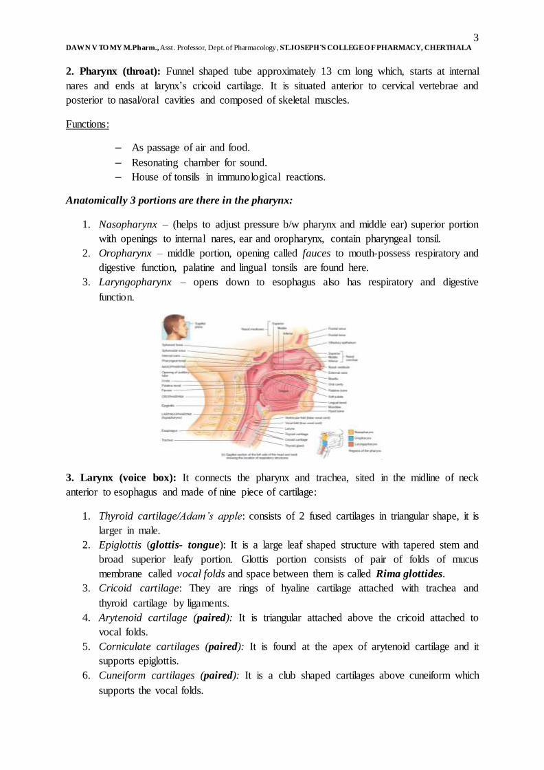
2. Pharynx (throat): Funnel shaped tube approximately 13 cm long which, starts at internal
nares and ends at larynx’s cricoid cartilage. It is situated anterior to cervical vertebrae and
posterior to nasal/oral cavities and composed of skeletal muscles.
Functions:
– As passage of air and food.
– Resonating chamber for sound.
– House of tonsils in immunological reactions.
Anatomically 3 portions are there in the pharynx:
1. Nasopharynx – (helps to adjust pressure b/w pharynx and middle ear) superior portion
with openings to internal nares, ear and oropharynx, contain pharyngeal tonsil.
2. Oropharynx – middle portion, opening called fauces to mouth-possess respiratory and
digestive function, palatine and lingual tonsils are found here.
3. Laryngopharynx – opens down to esophagus also has respiratory and digestive
function.
3. Larynx (voice box): It connects the pharynx and trachea, sited in the midline of neck
anterior to esophagus and made of nine piece of cartilage:
1. Thyroid cartilage/Adam’s apple: consists of 2 fused cartilages in triangular shape, it is
larger in male.
2. Epiglottis (glottis- tongue): It is a large leaf shaped structure with tapered stem and
broad superior leafy portion. Glottis portion consists of pair of folds of mucus
membrane called vocal folds and space between them is called Rima glottides.
3. Cricoid cartilage: They are rings of hyaline cartilage attached with trachea and
thyroid cartilage by ligaments.
4. Arytenoid cartilage (paired): It is triangular attached above the cricoid attached to
vocal folds.
5. Corniculate cartilages (paired): It is found at the apex of arytenoid cartilage and it
supports epiglottis.
6. Cuneiform cartilages (paired): It is a club shaped cartilages above cuneiform which
supports the vocal folds.

4 DAWN V TO MY M.Pharm., Asst. Professor, Dept. of Pharmacology, ST.JOSEPH’S COLLEGE O F PHARMACY, CHERTHALA

5 DAWN V TO MY M.Pharm., Asst. Professor, Dept. of Pharmacology, ST.JOSEPH’S COLLEGE O F PHARMACY, CHERTHALA
4. Trachea (wind pipe): It is the tubular passage for air, it is approximately 12 cm long and
2.5 cm in diameter and situated anterior to the esophagus. It starts from larynx and extends to
the 5th thoracic vertebra where it divides into 2 primary bronchi.
• 4 layered:
– Mucosa & sub mucosa - which provide protection from dust particles and
produces mucus.
– Hyaline cartilage: It consists of 16-20 horizontal, incomplete ‘C’ shaped rings
stalked upon each other, open ends has trachealis muscle which, helps to
adjust with esophagus expansion.
– Adventitia connect trachea to other surrounding tissues.
Adventitia is the outermost connective tissue covering of any organ, vessel, or other structure.
5. Bronchi (wind pipe): At the 5th thoracic vertebra trachea divides into 2 i.e. right and left
primary bronchi. The right enters into right lung and left into left lung. Primary bronchi also
consist of incomplete cartilages. The Internal ridge where right and left bronchi divide is
called carina where mucus membrane is most sensitive. The primary bronchi divides to
secondary (lobar) bronchi and then to tertiary (segmental) bronchi then to bronchioles and
then to terminal bronchioles. This extensive branching is called bronchial tree.
• Right pulmonary bronchus is more vertical, longer and wider. Aspired air enters more
into right lung. Right has 3 lobes.
• Branching of bronchi to bronchioles includes pseudo-stratified cuboidal epithelium.
The columnar becomes non ciliated simple cuboidal epithelium.
• No Goblet cells are present here. Goblet cells are glandular simple columnar epithelial
cells whose function is to secrete mucin a protein, which dissolves in water to form
mucus.
6 DAWN V TO MY M.Pharm., Asst. Professor, Dept. of Pharmacology, ST.JOSEPH’S COLLEGE O F PHARMACY, CHERTHALA
• Incomplete rings give away to cartilages and cartilage decreases leaving more and
more smooth muscle. This can result in muscle spasm.
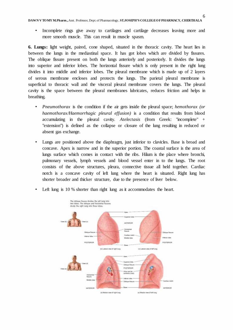
6. Lungs: light weight, paired, cone shaped, situated in the thoracic cavity. The heart lies in
between the lungs in the mediastinal space. It has got lobes which are divided by fissures.
The oblique fissure present on both the lungs anteriorly and posteriorly. It divides the lungs
into superior and inferior lobes. The horizontal fissure which is only present in the right lung
divides it into middle and inferior lobes. The pleural membrane which is made up of 2 layers
of serous membrane encloses and protects the lungs. The parietal pleural membrane is
superficial to thoracic wall and the visceral pleural membrane covers the lungs. The pleural
cavity is the space between the pleural membranes lubricates, reduces friction and helps in
breathing.
• Pneumothorax is the condition if the air gets inside the pleural space; hemothorax (or
haemothorax/Haemorrhagic pleural effusion) is a condition that results from blood
accumulating in the pleural cavity. Atelectasis (from Greek: "incomplete" +
"extension") is defined as the collapse or closure of the lung resulting in reduced or
absent gas exchange.
• Lungs are positioned above the diaphragm, just inferior to clavicles. Base is broad and
concave. Apex is narrow and in the superior portion. The coastal surface is the area of
lungs surface which comes in contact with the ribs. Hilum is the place where bronchi,
pulmonary vessels, lymph vessels and blood vessel enter in to the lungs. The root
consists of the above structures, pleura, connective tissue all held together. Cardiac
notch is a concave cavity of left lung where the heart is situated. Right lung has
shorter broader and thicker structure, due to the presence of liver below.
• Left lung is 10 % shorter than right lung as it accommodates the heart.

7 DAWN V TO MY M.Pharm., Asst. Professor, Dept. of Pharmacology, ST.JOSEPH’S COLLEGE O F PHARMACY, CHERTHALA
• Right lung has 3 lobes and the left lung has 2 lobes. The primary bronchi divide into
secondary bronchi and secondary to tertiary bronchi, each secondary bronchus divides
into 10 tertiary bronchi. Lobules consist of small compartment of bronchopulmonary
segment with lymphatic vessels, arterioles, venules and terminal bronchiole. Each
terminal bronchiole forms respiratory bronchiole which, divides into 2-11 alveolar
ducts.
• Around the alveolar ducts numerous alveoli and alveolar sacs are present. Alveolus is
a single cup shaped pouch. The alveolar sac consists of 2 or more alveolus with a
common opening.
• Alveolar cells are of 2 types. The type 1 simple squamous cells for gases exchange.
The type 2 or septal cells are rounded, few in number and secretes alveolar fluid
containing surfactant which is a mixture of complex phospholipids and lipoproteins. It
also contains alveolar macrophages which protect and remove the dust particles.

8 DAWN V TO MY M.Pharm., Asst. Professor, Dept. of Pharmacology, ST.JOSEPH’S COLLEGE O F PHARMACY, CHERTHALA
Respiratory membrane: The membrane wall between alveolar and capillary wall, having 0.5
micron thickness and formed of 4 layers.
• One layer of type 1, type 2 cells and macrophages. One layer of epithelial basement
membrane of alveolar wall and the capillary basement membrane fused to the
epithelial membrane.
• The capillary endothelium allows rapid diffusion of gases. The lungs consist of 300
million alveoli having a surface area of 70 m2 for gas exchange.
Blood supply:
• Lungs receive oxygenated blood supply from aorta via bronchial Arteries.
• Pulmonary artery carries deoxygenated blood from heart to lungs for oxygenation.
• Pulmonary Veins carry oxygenated blood from lungs back to heart for systemic
circulation.
• The circulation of blood from heart through pulmonary artery to lungs for
oxygenation and the circulation of the oxygenated blood back to heart by pulmonary
veins are known as pulmonary circulation.
• Ventilation perfusion coupling: It is constriction of arterioles in response to hypoxia
resulting in diversion of blood flow from poorly ventilated area to highly ventilated
areas for oxygenation.

9 DAWN V TO MY M.Pharm., Asst. Professor, Dept. of Pharmacology, ST.JOSEPH’S COLLEGE O F PHARMACY, CHERTHALA
3. (A) Voice production:
Structures:
Glottis portion of larynx consists of pair of folds of mucus membrane. The superior
pair of folds is called ventricular/false vocal folds and the inferior pair is called true vocal
cords and the space between ventricular folds is called rima glottides. The laryngeal muscles
are attached to cartilages and vocal folds to produce vibrations.
Process:
1. When laryngeal muscles contracts they pull posterior cricoarytenoid muscles and
moves vocal folds apart (abduction) and the rima glottidis becomes open.
2. When lateral cricoarytenoid muscles contracts moving the vocal folds together and
rima glottidis become narrow/closed (adduction). Other intrinsic muscles also can
elongate or shorten vocal folds.
3. Increased tension on vocal folds causes folds to vibrate rapidly at high pitch and vice
versa. Androgens cause male vocal folds to be thicker, so that it vibrates slowly and
have low pitch in males when compared with females. Pharynx, mouth, nasal cavity,
paranasal sinuses acts as resonating chambers and gives voice the individual quality.
Respiration: It is the process of gas exchange in our body. It includes 3 processes:
1. Pulmonary ventilation/breathing: It includes inhalation (inflow) of atmospheric air for
oxygenation and exhalation (outflow) of alveolar air of lungs after oxygenation.
2. External pulmonary respiration: It includes exchange of gases between alveoli of
lungs and blood in pulmonary capillaries across respiratory membrane. In this process
blood gains O2 and blood loses CO2.
10 DAWN V TO MY M.Pharm., Asst. Professor, Dept. of Pharmacology, ST.JOSEPH’S COLLEGE O F PHARMACY, CHERTHALA
3. Internal tissue respiration: It includes the exchange of gases between blood in
capillaries and tissue cells. In this process blood loses O2 and gains CO2.
Inhalation/inspiration/breathing in:
• It is an active process breathing in the atmospheric air is called inhalation/inspiration.
As per Boyle’s law, when volume increases the pressure decreases. Before respiration
the pressure inside the lungs is equal to air pressure of atmosphere which is equal to
760 mm of hg i.e. equal to1 atmosphere (1 atm.) Then pressure inside the lung
decreases by increased lung volume causing air to flow in. For inhalations lungs must
expand, this needs the contraction of muscles of inspiration.
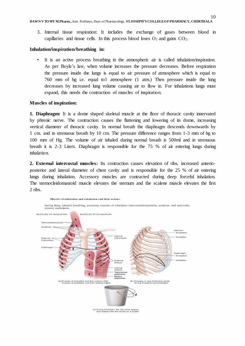
Muscles of inspiration:
1. Diaphragm: It is a dome shaped skeletal muscle at the floor of thoracic cavity innervated
by phrenic nerve. The contraction causes the flattening and lowering of its dome, increasing
vertical diameter of thoracic cavity. In normal breath the diaphragm descends downwards by
1 cm. and in strenuous breath by 10 cm. The pressure difference ranges from 1-3 mm of hg to
100 mm of Hg. The volume of air inhaled during normal breath is 500ml and in strenuous
breath it is 2-3 Liters. Diaphragm is responsible for the 75 % of air entering lungs during
inhalation.
2. External intercostal muscles: Its contraction causes elevation of ribs, increased anterio-
posterior and lateral diameter of chest cavity and is responsible for the 25 % of air entering
lungs during inhalation. Accessory muscles are contracted during deep forceful inhalation.
The sternocleidomastoid muscle elevates the sternum and the scalene muscle elevates the first
2 ribs.

11 DAWN V TO MY M.Pharm., Asst. Professor, Dept. of Pharmacology, ST.JOSEPH’S COLLEGE O F PHARMACY, CHERTHALA
Intra pleural pressure during the quiet inhalation is always sub atmospheric i.e. 756 mm of
hg, contraction of diaphragm and external inter coastal muscles further decreases it to 754
mm of Hg. Expansions of the parietal and visceral pleura occur and are pulled along the
thoracic cavity. The alveolar pressure of 760 also drops to 758 mm of Hg and a pressure
difference is established and the air enters into the lungs.
Exhalation or expiration: The process of breathing out is called exhalation and it is based
on the pressure gradient in the opposite direction i.e. the pressure inside the lung is more than
the atmospheric pressure of 760 mm of Hg.
Normal exhalation: It is a passive process and no muscle contraction is involved. It results
from elastic recoil of chest wall and lungs. Recoiling of the elastic fibers stretched during
inhalation occurs and inward pulls of surface tension due to the fill of alveolar fluid in the
alveoli. The relaxation of muscles of inhalation, diaphragm and the external intercostal
muscles decreases the lung volume. Hence the alveolar pressure increases to 762 mm of Hg
and as a result the air flows out.
During forceful exhalation: During the forceful exhalation like playing the wind instruments,
contracts the muscles of exhalation like the rectus abdominus and other abdominal muscles.
The contraction moves the inferior rib downwards and compresses the abdominal viscera
forcing diaphragm superiorly. The contraction of the internal Intercostal muscles pulls the rib
anteriorly and thereby reduces the surface area and volume which forces out the air inside the
lungs.
The other factors affecting the pulmonary ventilation are:
1. Surface tension of alveolar fluid: It causes the alveoli to assume the smallest possible
diameter and it also accounts for 2/3rd of elastic recoil of the lungs, which decreases the
size of alveoli during exhalation.
2. Compliance of lungs: It is the effort required to stretch the lung and chest wall. Higher
the compliance, the lungs expands rapidly and normally. In TB and pulmonary edema
the intercostal muscles are paralyzed, which decreases the compliance of the lungs.
3. Air way resistance: The resistance to the flow of air especially by bronchioles with the
signals from sympathetic system results in relaxation of air wall. The pathological
conditions like COPD, asthma and chronic bronchitis constricts/contracts the air ways.
4. Eupnoea: It is the normal pattern of breathing (inhalation and exhalation).
5. Coastal breathing: It is the shallow (not deep) chest breathing.
6. Diaphragmatic breathing: It is the deep abdominal breathing, where strong contraction
of diaphragm occurs.
7. Modified respiratory movements: It includes the movements like coughing, sneezing,
sighing, yawning, sobbing, crying, laughing, hiccoughing, valsalua maneuer.

12 DAWN V TO MY M.Pharm., Asst. Professor, Dept. of Pharmacology, ST.JOSEPH’S COLLEGE O F PHARMACY, CHERTHALA
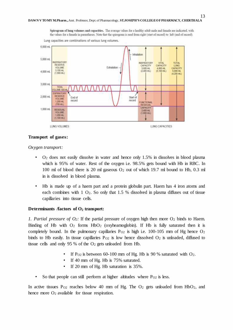
Lung volumes: The normal respiratory rate is 12 breathes per minute.
1. Tidal volume: It is the volume of one breath which is approximately equal to 500 ml.
2. Minute ventilation: It is the total volume air that enters in each minute which is equal to
the normal respirator rate multiplied by tidal volume (12 x 500 = 6 L).
3. Anatomic dead space: It is the conducting air ways where the air present does not
undergo respiratory exchange (30% of air = 150 ml).
4. Alveolar ventilation rate: It is the volume of air that reaches respiratory zone per
minute (350x12 =4200 ml).
5. Inspiratory reserve volume: It is the additional volume of air that can be inhaled by
taking a very deep breath (3100ml in male, 1900ml in female).
6. Expiratory reserve volume: It is the additional volume of air that can be exhaled
forcibly as far as possible after a normal inhalation (1200 ml in male, 700 ml in
female).
7. Forced expiratory volume FEV 1.01 second: The volume of air that can be exhaled
from the lungs in one second with maximal effort following a maximal inhalation.
8. Residual volume: The considerable volume of air remaining in the lungs even after
expiratory reserve volume cannot be measured by spirometer (1200 male, 1100
female).
9. Minimal volume: If thoracic cavity is cut opened, intra-pleural pressure rises and forces
out some residual volume, the air remaining after this is called minimal volume.
Fetal lung: If there is no air in the lungs of fetus, the condition is knows as still born and the
lung will not float in water.
Lung capacities: They are combinations of various or specific lung volumes.
• Inspiratory capacity: It is the sum of tidal volume and inspiratory reserve volume
(500+3100/1900).
• Functional residual capacity: It is the sum of residual volume and expiratory
reserve volume (1200+1200/1100+700).
• Vital capacity: It is the sum of inspiratory reserve volume, tidal volume and
expiratory reserve volume (3100+500+1200/1900+500+700=4800/3100).
• Total lung capacity: It is the sum of vital capacity and residual volume
(6000/4200).
Spirometer: A device to test how the lung is working (used for Pulmonary Function Tests)
to assess the effects of lung disease or the progress of treatment and the procedure is called
spirometry. The spirometer records the total volume of air breathed out – the forced vital
capacity. The machine also records the volume of air breathed out in one second i.e. the
forced expiratory volume. In diseases such as asthma, in which the airways are obstructed,
the ratio of the forced expiratory volume to the forced vital capacity is reduced.
13 DAWN V TO MY M.Pharm., Asst. Professor, Dept. of Pharmacology, ST.JOSEPH’S COLLEGE O F PHARMACY, CHERTHALA
Transport of gases:
Oxygen transport:
• O2 does not easily dissolve in water and hence only 1.5% in dissolves in blood plasma
which is 95% of water. Rest of the oxygen i.e. 98.5% gets bound with Hb in RBC. In
100 ml of blood there is 20 ml gaseous O2 out of which 19.7 ml bound to Hb, 0.3 ml
in is dissolved in blood plasma.
• Hb is made up of a haem part and a protein globulin part. Haem has 4 iron atoms and
each combines with 1 O2. So only that 1.5 % dissolved in plasma diffuses out of tissue
capillaries into tissue cells.
Determinants /factors of O2 transport:
1. Partial pressure of O2: If the partial pressure of oxygen high then more O2 binds to Haem.
Binding of Hb with O2 forms HbO2 (oxyheamoglobin). If Hb is fully saturated then it is
completely bound. In the pulmonary capillaries PO2 is high i.e. 100-105 mm of Hg hence O2
binds to Hb easily. In tissue capillaries PO2 is low hence dissolved O2 is unloaded, diffused to
tissue cells and only 95 % of the O2 gets unloaded from Hb.
• If PO2 is between 60-100 mm of Hg. Hb is 90 % saturated with O2.
• If 40 mm of Hg. Hb is 75% saturated.
• If 20 mm of Hg. Hb saturation is 35%.
• So that people can still perform at higher altitudes where PO2 is less.
In active tissues PO2 reaches below 40 mm of Hg. The O2 gets unloaded from HbO2, and
hence more O2 available for tissue respiration.
14 DAWN V TO MY M.Pharm., Asst. Professor, Dept. of Pharmacology, ST.JOSEPH’S COLLEGE O F PHARMACY, CHERTHALA
2. Acidity & PH:
• As the acidity increases pH of blood decreases. As a result more O2 dissociates from
Hb because affinity of Hb decreases. Lactic acid, carbonic acid etc. get increases
during exercise hence the O2 dissociation. Due to high concentration of H+ it will get
bind to Hb at the same time O2 diffuses out from Hb.
• Bohr Effect states that when pH increases Hb dissociation curve shifts to right side i.e.
at given PO2 Hb is less saturated with O2.
3. Partial pressure of CO2:
• The effect is similar to that of H+ as PCO2 increases, Hb releases O2 more readily, and
because CO2 also binds to Hb. CO2 itself increases acidity by formation of carbonic
acid.
• CO2 enters to the blood and temporarily converted to carbonic acid, by the enzyme
carbonic anhydrase, so that H+ is increased and O2 affinity gets decreased.
• CO2+H2O (water in plasma)H2CO3 (Carbonic acid)H++HCO3 (Bicarbonate ions).
4. Temperature:
• Increase in temperature leads to increased O2 release from Hb and heat is the
byproduct of all metabolic reactions. Metabolically active cells release acid along
with heat during metabolism which results in increased O2 release.
• In hypothermia only less O2 is released, so more O2 remain bounded to Hb.
5. BPG – 2, 3 BIPHOSPHOGLYCERATE:
• It is found in RBC, formed during glycolysis. When BPG binds to Hb – O2 binding to
Hb is decreased.
– Greater the BPG more O2 gets diffuses into the plasma. Thyroxine, Growth
Hormone, Nor-Adrenalin, Adrenalin, Testosterone increases BPG formation.
– At Higher altitudes people will have more BPG.
• Hb – HbA- adult, HbF-foetal differ in structure and affinity for O2, HbF more affinity
to O2, transfer more O2 at human placenta
• Carbon monoxide (CO) is 200 times stronger than O2; even at lower concentration
CO of 0.1% and PCO of 0.5 mm of hg it combines with half of Hb in blood hence can
be fatal. If CO is inhaled and intoxicated the lips, oral mucosa appears in bright red
color. Antidote includes giving pure O2.
Carbon dioxide transport:
• Deoxygenated blood, 100 ml = 53 ml gaseous CO2

15 DAWN V TO MY M.Pharm., Asst. Professor, Dept. of Pharmacology, ST.JOSEPH’S COLLEGE O F PHARMACY, CHERTHALA
• Transport in 3 forms
– Dissolved CO2 - 7 % in dissolution in blood plasma – diffuse in alveoli
– Carbamino compounds – 23% with amino groups of amino acids in protein in
Hb, Hb+CO2HbCO2 carboxyhaemoglobin. Carbaminohaemoglobin formed
at high PCO2 as high in tissues.
– Bicarbonate ions – 70 % as bicarbonate in plasma HCO3-
– CO2 enters RBC
– CO2 + H2O H2CO3 H+ + HCO3- by enzyme carbonic anhydrase.

16 DAWN V TO MY M.Pharm., Asst. Professor, Dept. of Pharmacology, ST.JOSEPH’S COLLEGE O F PHARMACY, CHERTHALA
• HCO3- diffuses out RBC
1. Down the concentration gradient &
2. Exchange – Cl- enters the cell.
• This exchange maintains the electrical balance b/w plasma and RBC – called chloride
shift.
• HCO3- in blood plasma reaches lungs, reaction reverses CO2 out.
• Haldane effect – relationship b/w CO2 transport and CO2 carrying capacity.
• Lower the oxyhaemoglobin higher the CO2 carrying capacity.
• Deoxy Hb binds to CO2 transport more CO2.
• Deoxy Hb buffers H+ more than O2, so remove H+ ions from solution and promote
CO2H2CO3
17 DAWN V TO MY M.Pharm., Asst. Professor, Dept. of Pharmacology, ST.JOSEPH’S COLLEGE O F PHARMACY, CHERTHALA
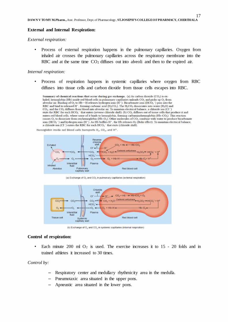
External and Internal Respiration:
External respiration:
• Process of external respiration happens in the pulmonary capillaries. Oxygen from
inhaled air crosses the pulmonary capillaries across the respiratory membrane into the
RBC and at the same time CO2 diffuses out into alveoli and then to the expired air.
Internal respiration:
• Process of respiration happens in systemic capillaries where oxygen from RBC
diffuses into tissue cells and carbon dioxide from tissue cells escapes into RBC.
Control of respiration:
• Each minute 200 ml O2 is used. The exercise increases it to 15 - 20 folds and in
trained athletes it increased to 30 times.
Control by:
– Respiratory center and medullary rhythmicity area in the medulla.
– Pneumotaxic area situated in the upper pons.
– Apneustic area situated in the lower pons.

18 DAWN V TO MY M.Pharm., Asst. Professor, Dept. of Pharmacology, ST.JOSEPH’S COLLEGE O F PHARMACY, CHERTHALA
Regulated by:
– Cortex of brain.
– Chemoreceptors situated centrally in the medulla, aorta and carotid bodies.
– Proprioceptors.
– Inflation reflux (HERING BREUR reflex).
Respiratory Centre: The nerve impulse from widely dispersed clusters of neurons alters the
size of thorax.
1. Medullary rhythmicity area: It is situated in medulla oblongata and controls the basic
rhythm of respiration. There are 2 areas: The inspiratory area responsible for inspiration and
the expiratory area responsible for expiration.
Quiet breathing (Duration is 5 seconds):
• First 2 seconds: The inspiration area nerve impulses are active for first 2 seconds
resulting in external intercostal and diaphragm contraction, which results in breathing
in and it gets inactive after 2 seconds stopping the inspiratory process.
• Last 3 seconds: Both inspiratory area and expiratory area are inactive and hence there
is no impulse, which results in the relaxation of muscles for inspiration and results in
breathing out.
Forceful breathing: At first the inspiratory area followed by active expiratory area are
triggered causing first contraction of external intercostal muscles and diaphragm followed
by contraction of internal intercostal and abdominal muscle during. The external
intercostal muscles and diaphragm relaxes during contraction of internal intercostal and
abdominal muscles.
2. Pneumotaxic area: It is present in the upper pons and coordinates transition between
inhalation and exhalation. It transmit inhibitory impulse to inspiratory area i.e. turn off
inspiratory area which makes the duration of inhalation shorter and breathing rate becomes
rapid.
3. Apnuestic area: It is present in the lower pons and sends stimulatory impulses to
inspiratory area which activates and prolongs inhalation resulting in long deep inhalations.
• The active pneumotaxic area overrides apneustic area.
Regulation of respiration: The basic rhythm of inspiratory area is modified by various
influences like:
The influence of cortex:
• Voluntarily we can inhibit the reflux to breathe, for a short time, protection from
water, irritating gases entering lungs etc. but is limited because increased CO2 and H+
activate inspiratory area overriding voluntary control.

19 DAWN V TO MY M.Pharm., Asst. Professor, Dept. of Pharmacology, ST.JOSEPH’S COLLEGE O F PHARMACY, CHERTHALA
• By hypothalamus and limbic system with emotional stimuli (laughing and crying)
modifies the respiratory rhythm.
Chemoreceptor regulations:
• Central chemoreceptors in medulla in CNS sense the changes in H+, PCO2 in CSF and
make changes in respiration rate and depth.
Peripheral nervous system:
• Aortic bodies are cluster of chemoreceptors in wall of arch of aorta part of
vagus nerve sense the percentage of oxygen in the oxygenated blood and
adjust the rate and depth of respiration by giving feedback mechanisms.
• Carotid bodies are oval nodules in wall of left and right common carotid
arteries, part of glossopharyngeal nerve sense the percentage of oxygen in the
oxygenated blood and adjust the rate and depth of respiration by giving
feedback mechanisms.
Sensitive to the changes of PO2, H+, PCO2 in blood:
• Normal PCO2 is 40 mm of Hg in the arteries and any short change in PCO2 leads to
hypercapnea, hypercarbia and regulates respiration.
Responds to deficiency of O2: If PCO2 falls below 50 mm of Hg and increase in PCO2 & H+,
participate in -ve feedback system cause hyperventilation till PCO2 & PO2 becomes normal.
Hypoxia: The condition in which there is a lower O2 level in tissues.
Proprioceptor stimulation:
• Exercise: It stimulates respiration (as PCO2 increase, PO2 decrease and H+ increase).
• From proprioceptors: The proprioceptor in joints and muscles stimulates respiration.
• Stretch sensitive: Sensitive to pressure differences e.g. baroreceptors in walls of
alveoli.
Proprioceptors: Sensory nerve endings in the muscles, tendons and joints which signal to the
brain their position relative to the outside world and the state of contraction of the muscle.
During movement, a regular flow of information to the brain from the proprioceptors, the
eyes and ears ensures that actions are coordinated and the body’s balance maintained.
Inflation reflex (Hering breur reflex): The over inflation of lungs during inspiration
stimulates stretch receptors and inspiratory area and stops further inhalation, as exhalation
begins lungs deplete and stretch receptors no longer stimulated. The inspiratory and apneustic
area are no longer inhibited and a new inflation begins, this protective mechanism for
preventing excessive inflation of lung is called inflation reflex.

20 DAWN V TO MY M.Pharm., Asst. Professor, Dept. of Pharmacology, ST.JOSEPH’S COLLEGE O F PHARMACY, CHERTHALA
Other factors influencing respiration:
1. Limbic system stimulation: The emotional changes activates excitatory signal to
inspiratory area.
2. Temperature: The increased temperature increases respiration.
3. Pain: Sudden pain and apnea or prolonged somatic pain can increase respiration.
4. Stretching of anal sphincter: The stretching of anal sphincter muscle increases
respiration. It is a technique used in the new born to increase respiration.
5. Irritation of airways: There are 2 types of irritations based on irritating agents: The
physical and chemical irritation, which will decrease respiration.
6. Blood pressure: The sudden rise in BP decreases respiration and drop in BP increases
respiration.
IMPORTANT QUESTIONS OF RESPIRATORY SYSTEM.
1. Describe the right lung. (5)
2. Regulation of respiration. (10)
3. Anatomy of respiratory tract and mention the functions of trachea. (5)
4. Explain the events that occur in inhalation and exhalation. (5)
5. Explain the exchange of gases in external and internal respiration. (5)
6. Mechanism of respiration. (5)
7. Draw and label the parts of respiratory system. Discuss the mechanism of respiration.
(10)
8. Structure and function of respiratory system. (5)
9. With the help of a neat labelled diagram outline the parts of respiratory system and
illustrate the process involved in CO2 transport in our body. (10)


![Respiratory system roadmap.pptx [Repaired] - Loginanatomical-sciences.health.wits.ac.za/roadmaps/Respiratory system... · DIVISION OF THE RESPIRATORY SYSTEM CONDUCTING PORTION Nasal](https://img.pdfslide.net/doc/110x75/5a78c3d87f8b9ae6228c9db0/respiratory-system-repaired-loginanatomical-scienceshealthwitsaczaroadmapsrespiratory.jpg)

![Respiratory System [โหมดความเข้ากันได้] · PATHOLOGY OF RESPIRATORY SYSTEM นพ. อรรณพ นาคะป ท Respiratory system U it](https://img.pdfslide.net/doc/110x75/5fa578efd4e80f055f6b3401/respiratory-system-aaaaaaaaaaaaaaaaaa-pathology.jpg)


![Anatomy and Physiology Respiratory System [Tab 2] Respiratory System](https://img.pdfslide.net/doc/110x75/56649ebd5503460f94bc631f/anatomy-and-physiology-respiratory-system-tab-2-respiratory-system.jpg)