Embed Size (px)
Citation preview
79
Editor: AssociateProfessorDr.SeowLiangLin BDS(Mal),MSc.(London),FDSRCS(England),PhD(Mal),FICD SchoolofDentistry InternationalMedicalUniversity 126,Jalan19/155B,BukitJalil 57000,KualaLumpur,Malaysia E-mail:[email protected]
AssistantEditor: Dr. Shahida Mohd SaidSecretary: Dr. Wey Mang ChekTreasurer: Dr. How Kim ChuanEx-Officio: Dr. S. Sivanesan
EditorialAdvisoryBoard: Professor Dr. Ong Siew Tin Professor Dr. Phrabhakaran Nambiar Professor Dr. Rosnah Mohd Zain Associate Professor Dr. Roslan Saub Dr. Elise Monerasinghe Dr. Lam Jac Meng Dr. Mohamad Muzafar Hamirudin Dr. Wey Mang Chek
The Editor of the Malaysian Dental Association wishes to acknowledge the tireless efforts of the following referees to ensure that the manuscripts submitted are of high standard.
Prof. Dr. Toh Chooi Gait Prof. Dr. Ong Siew Tin Dato’ Prof. Dr. Hashim b. Yaacob Prof. Dr. Lui Joo Loon Prof. Zubaidah Abdul Rahim Prof. Dr. Phrabhakaran NambiarDr. Zamros Yuzadi Prof. Dr. David Wilson Prof. Dr. Tara Bai Taiyeb AliDr. Elise Monorasinghe Assoc. Prof. Dr. Theunis Oberholzer Prof. Dr. Rahimah Abdul KadirDr. Lau Shin Hin Dr. Fathilah Abdul Razak Prof. Dr. Nik Noriah Nik HusseinDr. Loke Shuet Toh Dr. Lam Jac Meng Assoc. Prof. Dr. Datin Rashidah EsaDr. Shahida Said Dr. Nor Himazian Mohamed Assoc. Prof. Dr. Norsiah YunusDr. Zamri Radzi Dr. Norliza Ibrahim Assoc. Prof. Dr. Tuti Ningseh Mohd DomDr. Norintan Ab. Murat Dr. Mohd Fadhli Khamis Assoc. Prof. Dr. Roszalina RamliDr. Siti Adibah Othman Dr. Siti Mazlipah Ismail Assoc. Prof. Dr. Roslan Abdul Rahman Dr Nurul Asyikin Prof. Dr. Siar Chong Huat Dr. Mohd Muzafar HamirudinDr. Dalia Abdullah Dr. Wong Mei Ling Dr. Zeti Adura Che Abd. AzizDr. Wey Mang Chek Assoc. Prof. Dr. Shanmuhasuntharam Dr. Rohaya Megat Abdul Wahab
Malaysian Dental Journal (2008) 29(2) 79-80© 2008 The Malaysian Dental Association
MALAYSIANDENTALJOURNAL
80
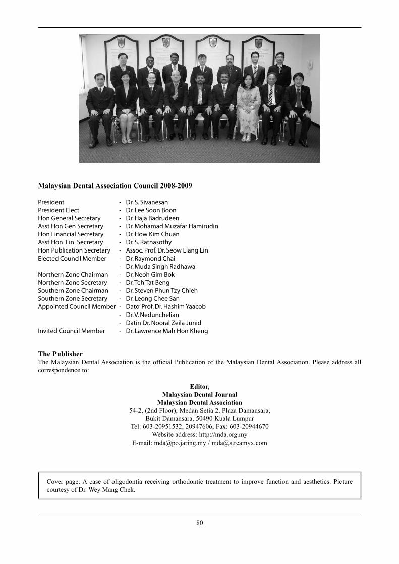
MalaysianDentalAssociationCouncil2008-2009
President - Dr.S.SivanesanPresidentElect - Dr.LeeSoonBoonHonGeneralSecretary - Dr.HajaBadrudeenAsstHonGenSecretary - Dr.MohamadMuzafarHamirudinHonFinancialSecretary - Dr.HowKimChuanAsstHonFinSecretary - Dr.S.RatnasothyHonPublicationSecretary - Assoc.Prof.Dr.SeowLiangLinElectedCouncilMember - Dr.RaymondChai - Dr.MudaSinghRadhawaNorthernZoneChairman - Dr.NeohGimBokNorthernZoneSecretary - Dr.TehTatBengSouthernZoneChairman - Dr.StevenPhunTzyChiehSouthernZoneSecretary - Dr.LeongCheeSanAppointedCouncilMember - Dato’Prof.Dr.HashimYaacob - Dr.V.Nedunchelian - DatinDr.NooralZeilaJunidInvitedCouncilMember - Dr.LawrenceMahHonKheng
ThePublisherThe Malaysian Dental Association is the official Publication of the Malaysian Dental Association. Please address all correspondence to:
Editor,MalaysianDentalJournal
MalaysianDentalAssociation54-2, (2nd Floor), Medan Setia 2, Plaza Damansara,
Bukit Damansara, 50490 Kuala LumpurTel: 603-20951532, 20947606, Fax: 603-20944670
Website address: http://mda.org.myE-mail: [email protected] / [email protected]
Cover page: A case of oligodontia receiving orthodontic treatment to improve function and aesthetics. Picture courtesy of Dr. Wey Mang Chek.
81
AimAndScopeThe Malaysian Dental Journal covers all aspects of work in Dentistry and supporting aspects of Medicine. Interaction with other disciplines is encouraged. The contents of the journal will include invited editorials, original scientific articles, case reports, technical innovations. A section on back to the basics which will contain articles covering basic sciences, book reviews, product review from time to time, letter to the editors and calendar of events. The mission is to promote and elevate the quality of patient care and to promote the advancement of practice, education and scientific research in Malaysia.
PublicationThe Malaysian Dental Journal is an official publication of the Malaysian Dental Association and is published half yearly (KDN PP4069/12/98)
SubscriptionMembers are reminded that if their subscription are out of date, then unfortunately the journal cannot be supplied. Send notice of change of address to the publishers and allow at least 6 - 8 weeks for the new address to take effect. Kindly use the change of address form provided and include both old and new address. Subscription rate: Ringgit Malaysia 60/- for each issue, postage included. Payment in the form of Crossed Cheques, Bank drafts / Postal orders, payable to Malaysian Dental Association. For further information please contact :
ThePublicationSecretaryMalaysianDentalAssociation
54-2,(2ndFloor),MedanSetia2,PlazaDamansara,BukitDamansara,50490KualaLumpur
BackissuesBack issues of the journal can be obtained by putting in a written request and by paying the appropriate fee. Kindly send Ringgit Malaysia 50/- for each issue, postage included. Payment in the form of Crossed Cheques, Bank drafts / Postal orders, payable to Malaysian Dental Association. For further information please contact:
ThePublicationSecretaryMalaysianDentalAssociation
54-2,(2ndFloor),MedanSetia2,PlazaDamansara,BukitDamansara,50490KualaLumpur
Copyright© 2007 The Malaysian Dental Association. All rights reserved. No part of this publication may be reproduced, stored in a retrieval system or transmitted in any form or by means of electronic, mechanical photocopying, recording or otherwise without the prior written permission of the editor.
MembershipandchangeofaddressAll matters relating to the membership of the Malaysian Dental Association including application for new member-ship and notification for change of address to and queries regarding subscription to the Association should be sent to Hon General Secretary, Malaysian Dental Association, 54-2 (2nd Floor) Medan Setia 2, Plaza Damansara, Bukit Damansara, 50490 Kuala Lumpur. Tel: 603-20951532, 20951495, 20947606, Fax: 603-20944670, Website Address: http://www.mda.org.my. Email: [email protected] or [email protected]
DisclaimerStatements and opinions expressed in the articles and communications herein are those of the author(s) and not necessarily those of the editor(s), publishers or the Malaysian Dental Association. The editor(s), publisher and the Malaysian Dental Association disclaim any responsibility or liability for such material and do not guarantee, warrant or endorse any product or service advertised in this publication nor do they guarantee any claim made by the manufacturer of such product or service.
Malaysian Dental Journal (2008) 29(2) 81© 2008 The Malaysian Dental Association
MALAYSIANDENTALJOURNAL
82
Malaysian Dental Journal (2008) 29(2) 82© 2008 The Malaysian Dental Association
MALAYSIANDENTALJOURNAL
CONTENT
MDJ : Publications And Key Performance Index 83 Seow LL
Are Cox-2 Inhibitors A Solution To Problems Associated With Current Oral Analgesics? 84 A Revisit With A Perspective Of Local Need. WC Ngeow, ST Ong
Oral Granular Cell Tumour: A Clinicopathological Study Of 7 Cases And A Brief Review Of The Literature 94 Ajura Abdul Jalil, Lau Shin Hui
Ability of Whitening Toothpastes in Removing Stains from Composite Resins 97 S Y Chong , T B Lim, L L Seow
Latest recommendations for dental radiography in general dental practice. 104 Kathiravan Purmal, Phrabhakaran Nambiar
Posterior Teeth Mesialization With Mini-implants In An Oligodontia Patient 113 Wey MC, Wu CL, Wong WK, Zamri R, Hägg U
The Reliability Of Bitemark Evidence: Analysis And Recommendations 119 In The Context Of Malaysian Criminal Justice System Shamsher Singh, Phrabhakaran Nambiar
Students Perception And Satisfactory Level In Preclinical Fixed Prosthodontic Teaching: Post And Core 128 Marlynda Ahmad, Natasya Ahmad Tarib
Inhibitory Effect of Mixed Paste of Ca(Oh)2 And Baso4 against Bacteria In Infected Root Canal. 135Halim H S, Iskandar, B, Liesan
Mothers’ Knowledge Of Fluoride Toothpaste Usage By Their Preschool- Children 140 HL Tay, N Jaafar
Management of Full Mouth Prosthodontic Rehabilitation Utilizing a Combination of Porcelain 149 Fused to Metal and High Strength Zirconium-oxide Crowns Ansgar C Cheng, Elvin WJ Leong, Helena Lee
What Expert Says … Periodontal Abscess 154 Norhidayah, Khamiza
Abstracts Of Scientific Papers Presented At The 65th Mda/agm Scientific Convention And Trade Exhibition, 158 20th - 22th June 2008
Continuing Professional Development Quiz 166
Instruction to contributors 168
83
Malaysian Dental Journal (2008) 29(2) 83© 2008 The Malaysian Dental Association
MALAYSIANDENTALJOURNAL
EDITORIAL:PUBLICATIONSANDKEYPERFORMANCEINDEX
Welcome to this issue of Malaysian Dental Journal.
As the official publication for Malaysian Dental Association, the Malaysian Dental Journal is slowly gaining popularity in this region. The electronic versions of the current and previous issues of MDJ have been made available at the secured e-journal section of the MDA website. We have received contribution of articles from authors in neighbouring countries i.e. Indonesia, India, Mongolia and Singapore. With more dental schools mushrooming in this country, contributions from local authors have been encouraging. There are ten dental schools in Malaysia at present i.e. six in public university and four in private university/college. Dental officers from the government sector have also been actively contributing as government has encouraged the dental officers to get involved in research, scientific paper presentation and publication.
Key performance index has been introduced to improve the efficiency and productivity of an organisation. Various layers in an organisation ranging from the top management to the supporting staff have to lay down measurable indices to bring the performance of the organisation to a greater height. Amongst the indices relevant to dentistry were improvement in patient charter, organisation and participation in community projects, creativity and innovations in teaching and learning activities, scientific paper presentations and publications.
With increasing emphasis on evidence-based practice to provide the best to the patients, scientific research, scientific presentations and publications have become an important triad to improve the provision of healthcare. This is especially so for academic institutions that train and produce the next generation of healthcare workers, much emphasis has been placed for staff, postgraduate students and also undergraduate students to conduct research and publish the scientific findings. The MDJ is currently a peer-reviewed, indexed journal and provide a good avenue for disseminating knowledge in the dental literature. It is hope that the academic institutions and government sector will encourage staffs to continue contributing articles to MDJ. It is also hope that academic staffs and specialists are willing to impart their expertise and spare their precious time in the manuscript reviewing process.
As the renewal of annual practicing certificate will be tied with CPD points in the near future, MDJ has provided another source for obtaining the CPD points. After reading through the articles in the journal, there is a short section of quizzes pertaining to the articles, upon answering the quiz, CPD points can be obtained from the MDA secretariat.
Thank you kindly for your warm support.
Associate Professor Dr. Seow Liang Lin
Editor
Malaysian Dental Journal
Are Cox-2 Inhibitors A Solution To Problems Associated With Current Oral Analgesics? A Revisit With A Perspective Of Local Need. WC Ngeow. BDS (Mal), FFDRCSI (OS), FDSRCS (Eng), MDSc (Mal)
ST Ong. BDS (Mal), FDSRCPS (Glasg) Department of Oral & Maxillofacial Surgery, Faculty of Dentistry, University of Malaya, 50603 Kuala Lumpur, Malaysia.
ABSTRACT
The primary obligation and ultimate responsibility of a dental surgeon is not only to restore aesthetic and function, but also to relieve pain which originates from dental pathology or surgical procedures performed. Post operative dental pain is mainly of inflammatory origin. Common traditional oral analgesics, namely salicylates, paracetamol and non-steroidal anti-inflammatory drugs have been the drugs of choice, but are increasingly being superseded by newer designer analgesics, the cyclooxygenase-2 (COX-2) inhibitors. This article reviews the advantages and disadvantages of prescribing common traditional oral analgesics as well as exploring the potential use of COX-2 inhibitors as an alternative to these analgesics for the control of post operative pain in dentistry.
Key words pain, analgesic, NSAIDs, COX-2 inhibitor
84
Malaysian Dental Journal (2008) 29(2) 84-93© 2008 The Malaysian Dental Association
INTRODuCTION
Pain can originate from dental pathology or asan outcome of trauma or surgical procedures performedon patients. Postoperative dental pain is mainly ofinflammatory origin and is caused mainly by increasedprostaglandin (PG) synthesis.1,2 Pain studies showed thatmajorityofpatientssufferedtheirhighestpainlevelontheday of operation, especially within the first 3 to 5 hourspostoperation.3,4 This happens irrespective of their age,operatingtime,whotheoperatorsare,typesofimpactionand presence or absence of pericoronitis during theprevious3weeks.3Itwassuggestedthatpainwashowever,influencedbythegenderofthepatients.3,5 Analgesicsmostcommonlyprescribedindentistryforacuteminororalsurgicalpainreliefincludesalicylates,the nonsteroidal anti-inflammatory drugs (NSAIDs),paracetamol and various opioid-containing analgesiccombinations. As these oral analgesics have been usedfor a long time and have well proven track records, theauthors wish to group them as “common traditionaloral analgesics”. Paracetamol and the NSAIDs such asmefenamicacidandibuprofen,areexamplesofanalgesicscommonlyprescribedforminororalsurgicalproceduresinMalaysia .6 These NSAIDs (salicylates included) andpresumably paracetamol act by inhibiting enzymecyclooxygenaseresponsiblefortheformationofPGsthatpromotepainandinflammation.7
Although NSAIDs are effective analgesics for mild tomoderatepain,theyareassociatedwithpotentiallyseriousside effects, including gastrointestinal (GI) haemorrhageand ulceration and alteration of platelet function.8 ThesehappenbecauseNSAIDsinhibitboththeconstitutive(COX-1) and inducible (COX-2) isoforms of cyclooxygenase(COX). TheinductionofCOX-2afterinflammatorystimulihasledtothehypothesisthatCOX-2inhibitionprimarilyaccountsforthetherapeuticpropertiesofNSAIDs.COX-2inhibitorsnowconstituteanewgroupofNSAIDswhich,atrecommendeddoses,blocktheproductionofPGbyCOX-2, but not COX-1. Two COX-2 inhibitors are currentlyavailable in Malaysia – celecoxib (Celebrex®, Pfizer),which is taken twice daily, and etoricoxib (Arcoxia®,MSD Merck), which is taken once daily. Celecoxiband etoricoxib show significantly lower incidences ofgastrotoxicitythannon-selectiveNSAIDsbutatthesametime show potent analgesic property.9,10-13 Moreover, incomparison with conventional NSAIDs, celecoxib andetoricoxibgenerallyhavea longerdurationof action;12hoursand22hoursrespectively. Thisarticlereviewstheadvantagesanddisadvantagesofprescribingcommontraditionaloralanalgesicsaswellas exploring the potential use of COX-2 inhibitors asan alternative to these analgesics for the control of postoperativepainindentistry.
MALAYSIAN DENTAL JOuRNAL
85
Ngeow / Ong
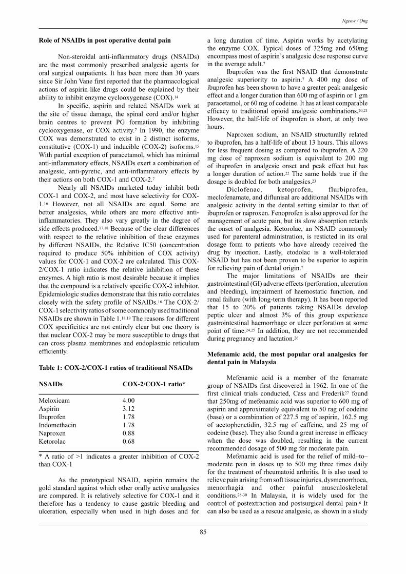
Role of NSAIDs in post operative dental pain Non-steroidal anti-inflammatory drugs (NSAIDs)are the most commonly prescribed analgesic agents fororal surgical outpatients. It has been more than 30 yearssinceSirJohnVanefirstreportedthatthepharmacologicalactions of aspirin-like drugs could be explained by theirabilitytoinhibitenzymecyclooxygenase(COX).14 In specific, aspirin and related NSAIDs work atthe site of tissue damage, the spinal cord and/or higherbrain centres to prevent PG formation by inhibitingcyclooxygenase, or COX activity.7 In 1990, the enzymeCOX was demonstrated to exist in 2 distinct isoforms,constitutive (COX-1) and inducible (COX-2) isoforms.15Withpartialexceptionofparacetamol,whichhasminimalanti-inflammatoryeffects,NSAIDsexertacombinationofanalgesic, anti-pyretic, and anti-inflammatory effects bytheiractionsonbothCOX-1andCOX-2.7 Nearly all NSAIDs marketed today inhibit bothCOX-1andCOX-2, andmosthave selectivity forCOX-1.16 However, not all NSAIDs are equal. Some arebetter analgesics, while others are more effective anti-inflammatories. They also vary greatly in the degree ofsideeffectsproduced.17,18Becauseofthecleardifferenceswith respect to the relative inhibition of these enzymesby different NSAIDs, the Relative IC50 (concentrationrequired to produce 50% inhibition of COX activity)valuesforCOX-1andCOX-2arecalculated.ThisCOX-2/COX-1 ratio indicates the relative inhibition of theseenzymes.AhighratioismostdesirablebecauseitimpliesthatthecompoundisarelativelyspecificCOX-2inhibitor.EpidemiologicstudiesdemonstratethatthisratiocorrelatescloselywiththesafetyprofileofNSAIDs.16TheCOX-2/COX-1selectivityratiosofsomecommonlyusedtraditionalNSAIDsareshowninTable1.18,19ThereasonsfordifferentCOXspecificitiesarenotentirelyclearbutone theory isthatnuclearCOX-2maybemoresusceptibletodrugsthatcan cross plasma membranes and endoplasmic reticulumefficiently.
Table 1: COX-2/COX-1 ratios of traditional NSAIDs
NSAIDs COX-2/COX-1 ratio*
Meloxicam 4.00Aspirin 3.12Ibuprofen 1.78Indomethacin 1.78Naproxen 0.88Ketorolac 0.68
*A ratio of >1 indicates a greater inhibition of COX-2thanCOX-1
As the prototypical NSAID, aspirin remains thegoldstandardagainstwhichotherorallyactiveanalgesicsare compared. It is relatively selective for COX-1 and ittherefore has a tendency to cause gastric bleeding andulceration, especially when used in high doses and for
a long duration of time. Aspirin works by acetylatingthe enzyme COX. Typical doses of 325mg and 650mgencompassmostofaspirin’sanalgesicdoseresponsecurveintheaverageadult.7
Ibuprofen was the first NSAID that demonstrateanalgesic superiority to aspirin.7 A 400 mg dose ofibuprofenhasbeenshowntohaveagreaterpeakanalgesiceffectandalongerdurationthan600mgofaspirinor1gmparacetamol,or60mgofcodeine.Ithasatleastcomparableefficacy to traditional opioid analgesic combinations.20,21However, the half-life of ibuprofen is short, at only twohours. Naproxen sodium, an NSAID structurally relatedtoibuprofen,hasahalf-lifeofabout13hours.Thisallowsforlessfrequentdosingascomparedtoibuprofen.A220mg dose of naproxen sodium is equivalent to 200 mgof ibuprofen in analgesic onset and peak effect but hasa longer duration of action.22The same holds true if thedosageisdoubledforbothanalgesics.23
Diclofenac, ketoprofen, flurbiprofen,meclofenamate,anddiflunisalareadditionalNSAIDswithanalgesic activity in the dental setting similar to that ofibuprofenornaproxen.Fenoprofenisalsoapprovedforthemanagementofacutepain,butitsslowabsorptionretardsthe onset of analgesia. Ketorolac, an NSAID commonlyused for parenteral administration, is resticted in its oraldosage form to patients who have already received thedrug by injection. Lastly, etodolac is a well-toleratedNSAIDbuthasnotbeenproventobesuperior toaspirinforrelievingpainofdentalorigin.7 The major limitations of NSAIDs are theirgastrointestinal(GI)adverseeffects(perforation,ulcerationand bleeding), impairment of haemostatic function, andrenalfailure(withlong-termtherapy).Ithasbeenreportedthat 15 to 20% of patients taking NSAIDs developpeptic ulcer and almost 3% of this group experiencegastrointestinalhaemorrhageorulcerperforationatsomepoint of time.24,25 In addition, they arenot recommendedduringpregnancyandlactation.26
Mefenamic acid, the most popular oral analgesics for dental pain in Malaysia Mefenamic acid is a member of the fenamategroupofNSAIDs firstdiscovered in1962. Inoneof thefirst clinical trials conducted, Cass and Frederik27 foundthat250mgofmefenamicacidwassuperiorto600mgofaspirinandapproximatelyequivalentto50ragofcodeine(base)oracombinationof227.5mgofaspirin,162.5mgof acetophenetidin, 32.5 rag of caffeine, and 25 mg ofcodeine(base).Theyalsofoundagreatincreaseinefficacywhen the dose was doubled, resulting in the currentrecommendeddosageof500mgformoderatepain. Mefenamic acid is used for the relief ofmild–to–moderate pain in doses up to 500 mg three times dailyforthetreatmentofrheumatoidarthritis.Itisalsousedtorelievepainarisingfromsofttissueinjuries,dysmenorrhoea,menorrhagia and other painful musculoskeletalconditions.28-30 In Malaysia, it is widely used for thecontrolofpostextractionandpostsurgicaldentalpain.6 Itcanalsobeusedasarescueanalgesic,asshowninastudy
86
Are COX-2 inhibitors a solution to problems associated with current oral analgesics? A revisit with a perspective of local need.
ontheeffectofperioperativeauricularelectroacupunctureafterthirdmolartoothextraction.31Inhumans,mefenamicacidismetabolisedbybothphaseIenzymesandthephaseII enzyme family UDP-glucuronosyltransferase. Threeglucuronideshadbeenidentifiedandisolatedfromhumanurineafteroraladministrationofmefenamicacid.32
The efficacy of mefenamic acid in the control ofpostsurgical pain has last been studied against aspirinand placebo more than a quarter century ago. Then, theresearchersfoundthatmefenamicacidwaswelltoleratedand was clearly superior to placebo and equalled orexceededtheabilityofaspirintocontrolpostsurgicalpainintheparametersmeasured.33
Theuseofmefenamicacidasapre-emptivedrughasbeenstudiedinobstetricandgynaecologicalmedicine,but no similar literatures could be found for dentistry.Nagele et al.34 assessed its efficacy as premedicationbeforehysteroscopyinadouble-blind,placebocontrolledtrial. They showed that 500 mg mefenamic acid givenone hour before hysteroscopy significantly reduce painafter hysteroscopy, though it had no significant benefitin the discomfort experienced during this procedure.They suggested that a larger dose or a longer intervalbetweenpremedicationandhysteroscopymaypossiblybeassociatedwithgreaterbenefits. Overtheyears,mefenamicacidgraduallybecomesunpopular in the western world because of concerns onits adverse effects, especially among the elderly. Thefollowingsideeffectsareimplicated:• Gastro-intestinalbleedingandulceration35,36
• Severeintestinaldamage37
• Hepatotoxicity35
• Nephrotoxicity including acute renal failure andtubulointerstitialnephritis35,38,39
• Fixeddrugeruption40
• Inductionofpseudoporphyria41
• Mefenamicacid-inducedbullouspemphigoid42
• Frank colitis in patients with no known predisposingfactors43
In fact, an authoritative pharmacological text had statedtherewerenoreasonsforcontinuingtoprescribemefenamicacidasthereisnowawiderangeofsaferandmoreeffectiveanalgesics.44 In thewesternworld, theuseofmefenamicacidhasbeensupersededbynewerandbetterNSAIDslikeibuprofen,naproxensodiumanddiclofenac. The incidence of adverse effects to mefenamicacidamongMalaysianisunknown.Perhapsitistimethatsuchastudyisdonetodeterminewhethermefenamicaciddoescauseasmuchcomplicationsasreportedinwesternliteratures.Bydoingso,wecanalsoascertainwhethertocontinueordiscontinueitsprescriptiontoourpatients.
Paracetamol, the reliable work-horse.
Paracetamolisacommonover-the-counteranalgesicthat is routinelyusedasmildanalgesic andantipyretic.45
It is effective in relieving mild to moderate pain.46 Itsanalgesic and anti-pyretic properties are found to becomparabletothatofaspirin.Skjelbredet al.47reportednoobservabledifferencewhencomparingtheanalgesiceffect
of paracetamol and aspirin, but their “patient preferencescores”showedatendencytofavourparacetamol.Anearlyplacebo controlled trial showed a significant analgesiceffect of paracetamol over the first three postoperativedays.48 The study by Seymour and Rawlins49 confirmedthisfinding,thoughtheysuggestedthatthemagnitudeoftheanalgesiceffectdemonstratedwas insufficient for theimmediate postoperativeperiod inmost patients.Severalother studies had compared the efficacy of paracetamolagainstcodeine,naproxensodiumandibuprofen.50-53
Analgesiaachievedwithparacetamolintheaverageadult becomes readily measurable at a dose of 300 mgand plateaus at 1 gm.7 Efficacy aside, the advantage ofusingparacetamolisthatitdoesnotelicitgastrointestinalirritationorprolongbleeding that is typicalof long termNSAIDs usage. It can be safely prescribed to pregnantpatients and those who developed hypersensitivity toNSAIDs.26,54 The disadvantage is that hepatotoxicity willoccurifthereisoverdosage.7 Though it is a well-accepted analgesic agent, itsmechanismofactionhasneverbeenproperlyunderstood.Itsassociationwithinhibitionofprostaglandinsynthetasewas first suggested more than thirty years ago.55 Morerecently,itsmechanismofactionhasbeenassociatedwiththe discovery of so-called COX-3, COX-1b or COX-1vofasplicevariantofCOX-1mRNA,retainingintron1.14Clinicallyachieveableconcentrationsofparacetamolhavebeen noted to be able to inhibit this so-called COX-3,COX-1borCOX-1v.56
ParacetamolexertsweakerinhibitionofperipheralPG synthesis than NSAIDs.57,58 Limited data availablesuggests that paracetamol may enhance analgesia whenaddedtoanNSAID,comparedtoNSAIDsalone.59Itisoneof thedrugsofchoiceforuseasrescuemedicineinpainstudy.31,60Ithasbeensuggestedtobetheviablealternativeto NSAIDs because of the low incidence of adverseeffects. It is also suggested to be the preferred choice inhigh-risk patients.59 Clinically, it is routinely prescribedas the analgesic of choice to supplement NSAIDs whenthese are expected to be ineffective to control patient’spain.61However,theuseofparacetamolassupplementaryanalgesic or rescue analgesic is not common amongMalaysianpatients.6Infact,itisthesecondmostroutinelyprescribedmainanalgesic,aftermefenamicacid,forpaincontrol following third molar surgery in this group ofpatients. Paracetamol,likeibuprofen,hasashorthalf-lifeofaround2-3hours.Asaresult,frequentdosingisnecessary.The recommendedregimen is500–1000mgevery4 to6 hours.62 So, in order to improve patient convenienceand compliance, especially for the benefit of patient atnight-time, a sustained release (SR) product containing665mgparacetamol(PanadolExtend®;GlaxoSmithKline,UnitedKingdom)which isdesigned toprovideanalgesiafor up to 8 hours after dosing has been introduced. Thetablethas abi-layerdesigncontaining immediate release(IR)paracetamolwithasecondlayerofSRparacetamol.Nevertheless,astudybyCoulthardetal.63failedtoshowadded advantage of SR paracetamol except for a longerduration of activity. The onset of analgesia and peak
87
Ngeow / Ong
analgesiceffectofSRparacetamolwasequivalenttothatofIRparacetamol. In summary, paracetamol and the non-selectiveNSAIDsdescribedaboveinhibitbothCOX-1andCOX-2,buthaveproventobehighlyeffectiveandsafeintheshort-term management of acute pain, such as dental pain.14Their role in the management of post operative dentalpain, either alone or in combination, will remain until“better”analgesicswithlessersideeffectsaredeveloped.Ofcourse,inthisquest,anewclassofNSAIDs,aclassofCOX-2 selective inhibitors (also termedCSIsorCoxibs)has been developed with the aim of reducing the GIadverse effects of traditional NSAIDs while maintainingtheireffectiveanti-inflammatoryandanalgesicproperties.
Cyclooxygenase-2 (COX-2) Inhibitors, the new kids on the block
As has been highlighted, COX-2 is a largelyinducibleisoformwhosesynthesisisactivatedindamagedor stimulated tissues that leads to the formation of pro-inflammatory PG. Hence, COX-2 plays a major role ininflammation and pain.64 Since its identification, two
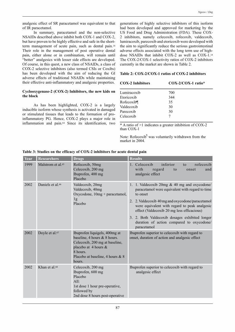
generations of highly selective inhibitors of this isoformhad been developed and approved for marketing by theUS Food and DrugAdministration (FDA). These COX-2 inhibitors, namely celecoxib, rofecoxib, valdecoxib,lumiracoxib,parecoxibandetoricoxibweredevelopedwiththeaimtosignificantlyreducetheseriousgastrointestinaladverseeffectsassociatedwiththelongtermuseofhigh-dose NSAIDs that inhibit COX-2 as well as COX-1.14TheCOX-2/COX-1selectivityratiosofCOX-2inhibitorscurrentlyinthemarketareshowninTable2.
Table 2: COX-2/COX-1 ratios of COX-2 inhibitors
COX-2 Inhibitors COX-2/COX-1 ratio*
Lumiracoxib 700Etoricoxib 344Rofecoxib¶ 35Valdecoxib 30Parecoxib 30Celecoxib 7
*Aratioof>1indicatesagreaterinhibitionofCOX-2thanCOX-1
Note:Rofecoxib¶wasvoluntarilywithdrawnfromthemarketin2004.
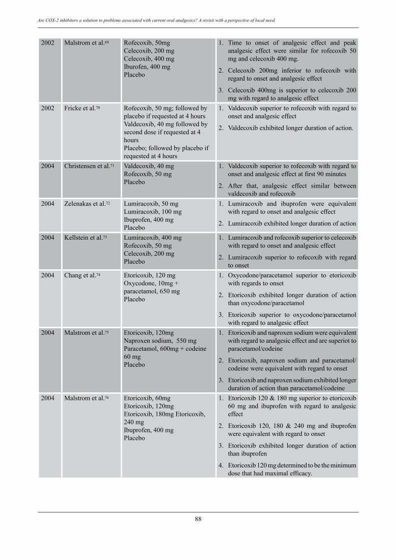
Table 3: Studies on the efficacy of COX-2 inhibitors for acute dental pain
Year Researchers Drugs Results1999 Malstrometal.65 Rofecoxib,50mg
Celecoxib,200mgIbuprofen,400mgPlacebo
Celecoxib inferior to rofecoxibwith regard to onset andanalgesiceffect
1.
2002 Danielsetal.66 Valdecoxib,20mgValdecoxib,40mgOxycodone,10mg+paracetamol,1gPlacebo
1.Valdecoxib20mg&40mgandoxycodone/paracetamolwereequivalentwithregardtotimetoonset
2.Valdecoxib40mgandoxycodone/paracetamolwere equivalent with regard to peak analgesiceffect(Valdecoxib20mglessefficacious)
2. Both Valdecoxib dosages exhibited longerduration of action compared to oxycodone/paracetamol
1.
2.
3.
2002 Doyleetal.67 Ibuprofenliquigels,400mgatbaseline,4hours&8hours.Celecoxib,200mgatbaseline,placeboat4hours&8hours.Placeboatbaseline,4hours&8hours.
Ibuprofensuperiortocelecoxibwithregardtoonset,durationofactionandanalgesiceffect
2002 Khanetal.68 Celecoxib,200mgIbuprofen,600mgPlaceboAll:1stdose1hourpre-operative,followedby2nddose8hourspost-operative
Ibuprofensuperiortocelecoxibwithregardtoanalgesiceffect
88
Are COX-2 inhibitors a solution to problems associated with current oral analgesics? A revisit with a perspective of local need.
2002 Malstrometal.69 Rofecoxib,50mgCelecoxib,200mgCelecoxib,400mgIburofen,400mgPlacebo
Time to onset of analgesic effect and peakanalgesic effect were similar for rofecoxib 50mgandcelecoxib400mg.
Celecoxib 200mg inferior to rofecoxib withregardtoonsetandanalgesiceffect
Celecoxib 400mg is superior to celecoxib 200mgwithregardtoanalgesiceffect
1.
2.
3.
2002 Frickeetal.70 Rofecoxib,50mg;followedbyplaceboifrequestedat4hoursValdecoxib,40mgfollowedbyseconddoseifrequestedat4hoursPlacebo;followedbyplaceboifrequestedat4hours
Valdecoxibsuperiortorofecoxibwithregardtoonsetandanalgesiceffect
Valdecoxibexhibitedlongerdurationofaction.
1.
2.
2004 Christensenetal.71 Valdecoxib,40mgRofecoxib,50mgPlacebo
Valdecoxibsuperiortorofecoxibwithregardtoonsetandanalgesiceffectatfirst90minutes
After that, analgesic effect similar betweenvaldecoxibandrofecoxib
1.
2.
2004 Zelenakasetal.72 Lumiracoxib,50mgLumiracoxib,100mgIbuprofen,400mgPlacebo
Lumiracoxib and ibuprofen were equivalentwithregardtoonsetandanalgesiceffect
Lumiracoxibexhibitedlongerdurationofaction
1.
2.
2004 Kellsteinetal.73 Lumiracoxib,400mgRofecoxib,50mgCelecoxib,200mgPlacebo
Lumiracoxibandrofecoxibsuperiortocelecoxibwithregardtoonsetandanalgesiceffect
Lumiracoxib superior to rofecoxib with regardtoonset
1.
2.
2004 Changetal.74 Etoricoxib,120mgOxycodone,10mg+paracetamol,650mgPlacebo
Oxycodone/paracetamol superior to etoricoxibwithregardstoonset
Etoricoxib exhibited longer duration of actionthanoxycodone/paracetamol
Etoricoxib superior to oxycodone/paracetamolwithregardtoanalgesiceffect
1.
2.
3.
2004 Malstrometal.75 Etoricoxib,120mgNaproxensodium,550mgParacetamol,600mg+codeine60mgPlacebo
Etoricoxibandnaproxensodiumwereequivalentwithregardtoanalgesiceffectandaresuperiottoparacetamol/codeine
Etoricoxib, naproxen sodium and paracetamol/codeinewereequivalentwithregardtoonset
Etoricoxibandnaproxensodiumexhibitedlongerdurationofactionthanparacetamol/codeine
1.
2.
3.
2004 Malstrometal.76 Etoricoxib,60mgEtoricoxib,120mgEtoricoxib,180mgEtoricoxib,240mgIbuprofen,400mgPlacebo
Etoricoxib120&180mgsuperiortoetoricoxib60 mg and ibuprofen with regard to analgesiceffect
Etoricoxib 120, 180 & 240 mg and ibuprofenwereequivalentwithregardtoonset
Etoricoxib exhibited longer duration of actionthanibuprofen
Etoricoxib120mgdeterminedtobetheminimumdosethathadmaximalefficacy.
1.
2.
3.
4.
89
Ngeow / Ong
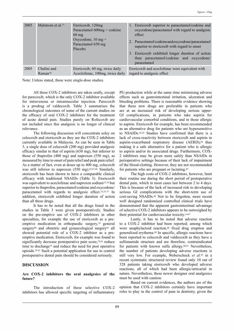
2005 Malstrometal.77 Etoricoxib,120mgParacetamol600mg+codeine60mgOxycodone,10mg+Paracetamol650mgPlacebo
Etoricoxibsuperior toparacetamol/codeineandoxycodone/paracetamolwithregardtoanalgesiceffect
Paracetamol/codeineandoxycodone/paracetamolsuperiortoetoricoxibwithregardtoonset
Etoricoxib exhibited longer duration of actionthan paracetamol/codeine and oxycodone/paracetamol
1.
2.
3.
2005 ChaliniandRaman78
Etoricoxib,60mg,twicedailyAceclofenac,100mg,twicedaily
Etoricoxibandaceclofenacwereequivalentwithregardtoanalgesiceffect
AlltheseCOX-2inhibitorsaretakenorally,exceptforparecoxib,whichistheonlyCOX-2inhibitoravailablefor intravenous or intramuscular injection. Parecoxibis a prodrug of valdecoxib. Table 3 summarises thechronologicaloutcomesofsomeofthecurrentstudiesonthe efficacy of oral COX-2 inhibitors for the treatmentof acute dental pain. Studies purely on Rofecoxib arenot included since this analgesic is no longer of clinicalrelevance. ThefollowingdiscussionwillconcentratesoleyoncelecoxibandetoricoxibastheyaretheCOX-2inhibitorscurrently available in Malaysia.As can be seen inTable3,asingledoseofcelecoxib(200mg)providedanalgesicefficacysimilartothatofaspirin(650mg),butinferiortothose of ibuprofen (400 mg) and naproxen (550 mg), asmeasuredbytimetoonsetofpainreliefandpeakpainrelief.Asamatteroffact,evenatdosesupto400mg,celecoxibwas still inferior to naproxen (550 mg).65,67,68 Similarly,etoricoxibhasbeenshown tohaveacomparableclinicalefficacy with traditional NSAIDs (Table 3). Etoricoxibwasequivalenttoaceclofenacandnaproxensodium75,78butsuperiortoibuprofen,paracetamol/codeineandoxycodone/paracetamol with regards to analgesic effect.74,75-77 Inaddition, etoricoxib exhibited longer duration of actionthanallthesedrugs. It has to be noted that all the drugs listed in thestudies in Table 3 were given postoperatively. Studieson the pre-emptive use of COX-2 inhibitors in otherspecialties, for example the use of etoricoxib as a pre-emptive medication in orthopaedic surgery,79 generalsurgery80 and obstetric and gynaecological surgery81 allshowed potential role of a COX-2 inhibitor as a pre-emptivemedication.Etoricoxib,forexamplewasfoundtosignificantlydecreasepostoperativepainscore,79,81reducetimetodischarge79andreducetheneedforpostoperativeopioids.79-81Suchapotentialapplicationforusetocontrolpostoperativedentalpainshouldbeconsideredseriously.
DISCuSSION
Are COX-2 inhibitors the oral analgesics of the future?
The introduction of these selective COX-2inhibitors has allowed specific targetingof inflammatory
Note:Unlessstated,theseweresingle-dosestudies.
PGproductionwhileatthesametimeminimisingadverseeffects such as gastrointestinal irritation, ulceration andbleedingproblems.Thereisreasonableevidenceshowingthat these new drugs are preferable in patients whoare at an increased risk of developing serious upper-GI complications, in patients who take aspirin forcardiovascularcomorbidconditions,and in thoseallergictoaspirin.Etoricoxibforexample,hasbeenrecommendedasanalternativedrugforpatientswhoarehypersensitiveto NSAIDs.82,83 Studies have confirmed that there is alackofcross-reactivitybetweenetoricoxibandaspirin inaspirin-exacerbated respiratory disease (AERD),84 thusmaking it a safe alternative for a patient who is allergictoaspirinand/oritsassociateddrugs.Furthermore,COX-2 inhibitors may be given more safely than NSAIDs inperioperativesettingsbecauseoftheirlackofimpairmentoftheblood-clotting.However,theyarenotrecommendedforpatientswhoarepregnantorlactating.85
ThehighcostsofCOX-2inhibitors,however,limittheir routineuseduring the shortperiodofpostoperativedentalpain,whichinmostcaseslastbetween2to4days.Thisisbecauseofthelackofincreasedrisktodevelopingserious GI complications with the short-term use ofcost-saving NSAIDs.86 Not to be forgotten, more recentwell designed randomised controlled clinical trials havedemonstrated that theapparentgastrointestinaladvantageofselectiveCOX-2inhibitorsappearstobeoutweighedbytheirpotentialforcardiovasculartoxicity.14,87
Lastly, it has to be noted that adverse reactionto a COX-2 inhibitor had been reported; among whichwere anaphylactoid reaction,88 fixed drug eruption andgeneralizederythema.89Inspecific,allergicreactionshavebeenreportedtocelecoxibandvaldecoxibastheyhaveasulfonamide structure and are therefore, contraindicatedfor patients with known sulfa allergy.90,91 Nevertheless,the number of patients developing adverse reactions isstill very low. For example, Weberschock et al.92 in arecent systematic structured review foundonly10outof328 patients taking etoricoxib who developed adversereactions, all of which had been allergic/urticarial innature.Nevertheless,thesenewerdesigneroralanalgesicsmustbeusedwithcaution. Basedoncurrentevidences, theauthorsareof theopinion that COX-2 inhibitors certainly have importantroles toplayin thecontrolofpainindentistry,giventhe
90
Are COX-2 inhibitors a solution to problems associated with current oral analgesics? A revisit with a perspective of local need.
natureof theirpharmacologicaldesignand longdurationofaction.Theseare:1. Potentially better patient compliance due to less
frequentdosing.2. Potentialuseinpaincontrolinconjuctionwithcertain
religiousreasone.g.theMuslimfastingmonth.Duetoitslong-actingeffect,apatientcanstillperformhis/herreligiousobligationwithoutinterference.
3. An alternative analgesic for patients with severe GIproblem.
4. Analternativetopatientswithallergytoaspirinand/orotherNSAIDs.
5. A potential pre-emptive premedication for patientsundergoingdentalsurgeriesundergeneralanaesthesiawithoutinterferingwithpre-operativefasting.
CONCLuSION
In conclusion, the common traditional analgesics,namely, paracetamol and NSAIDs (aspirin included) stillplay a major role in the management of post operativedentalpain.Astheyareonlygivenoverashortduration,theriskofdevelopingsevereorlong-termadversereactionsisminimised.Theyarewell-researched,ofreasonablecostand have proven to be as effective as the newer COX-2inhibitors in term of post operative dental pain efficacyand onset. The COX-2 inhibitors, while a novel idea, isstillplaquewith theworryofpossible long termadverseeffectpreviouslyundetectedinclinicaltrials,aswellasitshighercost.Perhapstheyarebestusedinconjuctionwiththeindicationssummarisedabove.
Disclaimer This review is intended to give a broad overviewofsomeof thecommonanalgesicsavailable inMalaysiabasedontheauthors’experienceandpastreports.Thelistsarenotexhaustiveandreadersareadvisedtoscrutinisethemanufacturers’latestrecommendationsforindicationsandcontraindications prior to prescribing the analgesic(s) ofconcern,especiallytopatientswithamedicalcondition.
REFERENCES
1. Dionne RA, Gordon SM. Non steroidal anti inflammatorydrugsforacutepaincontrol.DentalClinicsofNorthAmerica.1994;38:645-67.
2. ChiuWK,CheungLK.Efficacyofpreoperativeoralrofecoxibinpaincontrolforthirdmolarsurgery.OralSurgOralMedOralPatholOralRadiolEndod.2005;99:e47-53.
3. Fisher SE, Frame JW, Rout PG, McEntegart DJ. Factorsaffectingtheonsetandseverityofpainfollowingthesurgicalremoval of unilateral impacted mandibular third molar. BrDentJ.1988;164:351-4.
4. Roszkowski MT, Swift JQ, Hargreaves KM. Effect of nonsteroidal anti inflammatory drug administration on tissuelevelsof immunoreactiveprostaglandinE2, leukotrieneB4,and (S)-flurbiprofen following extraction of impacted thirdmolarpain.Pain.1997;73:339-45.
5. SeymourRA,BlairGS,WyatFA.Postoperativedentalpainand analgesic efficacy. Part I. Br J Oral Surgery 1983; 21:290-7.
6. HafisahM,LianCB,NgeowWC.AnauditofdrugsprescribedforminororalsurgeryintheFacultyofDentistry,UniversityofMalaya.MalaysDentJ.2002;23:40-4.
7. Huynh MP, Yagiela JA. Current concepts in acute painmanagement.JCalifDentAssoc.2003;31:419-27.
8. Joris J.Efficacyofnonsteroidalanti-inflammatorydrugs inpostoperative pain.ActaAnaesthesiol Belg. 1996; 47: 115-23.
9. Dubois RW, Melmed GY, Henning JM, Bernal M. Risk ofupper gastrointestinal injury and events in patients treatedwith cyclooxygenase (COX)-1/COX-2 nonsteroidal anti-inflammatory drugs (NSAIDs), COX-2 selective NSAIDs,andgastroprotectivecotherapy:anapraisalof theliterature.JClinRheumatol.2004;10:178-89.
10.El Miedany Y, Youssef S, Ahmed I, El Gaafary M. Thegastrointestinal safety and effect on disease activity ofetoricoxib,aselectivecox-2inhibitorininflammatoryboweldiseases.AmJGastroenterol.2006;101:311-7.
11.BarafHS,FuentealbaC,GreenwaldM,BrzezickiJ,O’BrienK,SofferB,PolisA,BirdS,KaurA,CurtisSP,EDGEStudyGroup.Gastrointestinalsideeffectsofetoricoxibinpatientswithosteoarthritis:resultsoftheetoricoxibversusdiclofenacsodiumgastrointestinaltolerabilityandeffectiveness(EDGE)trial.JRheumatol.2007;34:408-20.
12.LaineL,CurtisSP,CryerB,KaurA,CannonCP,MEDALSteering Committee. Assessment of upper gastrointestinalsafety of etoricoxib and diclofenac in patients withosteoarthritis and rheumatoid arthritis in the MultinationalEtoricoxib and Diclofenac Arthritis Long-term (MEDAL)programme: a randomised comparison. Lancet. 2007; 369:465-73.
13.BrooksPM,DayRO.COX-2 inhibitors.MedJAust2000;173:433-6.
14.HershEV,LallyET,MoorePA.Updateoncyclooxygenaseinhibitors: has a third COX isoform entered the fray? CurrMedResOpin.2005;21:1217-26.
15.Fu JY, Masferrer JL, Seibert K, RazA, Needleman P.Theinduction and suppression of prostaglandin H2 synthase(cyclooxygenase)inhumanmonocytes.JBiolChem.1990;265:16737-40.
16.MayN,EpsteinJ,OsborneB.SelectiveCOX-2inhibitors:areviewof their therapeuticpotentialandsafety indentistry.OralSurgOralMedOralPatholOralRadiolEndod.2001;92:399-405.
17.Cummings DM, Amadio PJr. A review of selected newernonsteroidal anti-inflammatory drugs. Am Fam Physician.1994;49:1197-1202.
18.CryerB,FeldmanM.Cyclooxygenase-1andcyclooxygenase-2 selectivityofwidelyusednonsteroidal anti-inflammatorydrugs.AmJMed.1998;104:413-21.
19.Lanza FL, Rack MF, Simon TJ, Quan H, Bolognese JA,Hoover ME, Wilson FR, Harper SE. Specific inhibition ofcyclooxygenase-2 with MK-0966 is associated with lessgastroduodenal damage than either aspirin or ibuprofen.AlimentPharmacolTher.1999;13:761-7.
20.Cooper SA, Engel J, Ladov M, Precheur H, RosenheckA, Rauch D. Analgesic efficacy of an ibuprofen-codeinecombination.Pharmacotherapy.1982;2:162-7.
21.HershEV,MoorePA,RossGL.Over-the-counteranalgesicsand antipyretics: a critical assessment.ClinTher2000; 22:500-48.
22.Kiersch TA, Halladay SC, Koschik M. A double-blind,randomizedstudyofnaproxensodium,ibuprofen,andplaceboinpostoperativedentalpain.ClinTher1993;15:845-54.
23.FrickeJR,HalladaySC,FranciscoCA.Efficacyandsafety
91
Ngeow / Ong
ofnaproxensodiumand ibuprofen forpain reliefafteroralsurgery.CurrTherRes.1993;54:619-27.
24.DaviesP,BaileyPJ,GoldenbergMM,Ford-HutchinsonAW.Theroleofarachidonicacidinpainandinflammation.AnnRevImmunol.1984;2:335-57.
25.CryerB,GottesdienerK,GertzB,HsiehP,DallobA,FeldmanM. Effects of a novel cyclooxygenase (COX)-2 inhibitoron gastric mucosal prostaglandin (PG) synthesis in healthyhumans.AmJGastroenterol.1996;91:1907.
26.MedicinesinPregnancyWorkingPartyoftheAustralianDrugEvaluation Committee. Prescribing medicine in pregnancy.AnAustraliancategorisationofriskofdruguseinpregnancy.4thed.1999.Canberra:CommonwealthofAustralia.
27.CassLJ,FrederikWS.ExperimentsinreliefofclinicalpainwithN-(2,3-xylyl)-anthranilicacid(CI-473;mefenamicacid).JPharmacolExpTher.1963;139:172-6.
28.Hart FD, Huskisson EC. Non steroidal anti–inflammatorydrugs. Current status and rational therapeutic use. Drugs.1984;27:232-55.
29.Hall P, Maclachlan N, Thorn N, Nudd NW, Taylor CG,GarriochDB.Controlofmenorrhagiabythecyclo–oxygenaseinhibitorsnaproxensodiumandmefenamicacid.BrJObstetGynaecol.1987;94:554–8.
30.Budoff PW. Mefenamic acid therapy in dysmennorrhea.JAMA.1979;241:2713–6.
31.Michalek-SaubererA, Heinzl H, Sator-Katzenschlager SM,Monov G, Knolle E, Kress HG. Perioperative auricularelectroacupuncture has no effect on pain and analgesicconsumptionafterthirdmolartoothextraction.AnesthAnalg.2007;104:542-7.
32.McGurkKA,RemmelRP,HosagraharaVP,ToshD,BurchellB.Reactivityofmefenamicacid1-o-acylglucuronidewithproteinsinvitroandexvivo.DrugMetabDispos.1996;24:842-9.
33.Rowe NH, Cudmore CL, Turner JL. Control of pain bymefenamic acid following removal of impacted molar. Adouble-blind,placebo-controlstudy.OralSurgOralMedOralPathol.1981;51:575-80.
34.Nagele F, Lockwood G, Magos AL. Randomised placebocontrolled trial of mefenamic acid for premedication atoutpatienthysteroscopy:apilotstudy.BrJObstetGynaecol.1997;104:842-4.
35.O’Brien WM, Bagby GF. Rare adverse reactions tononsteroidalantiinflammatorydrugs.JRheumatol.1985;12:13-20.
36.BatemanDN,KennedyJG.Non-steroidalanti-inflammatorydrugsandelderlypatients.BMJ1995;310:817-8.
37.IsaacsPE,SladenGE,FilipeI.Mefenamicacidenteropathy.JClinPathol.1987;40:1221-7.
38.Robertson CE, Ford MJ, Van Someren V, Dlugolecka M,PrescottLF.Mefenamicacidnephropathy.Lancet.1980;2:232-3.
39.TahaA, Lenton RJ, Murdoch PS, Peden NR. Non-oliguricrenalfailureduringtreatmentwithmefenamicacidinelderlypatients:acontinuingproblem.BMJ1985;291:661-2.
40.Long CC, Finlay AY, Marks R. Fixed drug eruption tomefenamicacid:areportofthreecases.BrJDermatol.1992;126:409–11.
41.O’HaganAH,IrvineAD,AllenGE,WalshM.Pseudoporphyriainducedbymefenamicacid.BrJDermatol.1998;139:1131–2.
42.Shepherd AN, Ferguson J, Bewick M, Bouchier IA.Mefenamicacid-inducedbullouspemphigoid.PostgradMedJ.1986;62:67-8.
43.HallRI,PettyAH,CobdenI,LendrumR.Enteritisandcolitis
associatedwithmefenamicacid.BMJ.1983;287:1182.44.DukesMNG.Meyler’ssideeffectsofdrugs.12thed.1992.
Amsterdam:Elsevier.45.Koch-WeserJ.Drugtherapy.Acetaminophen.NEnglJMed.
1976;295:1297-1300.46.MooreA,CollinsS,CarrollD,McQuayH,EdwardsJ.Single
doseparacetamol(acetaminophen),withorwithoutcodeine,forpostoperativepain.ChochraneDatabaseSystRev.2000;2:CD001547.
47.Skjelbred P, Album B, Lokken P. Acetylsalicyclic acid vsparacetamol: effects on post-operative course. Eur J ClinPharmacol.1977;12:257-64.
48.SkjelbredP,LokkenP.Paracetaomalversusplacebo:effectsonpost-operativecourse.EurJClinPharmacol.1979;15:27-33.
49.SeymourRA,RawlinsMD.Pharmacokineticsofparenteralparacetamolanditsanalgesiceffectsinpost-operativedentalpain.EurJClinPharmacol.1981;20:215-8.
50.Bentley KC, Head TW. The additive analgesic efficacy ofacetaminophen1000mg,andcodeine,60mg,indentalpain.ClinPharmacolTher.1987;42:634-40.
51.Cooper SA, Schachtel BP, Goldman E, Gelb S, Cohn P.Ibuprofen and acetaminophen in the reliefof acutepain:Arandomized, double-blind placebo-controlled study. J ClinPharmacol.1989;29:1026-30.
52.Mehlisch DR, Sollecito WA, Helfrick JF, Leibold DG,MarkowitzR,SchowCE,ShultzR,WaiteDE.Multicenterclinicaltrialofibuprofenandacetaminopheninthetreatmentofpostoperativedentalpain. JAmDentAssoc.1990;121:257-63.
53.KierschTA,HalladaySC,HormelPC.Asingle-dose,double-blind,comparisonofnaproxensodium,acetaminophen,andplacebo in postoperative dental pain. Clin Ther. 1994; 16:394-404.
54.NguyenAM,GrahamDY,GageT,GriffithsGR.Nonsteroidalanti-inflammatory drug use in dentistry: gastrointestinalimplications.GenDent1999;476:590-6.
55.FerreiraSH,VaneJR.Newaspectsinthemodeofactionofnon-steroidanti-inflammatorydrugs.AnnuRevPharmacol.1974;14:57-73.
56.Chandrasekharan NV, Simmons DL. The cyclooxygenases.GenomeBiol2004;5:241.
57.BergKJ,DjøselandO,GjellanA,HundalO,KnudsenER,RugstadHE,RønnebergE.Acuteeffectsofparacetamolonprostaglandinsynthesisandrenalfunctioninnormalmanandinpatientswithrenalfailure.ClinNephrol.1990;34:255–62.
58.Prescott LF, Mattison P, Menzies DG, Manson LM. Thecomparativeeffectsofparacetamolandindomethacinonrenalfunction inhealthy femalevolunteers.Br JClinPharmacol1990;29:403–12.
59.HyllestedM,JonesS,PedersonJL,KehletH.Comparativeeffect of paracetamol, NSAIDs or their combination inpostoperative pain management: a qualitative review. Br JAnesth2002;88:199-214.
60.MorseZ,TumpA,KevelhamE.Ibuprofenasapre-emptiveanalgesic is as effective as rofecoxib for mandibular thirdmolarsurgery.Odontology.2006;94:59-63.
61.McQuayH,MooreA.Anevidencebasedresourceforpain.1998.Oxford:OxfordUniversityPress.
62.British Medical Association and Royal PharmaceuticalSociety of Great Britain. British National Formulary 2000,Volume39.Oxon:PharmaceuticalPress:205-8.
63.CoulthardP,HillCM,FrameJW,BarryH,RidgeBD,BaconTH.Paincontrolwithparacetamolfromasustainedrelease
92
Are COX-2 inhibitors a solution to problems associated with current oral analgesics? A revisit with a perspective of local need.
formulation and a standard release formulation after thirdmolarsurgery:arandomisedcontrolledtrial.BrDentJ.2001;191:319-24.
64.Stoelting PK. Pharmacology & Physiology in AnestheticPractice. 1999. 3rd ed. Lippincott Williams & Wilkins,Philadelphia.
65.Malmstrom K, Daniels S, Kotey P, Seidenberg BC,DesjardinsPJ.Comparisonofrofecoxibandcelecoxib, twocyclooxygenase-2 inhibitors, in postoperative dental pain:a randomised placebo- and active- comparator controlledclinicaltrial.ClinTher.1999;21:1653-63.
66.DanielsSE,DesjardinsPJ,TalwalkerS,ReckerDP,VerburgKM. The analgesic efficacy of valdecoxib vs. oxycodone/acetaminophen after oral surgery. JAm DentAssoc. 2002;133:611-62.
67.DoyleG,JayawardenaS,AshrafE,CooperSA.Efficacyandtolerability of non-prescription ibuprofen versus celecoxibfordentalpain.JClinPharmacol.2002;42:912-9.
68.Khan AA, Brahim JS, Rowan JS, Dionne RA. In vivoselectivity of a selective cyclooxygenase 2 inhibitor in theoralsurgerymodel.ClinPharmacolTher.2002;72:44-9.
69.MalmstromK,FrickeJR,KoteyP,KressB,MorrisonB.Acomparison of rofecoxib versus celecoxib in treating painafter dental surgery: a single-center, randomized, double-blind, placebo- and active-comparator-controlled, parallel-group, single-dose studyusing thedental impactionmodel.ClinTher.2002;24:1549-60.
70.FrickeJ,VarkalisJ,ZwillichS,AlderR,ForesterE,ReckerDP, Verburg KM. Valdecoxib is more efficacious thanrofecoxibinrelievingpanassociatedwithoralsurgery.AmJTher.2002;9:89-97.
71.Christensen,K.S.,Cawkwell,G.D.(2004).Valdecoxibversusrofecoxibinacutepostsurgicalpain:resultsofarandomizedcontrolledtrial.JPainSymptomManage27,460-470.
72.ZelenakasK,FrickeJR,JaywardeneS,KellsteinD.Analgesiceffect of single oral doses of lumiracoxib and ibuprofen inpatientswithpostoperativedentalpain.IntJClinPract.2004;58:251-6.
73.Kellstein D, Ott D, Jayawardene S, Fricke J. Analgesicefficacy of a single dose of lumirocoxib compared torofecoxib, celecoxib and placebo in the treatment of post-operativedentalpain.IntJClinPract.2004;58:244-50.
74.Chang DJ, Desjardins PJ, King TR, Erb T, Geba GP. Theanalgesic efficacyof etoricoxib comparedwithoxycodone/acetaminophen in an acute postoperative pain model: arandomized,double-blindclinicaltrial.AnesthAnalg.2004;99:807-15.Erratumin:AnesthAnalg.(2005),101,644.
75.Malmstrom K, Kotey P, Coughlin H, Desjardins PJ. Arandomized,double-blind,parallel-groupstudycomparingtheanalgesiceffectofetoricoxib toplacebo,naproxensodium,andacetaminophenwithcodeineusingthedentalimpactionpainmodel.ClinJPain.2004;20:147-155.
76.MalmstromK,SapreA,CouglinH,AgrawalNG,MazenkoRS,FrickeJRJr.().Etoricoxibinacutepainassociatedwithdental surgery: A randomized, double-blind, placebo- andactivecomparator–controlleddose-rangingstudy.ClinTher.2004;26:667-79.
77.Malmstrom K,Ang J, Fricke JR, Shingo S, ReicinA.Theanalgesiceffectofetoricoxibrelativetothatofcetaminophenanalgesics: a randomized, controlled single-dose study inacutedentalimpactionpain.CurrMedResOpin.2005;21:141-9.
78.Chalini S, Raman U. Comparative efficacy of aceclofenacand etoricoxib in post extraction pain control: randomizedcontroltrial.IndJDentRes.2005;16:47-50.
79.Toivonen J, Pitko VM, Rosenberg PH. Etoricoxib pre-medicationcombinedwithintra-operativesubacromialblockforpainafterarthroscopicacromioplasty.ActaAnaesthesiolScand.2007;51:316-21.
80.Puura A, Puolakka P, Rorarius M, Salmelin R, LindgrenL. Etoricoxib pre-medication for post-operative pain afterlaparoscopic cholecystectomy. Acta Anaesthesiol Scand.2006;50:688-93.
81.Liu W, Loo CC, Tan HM, Ye TH, Ren HZ. Comparisonof preemptive analgesia efficacy between etoricoxib androfecoxibinambulatorygynecologicalsurgery.ZhongguoYiXueKeXueYuanXueBao.2004;26:666-70.
82.Muratore L, Ventura M, Calogiuri G, Calcagnile F, QuartaE, Muratore M, Ferrannini A. Tolerance to etoricoxib in37 patients with urticaria and angioedema induced bynonsteroidal anti-inflammatory drugs.AnnAllergyAsthmaImmunol.2007;98:168-71.
83.ViolaM,QuaratinoD,GaetaF,CarusoC,ValluzziR,RomanoA. Etoricoxib tolerability in patients with hypersensitivityto nonsteroidal anti-inflammatory drugs. Int Arch AllergyImmunol.2007;143:103-8.
84.ElMiedanyY,YoussefS,AhmedI,ElGaafarymM.Safetyofetoricoxib,aspecificcyclooxygenase-2inhibitor,inasthmaticpatients with aspirin-exacerbated respiratory disease. AnnAllergyAsthmaImmunol.2006;97:105-9.
85.ChanVS.A mechanistic perspective on the specificity andextentofCOX-2inhibitioninpregnancy.DrugSaf.2004;27:421-6.
86.CicconettiA,BartoliA,RipariF,RipariA.Cox-2selectiveinhibitors:Aliteraturereviewofanalgesicefficacyandsafetyinoral-maxillofacialsurgery.OralSurgOralMedOralPatholOralRadiolEndod2004;97:139-46.
87.RahmeE,NedjarH.RisksandbenefitsofCOX-2inhibitorsvs non-selective NSAIDs: does their cardiovascular riskexceed theirgastrointestinalbenefit?A retrospectivecohortstudy.Rheumatology(Oxford)2007;46:435-8.
88.Schellenberg RR, Isserow SH.Anaphylactoid reaction to acyclooxygenase-2 inhibitor in a patientwhohad a reactiontoacyclooxygenase-1 inhibitor.NEngl JMed.2001;345:1856.
89.AugustineM,SharmaP,StephenJ,JayaseelanE.Fixeddrugeruption and generalised erythema following etoricoxib.IndianJDermatolVenereolLeprol.2006;72:307-9.
90.WiholmBE.Identificationofsulfonamide-likeadversedrugreactions to celecoxib in the World Health Organizationdatabase.CurrMedResOpin.2001;17:210-6.
93
Ngeow / Ong
91.La Grenade L, Lee L, Weaver J, Bonnel R, KarwoskiC, Governale L, Brinker A. Comparison of reporting ofStevens-JohnsonsyndromeandtoxicepidermalnecrolysisinassociationwithselectiveCOX-2inhibitors.DrugSaf.2005;28:917-24.
92.WeberschockTB,MullerSM,BoehnckeS,BoehnckeWH.Tolerance to coxibs in patients with intolerance to non-steroidal anti-inflammatory drugs (NSAIDs): a systematicstructuredreviewoftheliterature.ArchDermatolRes.2007;299:169-75.
Address for correspondence:
Associate Professor Dr. WC Ngeow,Department of Oral & Maxillofacial Surgery,Faculty of Dentistry,University of Malaya,50603 Kuala Lumpur,Malaysia.Tel: 603-79674807Fax: 603-79674534E-mail: [email protected]

Oral Granular Cell Tumour: A Clinicopathological Study Of 7 Cases And A Brief Review Of The Literature Ajura Abdul Jalil. BDS, MClinDent (Mal) Stomology Unit, Institute for Medical Research, Kuala Lumpur.
Lau Shin Hui. BDS, FDSRCS (Eng), Stomatology Unit, Cancer Research Centre, Institute for Medical Research, Kuala Lumpur.
ABSTRACT
Oral granular cell tumour is a fairly rare lesion with a predilection for the tongue. Seven cases (6 females, 1 male) of oral granular cell tumour were seen during the 40 year period (1967-2006) in Stomatology Unit, Institute for Medical Research (IMR) in which 5 cases were located at the tongue. All the cases presented as a single swelling and excisional biopsies were carried out in all cases.
Key words oral granular cell tumour
94
Malaysian Dental Journal (2008) 29(2) 94-96© 2008 The Malaysian Dental Association
InTROdUCTIOn
Oral granular cell tumour is an uncommon lesionwithapredilectionforthetongue.Theaimofthisarticleistoreportsevenoralgranularcelltumourcasesseenduringthe40yearperiodfrom1967to2006ofStomatologyUnit,InstituteforMedicalResearch(IMR)andbrieflydiscussedtheclinicopathologicfeatures.
MATERIALS And METHOdS
TherecordsfromtheStomatologyUnit,IMRweresearchedforgranularcelltumourcasesfrom1967to2006.Congenitalepuliscases,anotherknowngranularcelllesionwereexcluded.Only7casesofgranularcelltumourwereretrieved.Therequestformsandhistologicalfeatureswerereviewed. Immunostaining for desmin, muscle specificactin and S100 protein using standard procedure werecarriedoutforallthecasesselected.
RESULTS
The patients’ characteristics are summarized inTable1.Therewere6femalesand1male,withameanageof29.4years(range11-62years).Thepatientscomprisedof3Malays,3Indiansand1Chinese.Fivecaseswerelocated
atthetongue,1inthebuccalmucosaand1atthebuccalsulcus. Most of the cases (n=5) presented as a painlessgrowth with durations ranging from 3 weeks to 2 years.The growths measured 0.5 to 1.5 cm in diameter. In allthecases,excisionalbiopsywasperformed.Histologicallyall the cases showed sheets of polygonal cells withgranular eosinophilic cytoplasm and small vesicularnucleiwhichextendedfromthesubepithelialzone to themuscular layer. The cells appeared cytologically benign.Pseudoepitheliomatoushyperplasiaof theepitheliumwasobservedin3cases. Immunohistochemistry staining was only carriedout in 6 cases since in one case, there was not enoughtissue left in the wax block. Immunostaining with S100protein showed 5 cases positive for the antibody. In onecase, the immunostaining result was inconclusive. Allcasesdemonstratednegativityfordesminandalsomusclespecificactin.
MALAYSIAn dEnTAL JOURnAL

95
Ajura / Lau
dISCUSSIOn Granular cell tumour is a rare soft tissue lesionwith a predilection for the oral cavity although it hasbeenreportedtooccur inotherpartsof thebodysuchasoesophagus1, thyroid2 and colon3. Intra-orally, the mostcommonsiteisthetongue4-8.However,oralgranularcelltumouroccurringintheparotidgland9,palate10,lowerlip11andfloorofmouth12hasbeenreported.Themostcommonsiteinthisstudywasthetongue(n=5,71.4%)ofwhich3caseswere locatedat thedorsalpartand2on the lateralborder. In this case series, the granular cell tumour wasmore prevalent among the females (n=6, 85.7%). Thisfindingconcurswithotherstudies4, 6-8. Oralgranularcelltumourshavebeenreportedinallagegroupsrangingfrom3years7to75years11,howeveritfrequentlyoccursinthethird and fourth decades of life4, 6, 8. The average age inthisstudywas29.4yearsandonlyonecasepresentedinachild. Clinically it is usually presents as a small, firm,rounded painless swelling with normal mucosal colourwhich generally gives an impression of a fibrous polyp.Thus, oral granular cell tumours are usually excisedimmediately by the surgeons. In our study, the clinicaldiagnosisgivenbythesurgeonsincludedfibroma,lipoma,neurofibroma, neurilemomma and papilloma. Granularcell tumour is a fairly slow growing benign lesion. In aseriesof8cases,Eguiaetal4reportedthedurationofthelesionintheoralcavityrangingfrom3monthsto2years.Oralgranularcelltumourusuallyhappenssinglyalthoughmultiple lesions occurring in the oral cavity have beenreported11, 13. Microscopically, the cells of granular cell tumourare rounded, polygonal with nuclei ranging from smalland dark to large with vesicular nuclei. The cytoplasmcontains fine to coarsely eosinophilicgranules.The cellsarearrangedinribbonsornestsdividedbyslenderfibrousconnectivetissueseptaorinlargesheetswithnoparticularcellulararrangement14.Otherhistologicalfeaturesincludepseudoepitheliomatoushyperplasiaoftheepithelium.Thisfeature may give a false impression of a squamous cellcarcinomaifabiopsywastakensuperficiallyandthuswillaffect themodeof treatment to thepatient. In this study,only3caseswereobservedtohavepseudoepitheliomatoushyperplasia. In a study done by Eguia et al4, 87.5% ofthe cases were noted to have pseudoepitheliomatous
hyperplasia in the overlying epithelium. The cause ofpseudoepitheliomatoushyperplasia isunknown.Granularcell tumours with pseudoepitheliomatous hyperplasiaexhibited increase in Ki-67 staining in the basal cells oftheoverlyingepitheliumanditmayrepresentaninductionphenomenonmediatedbyunidentifiedmoleculesproducedbythegranularcells8. AlthoughAbrikossoff first described granular celltumour (granular cell myoblastoma) 80 years ago andconsidereditasamuscletumour14,itiscurrentlybelievedtobeofneuralorigin.Immunohistochemistrydemonstratesthe cells are positive to S100 protein6, 8, 15-19 and neuronspecificenolase16, 17,butdonotreactwithchromogranin16,desmin20,actin20andmyoglobin20.Inourstudy,fivecaseswerepositiveforS100protein.Thisfindingconcurswithotherstudies6, 8, 15-19. A number of tumours have a granular appearancewhen seen microscopically. The differential diagnosisincludes alveolar soft part sarcoma and rhabdomyoma.Alveolar soft part sarcoma is characterized by thepseudoalveolar arrangement of large, oval to polyhedralcellusuallywithdistinctcellboundaries21.Rhabdomyomais composed of eosinophilic polygonal cells containinggranular cytoplasm with presence of cross striation14.Another lesion which contains granular cells is thecongenital epulis that happens in a newborn14. It is abenignlesionoccurringusuallyonthemaxillaryalveolarridgeofthenewborns.Althoughhistologicallycongenitalepulis and granular cell tumour have similar features,congenital epulis does not exhibit immunoreactivity forS100protein18, 22. Generally,thetreatmentfororalgranularcelltumouris straightforward by simple excision. The prognosis isgoodandrecurrenceisrare.Elevencasesoforalgranularcell tumours were treated by excisional resection usinglaser with no evidence of recurrence5. Granular celltumours can undergo malignant transformation. Lesionsareclassifiedasmalignantgranularcelltumoursifthreeormorecriteriaaremet:necrosis,spindling,vesicularnucleiwithlargenucleoli,increasedmitoticactivity,highnucleartocytoplasmicratioandpleomorphism23.InareviewdonebyAksoy et al24, out of 52 reported cases of metastaticgranularcelltumour,4casesoriginatedfromoralregion.
Table 1: Clinicopathological features of 7 cases of oral granular cell tumour
nO RACE SEX AGE SITE SIGn & SYMPTOM1 Malay F 34 dorsumoftongue painlessswelling2 Indian M 25 rightlateralborderoftongue painfulswelling3 Malay F 11 rightbuccalmucosa painlessswelling4 Indian F 62 leftlateralborderoftongue painlessswelling5 Malay F 22 buccalsulcusadjacentto14and15 painfulswelling6 Indian F 33 anteriordorsumtongue painlessswelling7 Chinese F 19 middorsumtongue painlessswelling
96
Oral Granular Cell Tumour: A Clinicopathological Study Of 7 Cases And A Brief Review Of The Literature
COnCLUSIOn
Although oral granular cell tumour is quite anuncommon lesion in the oral cavity, clinicians shouldconsideritintheirclinicaldifferentialdiagnosisofasinglelumpwhenseenclinicallyespeciallyifthelesionislocatedonthetongue.
ACKnOWLEdGEMEnT
The authors thank the Director General of HealthMalaysia and also to Director of Institute for MedicalResearch, Kuala Lumpur for their kind permission topublishthispaper.
REFEREnCES
1. Harikumar R, Simikumar, Aravindan SK, Thomas V.Abrikossoff’stumoroftheesophagus:casereportandreviewofliterature.TropGastroenterol2006:27(1):52-3.
2. BalochZW,MartinS,LivolsiVA.Granularcelltumorofthethyroid:acasereport.IntJSurgPathol2005;13(3):291-4.
3. SohnDK,ChoiHS,ChangYS,HuhJM,KimDH,KimDYetal.Granularcelltumorofcolon:reportofacaseandreviewofliterature.WorldJGastroenterol2004;10(16):2452-4.
4. EguiaA, UribarriA, Escoda CG, Crovetto MA, Martinez-Conde R, Aguirre JM. Granular cell tumor: report of 8intraoral cases. Med Oral Patol Oral Cir Bucal 2006; 11:E425-8.
5. AngieroF,CrippaR,StefaniM.Granularcelltumourintheoralcavity:reportofelevencasestreatedwithlasersurgery.MinnervaStomatol2006;55:423-30.
6. Xue JL, Fan MW, Wang SZ, Chen XM, Li Y. Aclinicopathological study of 14 cases of oral granular celltumor.ZhonghuaKouQiangXueZaZhi2005;40(4):302-5.
7. BrannonRB,AnandPM.Oralgranularcelltumors:ananalysisof10newpediatricandadolescentcasesandareviewoftheliterature.JClinPediatrDent2004;29(1):69-74.
8. ChrysomaliE,NikitakisNG,TosiosK,SaukJJ,PapinicolaouSI. Immunohistochemical evaluation of cell proliferationantigen Ki-67 and apoptosis-related proteins Bcl-2 andcaspase-3 in oral granular cell tumor. Oral Surg Oral MedOralPatholOralRadiolEndod2003;96:566-72.
9. Smith JL, Feehery JM, O’Hara BJ, RaoVM,Vernose GV.Granularcelltumoroftheparotid:Acasereportandliteraturereview.EarNoseThroatJ2001;80:454-7.
10.Boulos R, Marsot-Dupuch K, De Saint-Maur P, Meyer B,Tran Ba Huy P. Granular cell tumor of the palate: a casereport.AJNRAmJNeuroradiol2002;23(5):850-4.
11.WorsaaeN,SchwartzO,PindborgJJ.Follow-upstudyof14granularcelltumors.IntJSurg1979;8(2):133-9.
12.Tosios K, Rallis G, Vallianatou D, Vlachodimiropoulos D.Yellow-whitetumoronthefloorofthemouth.OralSurgOralMedOralPatholOralRadiolEndod2006;101:701-04.
13.JonesJK,KuoTT,GriffithsTM,ItharatS.Multiplegranularcelltumor.Laryngoscope1980:90:1646-51.
14.EnzingerFM,WeissSW.Softtissuetumours.4thed.Mosby;2001:p.1178-1187.
15.MaioranoE,FaviaG,NapoliA,RestaL,RiccoR,VialeG,AltiniM.Cellularheterogeneityofgranularcell tumours:acluetotheirnature?JOralPatholMed2000;284-90.
16.Williams HK,Williams DM. Oral granular cell tumours: a
histological and immunocytochemical study. J Oral PatholMed1997;26:164-9.
17.JunqueraLM,deVicenteJC,VegaJA,LosaJL,AlbertosJM.Granularcelltumours:animmunohistochemicalstudy.BrJOralMaxillofac1997;35:180-84.
18.KaiserlingE,RuckP,XiaoJC.Congenitalepulisandgranularcelltumor.Ahistologicandimmunohistochemicalstudy.OralSurgOralMedOralPatholOralRadiolEndod1995;80:687-97.
19.WangJ,ZhuXZ,ZhangRY.Malignantgranularcelltumor:a clinicopathologic analysis of 10 cases with review ofliterature.ZhonghuaBingLiXueZaZhi2004;33(6):497-502.
20.Stewart CM, Watson RE, Eversole LR, Werner F, LeiderAS. Oral granular cell tumors: a clinicopathologic andimmunocytochemicalstudy.OralSurgOralMedOralPathol1988;65:427-35.
21.ChristophersonWM,FooteFW,StewartFW.Alveolarsoft-partsarcomas.Structurallycharacteristictumorsofuncertainhistogenesis.Cancer1952;5:100-111.
22.Monteil RA, Loubiere R, Charbit Y, Gillet JY. Gingivalgranular cell tumor of the newborn: Immunoperoxidaseinvestigationwithanti-S-100antiserum.OralSurgOralMedOralPathol1987;64:78-81.
23.Fanburg-SmithJC,Meis-KindblomJM,FanteR,KindblomLG.Malignantgranularcelltumourofsofttissues:diagnosticcriteriaandclinicopathologiccorrelation.AmJSurgPathol1998;22:779-94.
24.Aksoy S,Abali H, Kilickap S, Harputluoglu H, Erman M.Metastaticgranularcell tumor:Acasereportandreviewoftheliterature:lettertotheeditor.ActaOncologica2006;45:91-94.
Address for correspondence:
dr. Ajura bt. Abdul Jalil,Stomatology Unit,Institute for Medical Research,50588 Jalan Pahang,Kuala Lumpur
Ability of Whitening Toothpastes in Removing Stains from Composite Resins Sum Yin Chong , BDS (Mal), Klinik Pergigian Ipoh, Perak, Malaysia
Tai Boon Lim, BDS (Mal), Klinik Pergigian Peringgit, Melaka, Malaysia
Liang Lin Seow, BDS (Mal), MSc (London), FDSRCS (Eng), PhD (Mal), School of Dentistry, International Medical University, Kuala Lumpur, Malaysia
ABSTRACT
Objectives: The objectives of the study were to assess: i) the staining susceptibility of composite resins, ii) the ability of whitening toothpastes in removing stains from composite resins. Materials and Methods: Thirty specimens from each composite resins: Filtek Z350 (3M ESPE), Filtek Z250 (3M ESPE) and Beautifil (Shofu Inc.) were fabricated. After polishing, specimens were immersed in coffee for 3 days. Specimens were then brushed twice a day for 2 weeks using Colgate Total (Colgate-Palmolive, control group), Colgate Advanced Whitening (Colgate-Palmolive, test group) and Darlie All Shiny White (Hawley & Hazel Chemical Co., test group). Colour changes (∆E*) were measured using Spectrophotometer at baseline, after coffee immersion and after brushing. Results were statistically analyzed using one way ANOVA and Tukey’s test. Results: There was significant difference in terms of colour changes for Filtek Z350, Filtek Z250 and Beautifil after coffee immersion (P<0.05). There was no significant difference in the ability to remove stains amongst the toothpastes investigated (P>0.05). Conclusions: Filtek Z350 was able to resist staining by coffee better than Filtek Z250 and Beautifil. The whitening toothpastes did not offer added advantage in terms of ability to remove stains compared to ordinary toothpaste.
97
Malaysian Dental Journal (2008) 29(2) 97-103 © 2008 The Malaysian Dental Association
INTROduCTION
Increasing demand for aesthetic restorations hasdrivencompositeresinstobeusedwidelyforbothanteriorand posterior teeth. Amongst the advantages offered bythis tooth-coloured restorative materials were strength,excellent aesthetics, ability to bond to tooth structureand enable minimal invasive tooth preparation. Newergeneration of materials such as packable and nanofilledcompositeshavebeenintroducedtomeetthechallengesinthedentalmarket1.Inaddition,pre-reactedglassionomercementtechnologyhasbeenutilizedtoaddtheadvantagesofglassionomercementtocompositeresins. Discolourationofcompositeresinrestorationsstillposes a problem especially for heavy smoker, coffeeand tea drinker2. Composite resins are susceptible todiscolourationbyaccumulatingstainsfromfooddyesanddietarycolourant3.Roughandirregularsurfaceisoneofthecontributoryfactorsforexternalstaininganddiscolourationand it is closely related to the type of composite resinmaterial4.Thestructureandthecharacteristicsofthefillerparticles in the composite resins have direct impact onsurfacesmoothnessandthusincreasethesusceptibilitytoextrinsic staining5. Once staining has occurred, brushingwith toothpastes and bleaching procedures may remove
thestainspartiallyortotally6.Ithasbeendemonstratedthattoothbrushingcanabradethesurfaceofcompositeresinswithathree-bodywearprocess7. In the last ten years, dentifrices have becomemore specialized and can be classified as therapeutic orcosmetic8. With regards to cosmetic function, dentifriceshavebecomeausefulsolutiontoremoveextrinsicstains,therefore whitened the teeth and also the surface ofrestorations9.Thedentifricescanactintwodifferentwaysi.e.mechanicallybytheactionofabrasivessuchassilicaorchemicallybytheeffectofwhiteningagentslikeperoxidesand sodium bicarbonate. The effectiveness of whiteningtoothpasteshasbeenwidelyinvestigated8,9.Itwouldbeofinteresttoevaluatetheeffectofthewhiteningtoothpastesontoothcolouredrestorativematerials.Thepresentstudy,therefore,wasconductedto:1.toassessthesusceptibilityofvarioustypesofcomposite
resinstostaining.2. to evaluate the ability of whitening toothpastes in
removingstainsfromcompositeresins.
MALAYSIAN dENTAL JOuRNAL
98
Ability of Whitening Toothpastes in Removing Stains from Composite Resins
MATERIALS ANd METHOdS
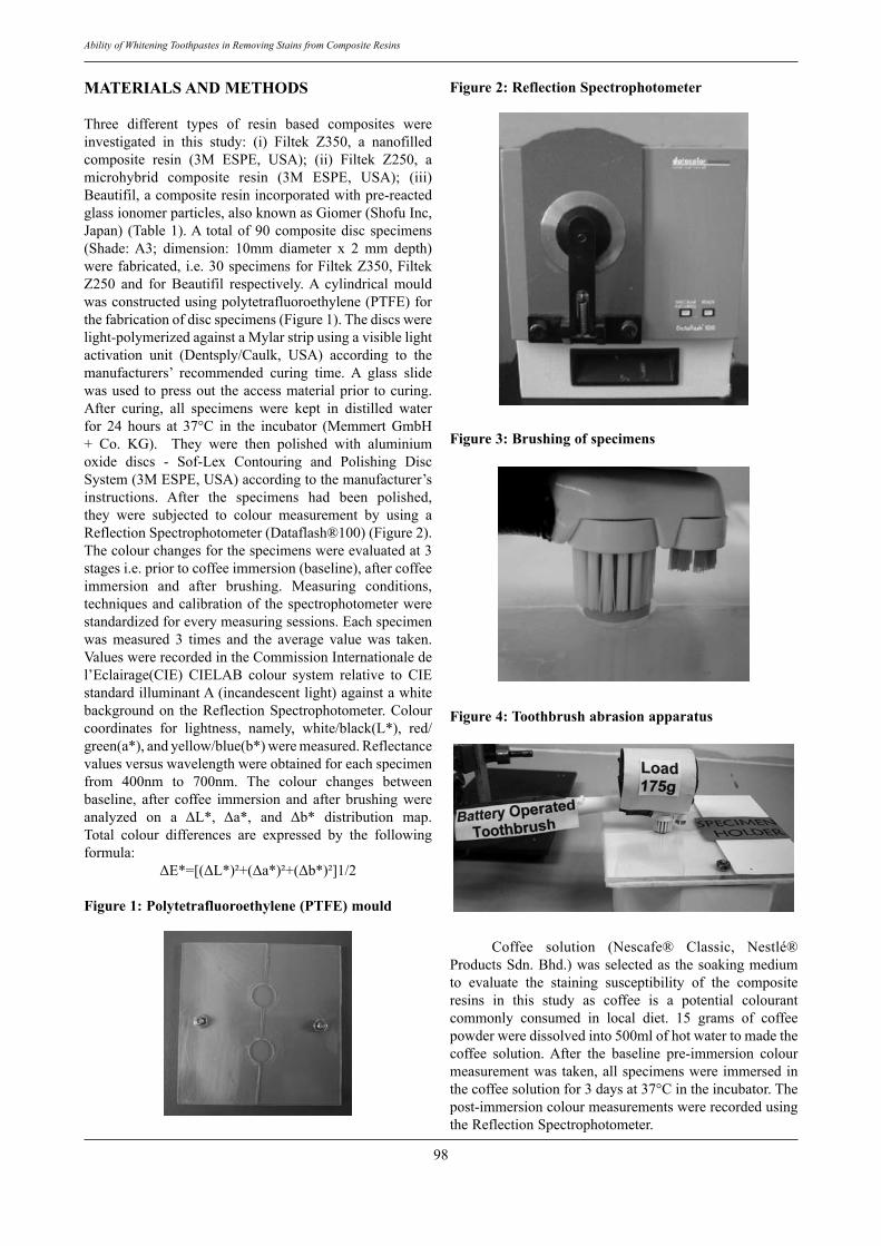
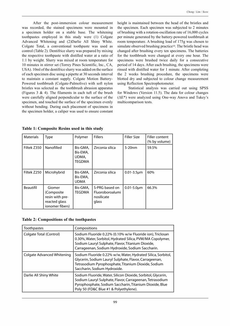
Three different types of resin based composites wereinvestigated in this study: (i) Filtek Z350, a nanofilledcomposite resin (3M ESPE, USA); (ii) Filtek Z250, amicrohybrid composite resin (3M ESPE, USA); (iii)Beautifil,acompositeresinincorporatedwithpre-reactedglassionomerparticles,alsoknownasGiomer(ShofuInc,Japan) (Table1).A totalof90compositediscspecimens(Shade:A3; dimension: 10mm diameter x 2 mm depth)werefabricated, i.e.30specimensforFiltekZ350,FiltekZ250 and for Beautifil respectively.A cylindrical mouldwasconstructedusingpolytetrafluoroethylene(PTFE)forthefabricationofdiscspecimens(Figure1).Thediscswerelight-polymerizedagainstaMylarstripusingavisiblelightactivation unit (Dentsply/Caulk, USA) according to themanufacturers’ recommended curing time. A glass slidewasusedtopressout theaccessmaterialprior tocuring.After curing, all specimens were kept in distilled waterfor 24 hours at 37°C in the incubator (Memmert GmbH+ Co. KG). They were then polished with aluminiumoxide discs - Sof-Lex Contouring and Polishing DiscSystem(3MESPE,USA)accordingtothemanufacturer’sinstructions. After the specimens had been polished,they were subjected to colour measurement by using aReflectionSpectrophotometer(Dataflash®100)(Figure2).Thecolourchangesforthespecimenswereevaluatedat3stagesi.e.priortocoffeeimmersion(baseline),aftercoffeeimmersion and after brushing. Measuring conditions,techniquesandcalibrationof thespectrophotometerwerestandardizedforeverymeasuringsessions.Eachspecimenwas measured 3 times and the average value was taken.ValueswererecordedintheCommissionInternationaledel’Eclairage(CIE) CIELAB colour system relative to CIEstandardilluminantA(incandescentlight)againstawhitebackgroundon theReflectionSpectrophotometer.Colourcoordinates for lightness, namely, white/black(L*), red/green(a*),andyellow/blue(b*)weremeasured.Reflectancevaluesversuswavelengthwereobtainedforeachspecimenfrom 400nm to 700nm. The colour changes betweenbaseline, after coffee immersion and after brushing wereanalyzed on a ΔL*, Δa*, and Δb* distribution map. Total colour differences are expressed by the followingformula:
ΔE*=[(ΔL*)²+(Δa*)²+(Δb*)²]1/2
Figure 1: Polytetrafluoroethylene (PTFE) mould
Figure 2: Reflection Spectrophotometer
Figure 3: Brushing of specimens
Figure 4: Toothbrush abrasion apparatus
Coffee solution (Nescafe® Classic, Nestlé®ProductsSdn.Bhd.)wasselectedas thesoakingmediumto evaluate the staining susceptibility of the compositeresins in this study as coffee is a potential colourantcommonly consumed in local diet. 15 grams of coffeepowderweredissolvedinto500mlofhotwatertomadethecoffee solution.After the baseline pre-immersion colourmeasurementwastaken,allspecimenswereimmersedinthecoffeesolutionfor3daysat37°Cintheincubator.Thepost-immersioncolourmeasurementswererecordedusingtheReflectionSpectrophotometer.
99
Chong / Lim / Seow
After the post-immersion colour measurementwas recorded, the stained specimens were mounted ina specimen holder on a stable base. The whiteningtoothpastes employed in this study were (1) ColgateAdvanced Whitening and (2)Darlie All Shiny White.Colgate Total, a conventional toothpaste was used ascontrol(Table2).Dentifriceslurrywaspreparedbymixingtherespectivetoothpastewithdistilledwaterataratioof1:1byweight.Slurrywasmixedatroomtemperaturefor10minutesinstirrerset(TorreyPinesScientific,Inc.,CA,USA).10mlofthedentifriceslurrywasaddedonthesurfaceofeachspecimendiscusingapipetteat30secondsintervalto maintain a constant supply. Colgate Motion Battery-Powered toothbrush (Colgate-Palmolive) with soft nylonbristleswasselectedasthetoothbrushabrasionapparatus(Figures 3 & 4).The filaments in each tuft of the brushwerecarefullyalignedperpendiculartothesurfaceofthespecimen,andtouchedthesurfaceofthespecimenevenlywithoutbending.Duringeachplacementofspecimens inthespecimenholder,acaliperwasusedtoensureconstant
height ismaintainedbetweentheheadof thebristlesandthespecimen.Eachspecimenwassubjectedto2minutesofbrushingwitharotation-oscillationrateof16,000cyclesperminutegeneratedbythebattery-poweredtoothbrushatroomtemperature.Abrushingloadof175gwaschosentosimulateobservedbrushingpractice10.Thebristleheadwaschangedafterbrushingeverytenspecimens.Thebatteriesfor the toothbrush were changed at every one hour. Thespecimens were brushed twice daily for a consecutiveperiodof14days.Aftereachbrushing,thespecimenswererinsedwithdistilledwaterfor1minute.Aftercompletingthe 2 weeks brushing procedure, the specimens wereblotted dry and subjected to colour change measurementusingReflectionSpectrophotometer. Statistical analysis was carried out using SPSSforWindows(Version11.5).Thedataforcolourchanges(ΔE*) were analyzed using One-way Anova and Tukey’s multicomparisontests.
Materials Type Polymer Fillers Filler Size Filler content(% by volume)
Filtek Z350 Nanofilled Bis-GMA,Bis-EMA,UDMA, TEGDMA
Zirconia silica 5-20nm 59.5%
Filtek Z250 Microhybrid Bis-GMA,Bis-EMA,UDMA
Zirconia silica 0.01-3.5µm 60%
Beautifil Giomer (Composite resin with pre-reacted glass ionomer fibers)
Bis-GMA,TEGDMA
S-PRG based on Fluoroboroaluminosilicateglass
0.01-5.0µm 66.3%
Table 1: Composite Resins used in this study
Table 2: Compositions of the toothpastes
Toothpastes Compositions
Colgate Total (Control) Sodium Fluoride 0.22% (0.10% w/w Fluoride ion), Triclosan 0.30%, Water, Sorbitol, Hydrated Silica, PVM/MA Copolymer, Sodium Lauryl Sulphate, Flavor, Titanium Dioxide, Carrageenan, Sodium Hydroxide, Sodium Saccharin.
Colgate Advanced Whitening Sodium Fluoride 0.22% w/w, Water, Hydrated Silica, Sorbitol, Glycerin, Sodium Lauryl Sulphate, Flavor, Carrageenan, Tetrasodium Pyrophosphate, Titanium Dioxide, Sodium Saccharin, Sodium Hydroxide.
Darlie All Shiny White Sodium Fluoride, Water, Silicon Dioxide, Sorbitol, Glycerin, Sodium Lauryl Sulphate, Flavor, Carrageenan, Tetrasodium Pyrophosphate, Sodium Saccharin, Titanium Dioxide, Blue Poly 50 (FD&C Blue #1 & Polyethylene).

100
Ability of Whitening Toothpastes in Removing Stains from Composite Resins
RESuLTS
The mean colour changes for all the compositeresins (Filtek Z350, Filtek Z250 and Beautifil) afterimmersion in coffee for 3 days were shown in Figure 5.The values of colour changes were recorded in NationalBureauofStandard (N.B.S.)units.FiltekZ350exhibitedthe least colour change followed by Filtek Z250 andBeautifil. Beautifil exhibited the greatest colour change(Figure5).Themeancolourchange forFiltekZ350was10.49N.B.S.unitswhilethecorrespondingdataforFiltekZ250andBeautifilwas11.35N.B.S.unitsand11.80N.B.Srespectively.Statisticallytherewasasignificantdifferencein terms of colour changes for Filtek Z350, Filtek Z250andBeautifilafterimmersionincoffee.Basedonastudycarried out by Ruyter et.al.11, colour change (∆E*) of more than (≥) 3.3 N.B.S. units was considered visually perceptible as well as clinically unacceptable. The alpharating according to United States Public Health Service(USPHS) clinical evaluation system corresponds to ∆E* valuesrangebetween2.2N.B.S.unitsto4.4N.B.S.units.Therefore,inaccordancewiththeaboveinformation,usingthe criterion for perceptible colour change of ∆E* ≥ 3.3 N.B.S. units, all specimens tested had perceptible colourchangesafterimmersedincoffeefor3days. The mean colour changes for Filtek Z350, FiltekZ250 and Beautifil after brushing with Colgate Total,ColgateAdvancedWhiteningandDarlieAllShinyWhiteweredepictedinFigure6.FiltekZ350specimensshowed4.23N.B.Sunits,4.38N.B.S.unitsand4.80N.B.S.unitsof mean colour change respectively after brushing withColgate Total, Colgate Advanced Whitening and DarlieAll Shiny White while Filtek Z250 demonstrated meancolour change of 4.40 N.B.S. units, 4.76 N.B.S. unitsand 5.27 N.B.S. units respectively. Beautifil showedmean colour change of 5.89 N.B.S. units, 6.08 N.B.S.units and 6.67 N.B.S. units after brushing with ColgateTotal,ColgateAdvancedWhiteningandDarlieAllShinyWhite.The trendfor thecolourchanges forFiltekZ350,FiltekZ250andBeautifilspecimensbrushedwithColgateTotal,ColgateAdvancedWhiteningandDarlieAllShinyWhite was fairly similar. Darlie All Shiny White groupexhibited greatest colour change, however, statisticalanalysisrevealedthatthemeancolourchangesofthethreetypes of composite resins after brushing with all threetoothpastes (Colgate Total as control, ColgateAdvancedWhiteningandDarlieAllShinyWhiteastestgroup)werenotsignificantlydifferentfromeachother(p>0.05).
Figure 5: Colour changes of Filtek Z350, Filtek Z250 and Beautifil after immersion in coffee solution for three days.
Figure 6: Colour changes of Filtek Z350, Filtek Z250 and Beautifil after brushing with Colgate Total, Colgate Advanced Whitening and Darlie All Shiny White
dISCuSSION Three commercially available composite resinswereselectedinthisstudyi.e.ananofilledcompositeresin(FiltekZ350,3MESPE,USA),amicrohybridcompositeresin(FiltekZ250,3MESPE,USA)andcompositeresinincorporated with pre-reacted glass ionomer particles,also known as Giomer (Beautifil, Shofu Inc, Japan).New composites integrating nanofiller technology havebeen introduced into clinical practice to achieve betterpolishabilityandwearresistance;however,thereislimitedinformation on their susceptibility to external stains.Giomerhasbeenintroducedinthelastdecadeandclaimedtohavetheadvantagesoffluoridereleasingandrecharging,goodwear resistance, excellent radiopacityandexcellentaesthetics.Withtheintroductionofthesenewmaterials,itiswarranttoevaluatetheirstainingsusceptibilitytoensurelong term clinical success as unsightly discolouration oftooth-coloured restorations has become one of the mainreasonsforreplacementoftheserestorations. It is important to select a valid colour-measuringinstrument to assess the colour changes of the test
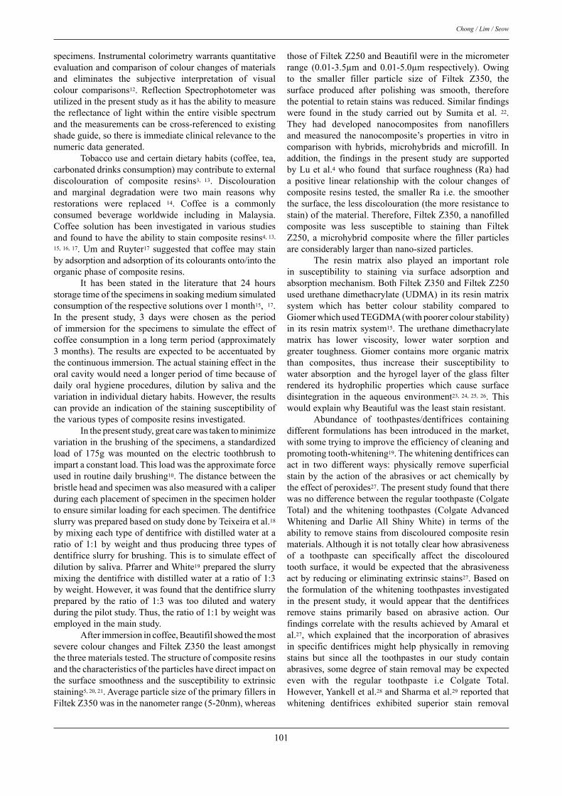
101
Chong / Lim / Seow
specimens.Instrumentalcolorimetrywarrantsquantitativeevaluationandcomparisonofcolourchangesofmaterialsand eliminates the subjective interpretation of visualcolour comparisons12. Reflection Spectrophotometer wasutilizedinthepresentstudyasithastheabilitytomeasurethe reflectanceof lightwithin theentirevisiblespectrumandthemeasurementscanbecross-referencedtoexistingshadeguide,sothereisimmediateclinicalrelevancetothenumericdatagenerated. Tobaccouseandcertaindietaryhabits(coffee,tea,carbonateddrinksconsumption)maycontributetoexternaldiscolouration of composite resins3, 13. Discolourationand marginal degradation were two main reasons whyrestorations were replaced 14. Coffee is a commonlyconsumed beverage worldwide including in Malaysia.Coffee solution has been investigated in various studiesandfoundtohavetheabilitytostaincompositeresins4, 13,
15, 16, 17.UmandRuyter17 suggested that coffeemaystainbyadsorptionandadsorptionofitscolourantsonto/intotheorganicphaseofcompositeresins. It has been stated in the literature that 24 hoursstoragetimeofthespecimensinsoakingmediumsimulatedconsumptionoftherespectivesolutionsover1month15,17.In the present study, 3 days were chosen as the periodof immersion for the specimens to simulate the effect ofcoffeeconsumptioninalongtermperiod(approximately3months).Theresultsareexpectedtobeaccentuatedbythecontinuousimmersion.Theactualstainingeffectintheoralcavitywouldneedalongerperiodoftimebecauseofdaily oral hygiene procedures, dilution by saliva and thevariationinindividualdietaryhabits.However,theresultscanprovideanindicationof thestainingsusceptibilityofthevarioustypesofcompositeresinsinvestigated. Inthepresentstudy,greatcarewastakentominimizevariationinthebrushingofthespecimens,astandardizedload of 175g was mounted on the electric toothbrush toimpartaconstantload.Thisloadwastheapproximateforceusedinroutinedailybrushing10.Thedistancebetweenthebristleheadandspecimenwasalsomeasuredwithacaliperduringeachplacementofspecimeninthespecimenholdertoensuresimilarloadingforeachspecimen.ThedentifriceslurrywaspreparedbasedonstudydonebyTeixeiraetal.18bymixingeachtypeofdentifricewithdistilledwaterataratioof 1:1byweight and thusproducing three typesofdentifriceslurryforbrushing.Thisistosimulateeffectofdilutionbysaliva.PfarrerandWhite19preparedtheslurrymixingthedentifricewithdistilledwaterataratioof1:3byweight.However,itwasfoundthatthedentifriceslurryprepared by the ratio of 1:3 was too diluted and wateryduringthepilotstudy.Thus,theratioof1:1byweightwasemployedinthemainstudy. Afterimmersionincoffee,BeautifilshowedthemostseverecolourchangesandFiltekZ350 the leastamongstthethreematerialstested.Thestructureofcompositeresinsandthecharacteristicsoftheparticleshavedirectimpactonthesurfacesmoothnessand thesusceptibility toextrinsicstaining5, 20, 21.AverageparticlesizeoftheprimaryfillersinFiltekZ350wasinthenanometerrange(5-20nm),whereas
thoseofFiltekZ250andBeautifilwereinthemicrometerrange (0.01-3.5µm and 0.01-5.0µm respectively). Owingto the smaller filler particle size of Filtek Z350, thesurface produced after polishing was smooth, thereforethepotentialtoretainstainswasreduced.Similarfindingswere found in the study carried out by Sumita et al. 22.They had developed nanocomposites from nanofillersand measured the nanocomposite’s properties in vitro incomparison with hybrids, microhybrids and microfill. Inaddition, the findings in the present study are supportedbyLuetal.4whofoundthatsurfaceroughness(Ra)hada positive linear relationship with the colour changes ofcomposite resins tested, the smaller Ra i.e. the smootherthesurface,thelessdiscolouration(themoreresistancetostain)ofthematerial.Therefore,FiltekZ350,ananofilledcomposite was less susceptible to staining than FiltekZ250, a microhybrid composite where the filler particlesareconsiderablylargerthannano-sizedparticles. The resin matrix also played an important rolein susceptibility to staining via surface adsorption andabsorptionmechanism.BothFiltekZ350andFiltekZ250usedurethanedimethacrylate(UDMA)initsresinmatrixsystem which has better colour stability compared toGiomerwhichusedTEGDMA(withpoorercolourstability)in its resin matrix system15.The urethane dimethacrylatematrix has lower viscosity, lower water sorption andgreater toughness. Giomer contains more organic matrixthan composites, thus increase their susceptibility towaterabsorptionandthehyrogellayeroftheglassfilterrendered its hydrophilic properties which cause surfacedisintegration in the aqueous environment23, 24, 25, 26. ThiswouldexplainwhyBeautifulwastheleaststainresistant. Abundance of toothpastes/dentifrices containingdifferent formulationshasbeen introduced in themarket,withsometryingtoimprovetheefficiencyofcleaningandpromotingtooth-whitening19.Thewhiteningdentifricescanact in two different ways: physically remove superficialstain by the action of the abrasives or act chemically bytheeffectofperoxides27.Thepresentstudyfoundthattherewasnodifferencebetweentheregulartoothpaste(ColgateTotal) and the whitening toothpastes (ColgateAdvancedWhitening and Darlie All Shiny White) in terms of theabilitytoremovestainsfromdiscolouredcompositeresinmaterials.Althoughitisnottotallyclearhowabrasivenessof a toothpaste can specifically affect the discolouredtooth surface, itwouldbe expected that the abrasivenessactbyreducingoreliminatingextrinsicstains27.Basedonthe formulationof thewhitening toothpastes investigatedin the present study, it would appear that the dentifricesremove stains primarily based on abrasive action. Ourfindingscorrelatewith the resultsachievedbyAmaraletal.27, which explained that the incorporation of abrasivesin specificdentifricesmight helpphysically in removingstains but since all the toothpastes in our study containabrasives,somedegreeofstainremovalmaybeexpectedeven with the regular toothpaste i.e Colgate Total.However,Yankelletal.28andSharmaetal.29reportedthatwhitening dentifrices exhibited superior stain removal
102
Ability of Whitening Toothpastes in Removing Stains from Composite Resins
propertycomparedtostandarddentifrices.ThedentifricesusedbyYankelletal.28andSharmaetal. 29maycontainingredients with chemical action. The importance ofchemicalactionhasalsobeendemonstratedbyKoertgeetal.,whoperformedalongitudinalclinicalstudyonbakingsodabaseddentifricesandfoundthattheyaresignificantlymore effective in removing extrinsic stain than standardsilica-baseddentifrices (abrasive type) although it is lessabrasivecomparedtothesilica-baseddentifrices. Koertgeetal.8hadstatedthatthewhiteningeffector stain removal effect of a dentifrice may not solelyrelated to its abrasiveness. In the present study, ColgateTotal,ColgateAdvancedWhiteningandDarlieAllShinyWhitedemonstratednosignificantdifferenceintheabilityto remove stains from three types of composite resinsi.e. Filtek Z350 (nanofilled), Filtek Z250 (microhybrid)and Beautifil (Giomer). Both Colgate Total and ColgateAdvanced Whitening were manufactured by the samemanufacturer.Itisimportanttotakenoteoftheincreaseinsurfaceroughnessofcompositeresinswhenbrushedwithwhiteningdentifriceswithhighabrasivity.Theincreaseinsurfaceroughnessafterlongtermtoothbrushingmayleadto easier absorption and adsorption of external stains30.Eventually, thismaynegate thepurposeof thewhiteningdentifriceandformedaviciouscycleofstainsformation.
CONCLuSION Within the limitations of the present study, it canbe concluded that coffee solution has the ability to staincomposite resins on prolonged and heavy consumptionleading todiscolouration thatcanbeperceivedclinically.Thecliniciansshouldwarnpatientswith largecompositerestorationstoreduceintakeofcoffeeorrinseimmediatelyafterconsumptiontoavoidunsightlydiscolourationoftherestorations. Filtek Z350 was more stain resistant thanFiltek Z250 and Beautifil. The whitening toothpastes(ColgateAdvancedWhiteningandDarlieAllShinyWhite)donotofferaddedadvantage to removestainscomparedtoordinarytoothpaste(ColgateTotal).Lastly,weadvocatefurtherstudytoevaluatetheeffectofwhiteningtoothpasteon surface roughness of composites resins and also theeffect of vicious cycle of brushing and consumption ofcoffeeonlongtermcolourstabilityofcompositeresins.
REFERENCES
1. Manhart J,ChenHY,HickelR.Thesuitabilityofpackableresin-basedcompositesforposteriorrestorations.JAmDentAssoc2001;132(5):639-645.
2. VanDijkenJWV.Directresincompositeinlays/onlays:an11yearfollowup.JDent2000;28:299-306.
3. DietschiD,CampanileG,HolzJ,MeyerJM.Comparisonofthe color stability of ten new-generation composites: an invitrostudy.DentMater1994;10:353-62.
4. Lu H, Roeder LB, Lei L, Powers JM. Effect of surfaceroughness on stain resistance of dental resin composites. JEsthetRestorDent2005;17:102-109.
5. Tjan AHL, Chan CA. The polishability of posterior
composites.JProthetDent1989;61:138-146.6. TürkünLS,TürkünM.Effectofbleachingand repolishing
proceduresoncoffeeandteastainremovalfromthreeanteriorcomposite veneering materials. J Esthet Restor Dent 2004;16:290-301.
7. Neme AL, Frazier KB, Roeder LB, Debner TL. Effect ofprophylacticpolishingprotocolsonthesurfaceroughnessofestheticrestorativematerials.OperDent2002;27(1):50-58.
8. Koertge TE, Brooks CN, SarbinAG, et al.A longitudinalcomparisonoftoothwhiteningresultingfromdentifriceuses.JClinDent1998;9:63-71.
9. Kleber CJ, Moore MH, Nelson BJ. Laboratory assessmentoftoothwhiteningbysodiumbicarbonatedentifrices.JClinDent1998;9(3):72-75.
10.Pugh BR. Toothbrush wear, brushing forces and cleaningperformance.JSocCosmetChem1978;29:423-431.
11.RuyterIE,Svendsen,SA.Remaininigmetacrylategroupsincomposite restorativematerials.ActaOdontol.Scand1987;36:75-82.
12.GrossMD,MoserJB.Acolorimetricstudyofcoffeeandteastaining of four composite resins. J Oral Rehabil 1977; 4:311-322.
13.AsmussenE,HansenEK.Surfacediscolorationofrestorativeresinsinrelationtosurfacesofteningandoralhygiene.ScandJDentRes1986;94(2):174-7.
14.MjörIA.Thereasonsforreplacementandtheageoffailedrestorations in general dental practice.Acta Odontol Scand1997;55(1):58-63.
15.Khokhar ZA, Razzoog ME, Yaman P. Colour stability ofcompositeresins.QuintessenceInt1991;22:733-737.
16.Yannikakis SA, ZissisAJ, Polyzois GL, Caroni C. Colourstabilityofprovisionalresinrestorativematerials.JProsthetDent1998;80:533-539.
17.Um CM, Ruyter IE. Staining of resin-based veneeringmaterialswithcoffeeandtea.QuintessenceInt1991;22:377-386.
18.Teixeira ECN, Thompson JL, Piascik JR et al. IN vitrotoothbrush-dentifriceabrasionoftworestorativecomposites.JEsthetRestorDent2005;17:172-182.
19.PfarrerAM,WhiteDJ.Anticariesprofilequalificationofanimprovedwhiteninigdentifrice.JClinDent2001;12:30-33
20.ShintaniH,SatouJ,SatouNetal.Effectsofvariousfinishingmethods on staining and accumulation of streptococcusmutansHS-6oncompositeresins.DentMater1985;1:225-227.
21.Hörsted-BindslevP,MjörIA.Estheticrestorations.In:Modernconcepts in operative dentistry. Copenhagen: Munksgaard.1988;Pp.190-246.
22.Sumita BM, Dong W, Brian NH. An application ofnanotechnology in advanced dental materials. JADA 2003;134:1382-1390.
23.GladysS,VanMeerbeekB,BraemM,LambrechtsP,VanherleG.Comparativephysico-mechanicalcharacterizationofnewhybridrestorativematerialswithconventionalglass-ionomerandresincompositematerials.JDentRes1997;76:883.
24.Cattani-LorenteMA,DupuisV,PayanJ,MoyaF,MeyerJM.Effectofwateronthephysicalpropertiesofresin-modifiedglass-ionomercements.DentMater1999;15:71.
25.ItotaT,CarrickTE,YoshiyamaM,McCabeJF.Fluoridereleaseandrechargeingiomer,compomerandresincomposite.DentMater2004;20:789-795.
26.GeurtsenW,LeyhausenG,Garcia-GodoyF.Effectofstoragemediaon the fluoride releaseandsurfacemicrohardnessof
103
Chong / Lim / Seow
four polyacid-modified composite resins (“compomer”).DentMater1999;15(3):196-201.
27.Amaral CM, Rodrigues JA, Erhardt MCG et al. Effect ofwhiteningdentifricesonthesuperficialroughnessofestheticrestorativematerials.JEsthetRestorDent2006;18(2):109-119.
28.Yankell SL, Shi X, Emling RC, Nelson BJ, Triol CW.Laboratoryevaluationsofthreedentifriceswithpolishingorbrushing.JClinDent1998;9:61-63.
29.SharmaN,GalustiansHJ,QaqishJetal.Comparativetoothwhiteningandextrinsic toothstainpreventionefficacyofanewdentifriceandacommerciallyavailabletoothwhiteningdentifrice: six-week clinical trial. J Clin Dent 2004; 15(2):52-57.
30.HachiyaY, Iwaku M, Hosoda H, Fusayama T. Relation offinishtodiscolourationofcompositeresins.JProsthetDent1984;52:811-814.
Address for correspondence:
Assoc. Prof. Dr. Liang Lin SeowSchool of DentistryInternational Medical University126, Jalan 19/155B, Bukit Jalil, 57000Kuala Lumpur, MalaysiaTel: 00603- 2731 7286e-mail: [email protected]
Latest recommendations for dental radiography in general dental practice. Kathiravan Purmal. BDS (Malaya), DGDP (UK), MFDSRCS (London), MOrth (Malaya), MOrth RCS (Edinburgh)
Phrabhakaran Nambiar. BDS (Mysore), BSc (Hons) (Adelaide), MScD (Adelaide), MScD(Western Cape), PG Dip Dent-Maxfac Rad (Stellenbosch)Department of General Dental Practice and Oral & Maxillofacial Imaging, Faculty of Dentistry, University of Malaya.
104
Malaysian Dental Journal (2008) 29(2) 104-112© 2008 The Malaysian Dental Association
INtroductIoN
Radiologyisimportantinthediagnosticassessment,treatmentandmonitoringprogressofpatientshavingdentalandmaxillofacialdiseases.Thecentralquestionindentalradiology iswhether there isany riskwith regard to lowdoses of radiation and what are the necessary protectionneededtopreventanyhazardousoutcomewithX-raying.Withtheemergenceofthenewmillennium,majorchangesare happening in the field of science and technology.More efficient equipments are being discovered andmore researches are being conducted to elicit betterunderstanding of the radiation process and its effecton the human health. This article will review the latestguidelinesforproperradiographicpracticefromtheUSAandEurope.Itwilladdresstopicsliketherisksfromdentalradiography,selectioncriteria,protectionforpatientsandstaff,improvementtoX-rayequipments,roomdimensionsandleadliningrequirements.Wehavelimitedourresearchto intraoral, panoramic and cephalometric radiographsbecause these are the radiographs or images that arecommonlytakeninthegeneralpracticesetting.
oBJEctIVES
1. To make the general dental practitioner aware ofthe latest trend and guidelines with regard to dentalradiography,particularlystandardsfromUnitedStatesofAmericaandEurope.
2. To dispel any myths and misinformation aboutradiographicprotection.
3. To analyze the reason and rationale for the proposedguidelineswithsupportiveresearcharticles.
4. Toeducatethegeneralpractitioneroftheneedtomakechangestolawspertainingtodentalradiation.
MEtHodS
Literatureresearchactivitieswereundertakenatthelibrary of University of Malaya. Pubmed was searched
usingtheterm“dentalradiographs”incombinationwithotherrelevantkeywordsincluding“guidelines”,“risks”,“protection”, “ thyroid” and “pregnancy”. Relevantresearchestojustifytheguidelineswerealsoperused.
risk from dental radiation doses.
Whathappensafterthepressofthebuttonandyouhear thefamiliarbeep?X-raysareproducedbyelectronsbombarding a target material (usually tungsten) and aresubsequently brought suddenly to rest. This happensinside a small evacuatedglass envelopecalled theX-raytube(Figure1).
Figure 1 : the anode enlarged showing the target and production of X-ray. Adapted from dental radiography. Principles and techniques 3rd Edition. Iannucci JM and Howrrton LJ.
ThisX-raywillpassthroughthepatientandhittheX-rayfilmordigitalreceptor.TheX-raywillaffectfilmemulsiontoproducethevisualimage(radiograph)orwiththedigitalreceptor,thedataistransferredtothecomputerto produce an image. When the X-ray passes throughhumantissue,itcanproducebiologicaldamagingeffects.Theseeffectscanbeclassifiedintotwomaincategories:-
a) Deterministiceffectsb) Stochasticeffects
MALAYSIAN dENtAL JourNAL
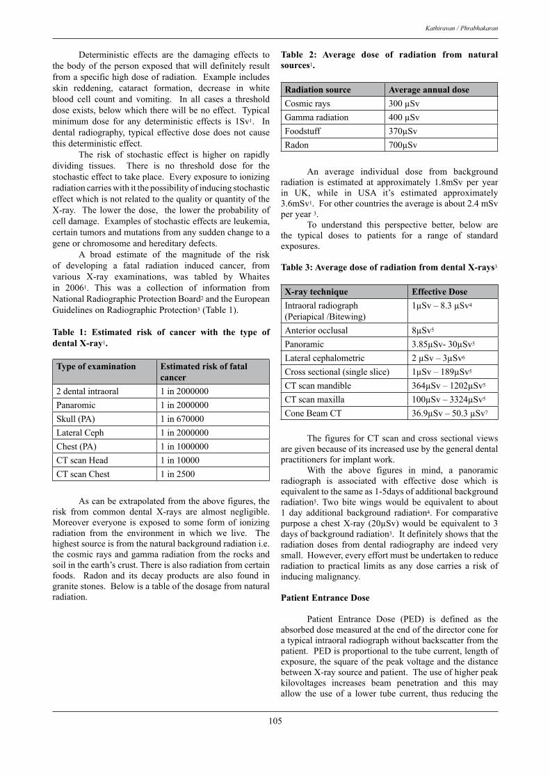
105
Kathiravan / Phrabhakaran
Deterministic effects are the damaging effects tothebodyof thepersonexposed thatwilldefinitely resultfromaspecifichighdoseofradiation.Exampleincludesskin reddening, cataract formation, decrease in whiteblood cell count and vomiting. In all cases a thresholddoseexists,belowwhichtherewillbenoeffect.Typicalminimum dose for any deterministic effects is 1Sv1. Indental radiography, typical effective dose does not causethisdeterministiceffect. The risk of stochastic effect is higher on rapidlydividing tissues. There is no threshold dose for thestochasticeffecttotakeplace.EveryexposuretoionizingradiationcarrieswithitthepossibilityofinducingstochasticeffectwhichisnotrelatedtothequalityorquantityoftheX-ray. The lower thedose, the lower theprobabilityofcelldamage.Examplesofstochasticeffectsareleukemia,certaintumorsandmutationsfromanysuddenchangetoageneorchromosomeandhereditarydefects. A broad estimate of the magnitude of the riskof developing a fatal radiation induced cancer, fromvarious X-ray examinations, was tabled by Whaitesin 20061. This was a collection of information fromNationalRadiographicProtectionBoard2andtheEuropeanGuidelinesonRadiographicProtection3(Table1).
table 1: Estimated risk of cancer with the type of dental X-ray1.
type of examination Estimated risk of fatal cancer
2dentalintraoral 1in2000000Panaromic 1in2000000Skull(PA) 1in670000LateralCeph 1in2000000Chest(PA) 1in1000000CTscanHead 1in10000CTscanChest 1in2500
Ascanbeextrapolatedfromtheabovefigures,therisk from common dental X-rays are almost negligible.Moreover everyone is exposed to some formof ionizingradiation from the environment in which we live. Thehighestsourceisfromthenaturalbackgroundradiationi.e.thecosmicraysandgammaradiationfromtherocksandsoilintheearth’scrust.Thereisalsoradiationfromcertainfoods. Radon and its decay products are also found ingranitestones.Belowisatableofthedosagefromnaturalradiation.
table 2: Average dose of radiation from natural sources1.
radiation source Average annual doseCosmicrays 300µSvGammaradiation 400µSvFoodstuff 370µSvRadon 700µSv
An average individual dose from backgroundradiation is estimated at approximately 1.8mSv per yearin UK, while in USA it’s estimated approximately3.6mSv1.Forothercountriestheaverageisabout2.4mSvperyear3. To understand this perspective better, below arethe typical doses to patients for a range of standardexposures.
table 3: Average dose of radiation from dental X-rays3
X-ray technique Effective doseIntraoralradiograph(Periapical/Bitewing)
1µSv–8.3µSv4
Anteriorocclusal 8µSv5
Panoramic 3.85µSv-30µSv5
Lateralcephalometric 2µSv–3µSv6
Crosssectional(singleslice) 1µSv–189µSv5
CTscanmandible 364µSv–1202µSv5
CTscanmaxilla 100µSv–3324µSv5
ConeBeamCT 36.9µSv–50.3µSv7
ThefiguresforCTscanandcrosssectionalviewsaregivenbecauseofitsincreasedusebythegeneraldentalpractitionersforimplantwork. With the above figures in mind, a panoramicradiograph is associated with effective dose which isequivalenttothesameas1-5daysofadditionalbackgroundradiation5.Two bite wings would be equivalent to about1 day additional background radiation4. For comparativepurposea chestX-ray (20µSv)wouldbe equivalent to3daysofbackgroundradiation3.Itdefinitelyshowsthattheradiation doses from dental radiography are indeed verysmall.However,everyeffortmustbeundertakentoreduceradiation to practical limits as any dose carries a risk ofinducingmalignancy.
Patient Entrance dose
Patient Entrance Dose (PED) is defined as theabsorbeddosemeasuredattheendofthedirectorconeforatypicalintraoralradiographwithoutbackscatterfromthepatient.PEDisproportionaltothetubecurrent,lengthofexposure,thesquareofthepeakvoltageandthedistancebetweenX-raysourceandpatient.Theuseofhigherpeakkilovoltages increases beam penetration and this mayallow the use of a lower tube current, thus reducing the

106
Latest recommendations for dental radiography in general dental practice.
dose.Increasingthefocustoskindistance(FSD)reducesthe dose greatly, for example if the FSD is doubled, thePEDwillbereducedbyafactoroffour8.ThemeanPEDfor45kVto55kVsetsusingEspeedfilmis4.1mGy(4.1µSv) and for machine with 60kV to 70kV sets using Espeed film is 1.8 mGy (1.8 µSv)9. When compared tothechestX-ray, theaveragePEDisonly0.24mGy(0.24µSv)9.Thisisduetothefocustoskindistanceinintraoralradiographswhichisaround200mmandthesamedistanceinchestX-rayis1500mm.Theimplicationofthisfactisalthoughtheeffectivedoseislessinintraoralradiography,the patient entrance dose (or skin exposed dose ) issubstantiallyhigher.Thereforeeveryeffortmustbetakentominimizethisexposureforthepatient.
Hormesis
Definitionofhormesis–theinductionofbeneficialeffectsbylowdosesofanotherwiseharmfulphysical(eg:9ionizingradiation)orchemicalagent. Theoverwhelmingmajorityofthecurrentavailableepidemiological data on populations exposed to ionizingradiation support the assumption that there is a linearnon-threshold dose-response relationship. Howeverepidemiological data fail to demonstrate detrimentaleffects of ionizing radiation absorbed doses smallerthan 100-200mSv. Risk estimates for these levels aretherefore based on extrapolations from higher doses.Sasaki et al 10 stated that, for mammalian cells, theoptimum dose for a radio-adaptive response is below100mSv. Adaptive response to radiation isaneffect thathas been convincingly demonstrated in cultured cells.There is however still doubt over how this influencesthe risk of multicellular organism and also over theduration of the radio-adaptive effect. If a hormeticeffect of radiation exists, it seems to be rather weakand inconsistent. Overall there is currently insufficientevidenceforradiationhormesistowarrantanyfarreachingchangeinthepresentradiationpolicyassociatedwithrisksofionizingradiation11.
Patient selection
Currently there are no clear guidelines to justify whenradiographs are necessary in Malaysia. Radiographs arerequestedbasedon the training that theparticulardentisthasundergoneandindividualpreference.In2002,Nambiarand Lim12 conducteda surveyof thepracticeofdentalradiographyinMalaysiaandfoundthatthegeneraldentalpractitionersinMalaysiaarenotverydependentondentalradiographsbecausethemajoritytooklessthan20intraoralfilmspermonth.Thereforewelookedintoguidelinesfromthe American Dental Association13 and The Faculty ofGeneral Dental Practitioners Royal College of SurgeonsLondon14 for guidance. It is essential that selection ofappropriateradiographisbasedontheindividualpatient’shistory and clinical examination. The ‘routine’ use ofradiographsonpatientsbasedonageneralizedorscreeningapproach isnot acceptable15. It is alsonot acceptable totakeradiographsformedicalrecords,trainingpurposelikethose practised in dental schools or for research purpose
whichdoesnotdirectlybenefitthepatient16.Listedbelowaresomeoftheconditionsthatmightrequiredentalradiographs.
1. DentalCariesDiagnosis The posterior bitewing radiographs are essentialadjunct to clinical examination14. However before thisradiograph is requested, the initial clinical examinationmust include an assessment of caries risk (as high,medium or low). If the risk of carious lesion is high,for example there are multiple carious lesions, extractedtooth, poor oral hygiene then bitewing radiographs canberequestedeverysixmonths.Ifthereismoderaterisk,radiographscanberequestedeveryyearandiftheriskislow,thepatientwouldneedbitewingsevery2yearsonly.The rationale for this time frame is early enamel lesionprogressesatarelativelyslowratetakingatleasttwoyearstoprogressintodentine17.Furthermore,intervalsbetweensubsequent bitewing radiographs must be reassessed foreach new period as individuals can move in and out ofcariousriskcategories.
2. Orthodontics Radiographs are needed following clinicalexamination in a proportion of orthodontic patients.A clinical examination is necessary to ensure that theradiographsrequestedwillbeappropriateforthepatient’sspecific orthodontic problem. Cephalometric lateralskull radiographs are not necessary if the patient is notindicated for fixed appliance or functional appliance.These radiographs are also not necessary if there is anobviousClassIskeletalpattern18.Thevalueofhandwristradiographs in clinical orthodontics has been questionedastheseviewslackthereliabilitytopredictgrowthspurts.Similarly radiographs for TMJ cannot be justified andfilmstakenforthisreasonhavenoimpactonthetreatmentplanning19.
BelowistheguidelinefortakingcephalometricradiographsfromBritishOrthodonticSociety18.
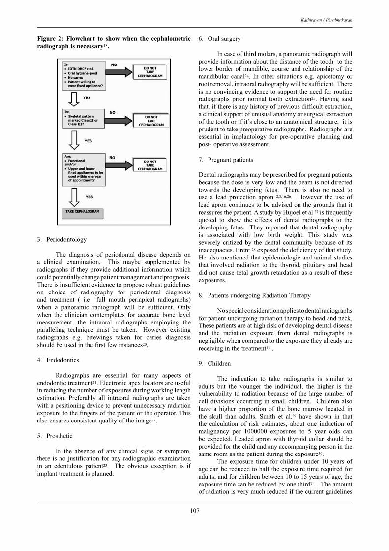
107
Kathiravan / Phrabhakaran
Figure 2: Flowchart to show when the cephalometric radiograph is necessary18.
3. Periodontology
The diagnosis of periodontal disease depends ona clinical examination. This maybe supplemented byradiographs if theyprovide additional informationwhichcouldpotentiallychangepatientmanagementandprognosis.Thereisinsufficientevidencetoproposerobustguidelineson choice of radiography for periodontal diagnosisand treatment ( i.e full mouth periapical radiographs)when a panoramic radiograph will be sufficient. Onlywhen the clinician contemplates for accurate bone levelmeasurement, the intraoral radiographs employing theparalleling technique must be taken. However existingradiographs e.g. bitewings taken for caries diagnosisshouldbeusedinthefirstfewinstances20.
4. Endodontics
Radiographs are essential for many aspects ofendodontictreatment21.Electronicapexlocatorsareusefulinreducingthenumberofexposuresduringworkinglengthestimation. Preferably all intraoral radiographs are takenwithapositioningdevicetopreventunnecessaryradiationexposuretothefingersofthepatientortheoperator.Thisalsoensuresconsistentqualityoftheimage22.
5. Prosthetic
In the absence of any clinical signs or symptom,there isno justificationforanyradiographicexaminationin an edentulous patient23. The obvious exception is ifimplanttreatmentisplanned.
6. Oralsurgery
Incaseofthirdmolars,apanoramicradiographwillprovideinformationaboutthedistanceofthetoothtothelowerborderofmandible, course and relationshipof themandibular canal24. In other situations e.g. apicetomy orrootremoval,intraoralradiographywillbesufficient.Thereisnoconvincingevidencetosupporttheneedforroutineradiographs prior normal tooth extraction25. Having saidthat,ifthereisanyhistoryofpreviousdifficultextraction,aclinicalsupportofunusualanatomyorsurgicalextractionofthetoothorifit’sclosetoananatomicalstructure,itisprudenttotakepreoperativeradiographs.Radiographsareessential in implantology for pre-operative planning andpost-operativeassessment.
7. Pregnantpatients
Dentalradiographsmaybeprescribedforpregnantpatientsbecausethedoseisverylowandthebeamisnotdirectedtowards the developing fetus. There is also no need touse a lead protection apron 2,3,16,26. However the use ofleadaproncontinues tobeadvisedonthegrounds that itreassuresthepatient.AstudybyHujoeletal27isfrequentlyquoted to show the effects of dental radiographs to thedeveloping fetus. They reported that dental radiographyis associated with low birth weight. This study wasseverely critizedby thedental communitybecauseof itsinadequacies.Brent28exposedthedeficiencyofthatstudy.Healsomentionedthatepidemiologicandanimalstudiesthat involved radiation to the thyroid, pituitary and headdidnotcausefetalgrowthretardationasaresultof theseexposures.
8. PatientsundergoingRadiationTherapy
Nospecialconsiderationappliestodentalradiographsforpatientundergoingradiationtherapytoheadandneck.Thesepatientsareathighriskofdevelopingdentaldiseaseand the radiation exposure from dental radiographs isnegligiblewhencomparedtotheexposuretheyalreadyarereceivinginthetreatment13.
9. Children
The indication to take radiographs is similar toadults but the younger the individual, the higher is thevulnerability to radiationbecauseof the largenumberofcell divisions occurring in small children. Children alsohave a higher proportion of the bone marrow located inthe skull than adults. Smith et al.29 have shown in thatthe calculation of risk estimates, about one induction ofmalignancy per 1000000 exposures to 5 year olds canbe expected.Leadedapronwith thyroid collar shouldbeprovidedforthechildandanyaccompanyingpersoninthesameroomasthepatientduringtheexposure30. The exposure time for children under 10 years ofagecanbereducedtohalftheexposuretimerequiredforadults;andforchildrenbetween10to15yearsofage,theexposuretimecanbereducedbyonethird31.Theamountofradiationisverymuchreducedifthecurrentguidelines

108
Latest recommendations for dental radiography in general dental practice.
on the X-ray equipment are adhered. The guidelines arepresentedbelow.
X-ray equipment
a) Kilovoltage
Developments in the design of dental X-rayequipment have improved both from the prespectiveof radiation hygiene and image quality. The operatingpotential of dental X-ray machine affects the radiationdose and backscatter radiation. Lower voltages producehigher contrast images and higher entrance skin doses.Highervoltagesproducelowercontrastimagesthatenablebetter separation of objects with different densities13.A kilovoltage of 60-70 Kvp is considered reasonablecompromiseinintraoralradiography26.ConstantpotentialX-raygeneration(directcurrent) isamodernalternativeto traditional pulsating kilovoltage for both intra andextraoraldentalX-rays.Suchamethodofx-raygenerationgivesfewerlowenergyX-raysandhencegivesreductiontoskindosageforpatients32.
b) Filtration
FiltrationoftheX-raybeamremovesthelowerenergizedphotons.Thereforeitreducestheskindosestothepatients.Filtration using aluminum is an essential component inthe new X-ray machines. Regulations require the totalfiltrationinthepathofdentalX-raybeamtobeequaltotheequivalentof1.5mmofaluminiumfor70kVpand2.5mmofalumuniumforallvoltageabove70kVp33.
c) Collimation
Collimation limits the amount of radiation, towhichthepatientisexposed.MostrecentdentalintraoralX-ray machines are manufactured with long open endedpositioningindicatingdevice(PID)orspacerconewithadiameterofatleast60mm.TheX-raytubeheadwithashortplasticpointedclosedspacerconeisunacceptablenowadaysbecausethescatterfromthepointerconeproducesalargediameterbeam(more than60mm)and the focus toskindistanceisshorterthan100mm.Thiswillgreatlyincreasethe radiation to the patient34. The use of long source toskin distances of 400 mm rather than short distances of200mmdecreasesexposureby10to25percent.Thereforeitisappropriatethatthetubelengthtobeinbetween200mmto400mm16. Cederbergetal 35havecalculated theeffectivedoseandtheestimatedriskfromtheuseofshortand long, round and rectangular open ended PIDs.Theyconcluded that long and short rectangular open endedPIDs collimation resulted in the lowest effective dosewithvalues3.5to5timeslessthanlongandshortroundopenendedPIDs.Theuseof rectangularcollimationhasbeenrecommendedboth inUK 2,36andUSA 13. Theuseofrectangularcollimationhoweverdefinitelyrequirestheuseoffilmholdingdeviceforbeamalignmenttopreventcone cuts. Rectangular collimation can be achieved byattaching a special rectangular collimating plate adapterto the end of the round PID or using a film holder that
incorporatesametalshield toblockradiationbeyondtheedgesofthefilm2. For panoramic radiography, the operator has nowayofcollimatingtheX-rays.Howevernewermachinesofferfieldsizetrimmingtoreducetheareaexposed.Somemachinescomewithchildimagingmodethatreducestheexposedsizefurtherby27-45%37. For cephalometric radiography, the dosage canbe reduced by the use of wedge collimation to removepartoftheskullfromthediagnosticarea37. Howeverthemanufacturesof themachines still havenot incorporatedthis into their machine. Soft tissue profile using wedgefilters is common on lateral cephalometric radiographs.Some amount of dose reduction can be achieved if thefilter is placed between the patient and the X-ray sourceratherthanbetweenthepatientandthefilm32.
d) FilmsandIntensifyingscreens
Another method to reduce the radiation dose tothe patient is by using faster film speed. Film speed forintraoral are available in D Speed, E speed and F speedwith D speed being the slowest and F speed the fastest.TheuseofEspeedcompared toDspeedfilmcanresultinupto50%percentdecreaseinexposuretothepatient.Therefore it is recommended film speed lower thanE speed should not be used for dental radiography2,38.Radiationcanalsobereducedforextraoralfilmsbyusinga “rare-earth” intensifying screen. Previous generationsofintensifyingscreenswerecomposedofphosphorssuchas calcium tungsten. Rare earth screens with film speedof400(imagespeedsystem)orgreatercanreducepatientradiationexposureby50%comparedtocalciumtungstateintensifyingscreens2.e) DigitalReceptors
There are two distinct types of sensors, cordedsensors which capture and digitize the image directlyand non- corded or wireless sensors which capture theimage indirectlyand thendigitize itwithascanner. Thecorded sensors have a computer chip receptor embeddedin them. Currently corded sensors use either of twotypes of chips: Charged Couple Device (CCD) chipsor Complementary Metal-Oxide semiconductor (CMOS)chips with Active Pixel Sensors (APC)39.CMOS-APS isthelatestdevelopmentindirectdigitalsensortechnology.Externally,CMOSsensorappearidenticaltoCCDdetectorsbut they use an active pixel technology and are lessexpensivetomanufacture40.TheAPStechnologyreducesbyafactorof100thesystempowerrequiredtoprocesstheimagecomparedtoCCD40.InadditiontheAPSsystemeliminatestheneedforchargetransferandmayimprovethereliabilityandlifespanofthesensor41.HoweverCMOS-APSsensorshavemorefixedpatternnoiseandasmalleractiveareaforimageacquisition42.IntheCCDandCMOSsystem,achipisusedasasensorfortheradiationimage.Acableconnectsthesensortothecomputerandtheimageisdisplayedonacomputermonitorafterexposureof thesensor.InStoragePlate(SP)system,aphosphorplateisexposedandalatentimageisstoredinthephosphorplate.
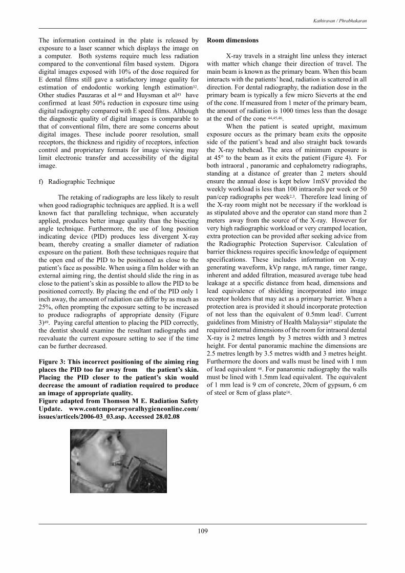
109
Kathiravan / Phrabhakaran
The information contained in the plate is released byexposure to a laser scannerwhichdisplays the imageona computer. Both systems require much less radiationcomparedtotheconventionalfilmbasedsystem.Digoradigitalimagesexposedwith10%ofthedoserequiredforE dental films still gave a satisfactory image quality forestimation of endodontic working length estimation32.OtherstudiesPauzarasetal 40andHuysmanetal43 haveconfirmedatleast50%reductioninexposuretimeusingdigitalradiographycomparedwithEspeedfilms.Althoughthe diagnostic quality of digital images is comparable tothat of conventional film, there are someconcerns aboutdigital images. These include poorer resolution, smallreceptors,thethicknessandrigidityofreceptors,infectioncontrol and proprietary formats for image viewing maylimit electronic transfer and accessibility of the digitalimage.
f) RadiographicTechnique
Theretakingofradiographsarelesslikelytoresultwhengoodradiographictechniquesareapplied.Itisawellknown fact that paralleling technique, when accuratelyapplied, produces better image quality than the bisectingangle technique. Furthermore, the use of long positionindicating device (PID) produces less divergent X-raybeam, thereby creating a smaller diameter of radiationexposureonthepatient.Boththesetechniquesrequirethattheopenendof thePID tobepositionedas close to thepatient’sfaceaspossible.Whenusingafilmholderwithanexternalaimingring,thedentistshouldslidetheringinasclosetothepatient’sskinaspossibletoallowthePIDtobepositionedcorrectly.ByplacingtheendofthePIDonly1inchaway,theamountofradiationcandifferbyasmuchas25%,oftenpromptingtheexposuresettingtobeincreasedto produce radiographs of appropriate density (Figure3)49.PayingcarefulattentiontoplacingthePIDcorrectly,the dentist should examine the resultant radiographs andreevaluate the current exposure setting to see if the timecanbefurtherdecreased.
Figure 3: this incorrect positioning of the aiming ring places the PId too far away from the patient’s skin. Placing the PId closer to the patient’s skin would decrease the amount of radiation required to produce an image of appropriate quality.Figure adapted from thomson M E. radiation Safety update. www.contemporaryoralhygieneonline.com/issues/articels/2006-03_03.asp. Accessed 28.02.08
room dimensions
X-raytravels inastraight lineunless theyinteractwith matter which change their direction of travel. Themainbeamisknownastheprimarybeam.Whenthisbeaminteractswiththepatients’head,radiationisscatteredinalldirection.Fordentalradiography,theradiationdoseintheprimarybeamistypicallyafewmicroSievertsattheendofthecone.Ifmeasuredfrom1meteroftheprimarybeam,theamountofradiationis1000timeslessthanthedosageattheendofthecone44,45,46. When the patient is seated upright, maximumexposure occurs as the primary beam exits the oppositeside of the patient’s head and also straight back towardsthe X-ray tubehead. The area of minimum exposure isat45° to thebeamas itexits thepatient (Figure4). Forbothintraoral ,panoramicandcephalometryradiographs,standing at a distance of greater than 2 meters shouldensuretheannualdoseiskeptbelow1mSVprovidedtheweeklyworkloadislessthan100intraoralsperweekor50pan/cepradiographsperweek2,3.ThereforeleadliningoftheX-rayroommightnotbenecessaryiftheworkloadisasstipulatedaboveandtheoperatorcanstandmorethan2metersawayfromthesourceoftheX-ray.Howeverforveryhighradiographicworkloadorverycrampedlocation,extraprotectioncanbeprovidedafterseekingadvicefromthe Radiographic Protection Supervisor. Calculation ofbarrierthicknessrequiresspecificknowledgeofequipmentspecifications. These includes information on X-raygeneratingwaveform,kVprange,mArange,timerrange,inherentandaddedfiltration,measuredaveragetubeheadleakageataspecificdistancefromhead,dimensionsandlead equivalence of shielding incorporated into imagereceptorholdersthatmayactasaprimarybarrier.Whenaprotectionareaisprovideditshouldincorporateprotectionof not less than the equivalent of 0.5mm lead2. CurrentguidelinesfromMinistryofHealthMalaysia47stipulatetherequiredinternaldimensionsoftheroomforintraoraldentalX-rayis2metreslengthby3metreswidthand3metresheight.Fordentalpanoramicmachinethedimensionsare2.5metreslengthby3.5metreswidthand3metresheight.Furthermorethedoorsandwallsmustbelinedwith1mmofleadequivalent48.Forpanaromicradiographythewallsmustbelinedwith1.5mmleadequivalent.Theequivalentof1mmleadis9cmofconcrete,20cmofgypsum,6cmofsteelor8cmofglassplate16.
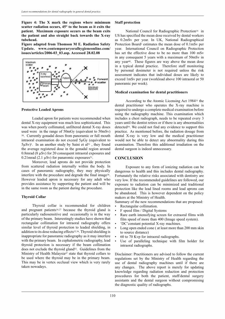
110
Latest recommendations for dental radiography in general dental practice.
Figure 4: the X mark the regions where minimum scatter radiation occurs, 45° to the beam as it exits the patient. Maximum exposure occurs as the beam exits the patient and also straight back towards the X-ray tubehead.Figure adapted from thomson M E. radiation Safety update. www.contemporaryoralhygieneonline.com/issues/articles/2006-03_03.asp. Accessed 28.02.08
Protective Leaded Aprons
LeadedapronforpatientswererecommendedwhendentalX-rayequipmentwasmuchlesssophisticated.Thiswaswhenpoorlycollimated,unfiltereddentalX-raydosesusedwereintherangeof50mGy(equivalentto50mSv)50.Currentlygonadaldosesfrompanoramicorfullmouthintraoral examinationdonotexceed5µGy (equivalent to5µSv)3. InananotherstudybySainietal51 , theyfoundtheaverage registereddose in thegonadal regionaround0.8mrad(8µSv)for20consequentintraoralexposuresand0.21mrad(2.1µSv)forpanaromicexposures51. Moreover, lead aprons do not provide protectionfrom scattered radiation internally within the body. Incases of panoramic radiography, they may physicallyinterferewiththeprocedureanddegradethefinalimage36.However leaded apron is necessary for any adult whoprovidesassistancebysupporting thepatientandwillbeinthesameroomasthepatientduringtheprocedure.
thyroid collar
Thyroid collar is recommended for childrenand pregnant patients16,27 because the thyroid gland isparticularlyradiosensitiveandoccasionallyisinthewayoftheprimarybeam.Interestinglystudieshaveshownthatrectangular collimation for intraoral radiography offerssimilar levelof thyroidprotection to leaded shielding, inadditiontoitsdosereducingeffects32,36.Thyroidshieldingisinappropriateforpanoramicradiographyasitmayinterferewiththeprimarybeam.Incephalometricradiography,leadthyroid protection is necessary if the beam collimationdoesnotexcludethethyroidgland52.GuidelinesfromtheMinistryofHealthMalaysia47statethatthyroidcollarstobe used where the thyroid may be in the primary beam.Thismaybeinvertexocclusalviewwhichisveryrarelytakennowadays.
Staff protection
NationalCouncilforRadiographicProtection16 inUShasspecifiedthemeandosereceivedbydentalworkersas 0.2mSv per year. In UK, National RadiographicalProtectionBoard2estimatesthemeandoseof0.1mSvperyear. International Council on Radiographic Protectionhas set the effective dose to be no more than 100 mSvinanyconsequent5yearswithamaximumof50mSvinany year48. These figures are way above the mean dosein a typical dental practice. Therefore staff monitoringby personal dosimeter is not required unless the riskassessment indicates that individual doses are likely toexceed1mSvperyear(workloadabove100intraoralor50panoramicperweek).
Medical examination for dental practitioners
AccordingtotheAtomicLicensingAct198453 thedental practitioner who operates the X-ray machine isrequiredtoundergoacompletemedicalexaminationbeforeusing the radiography machine. This examination whichincludesachestradiograph,needstoberepeatedevery3yearsuntilthedentistretiresorifthereisanyabnormalitiesdetected54.Wecouldnotfindanyevidencetosupportthispractice.Asmentionedbefore,theradiationdosagefromdental X-ray is very low and the medical practitionerwould not be able to detect any abnormality during thisexamination. Therefore this additional irradiation on thedentalsurgeonisindeedunnecessary.
coNcLuSIoN
Exposuretoanyformofionizingradiationcanbedangerous tohealthand this includesdental radiography.Fortunatelytherelativerisksassociatedwithdentistryareverylow.Iftherecommendedguidelinesarefollowed,ourexposure to radiation can be minimized and traditionalprotection like the lead lined rooms and lead aprons canbeabandoned. This ishoweverdependenton thepolicymakersattheMinistryofHealth.Summaryofthenewrecommendationsthatareproposed:• Rectangularcollimation.• Fspeedfilm/DigitalSystems• Rareearthintensifyingscreenforextraoralfilmswith
filmspeedofmorethan400(Imagespeedsystem).• ‘DC’constantpotentialX-raymachines.• Longopenendedcone(atleastmorethan200mmskin
tosourcedistance)• 60to70Kvpforintraoralradiographs.• Use of paralleling technique with film holder for
intraoralradiographs.
Disclaimer:Practitionersareadvisedtofollowthecurrentregulations set by the Ministry of Health regarding theuse of dental radiography machines until if there areany changes. The above report is merely for updatingknowledge regarding radiation reduction and protectionprocedures for both the patient, staff/dental surgeryassistants and the dental surgeon without compromisingthediagnosticqualityofradiographs.
111
Kathiravan / Phrabhakaran
rEFErENcES
1. Whaites E. Essentials of dental radiography and radiology.4thed.ChurchillLivingstone2006.
2. National Radiological Protection Board. Guidance notesfor dental practitioners on the use of X-ray equipment.DepartmentofHealthLondon.2001.
3. European Commission. European Guidelines on RadiationProtectioninDentalRadiology.Thesafeuseofradiographsin dental practice. Luxemborg 2004. Radiation Protection136.
4. Gijbels F R, Jacobs G, Sanderink E, De Smet B, NowakJ,Van Dam andVan Steenberghe D.A Comparison of theeffectivedosefromscanographywithperiapicalradiography.DentomaxillofacRadiol2002,31:159-63.
5. Dula K R, Mini P F, Van Der STelt and Buser D. Theradiographicassessmentofimplantpatients:decisionmakingcriteria.IntJOralMaxillofacImplants2001.16:80-9
6. MaillieHDandGildaJE.Radiationinducedcancerriskinradiographiccephalometrcy.OralSurgOralMedOralPathol1993.75:631-7
7. ScarfeWC,FarmanAG.Clinicalapplicationofconebeamcomputedtomographyindentalpractice.JCanDentAssoc2006;72(1):75-80
8. OsiboteOA,AzevedoDeACP.EstimationofadultpatientdosesforcommondiagnosticX-rayexaminationsinRioDeJaneiro,Brazil.PhysicaMedica2008;24:21-28
9. NapierID.Referencedosesfordentalradiography.BDentJour1999;186(8):392-396
10.Sasaki M S, EjimaY,TachibanaA,YamadaT, Ishizaki K,Shimizu T, Nomura T. DNA damage response pathway inradioadaptiveresponse.MutatRes2002;504:101-118
11. JohanssonL.Hormesis,anupdateofthepresentposition.EurJNuclMedicineandMolecImagi2003;30(6)921-933
12.NambiarPandLimSC.ThepracticeofdentalradiographyingeneraldentalclinicsintwostatesinMalaysia.MDJ2002.23(2):89-93
13.American Dental Association Report. The use of dentalradiographs.Updateandrecommendations.JAmDentAssoc2006.137:1304-12
14.Faculty of General Dental Practitioners, Royal College ofSurgeons,London.SAMSguidelines1998
15.Rushton V E, Horner K and Worthington H M. Screeningpanaromicradiographyofnewadultpatientsingeneraldentalpractice: diagnostic yield when combined with bitewingradiographyandidentificationofselectioncriteria.BrDentJ2002.192:275-279
16.NationalCouncilforRadiographicProtection&Measurements.Radiation protection in dentistry. Bethesda, Md.: NationalCouncilforRadiationProtection&Measurements;2003
17.Preventing Dental Caries in Children at High Caries Risk.ANationalClinicalguideline.SignPublicationnumber47.www.sign.ac.uk.Accessed20.12.07
18.Isaacson K G and Thom A R. Guidelines for the use ofradiographs in Clinical Orthodontics 2nd Edition. BritishOrthodonticSociety,London.2001
19.LukeLS,LeeP,AtchisonKAandWhiteSC.Orthodonticresidents’ indications for use of the lateral TMJ tomogramandtheposterioranteriorcephalogram.JDentEdu1997.61:29-36
20.TugnaitA,ClevehughVandHirschmannPN.Theusefulnessofradiographsindiagnosisandmanagementofperiodontaldiseases,areview.JDent2000.28:219-226
21.Fava L R D and Dummer P M H. Periapical radiographictechniques during endodontic diagnosis and treatment. IntEndJ1997.30:250-261
22.American DentalAssociation council on scientific matters.An update on radiographic practices. Information andrecommendations.JAmDentAssoc2001.132:234-8
23.BohayRW,StephensRGandKogonSL.Astudyof theimpactofscreeningorselectiveradiographyonthetreatmentandpostdeliveryoutcomeforedentulouspatients.OralSurgOralMedOralPatholOralRadiolEndod1998.86:353-9
24.Rushton V E, Horner K and Worthington. Aspects ofpanaromicradiographyingeneraldentalpractice.BrDentJ1999.186:342-4
25.DentalProtection.Thewholetoothandnothingbutthetooth.DentalNews1999.20:7-8
26.WoodRE,HarrisAMP,VanDerMerweJandNortje`CJ.Theleadedapronrevisited:Doesitreducegonadalradiationdose in dental radiology? Oral Surg Oral Med Oral Pathol1991.71:642-46
27.HujoelPP,BoldenAM,NoonanCJandDelAquilaMA.Antepartumdentalradiographyandinfantlowbirthweight.JAmMedAssoc2004.291(16):1987-93
28.Bernt R L. Commentary on JAMA article by Hujoel et al.HealthPhys2005.88(4):379-381
29.SmithNJ.Selectioncriteriafordentalradiography.BrDentJ1992.Sep5;173(4):120-1
30.EspelidI,MejareIandWeenheijmK.EAPDguidelinesforuseofradiographsinchildren.EurJPedDent2003.1:40-48
31.White S C, Pharoah M J. Oral Radiology Principles andInterpretation.5thed.St.Louis,Mo:Elsevier;2003:161.
32.HornerK.ReviewarticleonRadiationProtectioninDentalRadiology.BrJRadiol1994.67:1041-9
33.White S C, Pharoah M J. Oral Radiology Principles andInterpretation.5thed.St.Louis,Mo:Elsevier;2003:161.
34.Whaites E and Brown J.An update on dental imaging. BrDentJ1998.185(4):166-172
35.CederbergRA,FrederiksonNL,BensonBWandSokolowskiTW.Effectofthegeometryoftheintraoralpositionindicatingdeviceontheeffectivedose.OralSurgOralMedOralPatholOralRadiolEndod1997.84:101-9
36.Hirschmann P N. Guidelines on radiology standards forprimarydentalcare.RoyalCollegeofRadiologistsandTheNationalRadiologicalProtectionBoard.BrDentJ1995.178:165-7
37.Tsiklakis K C, Bakoyanni D, Tassopoulou M andKamenopoulou V. Absorbed radiation dose during lateralcephalometric radiography. Comparison of screen-filmsystemsandfield-sizecombinations.JClinPedDent2000.24:117-21
38.Sevenson B and PeterssonA. Questionnaire survey on theuse of dental X-ray film and equipment among generalpractitioners in the Swedish Public Dental Health Service.ActaOdontScand1997.53:230-5
39.EmmottL.M.Makingsenseofsensors.www.drlarryemmott.com/pdfs/Emmott0302.pdfaccessed18.2.08
40.Paurazas S B, Geist J R, Pink F E, et al. Comparison ofdiagnostic accuracy of digital imaging using CCD andCMOS-APS sensors with E-speed film in the detection ofperiapicalbonylesions.OralSurgOralMedOralPathOralRadiolEndod2000;89(3):356-62
41.Fossum E R. Active pixel sensors: Are CCDs dinosaurs?International Society of Optical Engineering (SPIE) 1993;1900:2-14
112
Latest recommendations for dental radiography in general dental practice.
42.Parks E T, Williamson G F. Digital Radiography: AnOverview.JContempDentPract2002;4(3):23-39
43.HuysmansMC,HintzeHandWenselA.EffectofExposureTimeOnInVitroCariesDiagnosisUsingtheDigoraSystem.EurJOralSci1997.105:15-20
44.DinerMH,MarcouxPandLegaultV.Intraoralradiographictechniquesfortheanesthetizedpatient.JIntAssocDentforChild1990.20:17-21
45.HirschmannPN.Thecurrentstatusofpanaromicradiography.IntDentJ1987.37:31-7
46.ReidJA,MacDonaldJC,DekkerTAandKuppersBU.RadiationexposuresaroundapanaromicdentalX-rayunit.OralSurgOralMedOralPath1993.75:780-2
47.Ministry of Health, Oral Health Division. Guidelines onRadiationinDentistry2006.
48.ICRP 73. Radiological Protection and Safety in Medicine1996.Vol26
49.Thomson M E. Radiation Safety Update. www.contemporaryoralhygieneonline.com/issues/articles/2006-03_03.asp.Accessed28.02.08
50.BudowskyJ,PiroJD,ZegarelliEV,KutscherAHandBarnettA.Radiationexposuretotheheadandabdomenduringoralroentgenography.JAmDentAssoc1956.52:555-559
51.SainiT,ManoharanV,AlAgilIAN.RadiationDosestothemalegonadalarea indental radiography.SaudiDentalJour1993;5(3):127-131
52.BeaconsfieldT,NicholsonR,ThortonAandAl-KutoubiA.WouldthyroidandbreastshieldingbebeneficialinCTofthehead?EurRadiol1998.8:664-7
53.Atomic Energy Licensing Act 1984 (License Regulation)1988
54.MinistryofHealthMalaysia.GuidelinestoobtainingClassCLicenceUndertheAtomicEnergyLicensingAct304,2000
Address for correspondence:
dr. Kathiravan PurmalDept of General Dental Practiceand Oral & Maxillofacial ImagingFaculty of DentistryUniversity of Malaya, Kuala Lumpuremail : [email protected]
Posterior Teeth Mesialization With Mini-implants In An Oligodontia Patient Wey MC. Wey Mang Chek, BDS (Malaya), FDSRCS (England), MOrthRCS (Edin), MOrth (HK), Senior Lecturer in Orthodontics, Faculty of Dentistry, Universiti Malaya
Wu CL. BDS (HK), MOrth (HK), MOrthRCS (Edin), FHKAM (Dental Surgery), FCDSHK (Orthodontics), Private Orthodontic Specialist, Hong Kong
Wong WK. BDS (HK), MOrth (HK), PhD (HK), FRACDS, MOrthRCS (Edin), FHKAM (Dental Surgery), FCDSHK (Orthodontics), Associate Professor in Orthodontics, Faculty of Dentistry, University of Hong Kong
Zamri R. DDS (Indon), MDent Sci (Leeds)), MOrth RCS (Eng), MOrth RCS (Edin), FFDRCSI, Associate Professor in Orthodontics, Faculty of Dentistry, Universiti Malaya
Hägg U. DDS (Lund), Cert Comp Orth (Sweden), Odont Dr (Lund), Hon FDSRCS (Edin), FHKAM (Dental Surgery), FCDSHK (Orthodontics), Chair Professor in Orthodontics, Faculty of Dentistry, University of Hong Kong
SUMMARY
A case report of a 16 year old male oligodontia patient who presented with a Class I malocclusion on a skeletal I base. He had multiple missing teeth of upper lateral incisors and all premolars except for lower right first premolar. Treatment involved fixed appliance with the aid of mini-implants to mesialize posterior teeth in order to reduce the number of prosthodontic replacement of the remaining missing teeth planned for the future. The application of the mini-implants in the sequence of treatment is presented.
Key words orthodontic mini-implant, mesialize posterior teeth, oligodontia
113
Malaysian Dental Journal (2008) 29(2) 113-118 ©2008 The Malaysian Dental Association
InTROdUCTIOn
definition Hypodontia is a developmental absence of only afew teeth, while oligodontia denotes congenital absenceof many teeth1. It is the most common craniofacialmalformation2.Ifsixormorepermanentteetharemissing,thetermoligodontiaisused3.
Prevalence and Etiology Hypodontia involving four or more congenitallymissingpermanentteethexcludingthirdmolarsisrelativelylesscommonthanthatinvolvinglessnumberofteethandreported to have a prevalence of 0.08 – 0.5%4,5,6,7, andthis has been found to be usually associated with somegeneralsystemiccondition.Theprevalenceofhypodontiain the permanent dentition among a Chinese populationhas been reported to be 6.9%, frequently involving thelower incisor, representing 60% of all missing teeth,followed by maxillary second premolars at 10% andmaxillary lateral incisors at 8% 8. The etiology is broadand includes environmental factors such as infection9,
trauma, drugs, chemotherapy or radiotherapy at youngage10, genetic factors11,12 and associated with conditionssuchasectodermaldysplasia,cleftlipandpalate,Down’ssyndromeandhemifacialmicrosomia13.
Features and Presenting Problems Malocclusions related to absence of teeth arecommon, such as rotations, tilting, drifting and spaces.Microdontia is a frequent association, significantly inhypodontia involving six or more congenitally missingteeth14,15. Other dental anomalies are transposition ofpermanent teeth16, impaction of permanent teeth17,18,taurodontism19andankylosis20.Asignificantretroclinationof incisors and an increased interincisal angle werealso observed with increasing severity of hypodontia21.Somestudieshavereportedflatorconcavefacialprofile,obtuse naso-labial angle, retrognathic maxilla, reducedanteriorfaceheightandmandibularplaneangleinseverehypodontiawithabsenceofsixormoreteeth4,22,23.
MALAYSIAn dEnTAL JOURnAL
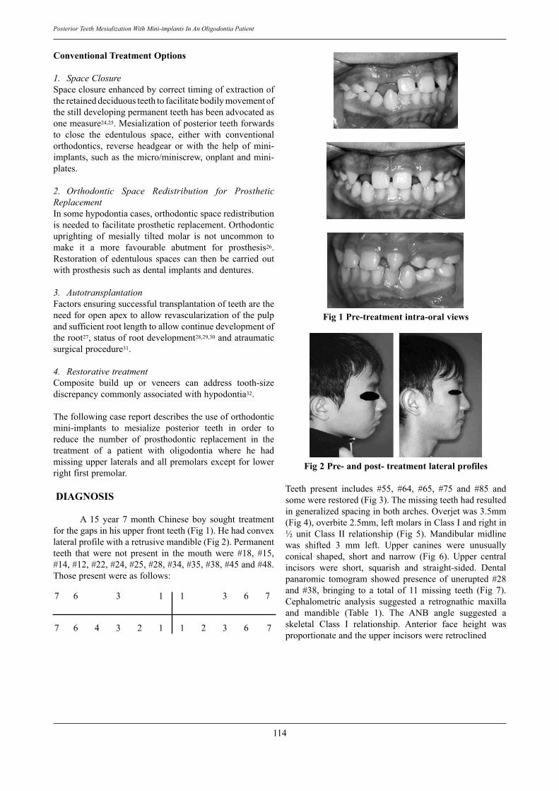
114
Posterior Teeth Mesialization With Mini-implants In An Oligodontia Patient
Conventional Treatment Options
1. Space Closure Spaceclosureenhancedbycorrecttimingofextractionoftheretaineddeciduousteethtofacilitatebodilymovementofthestilldevelopingpermanentteethhasbeenadvocatedasonemeasure24,25.Mesializationofposteriorteethforwardsto close the edentulous space, either with conventionalorthodontics, reverse headgear or with the help of mini-implants,suchasthemicro/miniscrew,onplantandmini-plates.
2. Orthodontic Space Redistribution for Prosthetic ReplacementInsomehypodontiacases,orthodonticspaceredistributionisneededtofacilitateprostheticreplacement.Orthodonticuprighting of mesially tilted molar is not uncommon tomake it a more favourable abutment for prosthesis26.Restorationof edentulous spaces can thenbe carried outwithprosthesissuchasdentalimplantsanddentures.
3. AutotransplantationFactorsensuringsuccessfultransplantationofteetharetheneedforopenapextoallowrevascularizationofthepulpandsufficientrootlengthtoallowcontinuedevelopmentoftheroot27,statusofrootdevelopment28,29,30andatraumaticsurgicalprocedure31.
4. Restorative treatmentComposite build up or veneers can address tooth-sizediscrepancycommonlyassociatedwithhypodontia32.
Thefollowingcasereportdescribestheuseoforthodonticmini-implants to mesialize posterior teeth in order toreduce the number of prosthodontic replacement in thetreatment of a patient with oligodontia where he hadmissingupperlateralsandallpremolarsexceptfor lowerrightfirstpremolar.
dIAGnOSIS
A 15 year 7 month Chinese boy sought treatmentforthegapsinhisupperfrontteeth(Fig1).Hehadconvexlateralprofilewitharetrusivemandible(Fig2).Permanentteeth that were not present in the mouth were #18, #15,#14,#12,#22,#24,#25,#28,#34,#35,#38,#45and#48.Thosepresentwereasfollows:
7 6 3 1 1 3 6 7
7 6 4 3 2 1 1 2 3 6 7
Fig 1 Pre-treatment intra-oral views
Fig 2 Pre- and post- treatment lateral profiles
Teeth present includes #55, #64, #65, #75 and #85 andsomewererestored(Fig3).Themissingteethhadresultedingeneralizedspacinginbotharches.Overjetwas3.5mm(Fig4),overbite2.5mm,leftmolarsinClassIandrightin½ unit Class II relationship (Fig 5). Mandibular midlinewas shifted 3 mm left. Upper canines were unusuallyconical shaped, short and narrow (Fig 6). Upper centralincisors were short, squarish and straight-sided. Dentalpanaromic tomogram showed presence of unerupted #28and #38, bringing to a total of 11 missing teeth (Fig 7).Cephalometric analysis suggested a retrognathic maxillaand mandible (Table 1). The ANB angle suggested askeletal Class I relationship. Anterior face height wasproportionateandtheupperincisorswereretroclined
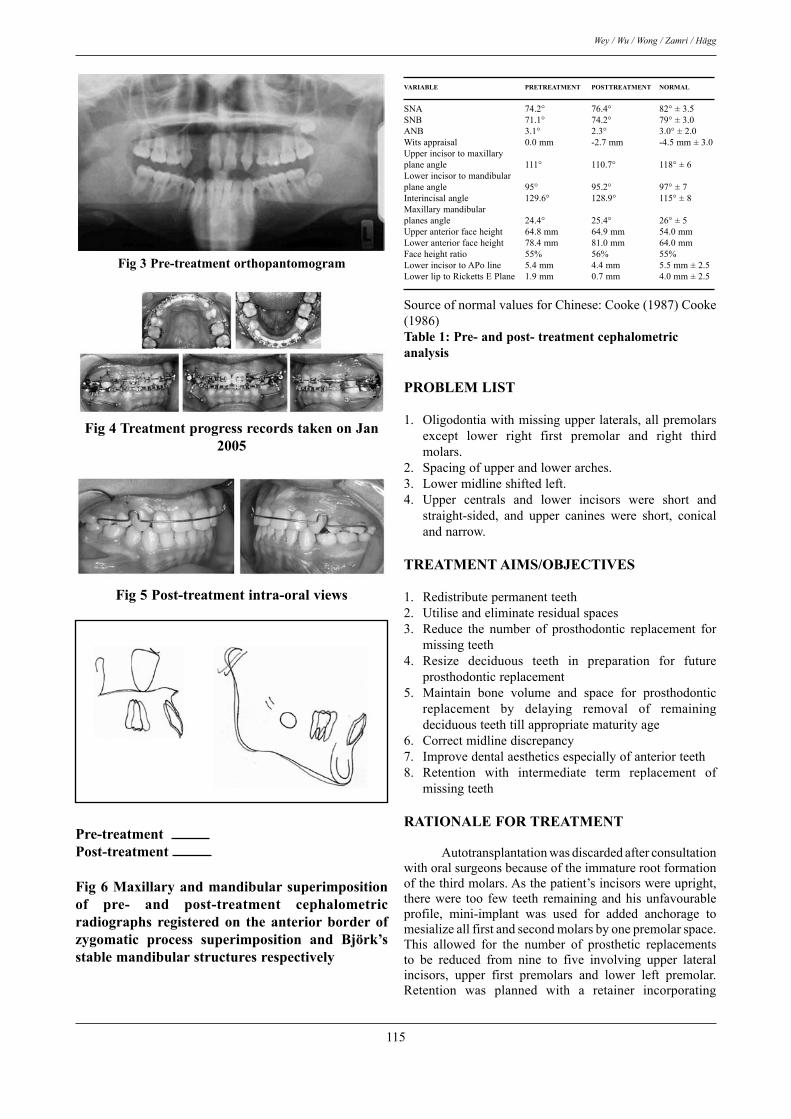
115
Wey / Wu / Wong / Zamri / Hägg
Fig 3 Pre-treatment orthopantomogram
Fig 4 Treatment progress records taken on Jan 2005
Fig 5 Post-treatment intra-oral views
Pre-treatment Post-treatment
Fig 6 Maxillary and mandibular superimposition of pre- and post-treatment cephalometric radiographs registered on the anterior border of zygomatic process superimposition and Björk’s stable mandibular structures respectively
VARIABLE PRETREATMEnT POSTTREATMEnT nORMAL
SNA 74.2° 76.4° 82°±3.5SNB 71.1° 74.2° 79°±3.0ANB 3.1° 2.3° 3.0°±2.0Witsappraisal 0.0mm -2.7mm -4.5mm±3.0Upperincisortomaxillaryplaneangle 111° 110.7° 118°±6Lowerincisortomandibularplaneangle 95° 95.2° 97°±7Interincisalangle 129.6° 128.9° 115°±8Maxillarymandibularplanesangle 24.4° 25.4° 26°±5Upperanteriorfaceheight 64.8mm 64.9mm 54.0mmLoweranteriorfaceheight 78.4mm 81.0mm 64.0mmFaceheightratio 55% 56% 55%LowerincisortoAPoline 5.4mm 4.4mm 5.5mm±2.5LowerliptoRickettsEPlane 1.9mm 0.7mm 4.0mm±2.5
SourceofnormalvaluesforChinese:Cooke(1987)Cooke(1986)Table 1: Pre- and post- treatment cephalometric analysis
PROBLEM LIST
1. Oligodontiawithmissingupperlaterals,allpremolarsexcept lower right first premolar and right thirdmolars.
2. Spacingofupperandlowerarches.3. Lowermidlineshiftedleft.4. Upper centrals and lower incisors were short and
straight-sided, and upper canines were short, conicalandnarrow.
TREATMEnT AIMS/OBJECTIVES
1. Redistributepermanentteeth2. Utiliseandeliminateresidualspaces3. Reduce the number of prosthodontic replacement for
missingteeth4. Resize deciduous teeth in preparation for future
prosthodonticreplacement5. Maintain bone volume and space for prosthodontic
replacement by delaying removal of remainingdeciduousteethtillappropriatematurityage
6. Correctmidlinediscrepancy7. Improvedentalaestheticsespeciallyofanteriorteeth8. Retention with intermediate term replacement of
missingteeth
RATIOnALE FOR TREATMEnT Autotransplantationwasdiscardedafterconsultationwithoralsurgeonsbecauseoftheimmaturerootformationofthethirdmolars.Asthepatient’sincisorswereupright,therewere too few teeth remainingandhisunfavourableprofile, mini-implant was used for added anchorage tomesializeallfirstandsecondmolarsbyonepremolarspace.This allowed for the number of prosthetic replacementsto be reduced from nine to five involving upper lateralincisors, upper first premolars and lower left premolar.Retention was planned with a retainer incorporating
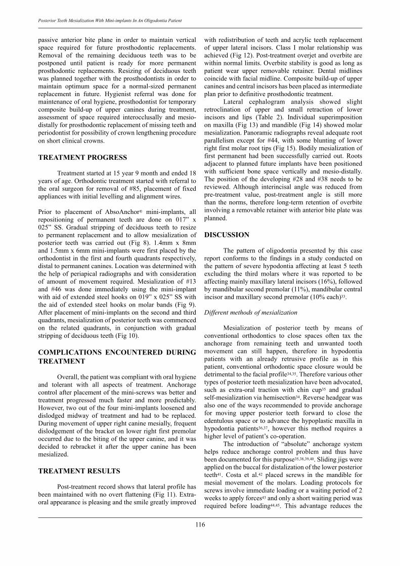
116
Posterior Teeth Mesialization With Mini-implants In An Oligodontia Patient
passive anterior bite plane in order to maintain verticalspace required for future prosthodontic replacements.Removal of the remaining deciduous teeth was to bepostponed until patient is ready for more permanentprosthodontic replacements. Resizing of deciduous teethwasplannedtogetherwiththeprosthodontists inorder tomaintain optimum space for a normal-sized permanentreplacement in future. Hygienist referral was done formaintenanceoforalhygiene,prosthodontistfortemporarycomposite build-up of upper canines during treatment,assessment of space required interocclusally and mesio-distallyforprosthodonticreplacementofmissingteethandperiodontistforpossibilityofcrownlengtheningprocedureonshortclinicalcrowns.
TREATMEnT PROGRESS Treatmentstartedat15year9monthandended18yearsofage.Orthodontictreatmentstartedwithreferraltothe oral surgeon for removal of #85, placement of fixedapplianceswithinitiallevellingandalignmentwires.
Prior to placement of AbsoAnchor® mini-implants, allrepositioning of permanent teeth are done on 017” x025” SS. Gradual stripping of deciduous teeth to resizeto permanent replacement and to allow mesialization ofposterior teeth was carried out (Fig 8). 1.4mm x 8mmand1.5mmx6mmmini-implantswerefirstplacedbytheorthodontistinthefirstandfourthquadrantsrespectively,distaltopermanentcanines.Locationwasdeterminedwiththehelpofperiapical radiographsandwithconsiderationof amount of movement required. Mesialization of #13and #46 was done immediately using the mini-implantwithaidofextendedsteelhookson019”x025”SSwiththe aid of extended steel hooks on molar bands (Fig 9).Afterplacementofmini-implantsonthesecondandthirdquadrants,mesializationofposteriorteethwascommencedon the related quadrants, in conjunction with gradualstrippingofdeciduousteeth(Fig10).COMPLICATIOnS EnCOUnTEREd dURInG TREATMEnT Overall,thepatientwascompliantwithoralhygieneand tolerant with all aspects of treatment. Anchoragecontrolafterplacementofthemini-screwswasbetterandtreatment progressed much faster and more predictably.However,twooutofthefourmini-implantsloosenedanddislodged midway of treatment and had to be replaced.Duringmovementofupperrightcaninemesially,frequentdislodgementof thebracketonlowerrightfirstpremolaroccurredduetothebitingoftheuppercanine,anditwasdecided to rebracket it after the upper canine has beenmesialized.
TREATMEnT RESULTS
Post-treatmentrecordshowsthatlateralprofilehasbeenmaintainedwithnoovert flattening(Fig11).Extra-oralappearanceispleasingandthesmilegreatlyimproved
with redistributionof teethandacrylic teeth replacementof upper lateral incisors. Class I molar relationship wasachieved(Fig12).Post-treatmentoverjetandoverbitearewithinnormallimits.Overbitestabilityisgoodaslongaspatient wear upper removable retainer. Dental midlinescoincidewithfacialmidline.Compositebuild-upofuppercaninesandcentralincisorshasbeenplacedasintermediateplanpriortodefinitiveprosthodontictreatment. Lateral cephalogram analysis showed slightretroclination of upper and small retraction of lowerincisors and lips (Table 2). Individual superimpositiononmaxilla(Fig13)andmandible(Fig14)showedmolarmesialization.Panoramicradiographsrevealadequaterootparallelism except for #44, with some blunting of lowerrightfirstmolarroottips(Fig15).Bodilymesializationoffirst permanent had been successfully carried out. Rootsadjacent toplannedfuture implantshavebeenpositionedwith sufficient bone space vertically and mesio-distally.The position of the developing #28 and #38 needs to bereviewed.Although interincisal angle was reduced frompre-treatment value, post-treatment angle is still morethan thenorms, therefore long-term retentionofoverbiteinvolvingaremovableretainerwithanteriorbiteplatewasplanned.
dISCUSSIOn The pattern of oligodontia presented by this casereport conforms to the findings in a study conducted onthepatternofseverehypodontiaaffectingat least5 teethexcluding the third molars where it was reported to beaffectingmainlymaxillarylateralincisors(16%),followedbymandibularsecondpremolar(11%),mandibularcentralincisorandmaxillarysecondpremolar(10%each)33.
Different methods of mesialization
Mesialization of posterior teeth by means ofconventional orthodontics to close spaces often tax theanchorage from remaining teeth and unwanted toothmovement can still happen, therefore in hypodontiapatients with an already retrusive profile as in thispatient, conventionalorthodontic spaceclosurewouldbedetrimentaltothefacialprofile34,35.Thereforevariousothertypesofposteriorteethmesializationhavebeenadvocated,such as extra-oral traction with chin cup35 and gradualself-mesializationviahemisection34.Reverseheadgearwasalsooneof thewaysrecommendedtoprovideanchoragefor moving upper posterior teeth forward to close theedentulousspaceortoadvancethehypoplasticmaxillainhypodontia patients36,37, however this method requires ahigherlevelofpatient’sco-operation. The introduction of “absolute” anchorage systemhelps reduce anchorage control problem and thus havebeendocumentedforthispurpose35,38,39,40.Slidingjigswereappliedonthebuccalfordistalizationofthelowerposteriorteeth41. Costa et al.42 placed screws in the mandible formesial movement of the molars. Loading protocols forscrewsinvolveimmediateloadingorawaitingperiodof2weekstoapplyforces43andonlyashortwaitingperiodwasrequired before loading44,45. This advantage reduces the

117
Wey / Wu / Wong / Zamri / Hägg
treatmentperiodand,thus,increasespatientacceptability.Success rate
Bernhart et al.46 placed 21 mini-implants on theparamedianregionofthepalateforprotractionofposteriorteeth,amongvariousotherpurposesandpresentedan86%successrateafter11.6monthsofuse.COnCLUSIOnS As a conclusion, mesialization of posterior teethwith mini-implants is a worthwhile treatment optionespeciallyinaseverehypodontiapatientinordertoreducecostofprosthodonticreplacementwithoutovertflatteningofpatient’sprofile.
REFEREnCES
1. ProffitWR,FieldsHW,ContemporaryOrthdoontics3rdedn.Mosby2000:118.
2. Kolenc-Fuse FJ. Tooth agenesis: in search of mutationsbehind faileddentaldevelopment.MedOralPatolOralCirBucal2004;9:390-5;385-90.
3. NunnJH,NECarter,TJGillgrass,etal.Theinterdisciplinarymanagementofhypodontia:backgroundandroleofpaediatricdentistry.BrDentJ2003;194:245-51.
4. SarnasKV,RuneB.Thefacialprofileinadvancedhypodontia:a mixed longitudinal study of 141 children. Eur J Orthod,1983;5:133-43.
5. PolderBJ,Van’tHofMA,VanderLindenFP,etal.Ameta-analysis of the prevalence of dental agenesis of permanentteeth.CommunityDentOralEpidemiol2004;32:217-26.
6. SchalkvanderWeideY.Oligodontia:aclinical,radiographicandgeneticevaluation.1992,UniversityofUtrecht:Utrecht.
7. RollingS,PoulsenS.OligodontiainDanishschoolchildren.ActaOdontolScand2001;59:111–112.
8. DavisPJ.HypodontiaandhyperdontiaofpermanentteethinHongKongschoolchildren.CommunityDentOralEpidemiol1987;15:218-20.
9. GulliksonJS.Toothmorphologyinrubellasyndromechildren.ASDCJDentChild1975;42:479-82.
10.daFonsecaMA.Pediatricbonemarrowtransplantation:oralcomplications and recommendations for care. Pediatr Dent1998;20:386-94.
11.VastardisH,KarimbuxN,GuthuaSW,etal.AhumanMSX1homeodomain missense mutation causes selective toothagenesis.NatGenet,1996;13:417-21.
12.StocktonDW,DasP,GoldenbergM,etal.MutationofPAX9isassociatedwitholigodontia.NatGenet.2000;24:18-9.
13.Silverman NE, Ackerman JL. Oligodontia: a study of itsprevalence and variation in 4032 children. ASDC J DentChild1979;46:470-7.
14.McKeown H.F, Robinson DL, Elcock C, et al. Toothdimensionsinhypodontiapatients,theirunaffectedrelativesandacontrolgroupmeasuredbyanewimageanalysissystem.EurJOrthod2002;24:131-41.
15.RuneB,SarnasKV.Toothsizeandtoothformationinchildrenwithadvancedhypodontia.AngleOrthod1974;44:316-21.
16.PeckS,PeckL,KatajaM.Site-specificityoftoothagenesisinsubjectswithmaxillarycaninemalpositions.AngleOrthod1996;66:473-6.
17.Pirinen, S, Arte S, Apajalahti S. Palatal displacement of
canineisgeneticandrelatedtocongenitalabsenceofteeth.JDentRes1996;75:1742-6.
18.Garn SM, Lewis AB. The gradient and the pattern ofcrown-size reduction in simple hypodontia. Angle Orthod1970;40:51-8.
19.Stenvik,A.,B.U.Zachrisson,B.Svatun,Taurodontismandconcomitanthypodontiainsiblings.OralSurgOralMedOralPathol,1972;33:841-5.
20.LaiPY,SeowWK.Acontrolledstudyof theassociationofvariousdentalanomalieswithhypodontiaofpermanentteeth.PediatrDent1989;11:291-6.
21.OgaardB,KrogstadO.Craniofacialstructureandsofttissueprofile in patients with severe hypodontia. Am J OrthodDentofacialOrthop1995;108:472-7.
22.BondaretsN,McDonaldF.Analysisoftheverticalfacialformin patients with severe hypodontia. Am J Phys Anthropol2000;111:177-84.
23.WisthPJ,ThunoldK,BoeOE.Thecraniofacialmorphologyof individuals with hypodontia. Acta Odontol Scand1974;32:293-302.
24. Lindqvist B. Extraction of the deciduous second molar inhypodontia.EurJOrthod,1980;2:173-81.
25.Mamopoulou A, Hägg U, Schroder U, et al. Agenesis ofmandibular second premolars. Spontaneous space closureafter extraction therapy: a 4-year follow-up. Eur J Orthod1996;18:589-600.
26.TullochCJF,AjunctiveTreatmentforadults,inContemporaryOrthodontics.Mosby:St.Louis2000:626-636.
27.BlakeyGH,WhiteR,Surgicaltreatment:Mandibularsurgery,in Contemporary Treatment of Dentofacial Deformity.Mosby:St.Louis2003:312-344.
28.Josefsson E, Brattstrom V, Tegsjo U, et al. Treatment oflower second premolar agenesis by autotransplantation:four-yearevaluationofeightypatients.ActaOdontolScand1999;57:111-5.
29.Lundberg, Isaksson TS. A clinical follow-up study of278 autotransplanted teeth. Br J Oral Maxillofac Surg1996;34:181-5.
30.Kristerson L. Autotransplantation of human premolars. Aclinicalandradiographicstudyof100teeth.IntJOralSurg1985;14:200-13.
31.Kristerson L, Andreasen JO. Autotransplantation andreplantationoftoothgermsinmonkeys.Effectofdamagetothedentalfollicleandpositionoftransplantinthealveolus.IntJOralSurg1984;13:324-33.
32.BoltonW.Theclinicalapplicationoftooth-sizeanalysis.AmJOrthod1962:504-529.
33.WongTY,McGrathC,McMillanAS.Oralhealthofsouthern
Chinesechildrenandadolescentswithseverehypodontia.IntJPediatrDent2005;15(4):256–263).
34.NorthwayWM.Thenutsandboltsofhemisectiontreatment:managingcongenitallymissingmandibularsecondpremolars.AmJOrthodDentofacialOrthop2005;127(5):606-10.
35.Kokich VG. Kokich VO. Congenitally missing mandibularsecondpremolars:clinicaloptions.AmJOrthodDentofacialOrthop2006;130(4):437-44.
36.GoodmanJ.Hypodontia:1.Dentalupdate1994:381-384.37.MillerJR.AdentalClassIIImalocclusiontreatedtoafull-
cusp Class II molar relationship.Am J Orthod DentofacialOrthop1990;97:10-9.
38.Block MS, Hoffman DR. A new device for absoluteanchoragefororthodontics.AmJOrthodDentofacialOrthop1995;107:251-8.
39.Sugawara J, Daimaruya T, Umemori M, et al. Distal
118
Posterior Teeth Mesialization With Mini-implants In An Oligodontia Patient
movement of mandibular molars in adult patients with theskeletalanchoragesystem.AmJOrthodDentofacialOrthop2004;125:130-8.
40.Umemori,SugawaraMJ,MitaniH,etal.Skeletalanchoragesystem for open-bite correction. Am J Orthod DentofacialOrthop1999;115:166-74.
41.ChungK,KimS,KookY.C-OrthodonticMicroimplantforDistalizationofMandibularDentitioninClassIIICorrection.AngleOrthod2005;75:119–128.
42.CostaA,RaffainlM,MelsenB.Miniscrewsasorthodonticanchorage: a preliminary report. Int J Adult OrthodonOrthognathSurg1998;13:201–209.
43.Ohashi E, Pecho OE, Moron M, Manuel O, Lagraverec.Implant vs Screw Loading Protocols in Orthodontics ASystematic Review. The Angle Orthodontist 2006;76:721–727.
44.ChengSJ,TsengIY,LeeJJ,KokSH.Aprospectivestudyoftheriskfactorsassociatedwithfailureofmini-implantsusedfor orthodontic anchorage. Int J Oral Maxillofac Implants2004;19:100–106.
45.LiouEJ,PaiBC,Lin JC.Dominiscrews remainstationaryunderorthodonticforces?.AmJOrthodDentofacialOrthop2004;126:42–47.
46.Bernhart T, Freudenthaler J, Dortbudak O, Bantleon HP,
WatzekG.Shortepitheticimplantsfororthodonticanchorageintheparamedianregionofthepalate.Aclinicalstudy.ClinOralImplantsRes2001;12:624–631.
Address for correspondence:
dr. Wey Mang ChekDepartment of Children’s Dentistry and Orthodontics,Faculty of Dentistry,Universiti Malaya,50603 Kuala Lumpur.Tel: 03-79674540, 017-6355746Fax: 03-79674530E-mail address: [email protected]
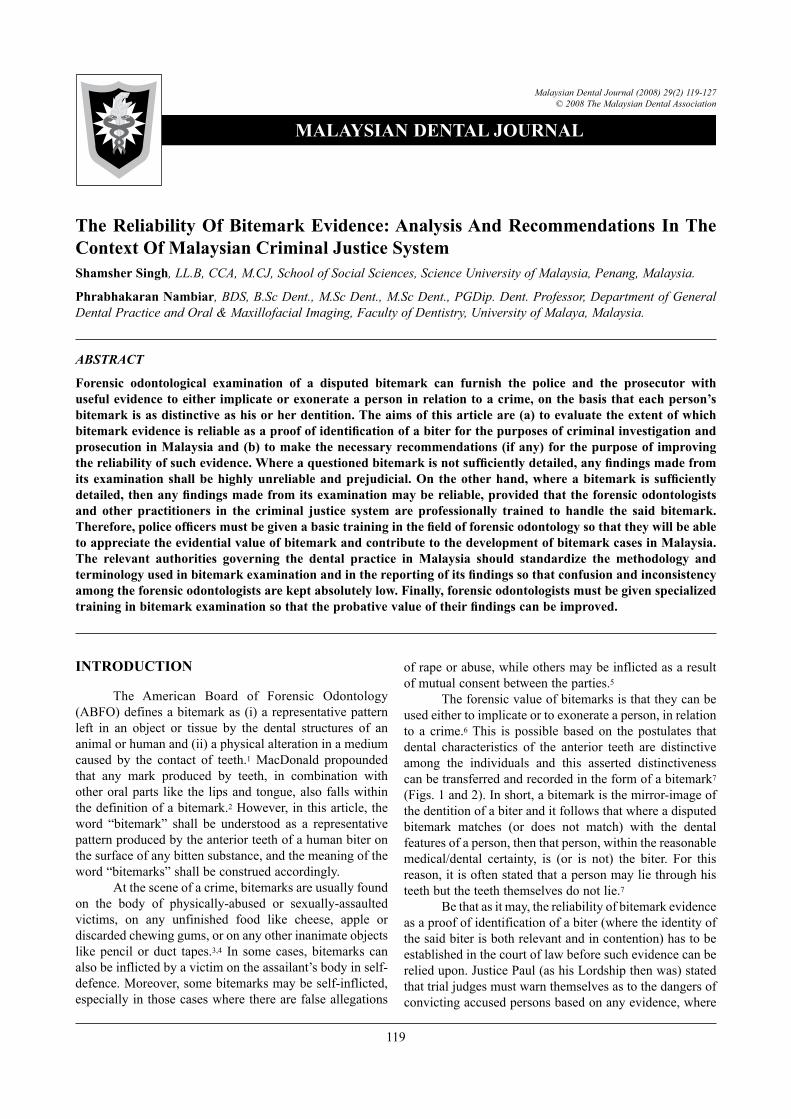
The Reliability Of Bitemark Evidence: Analysis And Recommendations In The Context Of Malaysian Criminal Justice System Shamsher Singh, LL.B, CCA, M.CJ, School of Social Sciences, Science University of Malaysia, Penang, Malaysia.
Phrabhakaran Nambiar, BDS, B.Sc Dent., M.Sc Dent., M.Sc Dent., PGDip. Dent. Professor, Department of General Dental Practice and Oral & Maxillofacial Imaging, Faculty of Dentistry, University of Malaya, Malaysia.
ABSTRACT
Forensic odontological examination of a disputed bitemark can furnish the police and the prosecutor with useful evidence to either implicate or exonerate a person in relation to a crime, on the basis that each person’s bitemark is as distinctive as his or her dentition. The aims of this article are (a) to evaluate the extent of which bitemark evidence is reliable as a proof of identification of a biter for the purposes of criminal investigation and prosecution in Malaysia and (b) to make the necessary recommendations (if any) for the purpose of improving the reliability of such evidence. Where a questioned bitemark is not sufficiently detailed, any findings made from its examination shall be highly unreliable and prejudicial. On the other hand, where a bitemark is sufficiently detailed, then any findings made from its examination may be reliable, provided that the forensic odontologists and other practitioners in the criminal justice system are professionally trained to handle the said bitemark. Therefore, police officers must be given a basic training in the field of forensic odontology so that they will be able to appreciate the evidential value of bitemark and contribute to the development of bitemark cases in Malaysia. The relevant authorities governing the dental practice in Malaysia should standardize the methodology and terminology used in bitemark examination and in the reporting of its findings so that confusion and inconsistency among the forensic odontologists are kept absolutely low. Finally, forensic odontologists must be given specialized training in bitemark examination so that the probative value of their findings can be improved.
119
Malaysian Dental Journal (2008) 29(2) 119-127 © 2008 The Malaysian Dental Association
INTROduCTION
The American Board of Forensic Odontology(ABFO)definesabitemarkas(i)arepresentativepatternleft in an object or tissue by the dental structures of ananimalorhumanand(ii)aphysicalalterationinamediumcaused by the contact of teeth.1 MacDonald propoundedthat any mark produced by teeth, in combination withotheroralparts like the lipsand tongue,alsofallswithinthedefinitionofabitemark.2However,inthisarticle,theword “bitemark” shall be understood as a representativepatternproducedbytheanteriorteethofahumanbiteronthesurfaceofanybittensubstance,andthemeaningoftheword“bitemarks”shallbeconstruedaccordingly. Atthesceneofacrime,bitemarksareusuallyfoundon the body of physically-abused or sexually-assaultedvictims, on any unfinished food like cheese, apple ordiscardedchewinggums,oronanyotherinanimateobjectslikepencilorduct tapes.3,4 In somecases,bitemarkscanalsobeinflictedbyavictimontheassailant’sbodyinself-defence.Moreover,somebitemarksmaybeself-inflicted,especiallyinthosecaseswheretherearefalseallegations
ofrapeorabuse,whileothersmaybeinflictedasaresultofmutualconsentbetweentheparties.5 Theforensicvalueofbitemarksisthattheycanbeusedeithertoimplicateortoexonerateaperson,inrelationto a crime.6This is possible basedon thepostulates thatdental characteristics of the anterior teeth are distinctiveamong the individuals and this asserted distinctivenesscanbetransferredandrecordedintheformofabitemark7(Figs.1and2).Inshort,abitemarkisthemirror-imageofthedentitionofabiteranditfollowsthatwhereadisputedbitemark matches (or does not match) with the dentalfeaturesofaperson,thenthatperson,withinthereasonablemedical/dental certainty, is (or is not) the biter. For thisreason,itisoftenstatedthatapersonmayliethroughhisteethbuttheteeththemselvesdonotlie.7
Bethatasitmay,thereliabilityofbitemarkevidenceasaproofofidentificationofabiter(wheretheidentityofthesaidbiterisbothrelevantandincontention)hastobeestablishedinthecourtoflawbeforesuchevidencecanbereliedupon.JusticePaul(ashisLordshipthenwas)statedthattrialjudgesmustwarnthemselvesastothedangersofconvictingaccusedpersonsbasedonanyevidence,where
MALAYSIAN dENTAL JOuRNAL
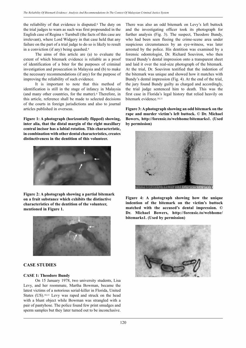
120
The Reliability Of Bitemark Evidence: Analysis And Recommendations In The Context Of Malaysian Criminal Justice System
the reliability of that evidence is disputed.8 The duty onthetrialjudgestowarnassuchwasfirstpropoundedintheEnglishcaseofReginavTurnbull(thefactsofthiscaseareirrelevant),whereLordWidgeryinthatcaseheldthatanyfailureonthepartofatrialjudgetodosoislikelytoresultinaconviction(ifany)beingquashed.9 The aims of this article are (a) to evaluate theextent of which bitemark evidence is reliable as a proofof identification of a biter for the purposes of criminalinvestigationandprosecutioninMalaysiaand(b)tomakethenecessaryrecommendations(ifany)forthepurposeofimprovingthereliabilityofsuchevidence. It is important to note that this method ofidentification is still in the stage of infancy in Malaysia(andmanyothercountries, for thematter).4Therefore, inthisarticle, referenceshallbemade to selecteddecisionsof the courts in foreign jurisdictions and also to journalarticlespublishedinoverseas.
Figure 1: A photograph (horizontally flipped) showing, inter alia, that the distal margin of the right maxillary central incisor has a labial rotation. This characteristic, in combination with other dental characteristics, creates distinctiveness in the dentition of this volunteer.
Figure 2: A photograph showing a partial bitemark on a fruit substance which exhibits the distinctive characteristics of the dentition of the volunteer, mentioned in Figure 1.
CASE STudIES
CASE 1: Theodore Bundy On15January1978,twouniversitystudents,LisaLevy, and her roommate, Martha Bowman, became thelatestvictimsofanotoriousserial-killerinFlorida,UnitedStates (US).10,11 Levy was raped and struck on the headwith a blunt object while Bowman was strangled with apairofpantyhose.Thepolicefoundfewprintsmudgesandspermsamplesbuttheylaterturnedouttobeinconclusive.
There was also an odd bitemark on Levy’s left buttockand the investigating officer took its photograph forfurther analysis (Fig. 3). The suspect, Theodore Bundy,who had been seen fleeing the crime-scene area undersuspicious circumstances by an eye-witness, was laterarrested by the police. His dentition was examined by aforensic odontologist, Dr. Richard Souviron, who thentracedBundy’sdentalimpressionontoatransparentsheetand laid itover the real-sizephotographof thebitemark.At the trial, Dr. Souviron testified that the indention ofthebitemarkwasuniqueandshowedhowitmatcheswithBundy’sdentalimpression(Fig.4).Attheendofthetrial,the jury foundBundyguilty as charged and accordingly,the trial judge sentenced him to death. This was thefirst case in Florida’s legal history that relied heavily onbitemarkevidence.10,11
Figure 3: A photograph showing an odd bitemark on the rape and murder victim’s left buttock. © dr. Michael Bowers, http://forensic.to/webhome/bitemarks1. (used by permission)
Figure 4: A photograph showing how the unique indention of the bitemark on the victim’s buttock matched with the accused’s dental impression. © dr. Michael Bowers, http://forensic.to/webhome/bitemarks1. (used by permission)
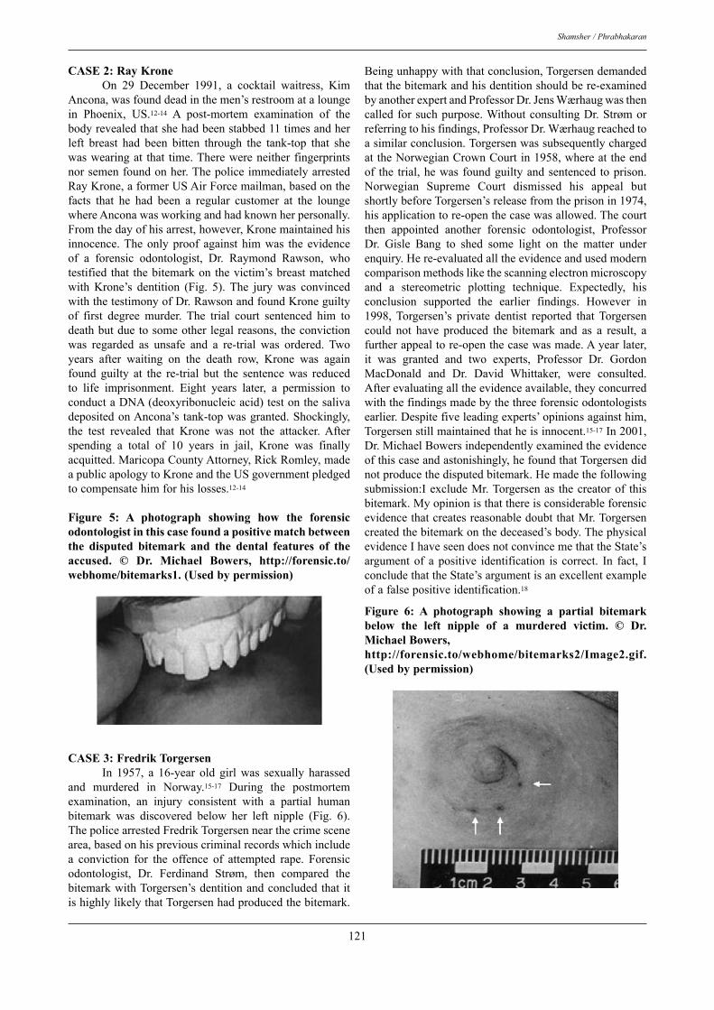
121
Shamsher / Phrabhakaran
CASE 2: Ray Krone On 29 December 1991, a cocktail waitress, KimAncona,wasfounddeadinthemen’srestroomataloungein Phoenix, US.12-14 A post-mortem examination of thebodyrevealedthatshehadbeenstabbed11timesandherleft breast had been bitten through the tank-top that shewaswearingat that time.Therewereneitherfingerprintsnorsemenfoundonher.ThepoliceimmediatelyarrestedRayKrone,aformerUSAirForcemailman,basedonthefacts that he had been a regular customer at the loungewhereAnconawasworkingandhadknownherpersonally.Fromthedayofhisarrest,however,Kronemaintainedhisinnocence.Theonlyproof against himwas the evidenceof a forensic odontologist, Dr. Raymond Rawson, whotestified that thebitemarkon thevictim’sbreastmatchedwith Krone’s dentition (Fig. 5). The jury was convincedwiththetestimonyofDr.RawsonandfoundKroneguiltyof first degree murder. The trial court sentenced him todeathbutduetosomeotherlegalreasons, theconvictionwas regarded as unsafe and a re-trial was ordered. Twoyears after waiting on the death row, Krone was againfound guilty at the re-trial but the sentence was reducedto life imprisonment. Eight years later, a permission toconductaDNA(deoxyribonucleicacid)testonthesalivadepositedonAncona’s tank-topwasgranted.Shockingly,the test revealed that Krone was not the attacker. Afterspending a total of 10 years in jail, Krone was finallyacquitted.MaricopaCountyAttorney,RickRomley,madeapublicapologytoKroneandtheUSgovernmentpledgedtocompensatehimforhislosses.12-14
Figure 5: A photograph showing how the forensic odontologist in this case found a positive match between the disputed bitemark and the dental features of the accused. © dr. Michael Bowers, http://forensic.to/webhome/bitemarks1. (used by permission)
CASE 3: Fredrik Torgersen In 1957, a 16-year old girl was sexually harassedand murdered in Norway.15-17 During the postmortemexamination, an injury consistent with a partial humanbitemark was discovered below her left nipple (Fig. 6).ThepolicearrestedFredrikTorgersennearthecrimescenearea,basedonhispreviouscriminalrecordswhichincludea conviction for the offence of attempted rape. Forensicodontologist, Dr. Ferdinand Strøm, then compared thebitemarkwithTorgersen’sdentitionandconcludedthat itishighlylikelythatTorgersenhadproducedthebitemark.
Beingunhappywiththatconclusion,Torgersendemandedthatthebitemarkandhisdentitionshouldbere-examinedbyanotherexpertandProfessorDr.JensWærhaugwasthencalledforsuchpurpose.WithoutconsultingDr.Strømorreferringtohisfindings,ProfessorDr.Wærhaugreachedtoasimilarconclusion.TorgersenwassubsequentlychargedattheNorwegianCrownCourtin1958,whereattheendof the trial,hewasfoundguiltyandsentenced toprison.Norwegian Supreme Court dismissed his appeal butshortlybeforeTorgersen’sreleasefromtheprisonin1974,hisapplicationtore-openthecasewasallowed.Thecourtthen appointed another forensic odontologist, ProfessorDr. Gisle Bang to shed some light on the matter underenquiry.Here-evaluatedalltheevidenceandusedmoderncomparisonmethodslikethescanningelectronmicroscopyand a stereometric plotting technique. Expectedly, hisconclusion supported the earlier findings. However in1998, Torgersen’s private dentist reported that Torgersencould not have produced the bitemark and as a result, afurtherappealtore-openthecasewasmade.Ayearlater,it was granted and two experts, Professor Dr. GordonMacDonald and Dr. David Whittaker, were consulted.Afterevaluatingalltheevidenceavailable,theyconcurredwiththefindingsmadebythethreeforensicodontologistsearlier.Despitefiveleadingexperts’opinionsagainsthim,Torgersenstillmaintainedthatheisinnocent.15-17In2001,Dr.MichaelBowersindependentlyexaminedtheevidenceofthiscaseandastonishingly,hefoundthatTorgersendidnotproducethedisputedbitemark.Hemadethefollowingsubmission:IexcludeMr.Torgersenas thecreatorof thisbitemark.MyopinionisthatthereisconsiderableforensicevidencethatcreatesreasonabledoubtthatMr.Torgersencreatedthebitemarkonthedeceased’sbody.ThephysicalevidenceIhaveseendoesnotconvincemethattheState’sargumentof a positive identification is correct. In fact, IconcludethattheState’sargumentisanexcellentexampleofafalsepositiveidentification.18
Figure 6: A photograph showing a partial bitemark below the left nipple of a murdered victim. © dr. Michael Bowers, http://forensic.to/webhome/bitemarks2/Image2.gif. (used by permission)
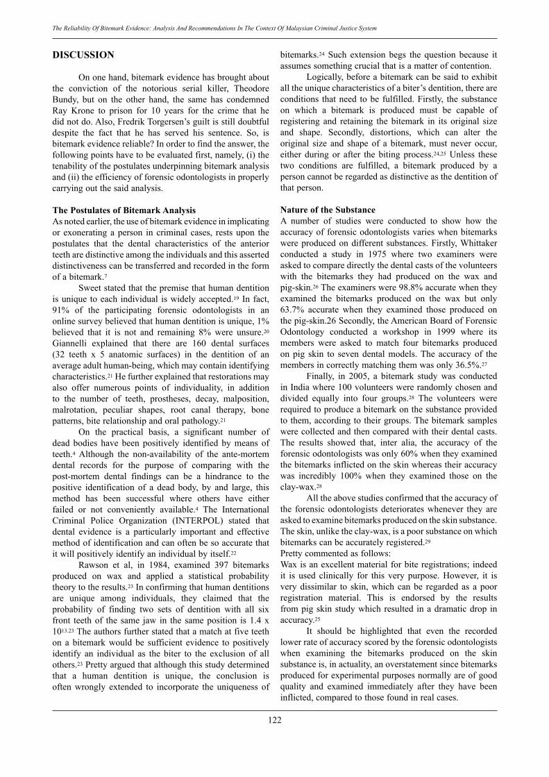
122
The Reliability Of Bitemark Evidence: Analysis And Recommendations In The Context Of Malaysian Criminal Justice System
dISCuSSION
Ononehand,bitemarkevidencehasbroughtaboutthe conviction of the notorious serial killer, TheodoreBundy, but on the other hand, the same has condemnedRay Krone to prison for 10 years for the crime that hedidnotdo.Also,FredrikTorgersen’sguiltisstilldoubtfuldespite the fact that he has served his sentence. So, isbitemarkevidencereliable?Inordertofindtheanswer,thefollowingpointshavetobeevaluatedfirst,namely,(i)thetenabilityofthepostulatesunderpinningbitemarkanalysisand(ii)theefficiencyofforensicodontologistsinproperlycarryingoutthesaidanalysis.
The Postulates of Bitemark Analysis Asnotedearlier,theuseofbitemarkevidenceinimplicatingor exoneratingaperson in criminal cases, restsupon thepostulates that the dental characteristics of the anteriorteetharedistinctiveamongtheindividualsandthisasserteddistinctivenesscanbetransferredandrecordedintheformofabitemark.7 Sweetstatedthatthepremisethathumandentitionisunique toeach individual iswidelyaccepted.19 Infact,91% of the participating forensic odontologists in anonlinesurveybelievedthathumandentitionisunique,1%believed that it is not and remaining 8% were unsure.20Giannelli explained that there are 160 dental surfaces(32 teeth x 5 anatomic surfaces) in the dentition of anaverageadulthuman-being,whichmaycontainidentifyingcharacteristics.21Hefurtherexplainedthatrestorationsmayalso offer numerous points of individuality, in additionto the number of teeth, prostheses, decay, malposition,malrotation, peculiar shapes, root canal therapy, bonepatterns,biterelationshipandoralpathology.21 On the practical basis, a significant number ofdead bodies have been positively identified by means ofteeth.4Although the non-availability of the ante-mortemdental records for the purpose of comparing with thepost-mortem dental findings can be a hindrance to thepositive identification of a dead body, by and large, thismethod has been successful where others have eitherfailed or not conveniently available.4 The InternationalCriminal Police Organization (INTERPOL) stated thatdental evidence is a particularly important and effectivemethodofidentificationandcanoftenbesoaccuratethatitwillpositivelyidentifyanindividualbyitself.22 Rawson et al, in 1984, examined 397 bitemarksproduced on wax and applied a statistical probabilitytheorytotheresults.23Inconfirmingthathumandentitionsare unique among individuals, they claimed that theprobability of finding two sets of dentition with all sixfront teethof the same jaw in the sameposition is1.4x1013.23Theauthorsfurtherstatedthatamatchatfiveteethon a bitemark would be sufficient evidence to positivelyidentify an individual as the biter to the exclusion of allothers.23Prettyarguedthatalthoughthisstudydeterminedthat a human dentition is unique, the conclusion isoften wrongly extended to incorporate the uniqueness of
bitemarks.24 Such extension begs the question because itassumessomethingcrucialthatisamatterofcontention. Logically,beforeabitemarkcanbesaidtoexhibitalltheuniquecharacteristicsofabiter’sdentition,thereareconditions thatneed tobe fulfilled.Firstly, thesubstanceon which a bitemark is produced must be capable ofregistering and retaining the bitemark in its original sizeand shape. Secondly, distortions, which can alter theoriginal size and shape of a bitemark, must never occur,either during or after the biting process.24,25 Unless thesetwo conditions are fulfilled, a bitemark produced by apersoncannotberegardedasdistinctiveasthedentitionofthatperson.
Nature of the SubstanceA number of studies were conducted to show how theaccuracyofforensicodontologistsvarieswhenbitemarkswereproducedondifferentsubstances.Firstly,Whittakerconducted a study in 1975 where two examiners wereaskedtocomparedirectlythedentalcastsofthevolunteerswith the bitemarks they had produced on the wax andpig-skin.26Theexaminerswere98.8%accuratewhentheyexamined the bitemarks produced on the wax but only63.7% accurate when they examined those produced onthepig-skin.26Secondly,theAmericanBoardofForensicOdontology conducted a workshop in 1999 where itsmembers were asked to match four bitemarks producedon pig skin to seven dental models.The accuracy of themembersincorrectlymatchingthemwasonly36.5%.27 Finally, in 2005, a bitemark study was conductedinIndiawhere100volunteerswererandomlychosenanddivided equally into four groups.28 The volunteers wererequiredtoproduceabitemarkonthesubstanceprovidedtothem,accordingtotheirgroups.Thebitemarksampleswerecollectedandthencomparedwiththeirdentalcasts.The results showed that, inter alia, the accuracy of theforensicodontologistswasonly60%whentheyexaminedthebitemarksinflictedontheskinwhereastheiraccuracywas incredibly 100% when they examined those on theclay-wax.28 Alltheabovestudiesconfirmedthattheaccuracyofthe forensicodontologistsdeteriorateswhenever theyareaskedtoexaminebitemarksproducedontheskinsubstance.Theskin,unliketheclay-wax,isapoorsubstanceonwhichbitemarkscanbeaccuratelyregistered.29Prettycommentedasfollows:Waxisanexcellentmaterialforbiteregistrations;indeedit is used clinically for this very purpose. However, it isverydissimilar to skin,whichcanbe regardedas apoorregistration material. This is endorsed by the resultsfrompigskinstudywhichresulted inadramaticdrop inaccuracy.25
It should be highlighted that even the recordedlowerrateofaccuracyscoredbytheforensicodontologistswhen examining the bitemarks produced on the skinsubstanceis,inactuality,anoverstatementsincebitemarksproducedforexperimentalpurposesnormallyareofgoodquality and examined immediately after they have beeninflicted,comparedtothosefoundinrealcases.
123
Shamsher / Phrabhakaran
Occurrence of distortions There are twokindsofdistortion,namely, (i) thatoccursduring thebitingprocess (primarydistortion) and(ii)thatoccursafterthat(secondarydistortion).30 Due to the fact that bitemarks in criminal casesmostlyaretheby-productsofviolentacts,especiallywhentheyare inflictedonhumanskin,primarydistortionmayoccur.Theskin ishighlyvariable in termsofanatomicallocation, underlying musculature, fat, curvature andloosenessoradherencetounderlyingtissues31andfurtherit is also highly visco-elastic, which allows stretching tooccurduringthebitingprocess.30Primarydistortionsmayalsooccurforotherreasons, liketheareabitten, thewaythebitingwasdone, the forceusedand thehealthof thevictim.Forthatreason,itispossiblethatasamebitermayleavemarksofdifferingappearanceonthesamevictim.30
Secondary distortions may occur naturally, forexample,duetotheself-healingprocessinalivingpersonand postmortem changes in a dead one, or unnaturally,when a victim seeks medical treatment.30 In 1974,Bioengineering Unit of the University of Strathclydeconducted a research to examine the features of a bitingprocess that are likely to impact upon the appearance ofbitemarksonhumanskin.Theauthorsconcludedthatthechangesinbitemarkappearancearelikelytobegreaterastheinjurygrowsolder.29Whittakerfoundinhisstudythattheaccuracyoftheforensicodontologistshaddroppedtoamere16%when they compared thedental casts of theputative biters with the photographs of the bitemarks onthe pig skin, which were taken after a 24-hour period.26Secondarydistortionsalsooccurduetothechangesintheposition of a body while examining a bitemark 32 (thusa round bitemark may become oval) and wrong cameraangulationsorlightingwhiletakingthephotographs.33.ConclusionEven if it is assumed that each man has a distinctivedentition, it cannot be so assumed that it will producean equally distinctive bitemark, especially when it ismade on skin. The reason is that skin, unlike clay-wax,is not a substance which is capable of registering andretainingbitemarkinitsoriginalsizeandshape.Moreover,distortionstoabitemarkmayoccurduringorafterthebitingprocess.Sincetherearemanyotherfactorsthatcanshapethecharacteristicsofabitemarkandnotjustthedentitionofabiter,abitemarkproducedonhumanskinmaynotbeasdistinctiveas thedentitionof thebiter.WilkinsonandGerughtyhaveputitrathercrudelyasfollows:There is no documented scientific data to support thehypothesisthatalatentbitemark,likealatentfingerprint,is a true and accurate reflection of this uniqueness. Tothe contrary, what little scientific evidence that doesexist clearly supports the conclusion that crime-relatedbitemarksaregrosslydistorted, inaccurateand thereforeunreliableasamethodofidentification.34
The Efficiency of Forensic OdontologistsDetailed, step-by-step procedures that are involved in a
bitemarkexaminationfalloutsidethelimitedscopeofthisarticle. It is sufficient tomention thatordinarily, thesaidexaminationinvolvesthreestages,namely,(i)registrationof a disputed bitemark and a putative biter’s dentition,(ii) comparison of the bitemark and the dentition, and(iii)evaluationof thepointsofsimilarityordissimilaritybetweenthem.19Thecontentiousissueisthatalthoughtheexperts based their conclusions on objective data, theiropinionsareessentiallysubjectiveones.35,36 Giannelliwrotethatthereisnoacceptedminimum(or maximum) number of points of similarity requiredfor a positive identification and the experts who havetestified in bitemark cases in US have used as low aseight points to as high as 52 points.21,37-39 Like firearmsidentification, bitemark identification is based on theexaminer’s experience and expertise.21 In Malaysia, therelevancy of scientific evidence (which is a conditionprecedent to the admissibility of that evidence in thecourt of law) is based on the expertise of the testifyingwitnessinthatscience.40Itisthisfactorthatoftenresultsindisagreementsamongbitemarkexperts.Forexample,inthecaseofPeoplevSmith,fourexpertsfortheprosecutiontestified that the mark on a murder victim’s body is abitemarkandithadbeenproducedbytheaccusedperson,whereasthreeexpertsforthedefencetestifiedthatitisnotat all a bitemark!41The conflict of opinions as such caneasily give rise to doubts and may be sufficient to showthatbitemarkevidenceisunreliable.42PROBLEMS ANd RECOMMENdATIONS
Sensibly,nothingcanbedonetoimprovethenatureof the skin or to control the amount of distortions thatmayoccurduringabitingprocess.Hence,ifaquestionedbitemark is not sufficiently detailed, then any findingsmade from its examination shall be highly unreliableand prejudicial. On the other hand, where a bitemark issufficiently detailed, then any findings made from itsexamination may be reliable, provided that the forensicodontologistsandotherpractitionersinthecriminaljusticesystemareprofessionallytrained.Inthissection,selectedproblemsfacedbythemshallbediscussed,followedbytheproposedrecommendations.
Problems Firstly, the investigating officers seldom give sufficientimportancetobitemarkswhencollectingevidence.Inonelocalcase,a22-yearoldIndonesianstudentsuccumbedtoherinjuriesfewdaysaftershehadbeenrapedandpushedfrom the balcony of her apartment by a serial rapist.43During the postmortem examination at the University ofMalayaMedicalCentre,anoval-shapedinjurypatternwasdiscovered on her left breast. In his examination report,ProfessorDr.PhrabhakaranNambiarmade the followingcomments:The almost oval-shaped contusion on the upper outerquadrant of the left breast was consistent with an adulthuman bite. However, as the site of injury was turning
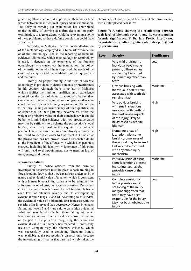
124
The Reliability Of Bitemark Evidence: Analysis And Recommendations In The Context Of Malaysian Criminal Justice System
greenish-yellowincolour,itimpliedthattherewasatimelapsedbetweentheinflictionofinjuryandtheexamination.The delay in carrying out examination has contributedto the inability of arriving at a firm decision. An earlyexamination,toagreatextentwouldhaveovercomesomeoftheseproblems,sothatadefiniteconclusioncouldhavearrivedat.43 Secondly, in Malaysia, there is no standardizationof themethodologyemployed inabitemarkexaminationor of the terminology used in the reporting of bitemarkevidence.Ultimately,whichmethodologyor terminologyis used, it depends on the experience of the forensicodontologist who carries out the examination, the policyoftheinstitutioninwhichheisemployed,theneedsofthecaseunderenquiryandtheavailabilityoftheequipmentsandmaterials. Thirdly,noproper training in the fieldof forensicodontologyisprovidedtodentalstudentsorpractitionersin this country. Although there is no law in Malaysiawhich specifies theminimumqualificationor experiencerequired on the part of dental practitioners before theycan conduct bitemark examinations or give evidence incourt,theneedforsuchtrainingisparamount.Thereasonis that any lacking or insufficiency of such qualificationor experience on their part may nevertheless affect theweightorprobativevalueof theirconclusion.44 It shouldbeborne inmind thatevidencewith lowprobativevaluemaynotbesufficienttodischargetheprosecution’slegalburden, which may result in the acquittal of a culpableperson.Thisisbecausethelawcompulsorilyrequiresthetrialcourt to recordanorder to thateffect if it finds thatthe prosecution has not proved beyond reasonable doubtalltheingredientsoftheoffencewithwhichsuchpersonischarged,includinghisidentity.45,46Ignoranceofthispointwill only lead to disappointment, not to mention lost oftime,energyandmoney.
Recommendations Firstly, all police officers from the criminalinvestigationdepartmentmustbegivenabasictraininginforensicodontologysothattheycanatleastunderstandthenatureandevidentialvalueofapatternwhichisconsistentwith a human bitemark and cause it to be examined bya forensic odontologist, as soon as possible. Pretty hascreated an index which shows the relationship betweeneach level of bitemark severity and its correspondingevidentialvalue (Figs.7and8).According to this index,theevidentialvalueofabitemarkfirst increaseswiththeseverityofitsinjuryandthendecreases.47Hence,bitemarksfallingintolevels3and4aresaidtocarryhighevidentialvalue and may be reliable but those falling into otherlevelsarenot.Asnotedinthelocalcaseabove,thefailureon the part of the police in recognizing the nature andevidentialvalueofabitemarkhasrendereditforensicallyuseless.43 Comparatively, the bitemark evidence, whichwas successfully used in convicting Theodore Bundy,was available at the prosecution’s disposal only becausetheinvestigatingofficerinthatcasehadwiselytakenthe
photograph of the disputed bitemark at the crime-scene,witharulerplacednearit.10,11
Figure 7: A table showing the relationship between each level of bitemark severity and its corresponding forensic significance. © dr. Iain Pretty, http://www.forensicdentistryonline.org/bitemark_index.pdf. (used by permission)
Level Severity Significance
1 Very mild bruising, no individual tooth marks present, diffuse arches visible, may be caused by something other than teeth
Low
2 Obvious bruising with individual, discrete areas associated with teeth, skin remains intact
Moderate
3 Very obvious bruising with small lacerations associated with teeth on the most severe aspects of the injury, likely to be assessed as definite bitemark
High
4 Numerous areas of laceration, with some bruising, some areas of the wound may be incised. Unlikely to be confused with any other injury mechanism
High
5 Partial avulsion of tissue, some lacerations present indicating teeth as the probable cause of the injury
Moderate
6 Complete avulsion of tissue, possibly some scalloping of the injury margins suggested that teeth may have been responsible for the injury. May not be an obvious bite injury
Low
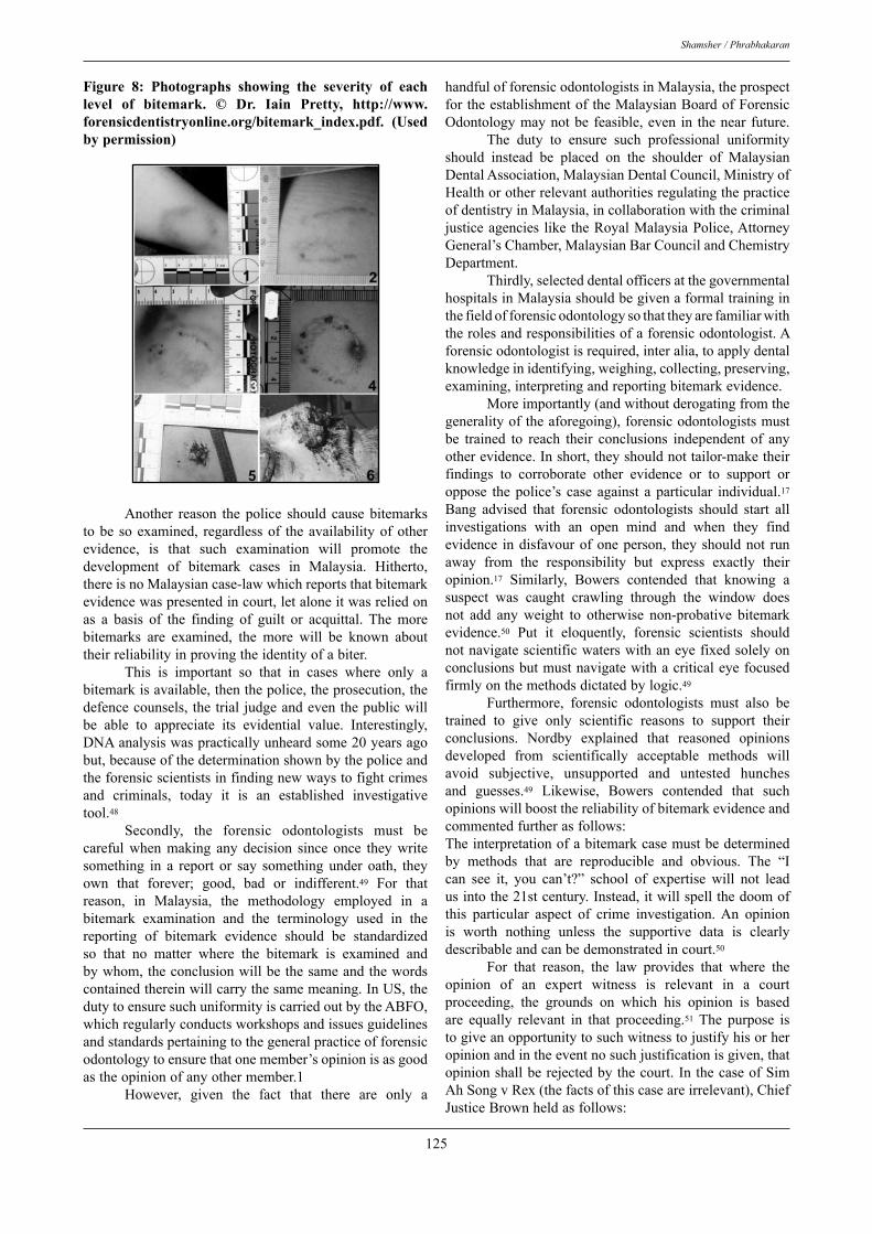
125
Shamsher / Phrabhakaran
Figure 8: Photographs showing the severity of each level of bitemark. © dr. Iain Pretty, http://www.forensicdentistryonline.org/bitemark_index.pdf. (used by permission)
Another reason the police should cause bitemarkstobe soexamined, regardlessof theavailabilityofotherevidence, is that such examination will promote thedevelopment of bitemark cases in Malaysia. Hitherto,thereisnoMalaysiancase-lawwhichreportsthatbitemarkevidencewaspresentedincourt,letaloneitwasreliedonas a basis of the finding of guilt or acquittal. The morebitemarks are examined, the more will be known abouttheirreliabilityinprovingtheidentityofabiter. This is important so that in cases where only abitemarkisavailable,thenthepolice,theprosecution,thedefencecounsels, thetrial judgeandeventhepublicwillbe able to appreciate its evidential value. Interestingly,DNAanalysiswaspracticallyunheardsome20yearsagobut,becauseofthedeterminationshownbythepoliceandtheforensicscientistsinfindingnewwaystofightcrimesand criminals, today it is an established investigativetool.48 Secondly, the forensic odontologists must becareful when making any decision since once they writesomething in a report or say something under oath, theyown that forever; good, bad or indifferent.49 For thatreason, in Malaysia, the methodology employed in abitemark examination and the terminology used in thereporting of bitemark evidence should be standardizedso that no matter where the bitemark is examined andbywhom,theconclusionwillbethesameandthewordscontainedthereinwillcarrythesamemeaning.InUS,thedutytoensuresuchuniformityiscarriedoutbytheABFO,whichregularlyconductsworkshopsandissuesguidelinesandstandardspertainingtothegeneralpracticeofforensicodontologytoensurethatonemember’sopinionisasgoodastheopinionofanyothermember.1 However, given the fact that there are only a
handfulofforensicodontologistsinMalaysia,theprospectfortheestablishmentoftheMalaysianBoardofForensicOdontologymaynotbe feasible,even in thenear future. The duty to ensure such professional uniformityshould instead be placed on the shoulder of MalaysianDentalAssociation,MalaysianDentalCouncil,MinistryofHealthorotherrelevantauthoritiesregulatingthepracticeofdentistryinMalaysia,incollaborationwiththecriminaljusticeagencies like theRoyalMalaysiaPolice,AttorneyGeneral’sChamber,MalaysianBarCouncilandChemistryDepartment. Thirdly,selecteddentalofficersatthegovernmentalhospitalsinMalaysiashouldbegivenaformaltraininginthefieldofforensicodontologysothattheyarefamiliarwiththerolesandresponsibilitiesofaforensicodontologist.Aforensicodontologistisrequired,interalia,toapplydentalknowledgeinidentifying,weighing,collecting,preserving,examining,interpretingandreportingbitemarkevidence. Moreimportantly(andwithoutderogatingfromthegeneralityoftheaforegoing),forensicodontologistsmustbe trained to reach their conclusions independent of anyotherevidence.Inshort,theyshouldnottailor-maketheirfindings to corroborate other evidence or to support oroppose thepolice’s case against aparticular individual.17Bang advised that forensic odontologists should start allinvestigations with an open mind and when they findevidence indisfavourofoneperson, theyshouldnot runaway from the responsibility but express exactly theiropinion.17 Similarly, Bowers contended that knowing asuspect was caught crawling through the window doesnot add any weight to otherwise non-probative bitemarkevidence.50 Put it eloquently, forensic scientists shouldnotnavigatescientificwaterswithaneyefixedsolelyonconclusionsbutmustnavigatewithacriticaleyefocusedfirmlyonthemethodsdictatedbylogic.49 Furthermore, forensic odontologists must also betrained to give only scientific reasons to support theirconclusions. Nordby explained that reasoned opinionsdeveloped from scientifically acceptable methods willavoid subjective, unsupported and untested hunchesand guesses.49 Likewise, Bowers contended that suchopinionswillboostthereliabilityofbitemarkevidenceandcommentedfurtherasfollows:Theinterpretationofabitemarkcasemustbedeterminedby methods that are reproducible and obvious. The “Ican see it, you can’t?” school of expertise will not leadusintothe21stcentury.Instead,itwillspellthedoomofthis particular aspect of crime investigation.An opinionis worth nothing unless the supportive data is clearlydescribableandcanbedemonstratedincourt.50
For that reason, the law provides that where theopinion of an expert witness is relevant in a courtproceeding, the grounds on which his opinion is basedare equally relevant in that proceeding.51 The purpose istogiveanopportunitytosuchwitnesstojustifyhisorheropinionandintheeventnosuchjustificationisgiven,thatopinionshallberejectedbythecourt.InthecaseofSimAhSongvRex(thefactsofthiscaseareirrelevant),ChiefJusticeBrownheldasfollows:
126
The Reliability Of Bitemark Evidence: Analysis And Recommendations In The Context Of Malaysian Criminal Justice System
A bare expression of the expert’s opinion has noevidential value at all. Unless he gives an explanationwhich supplies understanding of the subject which thecourt lacks, the court is innobetter position than itwasbefore.52 A proper training will definitely produce farmore competent dental practitioners who can contributeeffectivelyinthefieldofforensicodontology. Havingdiscussedthusfar,thereisanimportantpointthatneedsconsideration.Inanyforensicinvestigations,itis not infrequent that the practitioners in the criminaljusticesystemfailtoappreciatethefactthatthebiologicalsciences (includingdental science)cannot, in thepresentstateofknowledge,beexpected todeliver theexactitudeof the mathematical sciences.53 Furthermore, Prettyargued that it is healthy within any discipline that somecontentious issues exist, since without an inquisitorialapproachnosciencewouldadvance.24Therefore,althoughthe trainings should be aimed to improve the efficiencyof the forensic odontologists in Malaysia and create auniformityamongthem,bitemarkevidenceshouldnotberejectedasunreliableonthesolegroundthattherearestilldisagreementsamongtheexperts.
CONCLuSION The reliability of bitemark evidence depends ontwo factors, namely, (i) whether a disputed bitemark issufficientlydetailedand(ii)whetherforensicodontologistsand other practitioners in the criminal justice system areprofessionallytrainedtohandlethesaidbitemark.Duetothehostilenatureof theenvironment inwhichbitemarksare produced on human skin, such bitemarks may notbe sufficiently detailed and any findings made from itsexamination shall be highly unreliable and prejudicial.However,ifthedisputedbitemarksaresufficientlydetailed,thenprofessionallytrainedforensicteamcanmakeallthedifferencebetweenjusticeandinjustice. Therefore,selectedpoliceofficersmustbegivenabasictraininginthefieldofforensicodontologysothattheywillbeabletoappreciatetheevidentialvalueofbitemarkand contribute to the development of bitemark cases inMalaysia. The relevant authorities governing the dentalpractice inMalaysia should standardize themethodologyandterminologyusedinbitemarkexaminationandinthereportingofitsfindingssothatconfusionandinconsistencyamongtheforensicodontologistsarekeptabsolutelylow.Finally, forensic odontologists must be given specializedtraining in bitemark examination so that the probativevalueoftheirfindingscanbeimproved.
ACKNOWLEdGMENT
TheauthorsthankProfessorDr.KasinathanNadesan(JohnHunterHospital,NewSouthWales);Dr.IainPretty(UniversityofManchester);Dr.MichaelBowers(DiplomateoftheAmericanBoardofForensicOdontology);Dr.Hjh.NorainiNunNaharHj.Yunus(PaediatricInstitute,Kuala
LumpurHospital);Dr.JalZabdiMohd.Yusoff(FacultyofLaw,UniversityofMalaya);Ms.Gunamalar Joorindanjn(Advocate&Solicitor,MessrsGunamalarLawChambers)and Dato’Amiruddin Idris for their contributions duringthepreparationandwritingofthisarticle.
REFERENCES
1. Newbitemarkguidelines[Online].Colorado(UnitedStates):AmericanBoardofForensicOdontology.2006 [cited2008May7].Availablefrom:http://www.abfo.org/.
2. MacDonald G. Bitemark recognition and interpretation. JForensicSci.1974;14(3):229-33.
3. Pretty I, Sweet D. Anatomical location of bitemarks andassociated findings in 101 cases from the United States. JForensicSci.2000;45(4):812-4.
4. Nambiar P. Forensic odontology. In: Nadesan K, editor.ForensicmedicineandscienceforMalaysianlawyers.KualaLumpur(Malaysia):LexisNexis;2007.p145-60.
5. WarnickA, BiedrzyckiL, Russanow G. Not all bite marksareassociatedwithabuse,sexualactivitiesorhomicides:Acasestudyofaself-inflictedbitemark.JForensicSci.1987;32(3):788-92.
6. DinkelE.Theuseofbitemarkevidenceasaninvestigativeaid.JForensicSci.1973;19:535-47.
7. Hale A. The admissibility of bitemark evidence. SouthCaliforniaLawReview.1978;51(3):309-34.
8. Paul A. Evidence: Practice and procedure. 3rd ed. KualaLumpur(Malaysia):MalayanLawJournal;2003.
9. ReginavTurnbull.Queen’sBenchLawReport.1977;1:224-39.
10.RamslandK.Themostfamousbite[Online].Crimelibrary:Criminalmindsandmethods[cited2008Feb10].Availablefrom:http://www.crimelibrary.com/criminal_mind/forensics/bitemarks/1.html.
11.BundyvStateofFlorida.SouthernReporterSecond.1984;455:330-50.
12.Bowers M. Problem-based analysis of bitemarkmisidentifications:TheroleofDNA.ForensicSciInt.2006;159(1):104-9.
13.State ofArizona v Krone. Pacific Reporter Second. 1995;897:621-3.
14.HansenM.Theuncertainscienceofevidence.AmericanBarAssociationJournal.2005July:49-51.
15.Bang G. Analysis of tooth marks in homicide case. ActaOdontolScand.1976;34:1-11.
16.Keiser-NielsenS.Teeththattold.Odense(Denmark):OdenseUniversityPress;1992.p.52-6.
17.BangG.TheTorgersencase:After42yearsand5 forensicodontologists. In: Willems G, editor. Forensic odontology:Proceedings of the European IOFOS millennium meeting;2000 Aug 23-26; Leuven, Belgium. Leuven (Belgium):LeuvenUniversityPress;2000.p.183-90.
18.BowersM.Torgersen’scase [Online]. Issues inhumanandanimal bite mark analysis [cited 2008 May 10]. Availablefrom:http://forensic.to/webhome/bitemarks2/.
19.Sweet D. Human bitemarks: Examination, recovery, andanalysis. InBowersM,BellG,editors.Manualof forensicodontology. 3rd ed. Colorado (United States): AmericanSocietyofForensicOdontology;1995.p.148-69.
20.Pretty I. Web-based survey of odontologists’ opinionsconcerning bitemark analyses. J Forensic Sci. 2003; 48(5):1117-20.

127
Shamsher / Phrabhakaran
21.GiannelliP.Bitemarkanalysis[Online].Caseresearchpaperseries in legal studies; 2008 Jan; Case Western ReserveUniversity[cited2008May06].Availablefrom:http://papers.ssrn.com/sol3/papers.cfm?abstract_id=1086763.
22.Disaster victim identification guide. New York (UnitedStates):INTERPOL;1997.
23.Rawson R, Ommen R, Kinard G, Johnson J, Yfantis A.Statistical evidence for the individuality of the humandentition.JForensicSci.1984;29(1):245-53.
24.PrettyI.Unresolvedissuesinbitemarkanalysis.In:DorionR,editor.Bitemarkevidence.Colorado(UnitedStates):MarcelDekker;2005.p.547-63.
25.Pretty I. Reliability of bitemark evidence. In: Dorion R,editor.Bitemarkevidence.Colorado(UnitedStates):MarcelDekker;2005.p.531-45.
26.Whittaker D. Some laboratories studies on the accuracy ofbitemarkcomparisons.IntDentJ.1975;25(3):166-71.
27.Arheart K, Pretty I. Results of the 4th ABFO bitemarkworkshop–1999.ForensicSciInt.2001;124(2):104-11.
28.Gorea R, Jha M, Jasuja O, Vasudeva K, Aggarwal A.Marveloustoolsofidentification–bitemarks.In:SharmaR,editor.JMed-LegUpdate.2005;5(2):61-4.
29.BarbenelJ,EvansJ.Bitemarksinskin:Mechanicalactors.JForensicSci.1974;14(3):235-8.
30.Sheasby D, MacDonald G. A forensic classification ofdistortion in human bite marks. Forensic Sci Int. 2001;122(1):75-8.
31.Zhang Z, Monteiro-Riviere N. Comparison of integrins inhuman skin, pig skin and perfused skin: An in vitro skintoxicologymodel.JApplToxicol.1997;17(4):247-53.
32.DeVoreD.Bitemarksforidentification:Apreliminaryreport.MedSciLaw.1971;11(3):144-5.
33.Hyzer W, Krauss T. The bitemark standard reference scale–ABFONo.2.JForensicSci.1988;33(2):498-506.
34.WilkinsonA,GerughtyR.Bitemarkevidence:Itsadmissibilityis hard to swallow. Western State University Law Review.1985;12:519-61.
35.KoubleR,CraigG.Acomparisonbetweendirectandindirectmethodsavailableforhumanbitemarkanalysis.JForensicSci.2004;49(1):111-8.
36.SweetD,ParharM,WoodR.Computer-basedproductionofbitemarkcomparisonoverlays.JForensicSci.1998;43(5):1050-5.
37.Keiser-NielsonS.Forensicodontology.UniversityofToledoLawReview.1969;1:633-9.
38.State of North Carolina v Temple. South Eastern ReporterSecond.1981;273:273-9.
39.StateofMissourivSager.SouthWesternReporterSecond.1980;600:541-64.
40.EvidenceAct1950(Act56,Malaysia);s.45(1).41.PeoplevSmith.NorthEasternReporterSecond.1984;468:
879-86.42.Taroni F. Identification and use of probabilities in forensic
odontology:Aphilosophicaldiscussion.In:WillemsG,editor.Forensic odontology: Proceedings of the European IOFOSmillennium meeting; 2000 Aug 23-26; Leuven, Belgium.Leuven(Belgium):LeuvenUniversityPress;2000.p.67-71.
43.Policereportno:848/97(SubangJaya)and616/97(PetalingJaya).
44.PublicProsecutorvMuhamedbinSulaiman.MalayanLawJournal.1982;2:320-34.
45.CriminalProcedureCode (Act593,Malaysia); ss. 173 (m)and182A.
46.RexvSims.King’sBenchLawReport.1946;1:531-45.
47.Pretty I. Bitemark index [Online]. Forensic dentistryonline [cited 2008 Feb 20]. Available from: http://www.forensicdentistryonline.org/bitemark_index.pdf.
48.Miska S. Twenty years of DNA evidence [Online]. BBCnews;2006Oct9[cited2008Feb25].Availablefrom:http://news.bbc.co.uk/2/hi/uk_news/6031749.stm.
49.NordbyJ.Counteringchaos:Logic,ethicsand thecriminaljustice system. In: James S, Nordby J, editors. Forensicscience: An introduction to scientific and investigativetechniques.Florida(UnitedStates):CRC;2003.p.553-63.
50.BowersM.ThebalanceofDNAandbitemarks:Alawyer’spoint of view. In: Willems G, editor. Forensic odontology:Proceedings of the European IOFOS millennium meeting;2000 Aug 23-26; Leuven, Belgium. Leuven (Belgium):LeuvenUniversityPress;2000.p.99-108.
51.EvidenceAct1950(Act56,Malaysia);s.51.52.SimAhSongvRex.MalayanLawJournal.1951:150-7.53.HillI.Evidentialvalueofbitemarks.In:WillemsG,editor.
Forensic odontology: Proceedings of the European IOFOSmillennium meeting; 2000 Aug 23-26; Leuven, Belgium.Leuven(Belgium):LeuvenUniversityPress;2000.p.93-7.
Address for correspondence:
Mr. Shamsher Singh6623 Lorong Kurau 19Chai Leng Park13700 PeraiPenang, MalaysiaTel: (016) 474 1978Email: [email protected]

Students Perception And Satisfactory Level In Preclinical Fixed Prosthodontic Teaching: Post And CoreMarlynda Ahmad. BDS, MSc
Natasya Ahmad Tarib. BDS, MScDepartment of Prosthodontics, Faculty of Dentistry, Universiti Kebangsaan Malaysia, Malaysia.
ABSTRACT
Introduction: Preclinical teaching using simulation is very beneficial in training dental graduates. The use of laboratory simulation for its undergraduate training during the preclinical years has been used in dental education. Purpose: The aim of this study was to evaluate student’s perception, self evaluation and satisfactory level in preparing duralay burn-out post and core in preclinical fixed prosthodontics sessions. Materials and Methods: The participants comprised of 104 fourth year dental undergraduates in the Faculty of Dentistry Universiti Kebangsaan Malaysia. The students had undergone preclinical session for endodontics during the 3rd year and had already completed root canal treatment on single rooted tooth. The same tooth was used for preclinical post and core preparation. The gutta percha was partially removed and the root canal was prepared. They then proceeded with the preparation of duralay build-up/pattern based on the lecture, video demonstration and manual given. Once completed and satisfied with their work, students were asked to answer the questionnaires in the simulation manual. Results: Student response rate was 88.46% (92/104). Majority of the students were satisfied with their canal preparation, with about 5mm gutta percha left apically, appropriately shaped canal with sufficient retention and resistance form. They also thought that the surface of the duralay was good with no voids. With regards to the coronal preparation, majority of them incorporated ferrule effect and prepared preliminary crown preparation. More than half of the students claimed the level of difficulty of this procedure was moderate. Furthermore, majority of them said that the lecture and the preclinical manual were sufficient and helpful. The help from the supervisors was also benefit in preparing duralay burnout post and core. Conclusions: From this study, majority of fourth year dental students could perform appropriate canal preparation as well as duralay pattern post and core. Only one student did not feel competent and confident in doing canal preparation and duralay pattern post and core. Our teaching methods and aids were proven to help them in preparing these tasks.
Key words preclinical fixed prosthodontics, post and core, duralay build-up, satisfaction level, perception
128
Malaysian Dental Journal (2008) 29(2) 128-134© 2008 The Malaysian Dental Association
MALAYSIAN DENTAL JOURNAL
INTRODUCTION
Preclinical fixed prosthodontics is part of fixedprosthodontic curriculum designed to expand on thestudent’s skills in clinical fixed prosthodontics. Thepurposeof this course is to introduce the students to thebasic principles and techniques of fixed prosthodontics.Fixed prosthodontic educators must continually evaluatethe curriculum to ensure that the principles of evidencebasedteachingarebeingmet. Prosthodontic curriculum and laboratory surveysare useful tools in assessing prosthodontic education. 1Previoussurveyshaveshownthatthepreclinicaltechnicalexperienceofstudentsvariesgreatlyfromschooltoschool.
2, 5Schoolsstressdifferentphasesofpreclinicalexperience,and there is a great variety in the types of restorationsfabricated by the students. 2 The clinical experience islikewise diverse. 3-5 Some schools require the studentsto complete all laboratoryprocedures, someconsider thelaboratoryproceduresoptional forextracredit,andsomeoffertechnicalsupportforalllaboratoryprocedures.3
AttheFacultyofDentistryUniversitiKebangsaanMalaysia, the preclinical fixed prosthodontic trainingstartedatthebeginningofthefourthyear.Itisfocusedonproceduresthatarediscipline-basedandthestudentshavethe opportunity to work closely with the supervisors orlecturersineverystepintherequiredexercise.Therefore,students would gain more clinical experience during the
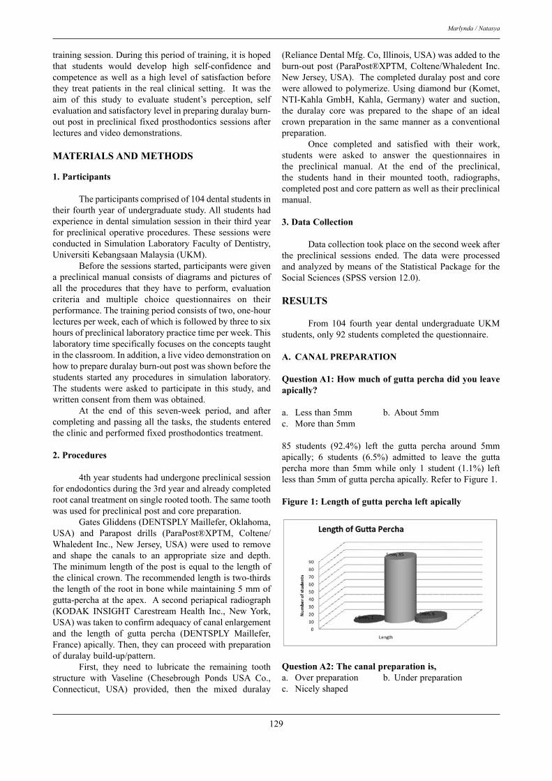
129
Marlynda / Natasya
trainingsession.Duringthisperiodoftraining,itishopedthat students would develop high self-confidence andcompetenceaswellasahigh levelof satisfactionbeforethey treat patients in the real clinical setting. It was theaim of this study to evaluate student’s perception, selfevaluationandsatisfactorylevelinpreparingduralayburn-outpost inpreclinical fixedprosthodontics sessionsafterlecturesandvideodemonstrations.
MATERIALS AND METHODS
1. Participants Theparticipantscomprisedof104dentalstudentsintheirfourthyearofundergraduatestudy.Allstudentshadexperienceindentalsimulationsessionintheirthirdyearforpreclinicaloperativeprocedures.These sessionswereconductedinSimulationLaboratoryFacultyofDentistry,UniversitiKebangsaanMalaysia(UKM). Beforethesessionsstarted,participantsweregivenapreclinicalmanualconsistsofdiagramsandpicturesofall the procedures that they have to perform, evaluationcriteria and multiple choice questionnaires on theirperformance.Thetrainingperiodconsistsoftwo,one-hourlecturesperweek,eachofwhichisfollowedbythreetosixhoursofpreclinicallaboratorypracticetimeperweek.Thislaboratorytimespecificallyfocusesontheconceptstaughtintheclassroom.Inaddition,alivevideodemonstrationonhowtoprepareduralayburn-outpostwasshownbeforethestudents started any procedures in simulation laboratory.The students were asked to participate in this study, andwrittenconsentfromthemwasobtained. At the end of this seven-week period, and aftercompletingandpassingallthetasks,thestudentsenteredtheclinicandperformedfixedprosthodonticstreatment.
2. Procedures 4thyearstudentshadundergonepreclinicalsessionforendodonticsduringthe3rdyearandalreadycompletedrootcanaltreatmentonsinglerootedtooth.Thesametoothwasusedforpreclinicalpostandcorepreparation. GatesGliddens(DENTSPLYMaillefer,Oklahoma,USA) and Parapost drills (ParaPost®XPTM, Coltene/Whaledent Inc.,NewJersey,USA)wereused to removeand shape the canals to an appropriate size and depth.Theminimumlengthofthepostisequaltothelengthoftheclinicalcrown.Therecommendedlengthistwo-thirdsthelengthoftherootinbonewhilemaintaining5mmofgutta-perchaat theapex. Asecondperiapicalradiograph(KODAK INSIGHT Carestream Health Inc., New York,USA)wastakentoconfirmadequacyofcanalenlargementand the length of gutta percha (DENTSPLY Maillefer,France)apically.Then,theycanproceedwithpreparationofduralaybuild-up/pattern. First, they need to lubricate the remaining toothstructure with Vaseline (Chesebrough Ponds USA Co.,Connecticut, USA) provided, then the mixed duralay
(RelianceDentalMfg.Co,Illinois,USA)wasaddedtotheburn-out post (ParaPost®XPTM, Coltene/Whaledent Inc.NewJersey,USA).Thecompletedduralaypostandcorewereallowedtopolymerize.Usingdiamondbur(Komet,NTI-Kahla GmbH, Kahla, Germany) water and suction,the duralay core was prepared to the shape of an idealcrownpreparation in the samemanner as a conventionalpreparation. Once completed and satisfied with their work,students were asked to answer the questionnaires inthe preclinical manual. At the end of the preclinical,the students hand in their mounted tooth, radiographs,completedpostandcorepatternaswellastheirpreclinicalmanual.
3. Data Collection
Datacollectiontookplaceonthesecondweekafterthe preclinical sessions ended. The data were processedandanalyzedbymeansof theStatisticalPackage for theSocialSciences(SPSSversion12.0).
RESULTS
From 104 fourth year dental undergraduate UKMstudents,only92studentscompletedthequestionnaire.
A. CANAL PREPARATION
Question A1: How much of gutta percha did you leave apically?
a. Lessthan5mm b. About5mm c. Morethan5mm
85 students (92.4%) left the gutta percha around 5mmapically; 6 students (6.5%) admitted to leave the guttapercha more than 5mm while only 1 student (1.1%) leftlessthan5mmofguttaperchaapically.RefertoFigure1.
Figure 1: Length of gutta percha left apically
Question A2: The canal preparation is,a. Overpreparation b. Underpreparationc. Nicelyshaped

130
Students Perception And Satisfactory Level In Preclinical Fixed Prosthodontic Teaching: Post And Core
78students(84.8%)claimedthecanalwasnicelyprepared;7students(7.6%)declaredthecanalwasunderpreparation;and another 7 students (7.6%) said the canal was overpreparation.RefertoFigure2.
Figure 2: The canal preparation
Question A3: What is your crown/root ratio?
a. 1:1 b.1:2 c. 1:3 d.2:1
48 students (52.2%) stated the crown/root ratio was 1:2;27 of the students claimed the crown/root ratio was 1:1;andtheremaining,17students(18.5%)confirmedthatthecrown/rootratiowas1:3.RefertoTable1.
Table 1: Crown / Root ratio
Crown Root Ratio 1:1 1:2 1:3 TotalNumberofStudent 27 48 17 92
B. RESISTANCE AND RETENTION
Question B1: What do you think of your preparation, in terms of resistance and retention?
a. Insufficientresistanceandretentionb. Sufficientresistanceandretention
Withregardstoresistanceandretentionformofthecanalpreparation (Figure 3), 84 students (91%) claimed thatresistanceandretentionofthecanalwassufficient.Only8students(9%)thoughtthatitwasinsufficient.
Figure 3: Retention and resistance form
C. DURALAY CORE/CORONAL PREPARATION
Question C1: What do you think of the duralay surface?
a. Voidsatapical1/3 b.Voidsatmiddle1/3c. Voidsatcoronal1/3 d.Perfectwithnovoids
49 students (53.3%) stated their duralay post and core’ssurface were perfect with no voids at all; 17 students(18.5%) claimed the voids were only at coronal third;15 students (16.3%) found voids at apical third; and theremaining12%ofthestudents(11)foundvoidsatmiddlethirdofthepostandcore’ssurface.RefertoFigure4.
Figure 4: Condition of duralay surface
Question C2: What is your duralay post diameter?
a. Smallerthan1/3thewidthoftherootb. About1/3thewidthoftherootc. Widerthan1/3thewidthoftheroot
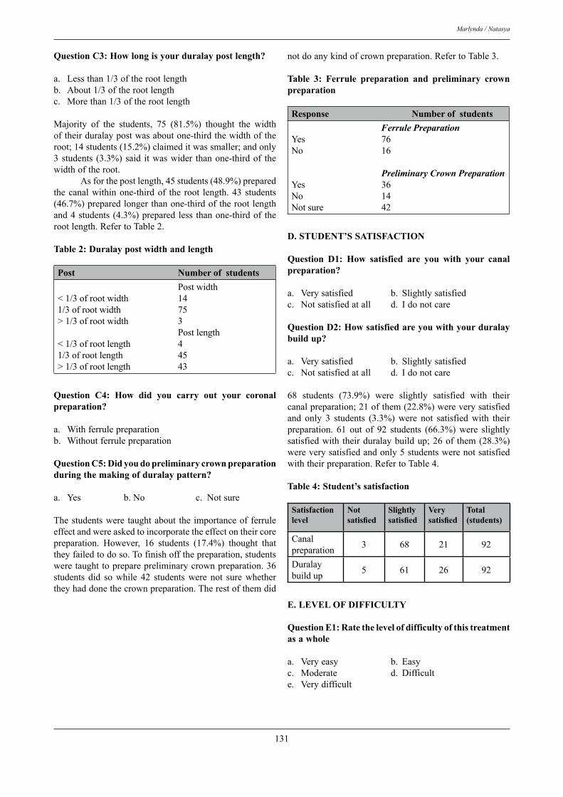
131
Marlynda / Natasya
Question C3: How long is your duralay post length?
a. Lessthan1/3oftherootlengthb. About1/3oftherootlengthc. Morethan1/3oftherootlength
Majority of the students, 75 (81.5%) thought the widthoftheirduralaypostwasaboutone-thirdthewidthoftheroot;14students(15.2%)claimeditwassmaller;andonly3students (3.3%)said itwaswider thanone-thirdof thewidthoftheroot. Asforthepostlength,45students(48.9%)preparedthecanalwithinone-thirdof the root length.43 students(46.7%)preparedlongerthanone-thirdoftherootlengthand4students(4.3%)preparedless thanone-thirdof therootlength.RefertoTable2.
Table 2: Duralay post width and length
Post Number of students Postwidth<1/3ofrootwidth 141/3ofrootwidth 75>1/3ofrootwidth 3 Postlength<1/3ofrootlength 41/3ofrootlength 45>1/3ofrootlength 43
Question C4: How did you carry out your coronal preparation?
a. Withferrulepreparation b. Withoutferrulepreparation
Question C5: Did you do preliminary crown preparation during the making of duralay pattern?
a. Yes b.No c. Notsure
Thestudentswere taughtabout the importanceof ferruleeffectandwereaskedtoincorporatetheeffectontheircorepreparation. However, 16 students (17.4%) thought thattheyfailedtodoso.Tofinishoffthepreparation,studentsweretaughttopreparepreliminarycrownpreparation.36students did so while 42 students were not sure whethertheyhaddonethecrownpreparation.Therestofthemdid
notdoanykindofcrownpreparation.RefertoTable3.
Table 3: Ferrule preparation and preliminary crown preparation
Response Number of students Ferrule PreparationYes 76No 16
Preliminary Crown PreparationYes 36No 14Notsure 42
D. STUDENT’S SATISFACTION
Question D1: How satisfied are you with your canal preparation?
a. Verysatisfied b. Slightlysatisfiedc. Notsatisfiedatall d. Idonotcare
Question D2: How satisfied are you with your duralay build up?
a. Verysatisfied b. Slightlysatisfiedc. Notsatisfiedatall d. Idonotcare
68 students (73.9%) were slightly satisfied with theircanalpreparation;21ofthem(22.8%)wereverysatisfiedand only 3 students (3.3%) were not satisfied with theirpreparation. 61 out of 92 students (66.3%) were slightlysatisfiedwith theirduralaybuildup;26of them(28.3%)wereverysatisfiedandonly5studentswerenotsatisfiedwiththeirpreparation.RefertoTable4.
Table 4: Student’s satisfaction
Satisfaction level
Not satisfied
Slightly satisfied
Very satisfied
Total (students)
Canalpreparation 3 68 21 92
Duralaybuildup 5 61 26 92
E. LEVEL OF DIFFICULTY
Question E1: Rate the level of difficulty of this treatment as a whole
a. Veryeasy b. Easy c. Moderate d. Difficult e. Verydifficult
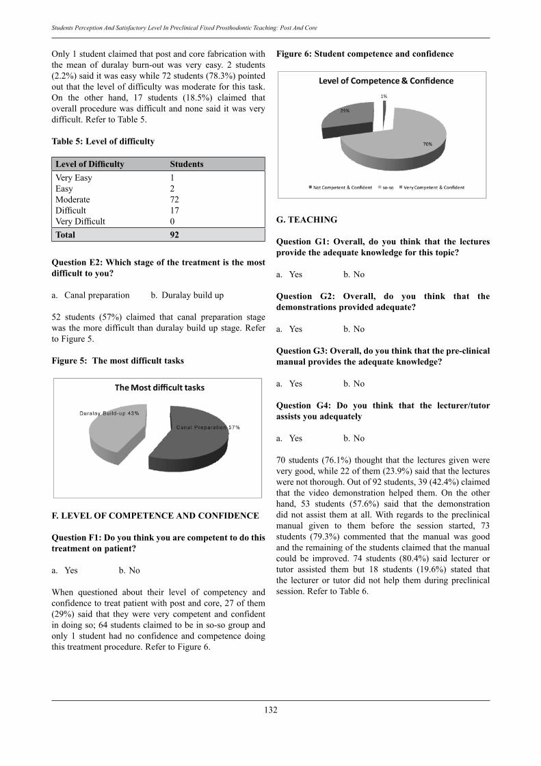
132
Students Perception And Satisfactory Level In Preclinical Fixed Prosthodontic Teaching: Post And Core
Only1studentclaimedthatpostandcorefabricationwiththe mean of duralay burn-out was very easy. 2 students(2.2%)saiditwaseasywhile72students(78.3%)pointedoutthatthelevelofdifficultywasmoderateforthistask.On the other hand, 17 students (18.5%) claimed thatoverallprocedurewasdifficultandnonesaiditwasverydifficult.RefertoTable5.
Table 5: Level of difficulty
Level of Difficulty StudentsVeryEasy 1Easy 2Moderate 72Difficult 17VeryDifficult 0Total 92
Question E2: Which stage of the treatment is the most difficult to you?
a. Canalpreparation b. Duralaybuildup
52 students (57%) claimed that canal preparation stagewas themoredifficult thanduralaybuildupstage.RefertoFigure5.
Figure 5: The most difficult tasks
F. LEVEL OF COMPETENCE AND CONFIDENCE
Question F1: Do you think you are competent to do this treatment on patient?
a. Yes b.No
When questioned about their level of competency andconfidencetotreatpatientwithpostandcore,27ofthem(29%) said that they were very competent and confidentindoingso;64studentsclaimedtobeinso-sogroupandonly1 student hadno confidence and competencedoingthistreatmentprocedure.RefertoFigure6.
Figure 6: Student competence and confidence
G. TEACHING
Question G1: Overall, do you think that the lectures provide the adequate knowledge for this topic?
a. Yes b.No
Question G2: Overall, do you think that the demonstrations provided adequate?
a. Yes b.No
Question G3: Overall, do you think that the pre-clinical manual provides the adequate knowledge?
a. Yes b.No
Question G4: Do you think that the lecturer/tutor assists you adequately
a. Yes b.No
70 students (76.1%) thought that the lecturesgivenwereverygood,while22ofthem(23.9%)saidthatthelectureswerenotthorough.Outof92students,39(42.4%)claimedthat the video demonstration helped them. On the otherhand, 53 students (57.6%) said that the demonstrationdidnot assist themat all.With regards to thepreclinicalmanual given to them before the session started, 73students (79.3%) commented that the manual was goodandtheremainingofthestudentsclaimedthatthemanualcould be improved. 74 students (80.4%) said lecturer ortutor assisted them but 18 students (19.6%) stated thatthe lecturer or tutor did not help them during preclinicalsession.RefertoTable6.

133
Marlynda / Natasya
Table 6: Teaching
Response Lectures Demonstration Manual Assistance
yesNo
70 39 73 74
22 53 19 18Total 92 92 92 92
DISCUSSION
Theresultsshowthatprosthodonticeducationandsimulationexperiencesreceivedvarybetweenparticipants.It is interesting to state that most of the participantsperceived and evaluated their canal preparation as goodto almost perfect leaving 5 mm gutta percha apicallywith acceptable crown/root ratio. At the end of postand core preclinical session, the participants excellentlyproducedanidealduralaypostandcorewithappropriatepost width and length, relatively smooth surface andsuperior resistance and retention. 82% of them preparedferruleeventhoughatthebeginningtheyhaddifficultyinunderstandingitsrationaleandtechnique.Simultaneously,the positive perception leads to better self evaluation aswellassatisfactionandcompetencelevel. Theparticipantsratedtheirlevelofsatisfactionforcanalpreparationandduralaybuild-upasverysatisfiedorslightly satisfied. There is always a possibility that theycouldperformbetterif theyweregivenasecondchance.Weighing between canal preparation and core build-up,most participants claimed that canal preparation is moredifficult to prepare compared to core build-up. This isprobably because they could not access directly into thecanalandalsofearofperforation.Withduralaybuild-up,onlydirectaccessisneededandthereisalwaysachancetorebuildandredoit. In this study, students’ perceptions of confidenceandcompetenceweremeasuredtoassesstheeffectofthepreclinical teaching. The participants had experience inthesimulationclinicforotherdisciplinessuchasoperativebut not for fixed prosthodontics, and it was anticipatedthattheirconfidencewouldbelowatthebeginningofthepreclinical course. Perhaps a more accurate evaluationwould have been obtained by measuring the students’perceptionsaftercompletionofthepreclinicalsession. As anticipated, prosthodontic educators in thisfaculty spent significantly more time interacting withstudents during their preclinical fixed prosthodontics.The total number of lecture hours for preclinical fixedprosthodonticsis36hourswhile42hourswerespareforpreclinical laboratory. Petropoulos et al stated that fixedprosthodonticslecturehours(42)scoredthehighestlectureshours followed by complete denture (28) and removablepartialdenture(21)amongUSdentalschools.4,5InUKM,lectures are available online, Fixed Prosthodontics e-Learning (FPeL), therefore the students can spend morehours reviewing the lectures after traditional classroom
lecture. Murphy in 2004 reported that dental studentsprefer visual learning at a higher percentage than thesamplepopulationmeasuredintheVARK(anacronymforVisual,Aural,Read/Write,andKinesthetic)website.6Thedistribution of dental student scores shows a preferencefor instructors who use strong visual presentations andfacilitatenote-takingduringlectures.6Rashedialsoreportedthat most US dental schools having live demonstrationsof laboratory procedures as well as prerecorded videodemonstrationsfortheseprocedures.4Unfortunately,onlyprerecorded livedemonstrationofclinicalprocedures forpost and core was available for the students. Students’involvementisnotneededineverylaboratorystep,sincemost laboratorywork isbeingdelegated.This resultwasconsistence with also Weintraub et al in 1997. 7 This isthe sole reason why we do not conduct any laboratorydemonstration.Dentaleducatorsshouldbeawareofthesedifferences in order to explore opportunities for makingtheeducationalexperiencemoreproductiveandenjoyable.However, our teachingmethods and aids did remarkablyhelptheparticipantsinperformingthesetasks.CONCLUSIONS It can be concluded from this study that almostall fourth year dental students can perform good canalpreparation as well as duralay pattern post and core.Unfortunately, one student did not feel competent andconfident enough in performing canal preparation andduralay pattern post and core. However, the teachingmethods were proven to help the students in preparingthese tasks. The survey results can only be interpretedwith respect to the questions that were used. There arelimitationstothisstudyandbiasinthequestionnairesduetothenatureandformulationofthequestions.
ACKNOWLEDGEMENT The authors thank Simulation Laboratory staffwho generously devoted their time and effort to ensurePreclinical Fixed Prosthodontics run smoothly. SpecialthankstoMrs.SitiZubaidahAnuar*forproofreadingthisreport.
*English Language Teacher, CELPAD, Centre forFoundation Studies, IIUM, Jalan Universiti, 46350,PetalingJaya,Selangor,Malaysia
REFERENCES
1. Goodacre C.J. Review of the literature predoctoral fixedprosthodonticseducation.JProsthetDent1990;64:319-325
2. TaylorM,AquilinoS.A,JordanR.D.Prosthodonticlaboratoryandcurriculumsurvey.PartIV:Fixedprosthodonticcurrirulumsurvey.JProsthetDent1985;53:267-270
3. PrestonJ.D.ProsthodonticeducationintheUnitedStates.IntJProsthodont1989;2:190-195
4. Rashedi B, Petropoulos V: Preclinical complete denturescurriculumsurvey.JProsthodont2003;12:37.

134
Students Perception And Satisfactory Level In Preclinical Fixed Prosthodontic Teaching: Post And Core
5. Petropoulos V.C, WeintraubA, Weintraub G.S. PredoctoralFixedProsthodonticsCurriculumSurvey.JProsthod1998;7:183-191.
6. Murphy RJ, Gray SA. Straja SR, Bogert MC. Studentlearningpreferencesandteachingimplications.JDentEduc2004;68(8):859-66.
7. Weintraub AM, Weintraub GS: The dental student astechnician: An 18-year follow-up of preclinical laboratoryprograms.JProsthodont1997;6:128.
Address for correspondence:
Dr. Marlynda Ahmad Department of Prosthodontics, Faculty of Dentistry UKM, Jalan Raja Muda Abdul Aziz, 50300, Kuala Lumpur. Phone: 603-40405748 Fax: 603-40405798Email: [email protected]

Inhibitory Effect of Mixed Paste of Ca(Oh)2 And Baso4 against Bacteria In Infected Root Canal. Halim H S , DDS.
Iskandar, B, DDS.
Liesan, DDS.Faculty of Dentistry, Trisakti University, Indonesia
ABSTRACT
The aim of this study was to evaluate the inhibitory effect of mixed paste of Ca(OH)2 and BaSO4 in various ratios against bacteria isolated from infected root canal. The isolated bacteria sample was taken from the lower first molar diagnosed with necrotic pulp. The sample of bacteria was selected by gram staining and further cultured in the brain heart infusion solution. Bacteria suspension in blood gel was given 5 holes with diameter of 5 mm to accommodate Ca(OH)2 and BaSO4 mixture with the ratio of 6: 1, 7:1, 8:1, 9:1 and 10:1 to observe the inhibitory zone with intervals of 24 hours, 48 hours, 72 hours, and 10 days. The addition of BaSO4 to Ca(OH)2 decreased the inhibitory effect against bacteria growth. Two-way analysis of variants test between the inhibitory diameter with the ratio of Ca(OH)2 and BaSO4 and time of incubation showed significant difference (p < 0.05). There was a significant difference between ratio of 6:1 to the rest of other ratios in the inhibition of bacterial growth. There was also a significant difference between the incubation periods- 10 days incubation period was different from other incubation periods. Calcium hydroxide was more effective in eliminating aerobic bacteria compared to the anaerobic. Within the limitations of the present study, it is concluded that addition of barium sulfate affected the efficacy of antibacterial effect and the time period of contact between calcium hydroxide with the micro-organisms also has an effect on the bacterial growth.
Key words bacteria inhibition, mixture of Ca(OH)2 and BaSO4, aerobe and anaerobe bacteria, infected root canal
135
Malaysian Dental Journal (2008) 29(2) 135-139© 2008 The Malaysian Dental Association
InTROduCTIOn
Microorganismsare themaincauseof endodonticinfection. Many attempts for endodontic success aredevotedtoeliminateasmanyaspossiblemicroorganismsthatexist in therootcanal. Inaddition to theappropriatepreparationoftherootcanals,medicamentofrootcanalsisrequiredtoachievetheobjective. The medicaments for root canals includechlorophenolcamphermenthol,formocresolandcresatine.These medicaments have adverse impacts due to theirantigenicandcytotoxicnatureandshorteffectiveness.Apartfromthesemedicament,calciumhydroxideCa(OH)2havebeenprovenitsefficacyclinicallyandpossessedbeneficialcharacteristicstobeusedasidealmedicamentforrootcanal.If the use of Ca(OH)2 for maintaining pulp capping andpulpotomy is consideredas abiologicalwounddressing,Ca(OH)2alsoservesasbiologicalroothealingatapexandperiapical tissue. Besides triggering lipopolysaccharidedegradation, Ca(OH)2 with pH of 11.5 plays the role as
powerfulalkalinesubstanceandisrecommendedbysomeresearchers to be used as medicament for root canal andcontains anti bacterial properties against most bacteriaspeciesfoundinendodonticinfection. The efficacy of Ca(OH)2 as medicament for rootcanal will be maximum if injected during the processand make sure it fills the root canal space thoroughly.Radiograph may be taken to confirm that the entire rootcanalhasbeen filledwith themedicament.Purecalciumhydroxide is not visible under any radiograph, and tomakethismaterialradio-opaque,itmustbeaddedwithacontrastivematerial,namelyBaSO4. The aim of this experiment was to find out theoptimalratiobetweenCa(OH)2andBaSO4inminimizing/inhibiting the growth of various bacteria that triggerinfectionintherootcanal. Safavi&Nakayama(2000)1haveshownthatpHofCa(OH)2is11.5andtoincreaseitspH,salineoranesthesiasolutionmaybeaddedandmixedwithCa(OH)2powder.Hydroxyl ions penetrate the dentinal tubule when smear
MALAYSIAn dEnTAL JOuRnAL
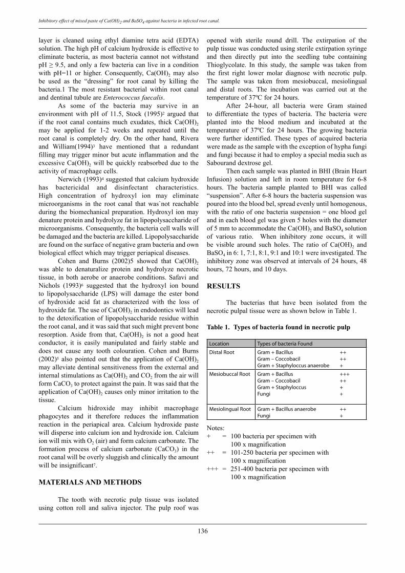
136
Inhibitory effect of mixed paste of Ca(OH)2 and BaSO4 against bacteria in infected root canal.
layer is cleaned using ethyl diamine tetra acid (EDTA)solution.ThehighpHofcalciumhydroxideiseffectivetoeliminatebacteria, asmostbacteria cannotnotwithstandpH ≥ 9.5, and only a few bacteria can live in a condition with pH=11 or higher. Consequently, Ca(OH)2 may alsobe used as the “dressing” for root canal by killing thebacteria.1 The most resistant bacterial within root canalanddentinaltubuleareEnterococcus faecalis. As some of the bacteria may survive in anenvironment with pH of 11.5, Stock (1995)2 argued thatif the root canal contains much exudates, thick Ca(OH)2may be applied for 1-2 weeks and repeated until theroot canal is completely dry. On the other hand, Riveraand William(1994)3 have mentioned that a redundantfillingmay triggerminorbutacute inflammationand theexcessiveCa(OH)2willbequicklyreabsorbeddue to theactivityofmacrophagecells. Nerwich (1993)4suggestedthatcalciumhydroxidehas bactericidal and disinfectant characteristics.High concentration of hydroxyl ion may eliminatemicroorganisms in the root canal that was`not reachableduring thebiomechanicalpreparation.Hydroxyl ionmaydenatureproteinandhydrolyzefatinlipopolysaccharideofmicroorganisms.Consequently,thebacteriacellwallswillbedamagedandthebacteriaarekilled.Lipopolysaccharidearefoundonthesurfaceofnegativegrambacteriaandownbiologicaleffectwhichmaytriggerperiapicaldiseases. Cohen and Burns (2002)5 showed that Ca(OH)2was able to denaturalize protein and hydrolyze necrotictissue, inbothaerobeoranaerobeconditions.SafaviandNichols (1993)6 suggested that the hydroxyl ion boundto lipopolysaccharide (LPS) will damage the ester bondof hydroxide acid fat as characterized with the loss ofhydroxidefat.TheuseofCa(OH)2inendodonticswillleadto thedetoxificationof lipopolysaccharideresiduewithintherootcanal,anditwassaidthatsuchmightpreventboneresorption.Aside from that, Ca(OH)2 is not a good heatconductor, it is easily manipulated and fairly stable anddoes not cause any tooth colouration. Cohen and Burns(2002)5 also pointed out that the application of Ca(OH)2mayalleviatedentinalsensitivenessfromtheexternalandinternalstimulationsasCa(OH)2andCO2fromtheairwillformCaCO3toprotectagainstthepain.ItwassaidthattheapplicationofCa(OH)2causesonlyminorirritationtothetissue. Calcium hidroxide may inhibit macrophagephagocytes and it therefore reduces the inflammationreaction in the periapical area. Calcium hydroxide pastewilldisperseintocalciumionandhydroxideion.CalciumionwillmixwithO2(air)andformcalciumcarbonate.Theformation process of calcium carbonate (CaCO3) in therootcanalwillbeoverlysluggishandclinicallytheamountwillbeinsignificant7.
MATERIALS And METHOdS
The tooth with necrotic pulp tissue was isolatedusing cotton roll and saliva injector. The pulp roof was
opened with sterile round drill. The extirpation of thepulptissuewasconductedusingsterileextirpationsyringeand then directly put into the seedling tube containingThioglycolate. In this study, the sample was taken fromthe first right lower molar diagnose with necrotic pulp.The sample was taken from mesiobuccal, mesiolingualand distal roots. The incubation was carried out at thetemperature of 37ºC for 24 hours. After 24-hour, all bacteria were Gram stained to differentiate the types of bacteria. The bacteria wereplanted into the blood medium and incubated at thetemperature of 37ºC for 24 hours. The growing bacteria were further identified. These types of acquired bacteriaweremadeasthesamplewiththeexceptionofhyphafungiandfungibecauseithadtoemployaspecialmediasuchasSabouranddextrosegel. TheneachsamplewasplantedinBHI(BrainHeartInfusion) solution and left in room temperature for 6-8hours. The bacteria sample planted to BHI was called“suspension”.After6-8hoursthebacteriasuspensionwaspouredintothebloodbel,spreadevenlyuntilhomogenous,withtheratioofonebacteriasuspension=onebloodgelandineachbloodgelwasgiven5holeswiththediameterof5mmtoaccommodatetheCa(OH)2andBaSO4solutionof various ratio. When inhibitory zone occurs, it willbe visible around such holes. The ratio of Ca(OH)2 andBaSO4 in 6: 1, 7:1, 8:1, 9:1 and 10:1 were investigated. The inhibitory zone was observed at intervals of 24 hours, 48 hours,72hours,and10days.
RESuLTS
The bacterias that have been isolated from thenecroticpulpaltissuewereasshownbelowinTable1.
Table 1. Types of bacteria found in necrotic pulp
Location Types of bacteria Found
Distal Root Gram + Bacillus ++Gram – Coccobacil ++Gram + Staphyloccus anaerobe +
Mesiobuccal Root Gram + Bacillus +++Gram – Coccobacil ++Gram + Staphyloccus +Fungi +
Mesiolingual Root Gram + Bacillus anaerobe ++Fungi +
Notes:+ = 100bacteriaperspecimenwith
100xmagnification++ = 101-250bacteriaperspecimenwith
100xmagnification+++ = 251-400 bacteria per specimen with
100xmagnification

137
Halim / Iskandar / Liesan
Table 2 showed that the inhibitory zone of all types ofbacteriaobservedbecomelargerwithincreasedobservationperiod of 24 hours, 48 hours, 72 hours and 10 days. In general, higher ratio of calcium hydroxide to BaSO4 haslargerinhibitoryeffecttoalltypesofbacteria. Basedonthediameterinhibitoryzonemeasurement,itwas shown that the largest inhibitoryeffectwas in theratioofCa(OH)2 :BaSO4=10:1andtheincubationtimewas10daysasshowninTable3.However,basedontheresultofmultiplecomparisontestwithHSDTukeytest,itwasshownthatsuchresultwasnotstatisticallysignificantdifferentattheratioofCa(OH)2 :BaSO4at 7:1,8:1and9:1 within the 10-day incubation. Theresultofstatistical testwithtwo-wayanalysisof variants between the diameter of blocking and ratioof the average magnitude of inhibitory zone during the4 periods in all types of anaerobe bacteria observed and invarious ratiosofCa(OH)2 : BaSO4 during incubation, indicatedasignificantdifference(p<0.05).Accordingly,amultipleposteriorcomparativetestwasadministeredusingthehonestlysignificantdifference(HSD)fromTukey. Prior to the test using HSD Tukey, a one-wayvariant analysis was administered among the treatmentgroupstoobtainthecriticalvaluethatwillbeusedasthemultiplier.Basedonone-wayanova,themultiplierof5.07was obtainedwith averagequadrate (Msc) of 2.68.Withsuch value, HSD value as the differential figure of 2.369 wasacquiredfromamongtheaveragegroups. Thereisastatisticalsignificantdifferencebetweenthe ratio of 6:1 and 7:1, 6:1 and 8:1, 6:1 and 9:1, 6:1 and10:1.TheadditionofBaSO4toCa(OH)2affecteditsinhibitory effect against bacteria that trigger endodonticinfection(Table3).Therewasalsoasignificantdifferencebetween incubationperiodof72hoursand10dayswithall various ratios of Ca(OH)2 : BaSO4 . There was nosignificant difference between incubation period of 24 hours, 48 hours and 72 hours all various ratios of Ca(OH)2:BaSO4 except 9: 1 and 10:1 at 48 and 72 hours.
Table 2 The average and standard deviation of the observation results of root canal bacteria inhibitory zone based on the ratio of Ca(OH)2 : BaSO4 and the seedling time.
Ratio of Ca(OH)2 : BaSO4
Seedling time Average ± standard deviation (mm)
6:1 24 hours48 hours72 hours10 days
1.208 ± 0.3341.625 ± 0.6062.625 ± 0.8824.083 ± 2.275
7:1 24 hours48 hours72 hours10 days
1.500 ± 0.6031.917 ± 0.9003.000 ± 1.2434.417 ± 2.592
8:1 24 hours48 hours72 hours10 days
1.833 ± 0.6512.208 ± 1.0763.125 ± 1.3164.625 ± 2.595
9:1 24 hours48 hours72 hours10 days
2.000 ± 0.7072.458 ± 1.3223.500 ± 1.7844.917 ± 2.721
10:1 24 hours48 hours72 hours10 days
2.042 ± 0.6892.542 ± 1.3223.625 ± 1.7855.292 ± 3.056
Table 3 The average diameter of inhibitory zone during the 4 observed intervals and various ratios of Ca(OH)2 and BaSO4
Ratio of Ca(OH)2 : BaSO4
Inhibitory zone (mm)
24 hours 48 Hours 72 Hours 10 days
6:1 1.208 1.625 2.625 4.083
7:1 1.500 1.917 2.958 4.375
8:1 1.792 2.208 3.083 4.583
9:1 1.917 2.458 3.458 4.875
10:1 2.083 2.583 3.583 5.292
Tables 4 and 5 showed that in general the inhibitory zones wwere smaller in anaerobic bacterias compared to theaerobic bacterias. This meant that Ca(OH)2 was moreeffective in eliminating aerobic bacterias compared toanaerobes.
Table 4 The average diameter of inhibitory zone for anaerobic bacteria during the 4 observed intervals and various ratios of Ca(OH)2 and BaSO4
Ratio of Ca(OH)2 : BaSO4
Inhibitory zone (mm)
24 hours 48 Hours 72 Hours 10 days
6:1 1.214 1.429 2.429 2.429
7:1 1.286 1.643 2.5 2.5
8:1 1.571 1.786 1.571 2.714
9:1 1.571 1.857 2.714 2.786
10:1 2.643 2.143 2.857 3
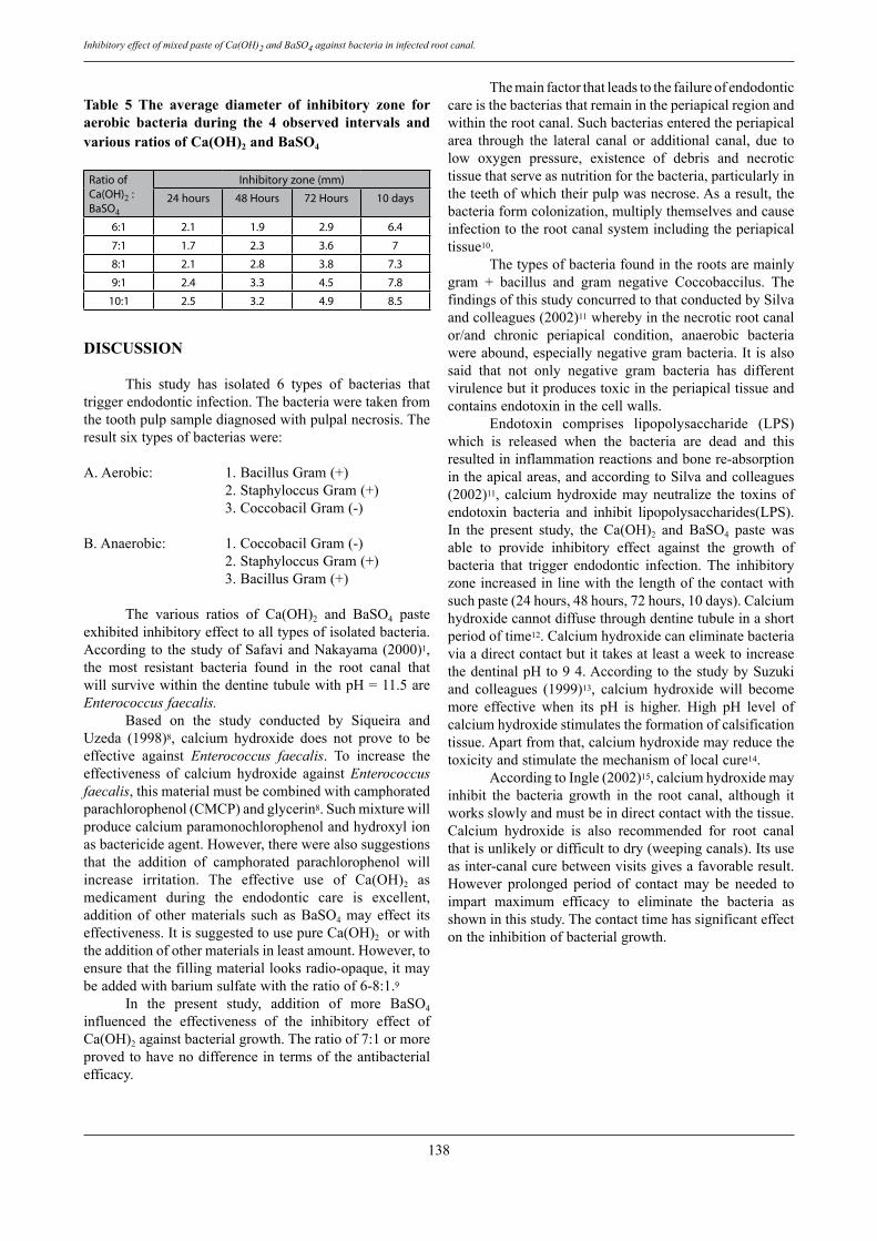
138
Inhibitory effect of mixed paste of Ca(OH)2 and BaSO4 against bacteria in infected root canal.
Table 5 The average diameter of inhibitory zone for aerobic bacteria during the 4 observed intervals and various ratios of Ca(OH)2 and BaSO4
Ratio of Ca(OH)2 : BaSO4
Inhibitory zone (mm)
24 hours 48 Hours 72 Hours 10 days
6:1 2.1 1.9 2.9 6.4
7:1 1.7 2.3 3.6 7
8:1 2.1 2.8 3.8 7.3
9:1 2.4 3.3 4.5 7.8
10:1 2.5 3.2 4.9 8.5
dISCuSSIOn This study has isolated 6 types of bacterias thattriggerendodonticinfection.Thebacteriaweretakenfromthetoothpulpsamplediagnosedwithpulpalnecrosis.Theresultsixtypesofbacteriaswere:
A. Aerobic: 1. Bacillus Gram (+) 2. Staphyloccus Gram (+) 3. Coccobacil Gram (-)
B. Anaerobic: 1. Coccobacil Gram (-) 2. Staphyloccus Gram (+) 3. Bacillus Gram (+)
The various ratios of Ca(OH)2 and BaSO4 pasteexhibitedinhibitoryeffecttoalltypesofisolatedbacteria.According to thestudyofSafaviandNakayama(2000)1,the most resistant bacteria found in the root canal thatwillsurvivewithinthedentinetubulewithpH=11.5areEnterococcus faecalis. Based on the study conducted by Siqueira andUzeda (1998)8, calcium hydroxide does not prove to beeffective against Enterococcus faecalis. To increase theeffectiveness of calcium hydroxide against Enterococcus faecalis,thismaterialmustbecombinedwithcamphoratedparachlorophenol(CMCP)andglycerin8.Suchmixturewillproducecalciumparamonochlorophenolandhydroxylionasbactericideagent.However,therewerealsosuggestionsthat the addition of camphorated parachlorophenol willincrease irritation. The effective use of Ca(OH)2 asmedicament during the endodontic care is excellent,addition of other materials such as BaSO4 may effect itseffectiveness.ItissuggestedtousepureCa(OH)2orwiththeadditionofothermaterialsinleastamount.However,toensurethatthefillingmateriallooksradio-opaque,itmaybeaddedwithbariumsulfatewiththeratioof6-8:1.9 In the present study, addition of more BaSO4influenced the effectiveness of the inhibitory effect ofCa(OH)2againstbacterialgrowth.Theratioof7:1ormoreprovedtohavenodifferenceintermsoftheantibacterialefficacy.
Themainfactorthatleadstothefailureofendodonticcareisthebacteriasthatremainintheperiapicalregionandwithintherootcanal.Suchbacteriasenteredtheperiapicalarea through the lateral canal or additional canal, due tolow oxygen pressure, existence of debris and necrotictissuethatserveasnutritionforthebacteria,particularlyintheteethofwhichtheirpulpwasnecrose.Asaresult,thebacteriaformcolonization,multiplythemselvesandcauseinfectiontotherootcanalsystemincludingtheperiapicaltissue10. Thetypesofbacteriafoundintherootsaremainlygram + bacillus and gram negative Coccobaccilus. ThefindingsofthisstudyconcurredtothatconductedbySilvaandcolleagues(2002)11wherebyinthenecroticrootcanalor/and chronic periapical condition, anaerobic bacteriawereabound,especiallynegativegrambacteria.Itisalsosaid that not only negative gram bacteria has differentvirulencebutitproducestoxicintheperiapicaltissueandcontainsendotoxininthecellwalls. Endotoxin comprises lipopolysaccharide (LPS)which is released when the bacteria are dead and thisresultedininflammationreactionsandbonere-absorptionintheapicalareas,andaccordingtoSilvaandcolleagues(2002)11, calciumhydroxidemayneutralize the toxinsofendotoxin bacteria and inhibit lipopolysaccharides(LPS).In the present study, the Ca(OH)2 and BaSO4 paste wasable to provide inhibitory effect against the growth ofbacteria that trigger endodontic infection. The inhibitoryzoneincreasedinlinewiththelengthofthecontactwithsuch paste (24 hours, 48 hours, 72 hours, 10 days). Calcium hydroxidecannotdiffusethroughdentinetubuleinashortperiodoftime12.Calciumhydroxidecaneliminatebacteriaviaadirectcontactbutittakesatleastaweektoincreasethe dentinal pH to 9 4. According to the study by Suzuki and colleagues (1999)13, calcium hydroxide will becomemore effective when its pH is higher. High pH level ofcalciumhydroxidestimulatestheformationofcalsificationtissue.Apartfromthat,calciumhydroxidemayreducethetoxicityandstimulatethemechanismoflocalcure14. AccordingtoIngle(2002)15,calciumhydroxidemayinhibit the bacteria growth in the root canal, although itworksslowlyandmustbeindirectcontactwiththetissue.Calcium hydroxide is also recommended for root canalthatisunlikelyordifficulttodry(weepingcanals).Itsuseasinter-canalcurebetweenvisitsgivesafavorableresult.However prolonged period of contact may be needed toimpart maximum efficacy to eliminate the bacteria asshowninthisstudy.Thecontacttimehassignificanteffectontheinhibitionofbacterialgrowth.

139
Halim / Iskandar / Liesan
COnCLuSIOn
Within the limitations of the present study, it is concluded that:
(1) The Ca(OH)2 and BaSO4 mixture paste showed the inhibitory effect to the growth of bacteria ininfected canal. The addition of BaSO4 affected the effectivenessofCa(OH)2atratioof6:1,higherratioof Ca(OH)2 : BaSO4 at 7:1, 8:1, 9:1 and 10:1 appear tohavesimilarefficacy.
(2) Calciumhydroxidewasmoreeffectiveineliminatingaerobicbacteriascomparedtoanaerobics.
(3) The time frame of contact for Ca(OH)2 is alsoimportant, as Ca(OH)2 needs sufficient time todiffuseinordertoimpartitsefficacy.
REFEREnCES
1. Safavi,K.andNakayamaT.A.InfluenceofMixingVehicleonDissociationofCalciumHydroxideinSolution.J.Endod.2000. 26: 649-651.
2. Stock, C.J.R, Gulabivala, K. Walker, R.T. Goodman, T.R. ColorAtlasandTextofEndodontics.SecondEdition.Mosby-Wolfe. London. 1995. 146-149.
3. Rivera, E.M. and William, K. Placement of CalciumHydroxide in Simulated Canals: Comparison of Glycerin Versus Water. J. Endod. 1994. 20 445-448.
4. Nerwich, A. Figdor, D. and Messer, HH. pH Changes in Root Dentin Over a 4-weeks Period Following Root Canal Dressing with Calcium Hydroxide. J. Endod. 1993 19. No. 6: 302-306.
5. Cohen,S.,Burns,R.C.PathwayofThePulp.8thEd..C.V.Mosby., St. Louis. 2002, 43-44, 508,559, 564-566.
6. Safavi, K.E. and Nichols, F.C. Effect of Calcium Hydroxide on Bacterial Lipopolysaccharide. J.Endod. 1993.19. No.2: 76-78.
7. Calts, Serper,A. and Ozcelik. PH changes and Calcium inDifusses From Calcium Hydroxide Dressing Materials Through Root Dentine. J. Endod. 1999, 25 : 329-331.
8. Siqueira, J. F. and Uzeda, M. Influence of Different Vehicles ontheAntibacterialEffectofCalciumHydroxide.J.Endod.1998. 24: 663-665.
9. Dumsha, T.C. and Gutmann, J.L. Clinician’s Endodontic Handbook,LexiComp.Inc.Cleveland.2000,178,183.
10. Mickel, A.K. and Wright, E.R. Growth Inhibition of StreptococcusAnginosus(Milleri)byThreeCalciuHydroxideSealers and One Zinc Oxide Eugenol Sealer. J. Endod. 1999. 25 : 34-37.
11. Silva, L.A.B. Nelson, Filho, P. Leonardo, M.R. Rossi, M.A and Pansani, G.A. Effect of Calcium Hydroxide on Bacterial Endotoxin in Vivo. J. Endod. 2002 28: 94-98
12. Beltes, P.G., Pissiotis, E., Koulaouzidou, E. and Kortsaris, A.H. In Vitro Release of Hydroxyl Ions from Six TypesCalcium Hydroxide Nonsetting Pastes. J. Endod. 1997, 23: 413-415.
13.Suzuki, K. Higuchi, N, Horiba, N. Matsumoto, T. andNakamura,H.AntimicrobialEffectofCalciumHydroxideonBacteria Isolated From Infected Root Canals. Dent. In Japan. 1999. 35: 43-47.
14. Pobdbielski, A., Sphar, A. and Haller, B. Additive Antimicrobial Activty of Calcium Hidroxyde and Chlorhexidine onCommon Endodontic Bacterial Pathogens. J. Endod. 2003.29: 340-345.
15. Ingle, J.I. Endodontics. Ed.5. Lea & Febiger. Philadelphia. 2002. 583-598, 653-656.
Address for correspondence:
dr. Herry Sofiandy HalimConservative DepartmentFaculty of DentistryCampus BTrisakti UniversityJl. Kyai Tapa, GrogolJakarta 11440, Indonesia. Tel : +62 21 563 2201 Mobile : +62 (0)816 1914682Fax : +62 21 662 8303Email : [email protected]
Mothers’ Knowledge Of Fluoride Toothpaste Usage By Their Preschool - ChildrenHL Tay, BDS, MCD, Senior Dental Officer, Kangar Dental Clinic, Kangar, Perlis.
N Jaafar, BDS, DDPH, MSC, PHD, Professor & Deputy Dean, Faculty of Dentistry, University of Malaya.
ABSTRACT
Background Mothers play an important role in preventing fluorosis due to inadvertent swallowing of fluoridated toothpaste and enhancing the effectiveness of toothbrushing amongst preschool children through proper supervision. Aim To investigate the knowledge of mothers with regards to the benefits and risks of fluoride toothpaste usage among preschool children and to assess the level of parental supervision during toothbrushing. In additional, we wish to investigate the toothpaste purchasing behaviour of mothers in relation to brand, price, flavour, fluoride content and the influence of advertisement. Methodology Cross-sectional study of a representative random sample of 373 mothers of 5-6 year old preschool children through self-administered questionnaires. Result The response rate was 90.3% (337). The majority (61.7%) of the mothers reported that the amount of toothpaste their children used was half-length. Most mothers (70.6%) claimed they usually apply toothpaste for their child. About one-half (50.4%) reported the children applied the toothpaste themselves. Only 41.2% of the respondents supervised their children every time during toothbrushing. The mean age at which the child started brushing and using toothpaste was about 34 months (S.D. 14.9) and 37 months (S.D.14.8) respectively. Almost all (95.8%) reported that their children rinsed their mouth after toothbrushing. The mothers’ choice of toothpaste for their child was influence by brand (91.4%), flavour (91.4%) and fluoride content (84.6%) with price being the least of the factors. The majority of the respondents (82.7%) had average to good overall knowledge scores. There was significant association (P=0.034) between the level of education of the mothers and their level of knowledge on fluoride toothpaste usage. Conclusion Future oral health messages for preschool children and mothers in Perlis should target areas found lacking in terms of knowledge and practices with regards to fluoride toothpaste usage. This includes regular supervision of preschool children during toothbrushing by parents and using only a small amount of toothpaste for young children.
140
Malaysian Dental Journal (2008) 29(2) 140-148© 2008 The Malaysian Dental Association
INTRODUCTION
Early childhood caries is common world wide1.In Malaysia, findings from the last nationwide surveyfound that caries prevalence in 5-year-olds was 87.1% 2and 80.6% in 6-year-olds 3. Only 19.4% of 6-year-oldswerecaries-free.This far lower than the target set in theNationalOralHealthGoalsfor2010wastohaveatleast30%caries-free. Toachievethisgoal,theOralHealthDivision(OHD)implemented nationwide community water fluoridationsince1972whichbenefitsabout64.8%ofthepopulationinMalaysia4. Another strategy for caries prevention adopted bytheOHDistoencouragetheuseoffluoridatedtoothpaste.WidespreaduseoffluoridatedtoothpastesisanimportantfactorforthefallincariesprevalenceinWesterncountries
5,6. In Malaysia, almost of the toothpastes sold locallycontainfluoride7.Althougheffectiveforcariesprevention,fluorideoverdoseduringthegrowthperiodinchildrencanleadtodevelopmentaldefectsofenamel. In Malaysia, a study among 16 year-oldschoolchildren found 74.7% prevalence of fluorosisin fluoridated areas compared to only 14.2% in non-fluoridatedareas9.Morethan40%ofthesechildrenstartedtousetoothpastebetweentheagesof2-4years.Morethanone-half (54.9%) of children used a full brush length oftoothpastebutonly5%usedapea-sizedtoothpaste.Therewasincreasingpublicconcernovertheeffectsoffluorideon teeth and general health which were expressed inlocalnewspapers.Howevertheeffectswereminimalandinsignificant.Theimpactsweremainlypsychologicalduetomild fluorosis.Only3.6%of thosewith fluorosis saiditaffected theirdecision togooutwith friends.Thus the
MALAYSIAN DENTAL JOURNAL
141
Tay /Jaafar
perceivedsocial impactwasmildanddidnotaffect theirlivesconsiderably10. Fluorideingestionbyyoungchildrenoccursbecausethey may inadvertently swallow some toothpaste duringtoothbrushing 11. Among the factors that increased therisksforfluorosisareusingfluoridetoothpastebeforetheageof6years12,noadultsupervisionwhilebrushingandchildrenwhousedmorethan0.5g(0.5mgF)oftoothpasteperbrushing13. Thus to reduce the risk of fluorosis, O’Mullane14 recommended that toothbrushing with fluoridetoothpaste should start no sooner than2 to2.5yearsoldto prevent fluorosis of maxillary central incisors. Konig15 recommended that parents brush their children’s teethwithapea-sizedtoothpasteonceaday,startingwhenthefirstdeciduousteetherupt.However,itwasshownthattheweightofapea-sizedtoothpastecanvaryacrosscultures.ForexampleinMalaysia,Amdah16measuredapea-sizedtoothpaste tobe equivalent to about0.35g.Apea-size inShanghaiChinawas0.25g17,whereasintheWest,Loverenetal13reportedthatasmallpeawasabout0.50g. On the other hand, Rock 18 reported that even apea-sizedquantityoftoothpastemaybetoomuchbecausechildrenmayswallowuptohalfofthetoothpastedispensed.Herecommendedthatyoungchildrenuseonlyasmearoflow fluoride toothpaste under parental supervision. Thuswhat is most important is parental supervision duringtoothbrushingtoensuresafetyandeffectiveness. The parents, especially mothers are influentialfiguresindeterminingchildren’sbehaviour.Mothersdecidethekindoftoothbrush,theamountoftoothpasteusedandthepatternofbrushingtheirchildrenadopt.Furthermore,theearliertheinfluence,themorelikelyitwilldeterminetheattitudeandbehaviouroftheirchildrenwhichmaybedifficulttochangelaterinlife.Withthisinview,theoralhealthprogrammeforantenatalmotherswassetupintheearly1970swherebyantenatalmothersattendingMaternaland Child Health Clinics for their check-ups are givendentalhealtheducation.Theywill thenbereferredtothedentalclinicforanoralexamination.Todatetheimpactoftheoralhealthmessagesgiveninthisantenatalprogrammeespeciallywithregardtotheusageoffluoridatedtoothpasteintoothbrushingpracticeshasneverbeenassessed. Assuch,theaimofthisstudywastoinvestigatetheknowledge,attitudeandbehaviouroftheparentsinrelationtousageoffluoridatedtoothpasteduringtoothbrushingbytheirpreschoolchildreninthestateofPerlis.Theobjectiveswere 1) to assess the level of supervision of preschoolchildren by their parents during toothbrushing, 2) toinvestigatetheknowledgeofmothersofpreschoolchildrenwithregardstothebenefitsandrisksoffluoridetoothpasteusageamongpreschoolchildrenand3) toinvestigatethetoothpastepurchasingbehaviourofmothersinrelationtobrandpreference,pricing,flavouring,fluoridecontentandtheinfluenceofadvertisement.
MATERIALS AND METHODS This is a cross-sectional study on the mothers orguardiansofpreschoolchildreninthestateofPerlisusingastratifiedrandomsamplerepresentativeofallpreschoolchildren in the state. The total population of preschoolchildrenwas7018from241preschools.Basedona95%confidencelevel,anestimatedprevalenceofabout40%ofpreschoolerswhoappliedpea-sizedamountof toothpaste16,theminimumsamplesizerequiredwascalculatedtobeabout 292 (Epi Info 3).To compensate for non-responsethe totalsamplesizewas inflated to373.Thepopulationwas first stratified by schools. A total of 14 preschoolswere then randomly selected fromwhichall themothersof5and6year-oldpreschoolchildrenwhowereenrolledwereincludedinthesample. Prior to data collection permission was obtainedfrom the relevant departments to conduct the surveyat the preschools. Data collection period was from 10January 2005 to early March 2005. The questionnaireswereadaptedfromapreviousstudy16.Thedatacollectedincluded demographic profiles, knowledge, attitude anddaily practice with regards to fluoride toothpaste usage.Thecontentsofthequestionnaireswerevalidatedbytwolecturers from the Department of Community Dentistry,FacultyofDentistry,UniversityMalaya. Pretest of the questionnaire for the mothers wasconducted among ten personnel who have preschoolchildren at the Kangar Dental Clinic. This was done tocheck forclarityof thequestionnaireand toevaluate therespondents’ understanding. Appropriate modificationswere made to the questionnaire forms after taking intoconsiderationsomeambiguitiesandtheirsuggestions.Theself-administeredquestionnairesforthemothersweresent by hand via the selected preschool children. Eachparentwasgivenaweektocompletethequestionnaire,atthe end of which the questionnaires were collected backthroughtheteachers. The returned questionnaires were checked forcompleteness. Incomplete questionnaires were eithersent back to the relevant parent through their preschoolchildren to be completed or contacted through phonefor clarification. The data were then analysed using theStatisticalPackageforSocialSciences(SPSS)version11.0programme. Analysis was limited to mainly descriptivestatistics.
RESULTS
Of the 373 self-administered questionnaires sentto the mothers/guardians via their preschool childrenfromtheselectedpreschools,337(90.3%)werecompletedand returned to the survey team. Table 1 refers to thesociodemographiccharacteristicsofthemothers/guardiansofthepreschoolchildreninvolvedinthesurvey.Themeanageofthemothers/guardianswas36.6years(SD=7)withthe majority (64.4%) being 35 years and above. Theirage ranged from22 to61yearsold.Themajorityof the
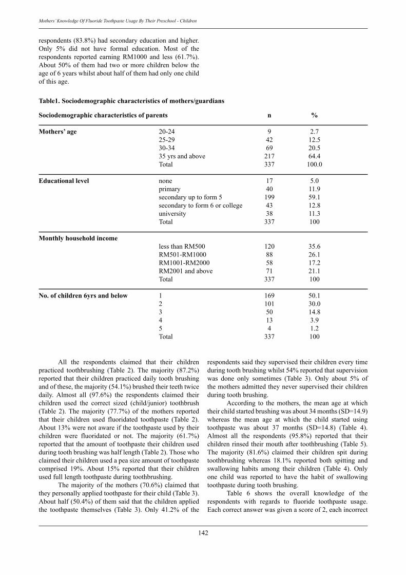
142
Mothers’ Knowledge Of Fluoride Toothpaste Usage By Their Preschool - Children
respondents(83.8%)hadsecondaryeducationandhigher.Only 5% did not have formal education. Most of therespondents reported earning RM1000 and less (61.7%).About50%of themhad twoormorechildrenbelow theageof6yearswhilstabouthalfofthemhadonlyonechildofthisage.
Table1. Sociodemographic characteristics of mothers/guardians
Sociodemographic characteristics of parents n %
Mothers’ age 20-24 9 2.7 25-29 42 12.5 30-34 69 20.5 35yrsandabove 217 64.4 Total 337 100.0
Educational level none 17 5.0 primary 40 11.9 secondaryuptoform5 199 59.1 secondarytoform6orcollege 43 12.8 university 38 11.3 Total 337 100
Monthly household income lessthanRM500 120 35.6 RM501-RM1000 88 26.1 RM1001-RM2000 58 17.2 RM2001andabove 71 21.1 Total 337 100
No. of children 6yrs and below 1 169 50.1 2 101 30.0 3 50 14.8 4 13 3.9 5 4 1.2 Total 337 100
All the respondents claimed that their childrenpracticed toothbrushing (Table 2). The majority (87.2%)reportedthattheirchildrenpracticeddailytoothbrushingandofthese,themajority(54.1%)brushedtheirteethtwicedaily. Almost all (97.6%) the respondents claimed theirchildren used the correct sized (child/junior) toothbrush(Table 2).The majority (77.7%) of the mothers reportedthat their children used fluoridated toothpaste (Table 2).About13%werenotawareifthetoothpasteusedbytheirchildren were fluoridated or not. The majority (61.7%)reportedthattheamountoftoothpastetheirchildrenusedduringtoothbrushingwashalflength(Table2).Thosewhoclaimedtheirchildrenusedapeasizeamountoftoothpastecomprised 19%. About 15% reported that their childrenusedfulllengthtoothpasteduringtoothbrushing. Themajorityof themothers (70.6%)claimed thattheypersonallyappliedtoothpastefortheirchild(Table3).Abouthalf(50.4%)ofthemsaidthatthechildrenappliedthe toothpaste themselves (Table 3). Only 41.2% of the
respondentssaidtheysupervisedtheirchildreneverytimeduringtoothbrushingwhilst54%reportedthatsupervisionwas done only sometimes (Table 3). Only about 5% ofthemothersadmittedtheyneversupervisedtheirchildrenduringtoothbrushing. According to the mothers, the mean age at whichtheirchildstartedbrushingwasabout34months(SD=14.9)whereas the mean age at which the child started usingtoothpaste was about 37 months (SD=14.8) (Table 4).Almost all the respondents (95.8%) reported that theirchildrenrinsedtheirmouthafter toothbrushing(Table5).The majority (81.6%) claimed their children spit duringtoothbrushing whereas 18.1% reported both spitting andswallowing habits among their children (Table 4). Onlyone child was reported to have the habit of swallowingtoothpasteduringtoothbrushing. Table 6 shows the overall knowledge of therespondents with regards to fluoride toothpaste usage.Eachcorrectanswerwasgivenascoreof2,eachincorrect
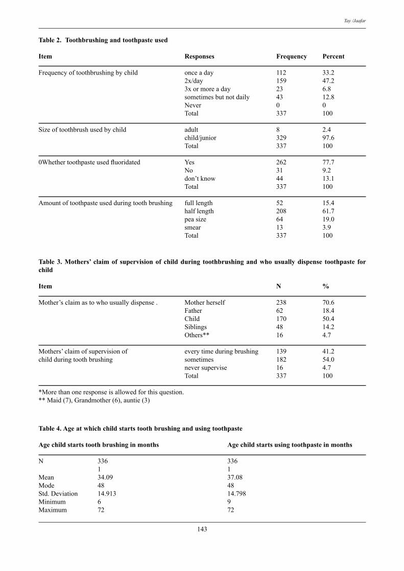
143
Tay /Jaafar
Table 2. Toothbrushing and toothpaste used
Item Responses Frequency Percent
Frequencyoftoothbrushingbychild onceaday 112 33.2 2x/day 159 47.2 3xormoreaday 23 6.8 sometimesbutnotdaily 43 12.8 Never 0 0 Total 337 100
Sizeoftoothbrushusedbychild adult 8 2.4 child/junior 329 97.6 Total 337 100
0Whether toothpaste used fluoridated Yes 262 77.7 No 31 9.2 don’tknow 44 13.1 Total 337 100
Amountoftoothpasteusedduringtoothbrushing fulllength 52 15.4 halflength 208 61.7 peasize 64 19.0 smear 13 3.9 Total 337 100
Table 3. Mothers’ claim of supervision of child during toothbrushing and who usually dispense toothpaste for child
Item N %
Mother’sclaimastowhousuallydispense. Motherherself 238 70.6 Father 62 18.4 Child 170 50.4 Siblings 48 14.2 Others** 16 4.7
Mothers’claimofsupervisionof everytimeduringbrushing 139 41.2childduringtoothbrushing sometimes 182 54.0 neversupervise 16 4.7 Total 337 100
*Morethanoneresponseisallowedforthisquestion.**Maid(7),Grandmother(6),auntie(3)
Table 4. Age at which child starts tooth brushing and using toothpaste
Age child starts tooth brushing in months Age child starts using toothpaste in months
N 336 336 1 1Mean 34.09 37.08Mode 48 48Std.Deviation 14.913 14.798Minimum 6 9Maximum 72 72
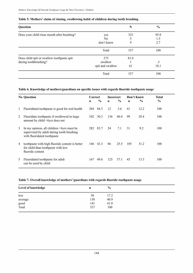
144
Mothers’ Knowledge Of Fluoride Toothpaste Usage By Their Preschool - Children
Table 5. Mothers’ claim of rinsing, swallowing habit of children during tooth brushing
Question N %
Doesyourchildrinsemouthafterbrushing? yes 323 95.8 No 5 1.5 don’tknow 9 2.7
Total 337 100
Doeschildspitorswallowtoothpastespit 275 81.6duringtoothbrushing? swallow 1 .3 spitandswallow 61 18.1
Total 337 100
Table 6. Knowledge of mothers/guardians on specific issues with regards fluoride toothpaste usage
No Question Correct Incorrect Don’t Know Total n % n % n % %
1 Fluoridatedtoothpasteisgoodfororalhealth 284 84.3 12 3.6 41 12.2 100
2 Fluoridatetoothpasteifswallowedinlarge 102 30.3 136 40.4 99 29.4 100 amountbychild<6yrsdoesnot
3 Inmyopinion,allchildren<6yrsmustbe 282 83.7 24 7.1 31 9.2 100 supervisedbyadultduringtoothbrushing with fluoridated toothpaste
4 toothpaste with high fluoride content is better 146 43.3 86 25.5 105 31.2 100 forchildthantoothpastewithlow fluoride content
5 Fluoridatedtoothpasteforadult 167 49.6 125 37.1 45 13.3 100 canbeusedbychild
Table 7. Overall knowledge of mothers’/guardians with regards fluoride toothpaste usage
Level of knowledge n %
low 58 17.2average 138 40.9good 141 41.8Total 337 100
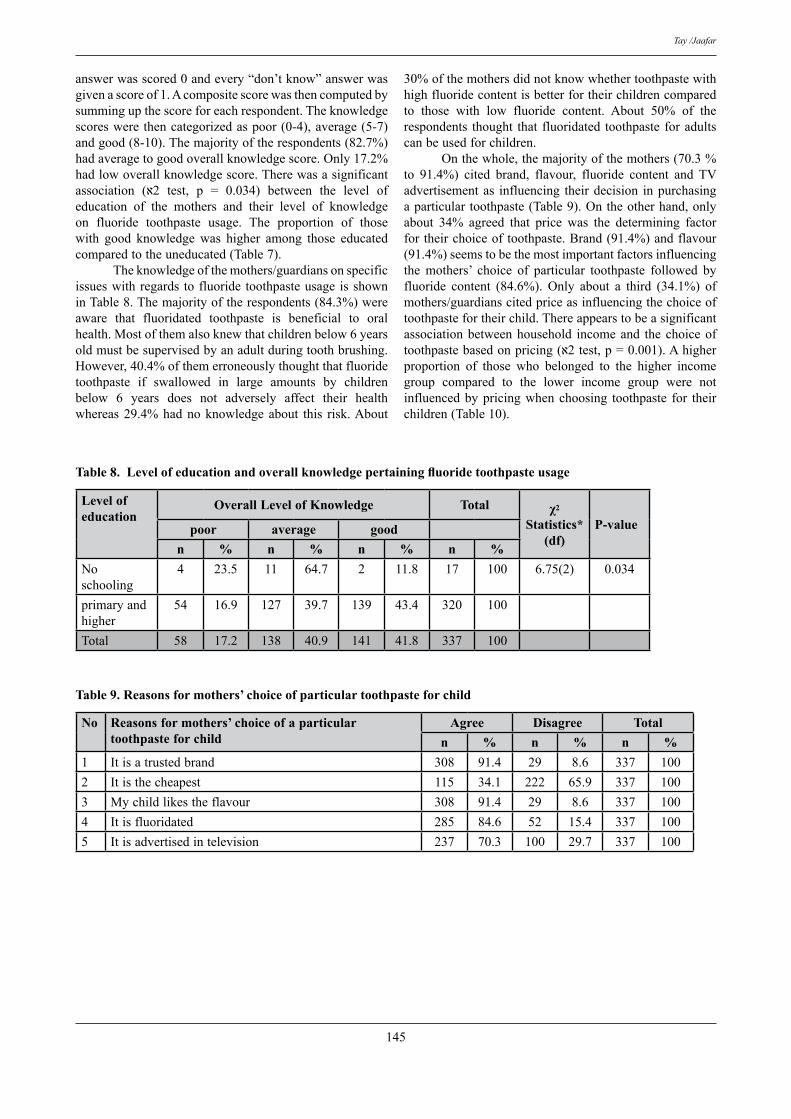
145
Tay /Jaafar
answerwasscored0andevery“don’tknow”answerwasgivenascoreof1.Acompositescorewasthencomputedbysummingupthescoreforeachrespondent.Theknowledgescoreswerethencategorizedaspoor(0-4),average(5-7)andgood(8-10).Themajorityoftherespondents(82.7%)hadaveragetogoodoverallknowledgescore.Only17.2%hadlowoverallknowledgescore.Therewasasignificantassociation (2א test, p = 0.034) between the level of education of the mothers and their level of knowledgeon fluoride toothpaste usage. The proportion of thosewith good knowledge was higher among those educatedcomparedtotheuneducated(Table7). Theknowledgeofthemothers/guardiansonspecificissueswithregards tofluoride toothpasteusage isshowninTable8.Themajorityoftherespondents(84.3%)wereaware that fluoridated toothpaste is beneficial to oralhealth.Mostofthemalsoknewthatchildrenbelow6yearsoldmustbesupervisedbyanadultduringtoothbrushing.However,40.4%ofthemerroneouslythoughtthatfluoridetoothpaste if swallowed in large amounts by childrenbelow 6 years does not adversely affect their healthwhereas29.4%hadnoknowledgeabout this risk.About
30%ofthemothersdidnotknowwhethertoothpastewithhighfluoridecontentisbetterfortheirchildrencomparedto those with low fluoride content. About 50% of therespondents thought that fluoridated toothpaste foradultscanbeusedforchildren. Onthewhole,themajorityofthemothers(70.3%to 91.4%) cited brand, flavour, fluoride content and TVadvertisementas influencing theirdecision inpurchasingaparticulartoothpaste(Table9).Ontheotherhand,onlyabout 34% agreed that price was the determining factorfortheirchoiceoftoothpaste.Brand(91.4%)andflavour(91.4%)seemstobethemostimportantfactorsinfluencingthe mothers’ choice of particular toothpaste followed byfluoride content (84.6%). Only about a third (34.1%) ofmothers/guardianscitedpriceasinfluencingthechoiceoftoothpastefortheirchild.Thereappearstobeasignificantassociationbetweenhousehold incomeand thechoiceoftoothpaste based on pricing (2א test, p = 0.001). A higher proportion of those who belonged to the higher incomegroup compared to the lower income group were notinfluencedbypricingwhenchoosing toothpaste for theirchildren(Table10).
Level of education
Overall Level of Knowledge Total χ2 Statistics*
(df)P-valuepoor average good
n % n % n % n %Noschooling
4 23.5 11 64.7 2 11.8 17 100 6.75(2) 0.034
primaryandhigher
54 16.9 127 39.7 139 43.4 320 100
Total 58 17.2 138 40.9 141 41.8 337 100
Table 8. Level of education and overall knowledge pertaining fluoride toothpaste usage
No Reasons for mothers’ choice of a particular toothpaste for child
Agree Disagree Totaln % n % n %
1 Itisatrustedbrand 308 91.4 29 8.6 337 1002 Itisthecheapest 115 34.1 222 65.9 337 1003 Mychildlikestheflavour 308 91.4 29 8.6 337 1004 Itisfluoridated 285 84.6 52 15.4 337 1005 Itisadvertisedintelevision 237 70.3 100 29.7 337 100
Table 9. Reasons for mothers’ choice of particular toothpaste for child
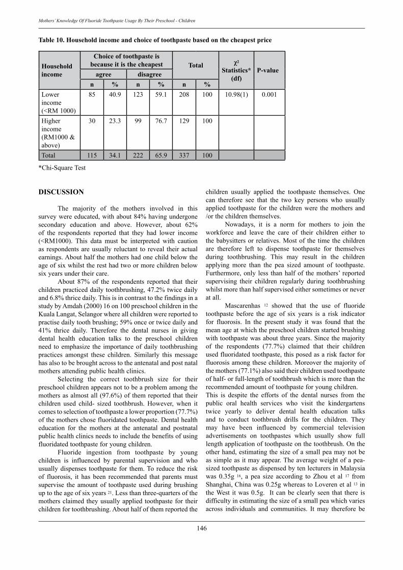
146
Mothers’ Knowledge Of Fluoride Toothpaste Usage By Their Preschool - Children
DISCUSSION
The majority of the mothers involved in thissurveywereeducated,withabout84%havingundergonesecondary education and above. However, about 62%of the respondents reported that they had lower income(<RM1000). This data must be interpreted with cautionas respondentsareusually reluctant to reveal theiractualearnings.Abouthalfthemothershadonechildbelowtheageofsixwhilsttheresthadtwoormorechildrenbelowsixyearsundertheircare. About 87% of the respondents reported that theirchildrenpracticeddailytoothbrushing,47.2%twicedailyand6.8%thricedaily.ThisisincontrasttothefindingsinastudybyAmdah(2000)16on100preschoolchildrenintheKualaLangat,Selangorwhereallchildrenwerereportedtopractisedailytoothbrushing;59%onceortwicedailyand41% thrice daily. Therefore the dental nurses in givingdental health education talks to the preschool childrenneed toemphasize the importanceofdaily toothbrushingpractices amongst these children. Similarly this messagehasalsotobebroughtacrosstotheantenatalandpostnatalmothersattendingpublichealthclinics. Selecting the correct toothbrush size for theirpreschoolchildrenappearsnottobeaproblemamongthemothersasalmostall(97.6%)ofthemreportedthattheirchildren used child- sized toothbrush. However, when itcomestoselectionoftoothpastealowerproportion(77.7%)ofthemotherschosefluoridatedtoothpaste.Dentalhealtheducation for the mothers at the antenatal and postnatalpublichealthclinicsneedstoincludethebenefitsofusingfluoridatedtoothpasteforyoungchildren. Fluoride ingestion from toothpaste by youngchildren is influenced by parental supervision and whousuallydispenses toothpaste for them.Toreduce the riskof fluorosis, it has been recommended that parents mustsupervise the amountof toothpasteusedduringbrushinguptotheageofsixyears21.Lessthanthree-quartersofthemothersclaimed theyusuallyapplied toothpaste for theirchildrenfortoothbrushing.Abouthalfofthemreportedthe
Household income
Choice of toothpaste is because it is the cheapest Total χ2
Statistics*(df)
P-valueagree disagreen % n % n %
Lowerincome(<RM1000)
85 40.9 123 59.1 208 100 10.98(1) 0.001
Higherincome(RM1000&above)
30 23.3 99 76.7 129 100
Total 115 34.1 222 65.9 337 100
*Chi-SquareTest
Table 10. Household income and choice of toothpaste based on the cheapest price
children usually applied the toothpaste themselves. Onecan therefore see that the two key persons who usuallyapplied toothpaste for the childrenwere themothers and/orthechildrenthemselves. Nowadays, it is a norm for mothers to join theworkforce and leave the care of their children either tothebabysittersorrelatives.Mostof the timethechildrenare therefore left to dispense toothpaste for themselvesduring toothbrushing. This may result in the childrenapplying more than the pea sized amount of toothpaste.Furthermore,only less thanhalfof themothers’ reportedsupervising their children regularly during toothbrushingwhilstmorethanhalfsupervisedeithersometimesorneveratall. Mascarenhas 12 showed that the use of fluoridetoothpaste before the age of six years is a risk indicatorfor fluorosis. In the present study it was found that themeanageatwhichthepreschoolchildrenstartedbrushingwithtoothpastewasaboutthreeyears.Sincethemajorityof the respondents (77.7%) claimed that their childrenusedfluoridatedtoothpaste,thisposedasariskfactorforfluorosisamongthesechildren.Moreoverthemajorityofthemothers(77.1%)alsosaidtheirchildrenusedtoothpasteofhalf-orfull-lengthoftoothbrushwhichismorethantherecommendedamountoftoothpasteforyoungchildren.This is despite the efforts of the dental nurses from thepublic oral health services who visit the kindergartenstwice yearly to deliver dental health education talksand to conduct toothbrush drills for the children. Theymay have been influenced by commercial televisionadvertisements on toothpastes which usually show fulllengthapplicationoftoothpasteonthetoothbrush.Ontheotherhand,estimatingthesizeofasmallpeamaynotbeassimpleasitmayappear.Theaverageweightofapea-sizedtoothpasteasdispensedbytenlecturersinMalaysiawas 0.35g 16, a pea size according to Zhou et al 17 fromShanghai,Chinawas0.25gwhereastoLoverenetal13intheWest itwas0.5g. Itcanbeclearlyseen that there isdifficultyinestimatingthesizeofasmallpeawhichvariesacross individuals and communities. It may therefore be
147
Tay /Jaafar
pertinenttoadopttherecommendationbyRock18thatthebrushbemerelysmearedwithtoothpasteasthecommonlyrecommendedpea-sizedquantityasperceivedbydifferentindividualsandcommunitiesmaybetoomuch. The amount of fluoride ingested from toothpastewassignificantlyreducedbyrinsingand/orspittinghabitsduring tooth brushing 13. Though the majority of thepreschoolchildrenwere reported to rinseandspitduringtoothbrushing,inadvertentingestionoftoothpastecanstilloccur due to the swallowing reflexes of young childrenbeingnotfullydeveloped. Knowledge on fluoride toothpaste usage does notseem to be a problem as the majority of them (83.7%)achieved average to good knowledge score. However,mothers seemed to be less knowledgeable with regardsto certain specific issues, i.e. the danger of accidentallyswallowing fluoride toothpaste in large amounts and theuseofthecorrectformulationoffluoridatedtoothpasteforchildren.About40%ofthemothersdidnotagreeandabout30%didnotknowthataccidentallyswallowingfluoridatedtoothpaste in large amounts can adversely affect dentalhealth.Many(37.1%)erroneouslythoughtthatfluoridatedtoothpasteforadultscanbeusedforchildrenwhilstsome13% were ignorant over this issue. Hence, future oralhealthmessagestothemothersbydentalpersonnelshouldincorporate and emphasise these specific shortcomings.Motherswillalsoneedtobeempoweredtomakeinformedchoicesespeciallywithregardsthepurchaseoffluoridatedtoothpaste.Morethanaquarterofthemthoughttoothpastewith higher fluoride content is better for children andaboutathirdwereignorantastowhethertoothpastewithhigherfluoridecontentisbetterforchildrenorviceversa.Several recommendations were put forth by a group ofexperts from four European countries who gathered atBasel, oneofwhichwas to increase the fluoride contentin toothpaste for toddlers from 250ppm to 500ppm15.Dentalpersonnelneed toempower themotherswith thisknowledge to help them make appropriate choices whenpurchasingtoothpasteforyoungchildrenundertheircare.ItnoteworthythattheMinistryofHealthhasalsotakenagoodproactivesteptoreducetherecommendedlevelforpublicwaterfluoridationfromarangeof0.5-0.7ppmtoarangeof0.4-0.6ppminordertoreducetheriskoffluorosiswhilstmaintainingthebeneficialeffectsofcariostasis. Brand and flavour seemed to be the two mainfactors influencing the toothpaste purchasing behaviourof themothersof thepreschoolchildren involved in thissurvey. The majority of the mothers tend to purchaseflavoured toothpaste which appeals to their children.Levy 22 reported that insomecases, theuseofdentifriceflavoured for children could put preschool children atincreasedriskfordentalfluorosis.However,inothercases,the special flavours could enhance children’s acceptanceofhavingtheirteethbrushedregularlytherebyreapingthepreventivebenefitsoffluoridedentifrice.Themajorityofthe mothers (84.3%) involved in this survey were awarethatfluoridatedtoothpasteisgoodfororalhealth.Thisisreflectedintheappropriatetoothpastepurchasingbehaviour
of84.6%oftherespondentswhocitedfluoridecontentasinfluencing their decision in the purchase of toothpaste.The toothpaste purchasing behaviour of more than twothirdsofthemothersseemedtobeinfluencedbytelevisionadvertisements. It is widely known that media has a farreachinginfluenceonthepublic.Thismayinpartexplainthepreferenceofmothersforcertainbrandsoftoothpaste.Interestingly, only about a-third of the respondents citedthat their purchasing behaviour is influenced by price.This is in contrast to the findings of a survey by Mat 16where almost all the mothers (96%) cited that price wasthedeterminingfactor in theirpurchaseof toothpaste fortheir children. However, there is a significant differencein purchasing behaviour with regards to pricing betweenthehigherincomeandthelowerincomegroup.Thismaybebecausethoseinthehigherincomegrouptendtolookforqualityorpreferenceoftheirchildrenwhenpurchasingtoothpasteasthepriceisnotanissuewiththisgroup.
CONCLUSION AND RECOMMENDATIONS More than80%of therespondentshadaverage togoodoverallknowledgescore.Mothersseemedtobelessknowledgeablewithregardstocertainspecificissues,i.e.thedangerofaccidentallyswallowingfluoridetoothpasteinlargeamountsandtheuseofthecorrectformulationoffluoridatedtoothpasteforchildren.Therewasasignificantassociationbetweenthelevelofeducationofthemothersandtheirlevelofknowledgeonfluoridetoothpasteusage.Abouthalfof them reported the childrenusually appliedthe toothpaste themselves. Only less than half of themothers’ reported supervising their children regularlyduring toothbrushing whilst the rest supervised eithersometimes or never at all. The majority of the motherssaid their children used toothpaste of half- or full-lengthof toothbrush which is more than the recommendedamount of toothpaste for young children. Brand andflavour seemed to be the two main factors influencingthetoothpastepurchasingbehaviourofthemothersofthepreschoolchildreninvolvedinthissurvey Future oral health messages for the preschoolchildren and mothers in Perlis will have to target areasfound lacking in terms of knowledge and practices inrelationtofluoridetoothpasteusage.Oralhealthmessagesfor mothers or guardians should emphasise on theimportance of parental supervision during toothbrushingandapplicationofpea-sizeorsmearamountoftoothpasteduring brushing, especially if they cannot afford to buyspecial lower fluoride toothpaste for children due toeconomic limitations. Mothers should be equipped withappropriateknowledgewithregardstofluoridetoothpasteusageandempowered tomake informedandappropriatechoiceswhenpurchasingtoothpastefortheirchildren.
148
Mothers’ Knowledge Of Fluoride Toothpaste Usage By Their Preschool - Children
ACKNOWLEDGEMENTS
We acknowledge the assistance of Dr Dahari DolPatah,DrShafinazbtMadArsyad,MdmNoayubtAdin,MdmManirahbtHashimandallpersonnelofPerlisOralHealth Services Department involved in this researchproject.
REFERENCES
1. MurrayJ.J.,PittsN.B.(1997).Trendsinoralhealth.In:PineC.M.(ed.),CommunityOralHealth.Oxford:Wright.
2. DentalServicesDivision,MinistryofHealthMalaysia(1995).Dental Epidemiological survey of Pre-school Children InMalaysia1995.p26.
3. Oral Health Division, Ministry of Health Malaysia (1997).Nationaloralhealthsurveyofschoolchildren1997(NOHSS’97).p44.MOH/K/GIG/6.98(RR),1998.
4. Oral Health Division, Ministry of Health Malaysia (2004).Annualreport2004.Healthmanagementinformationsystem(oral health sub-system), information and documentationunit.p48,
5. Glass, R.L.(1986). Fluoride dentifrice: the basis for thedeclineincariesprevalence.JRSocMedSuppl.79;15-17.
6 Brathall, D. Hansel Peterson, G, Sundberg, H. (1996).Reasonsforthecariesdecline.Whatdotheexpertsbelieve?EurJofOralScience104,416-422.
7. Abdul-Kadir R., Abdol-Latif L. (1998). Fluoride Level inDentifrices.AnnalsDentUnivMalaya5,2-5.
8. Horowitz,H.S.(1992).Theneedfortoothpasteswithlowerthanconventionalfluorideconcentrationforpreschool-agedchildren.JofPublicHealthDent52,216-221.
9. Oral Health Division, Ministry of Health Malaysia (2001).Fluoride enamel opacities in 16-year-old school children.p40.MOH/GIG/2.2001(RR),2001.
10.Maznah,M.N.,Sheiham,A.,Tsakos,G.(2006).PsychologicalimpactsofdentalfluorosisamongMalaysianschoolchildren.Abstract Oral Health Research Conference, Oral healthDivision,MinistryofHealth,Malaysiap15.
11.Bently, E.M. (1999). Elwood, R., Davies, R.M. FluorideIngestion from toothpaste by Young Children. Br Dent J 186, 222-226.
12.Mascarenhas,A.K., Burt, B.A. (1998). Fluorosis risk fromearlyexposuretofluoridetoothpaste.CommunityDentandOralEpidemiol26,241-248.
13.Loveren,C.V.,Ketley,C.E.,Cochran,J.A.,Duckworth,R.M.,O’Mullane,D.M.(2004).Fluorideingestionfromtoothpaste:fluoriderecoveredfromthetoothbrush, theexpectorateandtheafter-brushrinses.CommunityDentandOralEpidemiol32(suppl.),154-161.
14.O’Mullane,D.M.(2004)Fluorideingestionfromtoothpaste:conclusionsofEuropeanUnion-fundedmulticentreproject.CommunityDentandOralEpidermiol32(suppl.1),74-76.
15.Konig, K.G. (2002). New recommendations concerningfluoride content of toddler toothpaste- consequences forsystemicapplicationoffluoride.Gesundheitswesen,64,33-38.
16 AmdahM.(2000).Fluoridetoothpasteusageamongpreschoolchildren. MCD Thesis, Faculty of Dentistry, University ofMalaya,KualaLumpur.
17. Zhou, J., Feng, X.P., Liu, Y.L. (2002). Effects of different quantities of fluoride toothpaste on fluoride intake bypreschool children. Shanghai Kou Qiang Yi Xue 11(1), 13-15.0.
18. Rock, W.P. (1994) Young Children and Fluoride toothpaste. BrDentJ1,17-20.
19.Curnow, M.M.T.. Pine, C.M., Burnside, G., Nicholas, J.A.,ChestersR.K.,HuntingtonE.(2002).Arandomisedcontrolledtrialoftheefficacyofsupervisedtoothbrushinginhighcariesriskchildren.CariesRes36,294-300.
20.Blinkhorn A.S. (1978). Influence of social norms ontoothbrushingbehaviourof preschool children.CommunityDentOralEpidemiol6,222-226.
21.HoltR.D.,NunnJ.H.,RockW.P.,andPageJ.BritishSocietyofPaediatricDentistry:Apolicydocumentonfluoridedietarysupplementsandfluoridetoothpastesforchildren.Int.J.Paed.Dent.1996;6:139-142.
22.Levy,S.M.,Maurice,T.J.,Jakobsen,J.R.(1992).APilotStudyOfPreschoolers’UseOfRegular-FlavouredDentifricesAndThoseFlavouredForChildren.PaediatricDent14,388-391.
Address for correspondence:
Dr Tay Hong LukKlinik Pergigian, Klinik Kesihatan Kangar,Jalan Kolam, 01000Kangar, Perlis, Malaysia.Tel: 04-9778333 ext 137Fax: 04-9772710E-mail: [email protected]
Management of Full Mouth Prosthodontic Rehabilitation Utilizing a Combination of Porcelain Fused to Metal and High Strength Zirconium-oxide CrownsAnsgar C Cheng , BDS, MS, FRCDC, FRACDS, FAMS, Consultant Prosthodontist, Specialist Dental Group, Mount Elizabeth Hospital, Singapore, Adjunct Associate Professor, National University of Singapore
Elvin WJ Leong , BDS, MDS, MRDRCS, FAMS, Consultant Prosthodontist, Specialist Dental Group, Mount Elizabeth Hospital, Singapore
Helena Lee, BDS, MSc, Consultant Periodontist, Specialist Dental Group, Mount Elizabeth Hospital, Singapore
ABSTRACT
Full mouth fixed prosthodontic rehabilitation of a compromised occlusion is always a clinical challenge. Precise diagnosis, prudent choice of prosthodontic materials, and meticulous treatment execution are essential for successful treatment outcome over a long period of time. The prosthodontic treatment of a partially edentulous oral cavity with loss of vertical dimension of occlusion is presented. Innovative prosthodontic materials were used in this report.
149
Malaysian Dental Journal (2008) 29(2) 149-153© 2008 The Malaysian Dental Association
InTroducTIon
Prudent clinical judgement, careful considerationof risks and benefits of various treatment options areessentialforthetreatmentplanningandlong-termsuccessof prosthodontic treatment.1 It is known that loss ofvertical dimension of occlusion may pose significantclinical difficulties in prosthodontic treatment.2,3 There-establishment and maintenance of a new verticaldimensionofocclusionisseldomtaughtintheundergraduatedentalcurriculum. Vertical dimension of occlusion is defined as thevertical measurement of the face between two selectedpoints superior and inferior to the oral cavity when theoccludingmembersareincontact.4Variousmethodshavebeenproposedfortheassessmentandre-establishmentoftreatment vertical dimension.3 The vertical measurementofphysiological restposition shouldhaveahighervaluethan the vertical dimension of occlusion; the differenceis referred to as the interocclusal rest space4 , which isessentialfornormalpatientfunction. It isknownthatasteetharewornout thealveolarbone may undergo an adaptive process which maycompensate for the loss of tooth structure.5 Therefore,the vertical dimension of occlusion should be carefullyassessed before the initiation of restorative procedures.In general, alteration of vertical dimension of occlusionshould be approached with great care5, 6 and excessivechanges of vertical dimension of occlusion should beavoided.6
One of the challenges in full mouth fixedprosthodontic rehabilitation is in obtaining an accurateimpressionofmultipleabutmentteeth.Dentalimpressionssent to commercial laboratories for conventional fixedprostheseshavecommonlybeen found tobedeficient inseveralrespects.7Oneofthemajordeficienciesisthatthemarginsof toothpreparationsare inadequately registeredin the definitive impression. Since the master blueprintfor crown restorations is the definitive impression,8 it iscrucial thatagood impression techniquebeemployed toobtain an accurate impression that will allow fabricationofprecisely-fittingindirectrestorations,whichmayinturndeterminetherestorations’longevity.9
Traditionalporcelain-fused-to-metalanteriorcrownrestorationsrequiretheplacementoflabialcrownmarginswithinthegingivalsulcusinordertomaskthehueandvaluetransitionbetweentherootsurfaceandtheporcelain-fused-to-metal restoration. However, intracrevicular placementofcrownmarginsarerelatedtoadverseperiodontaltissueresponse.10-12Fromaperiodontalpointofview,preparationmarginsarebestkeptawayfromthegingivalmargin.12
The dentition, the masticatory muscles and thetemporomandibular joints form a class 3 lever system.In a class 3 lever system, functional load is inverselyproportionaltothelengthoftheleverarm.Anteriorteethare under less functional load compared with posteriorteeth.Porcelain-fused-to-metalrestorationsarecommonlyusedintheposteriorteethbecauseofit’swell-documentedlong term clinical track record in anterior and posterior
MALAYSIAn dEnTAL JournAL
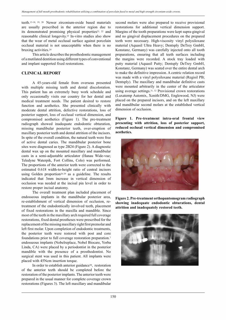
150
Management of full mouth prosthodontic rehabilitation utilizing a combination of porcelain fused to metal and high strength zirconium-oxide crowns.
teeth.13-18, 19, 20 Newer zirconium-oxide based materialsare usually prescribed in the anterior region due toits demonstrated promising physical properties21, 22 andreasonableclinical longevity.23 In-vitro studiesalso showthat the wear of metal occlusal surface against porcelainocclusal material is not unacceptable when there is nobruxingactivities.24
Thisarticledescribestheprosthodonticmanagementofamutilateddentitionusingdifferenttypesofconventionalandimplantsupportedfixedrestorations.
cLInIcAL rEPorT
A 45-years-old female from overseas presentedwith multiple missing teeth and dental discoloration.This patient has an extremely busy work schedule andonly occasionally visits our country for her dental andmedical treatment needs. The patient desired to restorefunction and aesthetics. She presented clinically withmoderate dental attrition, defective restorations, loss ofposteriorsupport,lossofocclusalverticaldimension,andcompromised aesthetics (Figure 1). The pre-treatmentradiograph showed inadequate endodontic obturation,missing mandibular posterior teeth, over-eruption ofmaxillaryposteriorteethanddentalattritionoftheincisors.Inspiteoftheoverallcondition,thenaturalteethwerefreeof active dental caries. The mandibular posterior bonesiteswerediagnosedastype2B24(Figure2).Adiagnosticdentalwaxupon themountedmaxillaryandmandibularcasts in a semi-adjustable articulator (Hanau Wide-vue;Teledyne Waterpik, Fort Collins, Colo) was performed.Theproportionsoftheanteriorteethwerecorrectedtotheestimated 0.618 width-to-height ratio of central incisorsusing Golden proportion26-29 as a guideline. The resultsindicated that 3mm increase in vertical dimension ofocclusion was needed at the incisal pin level in order torestoreproperincisalanatomy. The overall treatment plan included placement ofendosseous implants in the mandibular posterior area,re-establishment of vertical dimension of occlusion, re-treatmentoftheendodonticallyinvolvedteeth,placementof fixed restorations in the maxilla and mandible. Sincemostoftheteethinthemaxillaryarchrequiredfullcoveragerestorations,fixeddentalprostheseswereprescribedforthereplacementofthemissingmaxillaryrightfirstpremolarandleftfirstmolar.Uponcompletionofendodontictreatments,the posterior teeth were restored with post and corefoundationspriortofullcoveragerestorationpreparation.7endosseousimplants(Nobelreplace,NobelBiocare,YorbaLinda,CA)wereplacedbyaperiodontistintheposteriormandible with the presence of a prosthodontist. Nosurgical stentwasused in thispatient.All implantswereplacedwith45Ncminsertiontorque. Inordertoestablishanteriorguidance30,restorationof the anterior teeth should be completed before therestorationoftheposteriorimplants.Theanteriorteethwerepreparedintheusualmannerforcompletecoveragecrownrestorations(Figures3).Theleftmaxillaryandmandibular
second molars were also prepared to receive provisionalrestorations for additional vertical dimension support.Marginsofthetoothpreparationswerekeptsupra-gingivalandnogingivaldisplacementproceduresonthepreparedteeth were necessary. High-viscosity vinyl polysiloxanematerial (AquasilUltraHeavy;DentsplyDeTreyGmbH,Konstanz,Germany)wascarefullyinjectedontoalltoothpreparations, ensuring that all teeth surfaces includingthe margins were recorded. A stock tray loaded withputty material (Aquasil Putty; Dentsply DeTrey GmbH,Konstanz,Germany)wasseatedovertheentiredentalarchtomakethedefinitiveimpression.Acentricrelationrecordwasmadewithavinylpolysiloxanematerial(RegisilPB;Dentsply).Themaxillaryandmandibulardefinitivecastswere mounted arbitrarily in the center of the articulatorusingaveragesettings.31, 32Provisionalcrownrestorations(LuxatempAutomix,Xenith/DMG,Englewood,NJ)wereplacedonthepreparedincisors,andontheleftmaxillaryandmandibular secondmolars at the establishedverticaldimensionofocclusion.
Figure 1. Pre-treatment intra-oral frontal view presenting with attrition, loss of posterior support, reduced occlusal vertical dimension and compromised aesthetics.
Figure 2. Pre-treatment orthopantomogram radiograph showing inadequate endodontic obturations, dental attrition and inadequately restored teeth.
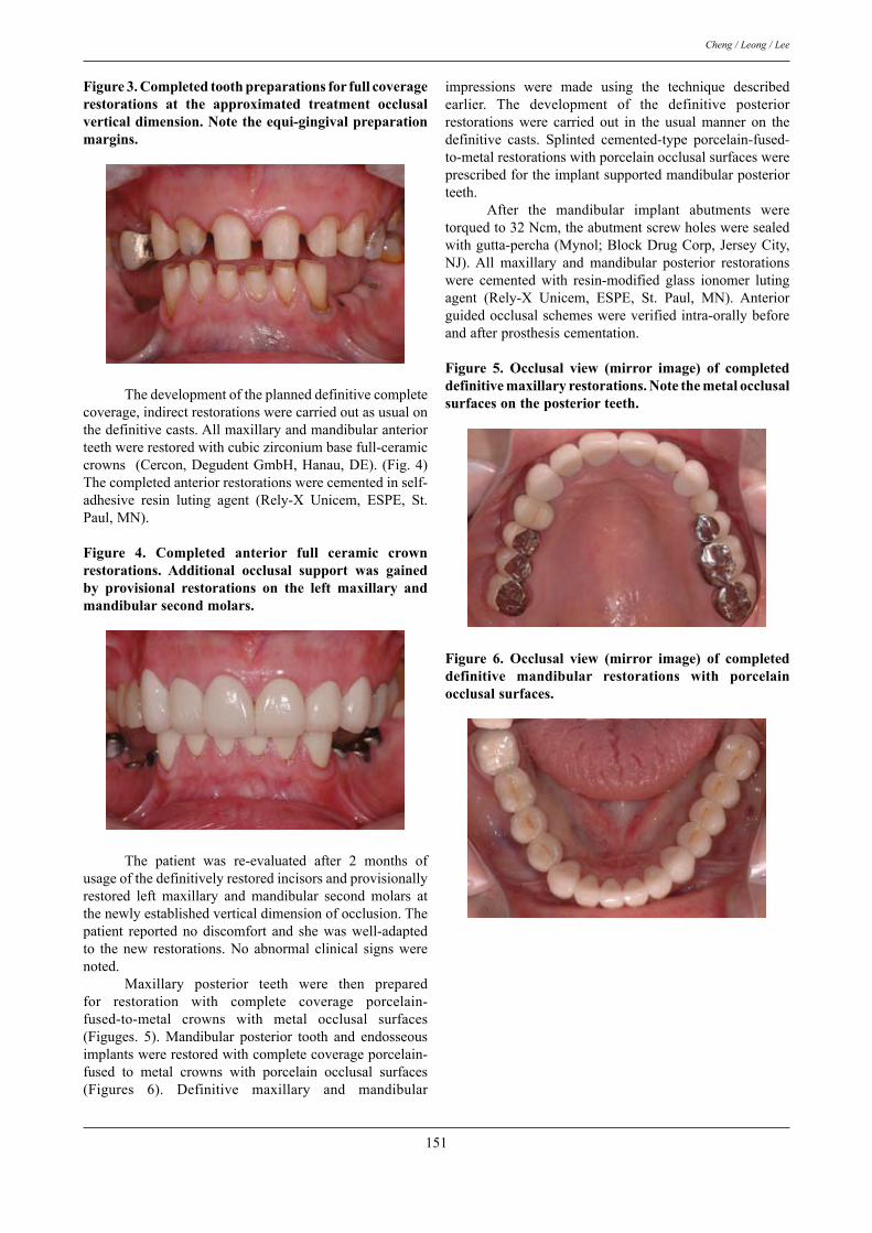
151
Cheng / Leong / Lee
Figure 3. completed tooth preparations for full coverage restorations at the approximated treatment occlusal vertical dimension. note the equi-gingival preparation margins.
Thedevelopmentoftheplanneddefinitivecompletecoverage,indirectrestorationswerecarriedoutasusualonthedefinitivecasts.Allmaxillaryandmandibularanteriorteethwererestoredwithcubiczirconiumbasefull-ceramiccrowns (Cercon,DegudentGmbH,Hanau,DE).(Fig.4)Thecompletedanteriorrestorationswerecementedinself-adhesive resin luting agent (Rely-X Unicem, ESPE, St.Paul,MN).
Figure 4. completed anterior full ceramic crown restorations. Additional occlusal support was gained by provisional restorations on the left maxillary and mandibular second molars.
The patient was re-evaluated after 2 months ofusageofthedefinitivelyrestoredincisorsandprovisionallyrestored left maxillary and mandibular second molars atthenewlyestablishedverticaldimensionofocclusion.Thepatient reportednodiscomfort and shewaswell-adaptedto thenew restorations.Noabnormal clinical signswerenoted. Maxillary posterior teeth were then preparedfor restoration with complete coverage porcelain-fused-to-metal crowns with metal occlusal surfaces(Figuges. 5). Mandibular posterior tooth and endosseousimplantswererestoredwithcompletecoverageporcelain-fused to metal crowns with porcelain occlusal surfaces(Figures 6). Definitive maxillary and mandibular
impressions were made using the technique describedearlier. The development of the definitive posteriorrestorations were carried out in the usual manner on thedefinitive casts. Splinted cemented-type porcelain-fused-to-metalrestorationswithporcelainocclusalsurfaceswereprescribedfortheimplantsupportedmandibularposteriorteeth. After the mandibular implant abutments weretorquedto32Ncm,theabutmentscrewholesweresealedwithgutta-percha(Mynol;BlockDrugCorp,JerseyCity,NJ).All maxillary and mandibular posterior restorationswere cemented with resin-modified glass ionomer lutingagent (Rely-X Unicem, ESPE, St. Paul, MN). Anteriorguidedocclusalschemeswereverifiedintra-orallybeforeandafterprosthesiscementation.
Figure 5. occlusal view (mirror image) of completed definitive maxillary restorations. note the metal occlusal surfaces on the posterior teeth.
Figure 6. occlusal view (mirror image) of completed definitive mandibular restorations with porcelain occlusal surfaces.

152
Management of full mouth prosthodontic rehabilitation utilizing a combination of porcelain fused to metal and high strength zirconium-oxide crowns.
Figure 7. Post-operative radiograph showing the combination of prosthodontic treatment modalities. The radio-opaque nature of the full ceramic restrations allow radiographic assessment of restoration fit.
dIScuSSIon
The re-establishment of a new vertical dimensionof occlusion is a crucial element of this report. It wasnecessary tomake impressionswhichregisteredall toothpreparationsintheanteriorsegmentatonce. As the patient desired a high level of aesthetics,full ceramic restorations were chosen for all anteriorrestorations. Since the minimum core thickness for thisfullceramicsystemis0.4mm, thisenabledconservationof tooth structure while achieving reasonable aestheticssimultaneously. By prescribing full ceramic restorations, intra-sulcularplacementofcrownmarginsonthelabialsurfacebecomeslessimportantfromanestheticpointofview.Inthis report, theanterior teethwereessentiallycaries free,teethpreparationmarginsweremadeatthesupra-gingivallevel andgingival retractionprocedureswereeliminated.As gingival retraction cord placement was not required,therewaslessphysicaltraumatothegingivaltissuesandlessclinicaltimewasneeded.Thisisparticularlybeneficialforthingingivalbiotypes. Full mouth rehabilitation using fixed prosthesesusually require a longer term provisional restorationin order to facilitate predictable treatment outcome. Inthis patient, due to her busy travel schedule, long termprovisional restoration for the purpose of verifying heradaptability and multiple visits for professional clinicaladjustment of provisional restorations established at thenew vertical dimension of occlusion was not feasible.The anterior teeth were restored based on the diagnosticwax up without long term provisional restoration beforedefinitivecementationofthedefinitivecrownrestorations.This treatment sequence left almost no room for clinicalerrorsintheexecutionoftheplannedtreatment.Intra-oralverification of the new occlusal scheme and detailed in-situ clinical adjustment of the restorations on the day ofprostheses insertion were essential for proper treatmentexecution. In this unique treatment approach, the patientshould be informed of the potential financial and timeimplicationsifthereisanyneedforre-fabricationofthe
definitiverestorations. In order to maximize the aesthetic outcome33,porcelain occlusal surfaces were prescribed in themandibular teeth. In the maxillary posterior teeth, metalocclusalsurfaceswereprescribedforit’seaseoffabricationandsuperiorstructuralstrength.
concLuSIon
The functional management of a complexprosthodonticrehabilitationisaclinicalchallenge.Variousrestorativematerialswereusedinthisreport.Acombinationof high strength full ceramic restorations and porcelainfused to metal restorations with metal and porcelainocclusal surfaces enhances the overall aesthetic outcomeaswell as functionalpredictabilityover a longperiodoftime.
rEFErEncES
1. GoodacreCJ,CampagniWV,AquilinoSA.Toothpreparationsfor complete crowns: An art . form based on scientificprinciples.JProsthetDent2001;85:363-376.
2. Torabinejad M,Anderson P, Bader J, Brown LJ, Chen LH,Goodacre CJ, Kattadiyil MT, Kutsenko D, Lozada J, PatelR,PetersenF,PutermanI,WhiteSN.Outcomesofrootcanaltreatment and restoration, implant-supported single crowns,fixedpartialdentures,andextractionwithoutreplacement:Asystematicreview.JProsthetDent2007;98:285-311.
3. Turrell AJW. Clinical assessment of vertical dimension. JProsthetDent2006;96:79-83.
4. Academy of Prosthodontics. The glossary of prosthodonticterms.(8thed)JProsthetDent94:2005:10-92.
5. Dawson P. Evaluation, diagnosis, and treatment of occlusalproblems.In:2nded.St.Louis:CVMosby;1989;p.56–71500-10.
6.TurnerKA,MissirlianDM.Restorationoftheextremelyworndentition.JProsthetDent1984;52:467-74.
7. Winstanley RV, Carrotte PV, Johnson A. The quality ofimpressionsforcrownsandbridgesreceivedatcommercialdentallaboratories.BrDentJ1997;183:209.
8. Donovan TE, Chee WWL. A review of contemporaryimpressionmaterialsandtechniques.DentClinNAm2004;48:445-470.
9.SorensenSE,LarsenIB,JorgensenKD.Gingivalandalveolarbonereactiontomarginalfitofsubgingivalcrownmargins.ScandJDentRes1986;94:109-14.
10.FeltonDA,KanoyBE,BayneSC,WirthmanGP.Effectofinvivocrownmargindiscrepanciesonperiodontalhealth.JProsthetDent1991;65:357–364.
11.KnoernschildKL,CampbellSD.Periodontaltissueresponsesafterinsertionofartificialcrownsandfixedpartialdentures.JProsthetDent.2000;84:492–498.
12.ReitemeierB,HänselK,WalterMH,KastnerC,ToutenburgH.Effectofposterior crownmarginplacementongingivalhealth.JProsthetDent2002;87:167-172.
13.HolmC,TidehagP,TillbergA,MolinM.LongevityandqualityofFPDs:aretrospectivestudyofrestorations30,20,and10yearsafterinsertion.IntJProsthodont2003;16:283–289.
14.WaltonTR.Anupto15-yearlongitudinalstudyof515metal-ceramicFPDs:Part2.Modesoffailureandinfluenceofvarious
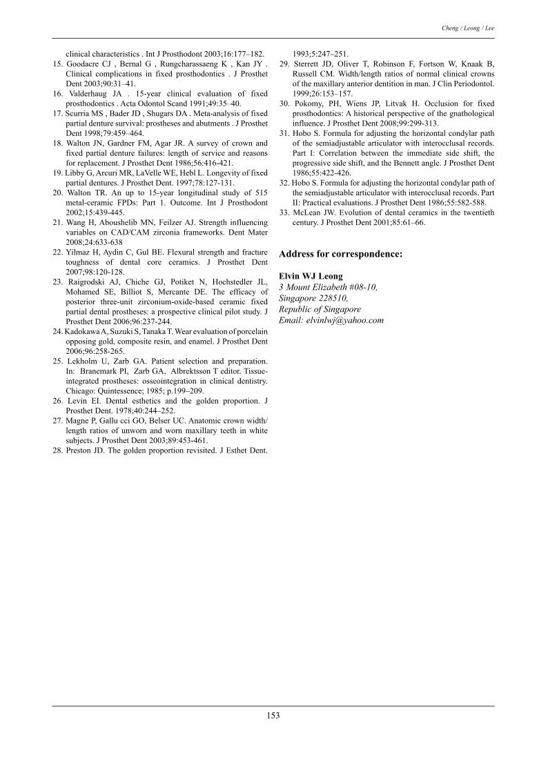
153
Cheng / Leong / Lee
clinicalcharacteristics.IntJProsthodont2003;16:177–182.15. Goodacre CJ , Bernal G , Rungcharassaeng K , Kan JY .
Clinical complications in fixed prosthodontics . J ProsthetDent2003;90:31–41.
16. Valderhaug JA . 15-year clinical evaluation of fixedprosthodontics.ActaOdontolScand1991;49:35–40.
17.ScurriaMS,BaderJD,ShugarsDA.Meta-analysisoffixedpartialdenturesurvival:prosthesesandabutments.JProsthetDent1998;79:459–464.
18.Walton JN, Gardner FM,Agar JR.A survey of crown andfixedpartialdenture failures: lengthof service and reasonsforreplacement.JProsthetDent1986;56:416-421.
19.LibbyG,ArcuriMR,LaVelleWE,HeblL.Longevityoffixedpartialdentures.JProsthetDent.1997;78:127-131.
20. Walton TR. An up to 15-year longitudinal study of 515metal-ceramic FPDs: Part 1. Outcome. Int J Prosthodont2002;15:439-445.
21.Wang H,Aboushelib MN, FeilzerAJ. Strength influencingvariables on CAD/CAM zirconia frameworks. Dent Mater2008;24:633-638
22.YilmazH,AydinC,GulBE.Flexuralstrengthandfracturetoughness of dental core ceramics. J Prosthet Dent2007;98:120-128.
23. Raigrodski AJ, Chiche GJ, Potiket N, Hochstedler JL,Mohamed SE, Billiot S, Mercante DE. The efficacy ofposterior three-unit zirconium-oxide-based ceramic fixedpartialdentalprostheses:aprospectiveclinicalpilotstudy.JProsthetDent2006;96:237-244.
24.KadokawaA,SuzukiS,TanakaT.Wearevaluationofporcelainopposinggold,compositeresin,andenamel.JProsthetDent2006;96:258-265.
25. Lekholm U, Zarb GA. Patient selection and preparation.In:BranemarkPI,ZarbGA,AlbrektssonTeditor.Tissue-integrated prostheses: osseointegration in clinical dentistry.Chicago:Quintessence;1985;p.199–209.
26. Levin EI. Dental esthetics and the golden proportion. JProsthetDent.1978;40:244–252.
27.MagneP,GallucciGO,BelserUC.Anatomiccrownwidth/length ratios of unworn and worn maxillary teeth in whitesubjects.JProsthetDent2003;89:453-461.
28.PrestonJD.Thegoldenproportionrevisited.JEsthetDent.
1993;5:247–251.29. Sterrett JD, Oliver T, Robinson F, Fortson W, Knaak B,
Russell CM.Width/length ratios of normal clinical crownsofthemaxillaryanteriordentitioninman.JClinPeriodontol.1999;26:153–157.
30. Pokomy, PH, Wiens JP, Litvak H. Occlusion for fixedprosthodontics:Ahistoricalperspectiveofthegnathologicalinfluence.JProsthetDent2008;99:299-313.
31.HoboS.Formulaforadjustingthehorizontalcondylarpathof the semiadjustable articulatorwith interocclusal records.Part I: Correlation between the immediate side shift, theprogressivesideshift,andtheBennettangle.JProsthetDent1986;55:422-426.
32.HoboS.Formulaforadjustingthehorizontalcondylarpathofthesemiadjustablearticulatorwithinterocclusalrecords.PartII:Practicalevaluations.JProsthetDent1986;55:582-588.
33. McLean JW. Evolution of dental ceramics in the twentiethcentury.JProsthetDent2001;85:61–66.
Address for correspondence:
Elvin WJ Leong3 Mount Elizabeth #08-10, Singapore 228510, Republic of SingaporeEmail: [email protected]
What Expert Says … Periodontal Abscess Norhidayah. BDS(Malaya), Dental Officer, Selama Dental Clinic,Perak
Khamiza. BDS (Malaya), MClinDent (Perio)(Malaya), Periodontal Specialist Clinic, Taiping Hospital, Perak
154
Malaysian Dental Journal (2008) 29(2) 154-157© 2008 The Malaysian Dental Association
MALAYSIAN DENTAL JOURNAL
PERIODONTAL ABSCESS
Abscess of the periodontium is a localizedpurulent inflammation of the periodontal tissues1. It hasbeen classified into three categories which are gingivalabscess, periodontal abscess and pericoronal abscess2,
3. A periodontal abscess can be defined as a localizedpurulent infection affecting the tissues surrounding aperiodontal pocket that can lead to the destruction ofsupporting structures4 including tortuous periodontalpockets, furcation involvement, and intrabony defects5.The lesion may be acute or chronic abscess.A localizedacuteabscessmayprogresstoachronicabscessifthepusdrains througha fistula into theoutergingivalsurfaceorintotheperiodontalpocket6.
PATHOGENESIS AND MICROBIOLOGY
Thebasicpathogenicmechanismsoftheperiodontalabscess do not differ from other abscesses in the body.The formation of the periodontal abscess is initiated bythe multiplication and invasion of periodontal tissuesby selected species of periodontal bacteria. It has beenreported,theincreasedbacterialactivitycouldbetheresultofanimbalanceinbacterialhemostasis,destructionoftheepitheliumbarrier,and/orbyrandomevents7. The microbiota of periodontal abscess has beencharacterized by the presence of periodontal pathogenspresent inchronicandaggressiveperiodontitis. Jaramilloetal2005reported inhisstudy thatFusobacterium spp., P. intermedia/nigrescens and P. gingivalis were found tobe the most prevalent microorganisms associated withperiodontal abscess. These were also similar findings inpreviousreports8.9'10. Jaramilloetal.2005alsoconcludedthatthepresenceofGram-negativeentericrodsmaybeofclinicalimportance.However,theisolatedmicroorganismscan differ among patients, sites, and even at the samesite4'¹¹. Therefore rationale use of antibiotic adjunctivetherapy in abscess treatment must be taken into moreserious consideration. These findings also emphasizedthe suggestions that use of antibiotics must be based onsusceptibility testing, instead of a unique protocol ofadjunctiveantimicrobialregime.
ETIOLOGY
Different etiologies have been proposed, someof them related to the exacerbation of a non-treated pre-existing cases of chronic periodontitis, precipitated bychangesinthesubgingivalmicrofloraand/ordecreasehostresistance5’¹² , antibioticuse inuntreatedperiodontitis4’¹³and can also be associated with periodontal trauma14,occlusion of pocket orifices, furcation involvement, anddiabetes5. Closure or occlusion of the periodontal pocketby local factors such as impaction of food or foreignbodies15,16, isbelieved toreduce theclearanceofbacteriaand accumulation of host cells17,18. This may lead tospreading of infection from the pocket into supportingtissuesandisthenlocalizedformingperiodontalabscess. Smith RG & Davies RM20 stated in their studythat the incidenceofacuteperiodontalabscesses,clinicaland/or radiologic evidence of furcation involvement wasnoted in majority periodontal abscess in molars. This isin agreement with another study that most periodontalabscessoccurredinmolars(37of40cases,92.5%)21. Insomecases,multipleperiodontalabscessescannotbeexplainedbylocalfactorsalone.Unnecessarysystemicantibioticadministrationtopatientwithuntreatedadvancedperiodontaldiseasemaytriggerabscessformation13'8.Thismaybeduetosuperinfectionwithopportunisticorganismsresultingindevelopmentofperiodontalabscesses. Patients with diabetes mellitus are more proneto acute periodontal abscess due to systemic alterationsthat causes low host resistance such as impaired cellularimmunity,decreasedleukocytechemotaxis/phagocytosisand bacterial activity. Diabetes mellitus causes vascularchangesandalteredcollagenmetabolismthatmayincreasesusceptibilitytoabscessformation²²intheoralcavity. Other factors thatmay triggerperiodontal abscessformationincludetraumatothetoothsuchasperforationto the lateral wall of the root in endodontic therapy andanatomicdentalanomaliessuchasenamelpearlsinmolarfurcations and invaginated root5. These situations maycontributetotheinitiationofperiodontaldisease.Enamelpearlsforexampleareectopicdepositsofenamelthatcanbe located at furcation.The presence of enamel in theselocations prevents connective tissue attachment, thuspredisposed the involvedareas toperiodontalbreakdownwithpresenceofdentalplaqueinsusceptibleindividuals.

155
Norhidayah / Khamiza
CLINICAL FEATURES
A periodontal abscess may be acute in itspresentation,orchronicinnature.Howeverchroniclesionsmay become acute abscesses if the orifice of the sinustractbecomesoccluded23.Thefollowingcouldbeusedasaguideon the sign and symptoms related toperiodontalabscess24:• edemaandrednessattheaffectedsite.• increasedmobilityoftheaffectedtooth.• increasedprobingdepth.• bleedingorpurulentexudateonprobing.• bonelossdeterminedradiographically
Anacuteperiodontalabscesscanbeseenclinicallyasavoidelevationofthegingivaalongthelateralaspectsof the root. The gingiva usually is edematous and redwith a smooth shiny surface. In most cases, pus may beexpressedwhentissueispalpated.Patientmayexperiencepain,swellingwith“pressureinthegums”,increasedtoothmobilityand tendernessonmastication5.Other signsandsymptomsaredeepeningperiodontalpocketdepths,toothelevation in socket, exudation, elevated temperature andpresenceofregionallymphadenopathy. A chronic periodontal abscess can present as nopain or dull pain, with localized inflammatory lesion,intermittentexudationandslighttoothelevation.Clinically,thereispresenceoffistuloustractoftenassociatedwithadeeppocketandusuallywithoutsystemicinvolvement.
TREATMENT
Once a periodontal abscess has been diagnosed,emergency treatment needs to be provided primarily toresolve the infection. Generally, treatment options forperiodontalabscess,whetheracuteorchronicphaseare:1. drainage of suppuration through pocket retraction or
incision2. scalingandrootdebridement3. periodontalsurgery4. systemic antibiotics as an adjunct to mechanical
debridement5. toothremoval Theacuteabscessistreatedtoalleviatesymptoms,control the spread of infection, and establish drainage.Patient’s medical history, dental history, and systemiccondition are reviewed and evaluated to assist in thediagnosis and to determine the need for systemicantibiotics. Drainagethroughperiodontalpocketisthefirstlinetreatment in managing acute periodontal abscess. Localanesthesia is given to provide some comfort to patientand may not ensure total pain control, depending on theseverity and spread of the infection. The pocket wall isthen gently retracted with a periodontal probe or curettein an attempt to promote drainage through the pocketorifice. Gentle pressure and abundant irrigation withwater or antimicrobials may assist to express exudatesandclearthepocket.Thiscanbeeasilydonewithuseofultrasonic irrigation.Deep (subgingival) scaling and root
planning may be undertaken if the lesion is small andaccessisuncomplicated.Ifthelesionislargeand/ordeepand drainage cannot be established, root debridement bynon-surgical or surgical access should be delayed untilmajor clinical signs have subsided25. In these patients,useofsystemicantibioticssuchastetracycline,penicillin,metronidazole, amoxicillin/clavulanic and azythromycin,isrecommended26,27forareasonableduration. Suppurationdrainagecanalsobeachievedthroughexternal incision if there is no communication with theoral cavity thorough the periodontal pockets or sinuses.A vertical cut is made through the most fluctuant centreof a gingival abscess. The tissue lateral to the incisioncan be separated with a curette or periosteal elevator.Fluctuant matte is expressed and the wound edgesapproximated under light digital pressure with moistgauze pad. In abscesses presenting with severe swellingandinflammation,aggressivemechanical instrumentationshould be delayed in favor of antibiotic therapy so as toavoiddamagetohealthycontiguousperiodontaltissues. Itisworthnotingthatwhenaninadequatetreatmentis carried out (e.g., antibiotic therapy without abscessdrainage associated with supragingival scaling), theexacerbationoftheabscesswillbeprolongedandclinicalsignsmaypersist24'28.Thusthisemphasizestheindicationof antibiotic therapy to be restricted only for patientswith systemic signs including lymphadenopathy, fever,malaise29 , medically compromised, or when a diffuseinfectionisobserved30'7. In addition, post treatment instruction includesfrequentrinsingwithwarmsaltwater(saline)andperiodicapplicationofchlorhexidineforshortdurationbetween1-2weekscanofferanalternativetoantibiotics.Analgesiccanbeprescribedforpatient’scomfort.Thisoftenresultsin satisfactory healing and the lesion can be treated as achronicabscess29. Achronicperiodontalabscesscansimplybetreatedwithscalingandrootplanning,orsurgicaltherapy.Surgicalintervention is usually considered when deep verticalor furcation defects are encountered and are beyond thetherapeuticcapabilitiesofnon-surgicalinstrumentation29.This invasive treatment approach following reassessmentafter initial therapy is important to restore function,and aesthetics and to enable patient to maintain healthof periodontium. Nevertheless, definitive periodontaltreatment should be in accordance to treatment needs ofthepatientasindicatedintheCPITNIndex31andoptimaloralhygienecareisanessentialpre-requisite.
CONCLUSION
A periodontal abscess is a localized purulentinfection affecting the tissues surrounding a periodontalpocket that can lead to the destruction of supportingstructures.Generally,firstlinemanagementforperiodontalabscess whether acute or chronic phase are control ofinfection and drainage through pocket retraction orincision, scaling and root debridement with or withoutsystemic antibiotics, depending on the needs. Toothremovalisindicatedwhenprognosisofthetoothispoor.Antibioticprescriptionalonewithoutlocaldebridementisnotrecommended.
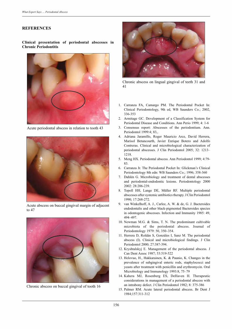
156
What Expert Says … Periodontal Abscess
REFERENCES
Clinical presentation of periodontal abscesses in Chronic Periodontitis
Acuteperiodontalabscessinrelationtotooth43
Acuteabscessonbuccalgingivalmarginofadjacentto47
Chronicabscessonbuccalgingivaloftooth16
Chronicabscessonlingualgingivalofteeth31and41
1. Carranza FA, Camargo PM. The Periodontal Pocket In:Clinical Periodontology, 9th ed, WB Saunders Co.; 2002,336-353
2. Armitage GC. Development of a Classification System forPeriodontalDiseaseandConditions.AnnPerio1999;4:1-6
3. Consensus report: Abscesses of the periodontium. Ann.Periodontol1999:4;83,.
4. Adriana Jaramillo, Roger Mauricio Arce, David Herrera,Marisol Betancourth, Javier Enrique Botero and AdolfoContreras. Clinical and microbiological characterization ofperiodontal abscesses. J Clin Periodontol 2005; 32: 1213–1218.
5. MengHX.Periodontalabscess.AnnPeriodontol1999;4:79-83.
6. CarranzaJr.ThePeriodontalPocketIn:Glickman’sClinicalPeriodontology8thedn:WBSaundersCo.;1996;358-360
7. Dahlén G. Microbiology and treatment of dental abscessesand periodontal-endodontic lesions. Periodontology 20002002:28:206-239.
8. Topoll HH, Lange DE, Müller RF. Multiple periodontalabscessesaftersystemicantibioticstherapy.JClinPeriodontol1990;17:268-272.
9. vanWinkelhoff,A.J.,Carlee,A.W.&de,G.J.Bacteroidesendodontalisandotherblack-pigmentedBacteroidesspeciesinodontogenicabscesses.InfectionandImmunity1985:49,494–497.
10.Newman M.G. & Sims, T. N. The predominant cultivablemicrobiota of the periodontal abscess. Journal ofPeriodontology1979:50,350–354.
11.HerreraD,RoldánS,Gonzáles I,SanzM.Theperiodontalabscess (I). Clinical and microbiological findings. J ClinPeriodontol2000;27:387-394.
12.Kryshtalskyj E. Management of the periodontal abscess. JCanDentAssoc1987;53:519-522
13.Helovuo,H.,Hakkarainen,K.&Paunio,K.Changesintheprevalence of subgingival enteric rods, staphylococci andyeastsaftertreatmentwithpenicillinanderythromycin.OralMicrobiologyandImmunology1993:8,75–79
14.Kahera MJ, Rosenberg ES, DeHaven H. Therapeuticconsiderationsinmanagementofaperiodontalabscesswithanintrabonydefect.JClinPeriodontol1982;8:375-386
15.Palmer RM. Acute lateral periodontal abscess. Br Dent J1984;157:311-312

157
Norhidayah / Khamiza
16.McFallWT.Theperiodontalabscess.JNCarolinaDentSoc1964;47:34-39
17.CarranzaJr.ThePeriodontalPocketIn:Glickman’sClinicalPeriodontology8thedn:WBSaundersCo.;1996:292-295
18.vanWinkelhoffAJ, van SteenbergenTJM, de Graff J.Theroleofblack-pigmentedBacteroidesinhumanoralinfections.JClinPerodontol1988;15:145-155.
19.Prichard JF. Management of the periodontal abscess. In:Advanced Periodontal Disease, 2nd ed. Philadelphia: WBSaundersCo.;1972:602-637
20.Smith,R.G.&Davies,R.M.(1986)Acutelateralperiodontalabscesses.BritishDentalJournal161,176–178.
21.Yang BQ. Analysis of bacterial cultures from 40 cases ofperiodontal abscess and their drug sensitivity. Chung HuaKouChiangHsuehTsaChih1987;22:327-328,369.
22.UetaEOsakiT,YonedaK,YamamotoT.Theprevalenceofdiabetesmellitus inodontogenic infectionsandcandidiasis:an analysis of neutriphil suppression. J Oral Pathol Med1993;22:168-174
23.CorbetEF.Diagnosisofacuteperiodontallesions,Periodontol200034:204,2004.
24.Silva GLM, Soares RV, Zenóbio EG. Periodontal Abscessduring Supportive Periodontal Therapy: A Review of theLiterature.JContempDentPract2008September;(9)6:082-091
25.Lewis MA, MacFarlane TW. Short-course high dosageamoxicillininthetreatmentofacutedento-alveolarabscess,BrDentJ1986:299,1986
26.HerreraD,RoldánS,O’ConnorA,SanzM.Theperiodontalabscess(II).Short-termclinicalandmicrobiologicalefficacyof2systemicantibioticsregimes*.JClinPeriodontol2000;27:395-404
27.Hafstrom CA, Wikstrom MB, Renvert SN, Dahlen GG.Effect of treatment on some periodontopathogens and theirantibodylevelsinperiodontalabscesses,JPeriodontol1994:65:1022
28.Ammons WF. In: Wilson TG, Kornman KS. FundamentalsofPeriodontics,QuintessencePublishingCo,CarolStream;
1996:423-451.29.PhilipRMelnic ,HenryHTakei.TreatmentofPeriodontal
Abscess In:ClinicalPeriodontology10thed,Philadelphia :WBSaundersCo.2006:714-721
30. Position paper. Systemic antibiotics in periodontics. JPeridontol2004;75:1553-1565.
31.AinamoJ,BarmesD,BeagrieG,CutressT,MartinJ,Sardo-Infirri J. WHO Index for Treatment Needs. InternationalDentalJournal1978:Vol.32(3),281-291
32.David Keys, Mark Bartold.Australian Dental Force Health2000;1:114-118
Address for correspondence:
Dr.Khamiza Zainol Abidin Pakar Klinikal PeriodontikKlinik Pakar Periodontik,Hospital Taiping,Jln. Taming Sari,34000 Taiping,Perak. Tel: 05- 8408103Fax: 05- [email protected]
158
ABSTRACTS OF SCIENTIFIC PAPERS PRESENTED AT THE 65TH MDA/AGM SCIENTIFIC CONVENTION AND TRADE EXHIBITION, 20th - 22th JUNE 2008
MEDIATION – THE MDA EXPERIENCEPurmal K*, Nambiar P
Faculty of Dentistry, University Malaya
Objectives: To review the cases that was mediated by MDA complaints bureau from 2004 until 2007.
Methods: This is a retrospective analysis of the complaints from the files of MDA.
Results: There was 41 cases from that time period. Most of the cases involves oral surgical procedures (10) and endodontics (9). 69% of the cases where from clinics in the Klang Valey. 69% of the cases was successfully mediated, 10% unresolved and the 21% are in various stages of mediation.
Conclusion: The complaints that are mediated by MDA is steadily increasing but we manage to settle most of the cases.
Keywords: dental negligence, mediation, risks
REHABILITATION OF THE PERIODONTAL PATIENTSK Lim
Periodontic Unit, Government Dental Clinic, Jalan Perak, Pulau Pinang
Periodontal diseases often result in the loss of teeth and/or supporting tissues. Migration of teeth with derangement of occlusion result in compromised function and aesthetics. The author will illustrate, by means of clinical examples, the complex interdisciplinary treatment modalities used to rehabilitate patients with advanced periodontitis. Dental implants, aesthetic dentistry, orthodontics, endodontics and periodontics are used in a planned and staged manner to achieve the best possible outcome.
Keywords: Periodontitis, ortho-perio interface, dental implants
THE EFFECT OF DENTURE LABELLING ON THE FLEXURAL STRENGTH OF RESIN ACRYLIC DENTURENur Azliani AH, Khamis MF*
School of Dental Sciences, Health Campus, Universiti Sains Malaysia, Kelantan
Objective: The aim of the study was to compare the flexural strength of conventional resin acrylic denture with flexural strength of labelled resin acrylic denture fabricated using two different techniques; heat cured and cold cured.
Materials and Methods: Forty-five resin acrylic blocks were fabricated with fixed dimensions 64 mm x 10 mm x 2.5 mm. There were equally divided into three groups; conventional (no labelling included), and heat and cold cured labelled resin acrylic. The flexural strength of each block was measured using INSTRON 8874 Universal Testing Machine (INSTRON Inc, USA). One-way ANOVA and Tukey Post-Hock Test, and one sample t-test (test value=55N) were used for statistical analyses. The significance level was set at 5%.
Malaysian Dental Journal (2008) 29(2) 158-165© 2008 The Malaysian Dental Association
MALAYSIAN DENTAL JOURNAL
159
Results: The flexural strength can be arranged in descending order of cold-cured>conventional>heat-cured techniques. All multiple comparisons were significant, p<0.05. The peak load for conventional and cold-cured labelled resin acrylic was significantly better than minimum peak load 55N (both p<0.001) while heat-cured peak load was equal to minimum peak load.
Conclusion: Both techniques are acceptable for application with cold-cured labelling technique is more recommended since its flexural strength is even better than conventional resin acrylic.
Keywords: denture labelling, flexural strength, resin acrylic denture
A STUDY OF SATISFACTION WITH SCHOOL DENTAL SERVICES AMONG 12-YEARS-OLD SCHOOL CHILDREN IN JOHORHabibah Y1*, Loh KH2, Sheila AR2
1Klinik Pergigian Bandar Maharani, Poliklinik Komuniti Bandar Maharani, Muar, Johor2Pejabat Timbalan Pengarah Kesihatan Negeri Johor (Pergigian), Johor
Objectives: To evaluate the students’ perception on Johor School Dental Services and to make recommendations for corrective actions or continual improvements, if necessary.
Methods: This is a cross-sectional study of satisfaction survey among 2170 12-years-old school children in Johor. The Dental Satisfaction Questionnaire developed by Johor Dental Services was used to assess the satisfaction level. The questionnaire, consisting of fifteen items, including 5 important domains; tangibles, reliability, responsiveness, assurance and empathy, was distributed to the selected schoolchildren after receiving dental checkup/treatment in their school.
Results: The response rate was 84.9%. Majority of the 12-years-old school children (96.9%) were satisfied with the quality of care they received. Only small percentage (3%) was dissatisfied with the dental care. The following items explained most of the variance of satisfaction with school dental care: “Pegawai Pergigian/ Jururawat pergigian sentiasa prihatin dengan pesakit” (Dental officer/dental nurses always care about the patients), “Pegawai Pergigian/Jururawat Pergigian dan kakitangan pergigian lain sentiasa bersedia memberi bantuan bagi semua masalah pergigian murid” (Dental officer/dental nurses and other dental staff are ever willing to help solve all students’ dental problems), “Kakitangan pergigian menjalankan tugas mereka dengan bersungguh-sungguh dan berdedikasi”(Dental staff are hardworking and dedicated). Dissatisfactions, meanwhile, were associated with the following items: “Jika saya perlu dirujuk kepada pegawai lain, tempoh temujanji yang diberi adalah memadai”(If I should be referred to other dental officer, the appointment period given is reasonable),and “Maklumat-maklumat mengenai kesihatan pergigian mudah didapati”(Oral health information is easily available).
Conclusion: Overall, most of the 12 years-old school children were satisfied with the quality of dental care they received. The high percentage of satisfaction, well above the state set target (85%), is a good achievement to be proud of. This only serves to motivate us for even better services to be rendered in the future.
Keywords: satisfaction, dental satisfaction questionnaire, quality of dental care
THE ALL-ON-FOUR CONCEPT FOR DENTAL IMPLANT PLACEMENT IN THE EDENTULOUS JAWChong SMY*, Abraham MTDepartment of Oral &Maxillofacial Surgery, Hospital Tengku Ampuan Rahimah, Klang, Selangor
Objectives: To highlight some the clinical aspect of the All-on-Four technique applied in few of the cases done in our setting, while discussing the advantages and disadvantages this technique.
Methods: Review of literature and clinical cases of implant placement using the All-on-Four technique.
Results: The All-on-Four clinical solutions for implants placement in the edentulous jaw was developed by Dr. Paulo Málo from Portugal. The technique uses only four implants in the edentulous jaw by tilting posterior implants to increase inter-implant distances and increasing implant anchorage in bone.
Conclusion: With this technique, it is possible to provide optimal prosthesis support and allow immediate function even in cases with minimal bone volume.
Keywords: All-on-Four technique, dental implant, edentulous
160
SURFACE ROUGHNESS AND STAINING SUSCEPTIBILITY OF COMPOSITE RESINSChan GL1*, Koh KH2, Seow LL3
1Klinik Pergigian Butterworth, Pulau Pinang2Klinik Pergigian Jalan Perak, Pulau Pinang3School of Dentistry, International Medical University
Objectives: The objectives of the study were to evaluate (i)the surface roughness of composite resins polished with various polishing systems; (ii)susceptibility of composite resins to staining after immersion in various dietary dyes and (iii)the correlation between surface roughness and susceptibility to staining.
Methods: One hundred and eighty specimens of two composite resins were fabricated: Filtek Z350 (3M Co., USA; ninety specimens) and Solare (GC Dental Products Corp, Japan; ninety specimens). The specimens were polished with Sof-Lex™ Discs (3M Co., USA) and Enhance™ Polishing System (Dentsply International Inc., USA) with curing against Mylar strip as control. The surface roughness (Ra) was measured with Atomic Force Microscope (AFM). Subsequently, ten discs from each polishing system and control group were immersed in coffee, curry and thick soya sauce and colour changes (∆E*ab) were measured using a spectrophotometer. Results were statistically analyzed using one way ANOVA and Tukey test.
Results: There was a significant difference in the surface roughness between Mylar and the other 2 polishing systems for both composite resins (p<0.05). Findings also showed that there was no significant difference between Sof-Lex and Enhance for Filtek Z350 but there was a significant difference for Solare (p<0.05). There was no significant difference in colour changes between the control group and the test groups when immersed in coffee and thick soya sauce (p>0.05). However there was a significant difference when the test groups were immersed in curry (p<0.05).
Conclusions: Both polishing systems provide equally good surface finishing for Solare and Filtek Z350. Curry produces significant discolouration on composite resins.
Clinical significance: It is advisable for patients with large anterior composite restorations to brush or rinse their mouth after consuming curry.
Keywords: composite resins, surface roughness, staining susceptibility
THE SEVERITY OF GINGIVAL ENLARGEMENT INDUCED BY CYCLOSPORIN-A AND NIFIDIPINE AN EXPERIMENTAL STUDY IN RABBITAL-Bayaty F.H1, Basma O. AL-Tay2, Salah S. AL-Kushali2, Likaa Mahmmod2
1Department of Oral pathology, Oral Medicine &Periodontology, Faculty of Dentistry, University Malaya 2College of Dentistry, University of Baghdad
Objectives: The present study was undertaken to estimate the severity of gingival enlargement induced by Cyclosporin – A and Nifedipine separately and in combination.
Material & Methods: Thirty adult rabbits were used in this study. , divided equally into 3 main experimental groups. The first group received 10mg/kg/day Nifedipine, The second received 10mg/kg/day CsA, the third received combination of 10mg/kg/day Nifedipine and CsA by gastric feeding from day 1 till day 70. Alginate impressions were taken for the anterior segments of the maxilla and mandible for each animal on the days 0, 30, 70, and110. Gingival enlargement was assessed on stone study models according to Seymour’s method.
Results: revealed a highly significant difference in the mean values of the gingival enlargement scores in the experimental group that received combination of CsA and Nifedipine, compared to that received either CsA or Nifedipine,and in the group that receive CsA compared to that received Nifedipine on day 70 of the experiment.
Conclusion: the effect of CsA in causing gingival enlargement was significantly more than the effect of Nifedipne, While the effect of the combination of the two drugs caused significantly more gingival enlargement than the effect of one of the drugs alone, Drug withdrawal shows partial regression of gingival enlargement on day 110 in the experimental groups.
Keywords: Cyclosporin – A, Nifedipine, gingival enlargement, rabbit
161
COMPLETE DENTURE DELIVERED BY UNIVERSITI KEBANGSAAN MALAYSIA’S (UKM) DENTAL STUDENTS – A RETROSPECTIVE STUDY.Dahari N1*, Zaharudin N I1, Hamirudin M M2, Tarib N2, Marlynda A2
1Specialist Clinic, Hospital Umum, Kuching, Sarawak.2Department of Prosthodontics, Faculty of Dentistry, UKM.
Abstract: This study is aim to evaluate quality of complete dentures delivered by UKM dental students from 2001 to 2005.
Objectives: The objectives of this study are to identify complications, failures and success related to complete dentures made by UKM dental students and to assess patient’s satisfactory level who received complete dentures made by UKM dental students.
Materials and Methods: Methods used in this study are including references by using electronic databases for articles and studies, systematic folder searching from 40 000 patient’s folder, pilot study through cases managed in 2006/2007, clinical examination, patient’s satisfaction and data analysis by using SPSS version 12.0.
Result: Result shows that overall quality of complete dentures delivered by UKM dental students is good. As for the patient’s satisfactorily level, most of the subjects satisfied with their denture.
Conclusion: Majority of complete dentures delivered by UKM dental students is in a good quality and most of the patient satisfied with their denture.
Keywords: complete denture, edentulous, psychology, satisfaction level
REVIEW OF OUTCOMES OF THE ORAL CANCER IN THE KLANG VALLEY REGION.Hamzah SH*, Abraham MT Department of Oral & Maxillofacial Surgery, Hospital Tengku Ampuan Rahimah,Klang, Selangor. Objectives: To highlight the outcomes of oral cancer patients in Klang Valley from 2000 to May 2008.
Methods: This is a retrospective analysis of 178 patients’ record that was diagnosed to have oral cancer in Klang Valley. We analyzed the age group, gender, ethnic and type of cancer, the stage of the patients when they were referred, the type of treatment involved, the years and the outcomes after the treatment.
Results: Oral cancer commonly diagnosed in the ethnic of India (66%) with the female group (102) .The mean age group is 60.02. 95% of the patient was diagnosed to have squamous cell carcinoma and the site of oral cancer that commonly involved was buccal mucosa (33%), followed by tongue (32%). 84% of the patient were diagnosed at stage IV while the most common modality of treatment are surgery (20%), followed by surgery, chemotherapy and radiotherapy (18%) and also palliative radiotherapy and chemotherapy (18%). About 41% of the patients survive within the first 5 years after the treatment.
Conclusions: Majority of patients had their tumours diagnosed at the advanced stage with the mortality increases in relation to the stage at which diagnosis is made. Keywords: oral cancer, squamous cell carcinoma, surgery
162
OVERCOMING PROBLEMS ASSOCIATED WITH TRANSPORT DISC DISTRACTION OSTEOGENESIS IN MANDIBULAR RECONSTRUCTIONThavamalar M*, Thomas Abraham
Department of Oral & Maxillofacial Surgery, Hospital Tengku Ampuan Rahimah Klang, Selangor
Transport disc distraction osteogenesis (TDDO) is an alternative method to reconstruct mandible following tumor ablation. This is method of mandibular reconstruction was successfully done for the first time in Malaysia. This paper presents a few cases of reconstruction of the mandible using TDDO. Resected segment of the mandible were successfully reconstructed using distraction osteogenesis without the need for complicated bone grafts. Also discussed in this paper are some of the steps taken to overcome the problems encountered during distraction osteogenesis and what could be done to prevent such problems from recurring. Advantages and disadvantages of these procedures over other methods of mandibular reconstruction have been highlighted.
Keywords: transport disc distraction osteogenesis, mandibular reconstruction, resection
SPORTS RELATED MAXILLOFACIAL FRACTURES: A PRELIMINARY STUDYMustafa WM1, Yuen KM2, Ma BC3, Sockalingam G 3, Rahman RA4, Razali AR5, Tan HL1* 1 Department of Oral Surgery, Hospital Kuala Lumpur2 Department of Oral Surgery, Hospital Ipoh3 Department of Oral Surgery, Hospital Sultanah Aminah Johor Bahru4 Department of Oral Surgery, Hospital Sultanah Zainab Kota Bahru5 Department of Oral Surgery, Hospital Seberang Jaya
Objectives: To assess the spectrum of maxillofacial fractures sustained during sports in Ministry of Health Hospitals Malaysia.
Methods: From June 1st 2002 to May 31st 2003 detailed records of facial trauma patients in 23 major Ministry of Health Hospitals were recorded prospectively on a modified proforma by Meaders and Sulivan . The study collected data regarding age group, gender, the aetiology of the fracture, the treatment employed and the complications following treatment. Patients were followed up for a period of 3 months.
Results: A total of 1862 cases of maxillofacial fractures were recorded and of these only 18 cases were sports related. The highest incidence of sports related facial fracture was in the age group 10-19 years (11 cases). All the cases recorded were in male patients and no female. The bone most commonly fractures is the mandible (7cases), followed by dento-alveolar fracture of the maxillary bone (4 cases). Closed reduction was the most common mode of treatment (7 cases) followed by conservative method (6 cases). All patients recovered uneventful.
Conclusions: This study provides an initial data base for facial fractures related to sports in the Malaysia population. Sports injuries are likely to be more common in the future as a result of an increase in leisure time. Protective measures need to be considered in the high contact sports.
Keywords: Maxillofacial trauma, sports, injury.
EFFECT OF TWO POLISHING SYSTEMS ON THE SURFACE ROUGHNESS OF COMPOSITESSalim MR*, Yahya NA, Abu Kasim NH
Faculty of Dentistry, University of Malaya, Kuala Lumpur
Objectives: To determine the surface roughness of 3 types of dental composites when polished with 2 types of polishing systems.
Methods: 60 specimens of 10 mm in diameter and 2 mm thick were made for each composites, Filtek™Z350 (3M ESPE, St Paul Minn USA), Grandio (Voco,Germany) and Glacier (ISD, Australia). The specimens were divided into three groups: group 1-Mylar matrix (control), group 2-Sof-Lex™ System (3M ESPE, St Paul, U.S.A) and group 3-Astrobrush (Ivoclar Vivadent, Liechtenstein).The specimens were polished according to the manufacturers’ instructions and the mean surface roughness (Ra) were determined using a profilometer (SurfTest SV400, Mitotoyo,

163
Japan) and Universal Scanning Probe Microscope,USPM (Ambious Technology). The data were subjected to analysis of variance (ANOVA) and Dunnett T3 post hoc test.
Results: Two way ANOVA showed significant interaction between polishing agents and composites (p<.05). Dunnett T3, showed that the surface roughness of composites produced by polishing agents was significantly different with Mylar producing the smoothest surface followed by Sof-Lex™ and Astrobrush. When the types of composites were compared, Dunnett T3 showed that no significant difference was observed between Grandio and Filtek, but significantly smoother compared to Glacier (p<.05). One way ANOVA was carried out to test significant levels between polishing agents for each individual composites. For Glacier, significant difference were detected, Mylar exhibiting the smoothest surface (0.10±0.02) followed by Sof-Lex™ (0.29±0.05) and Astrobrush (1.04±0.32). Similar trend was observed for Filtek. As for Grandio, Mylar produced the smoothest surface (0.07±0.02); however there was no significant difference was detected between Astrobrush (0.30±0.05) and Sof-Lex™ (0.28±0.03).
Conclusions: Mylar produced the smoothest suface for all composites tested. Both Sof-Lex™ and Astrobrush produce equally smooth surface for Grandio, however Sof-Lex™ produce smoother surface compared to Astrobrush for Filtek.
Keywords: fluoride readings, temperature, storage time
THE EFFECT OF TEMPERATURE AND STORAGE TIME ON FLUORIDE LEVEL READINGS OF WATER SAMPLES USING HACH’S COLORIMETER IN JOHOR BAHRU AND KOTA TINGGI DISTRICTSMuz’ini M1*, Noridah A2
1 Klinik Pergigian Kota Tinggi, Jalan Tun Habab, Kota Tinggi, Johor2 Klinik Pergigian Jalan Abdul Samad, Johor Bahru, Johor
Objectives: To study the association between storage time on fluoride level reading of water sample and to study the association between temperature on fluoride level reading of water sample.
Methods: This is a cross sectional study of fluoride level readings of water samples collected from 10 water fluoridation plants and 10 reticulation points in Johor Bahru and Kota Tinggi districts from March 2006 to September 2006.
Results: There is no significant association between storage time of water samples and fluoride level readings using a Hach’s colorimeter. However, there is a significant association between temperature and fluoride level readings of water samples. Fluoride level reading is negatively correlated with temperature of water sample (p < 0.01). Regression analysis between both temperature and storage time of water samples shows a significant negative relationship between temperature of water samples and fluoride level reading (p<0.01) and no significant relationship between storage time and fluoride level reading.
Conclusion: Findings from this study demonstrate that temperature of water samples affect the fluoride level reading during analysis using a Hach’s colorimeter. There is a need to standardise temperature of water samples when analysing for fluoride level using a Hach’s colorimeter. Otherwise, a standard fluoride level analyser controlling for temperature is necessary to demonstrate true readings of fluoride level in water samples.
Keywords: fluoride readings, temperature, storage time
164
ORAL CANDIDA AND DISEASE CONTROL IN NON-INSULIN DEPENDENT DIABETIC MELLITUS PATIENTSShareef BT*, Roni N M, Harun A
School of Dental Sciences, Universiti Sains Malaysia, Kubang Kerian, Kelantan, Malaysia.
Objective: This study was designed to assess the relationship of oral Candida colonization, carriage and infection to the glycaemic control in Non-Insulin Dependent Diabetes Mellitus (NIDDM) patients. Determination of the relationship of candidal carriage in smokers and denture wearers to the glycaemic control among NIDDM patients was also carried out.
Materials & Methods: NIDDM patients were recruited randomly from Diabetic Clinic in USM. A total of one hundred and two (102) NIDDM patients, on treatment and follow up were included in this study. All patients underwent oral examination after being interviewed. Relevant information was taken from the medical records for each of the patients. Oral rinse technique was performed for isolation of fungi species. Data were registered and analyzed by using SPSS version 11.0.
Results: It was found that the candidal carriage and Candida colonization was significantly higher in poor-glycaemic control compared with well-controlled group.Candida albicans species was found to be the most common fungi recovered from diabetic patients (73.4%), followed by Candida stellatoidea species (10.8%). Only two (2) diabetic patients were found to have oral candidal lesions. A significant relationship was found between denture wearing and glycaemic control, whereas non-significant relationship was observed between smoking habits and glycaemic control.
Conclusion: From this study, it was concluded that the status of glycaemic control play a role in candidal carriage and colonization among NIDDM patients.
Keywords: Diabetes mellitus, Candidal species , glycaemic control .
DENTURE TEETH SET-UP MOULD Jacob J1*, Azizah Y2, Mat Yusoh S3, Yahaya MN1, Che Maziah H4, Asma A4
1Tendong Dental Clinic, 17030 Pasir Mas, Kelantan, Malaysia 2 formerly, Senior Dental Officer of Pasir Mas, 3 Rantau Panjang Dental Clinic, Pasir Mas, 4Pasir Mas Town Health Centre, Pasir Mas.
Objective: It takes about 90 minutes to complete an upper and lower complete denture teeth set-up. This restricts the number of denture appointment in the government dental clinics. Denture Set-up Mould fabrication was taken up to overcome this problem.
Method: The moulds are of different sizes according to the teeth and jaw size. The teeth are set according to the principles of teeth setting. The mould is reusable and can be disinfected.
Results: A six-month study before and after use of denture set-up mould showed that by using the mould, time for teeth setting was reduced to 30 minutes. Denture appointments could be increased from 7 to 12 patients per day. The waiting time for new denture patients was reduced from 5-6 months to less than 2 months. The number of dentures fabricated by each technician monthly increased from 20 to 36 units. This led to increase in revenue from issue of dentures by 77%. However, some limitations observed were the mould could not be used in situations where the vertical dimension is reduced and there is no provision for edge-to-edge or cross bite conditions.
Conclusion: With the reduction in working time for every case of complete denture teeth set-up, the customer satisfaction level is dramatically improved. Despite certain limitations, denture set-up mould benefits all its stake holders including the dental service providers, its customers as well as the government.
Keywords: Complete denture, teeth set-up, denture mould
165
REFERRAL TREND OF PATIENTS REFERRED TO UNIT PAKAR PERIODONTIK, KLINIK PERGIGIAN PERINGGIT, POLIKLINIK KOMUNITI PERINGGIT, MELAKA.Yusoff S ¹*, Asari ASM²
¹Unit Pakar Periodontik, Klinik Pakar Pergigian, Hospital Kajang, Kajang, Selangor.²Unit Pakar Periodontik, Klinik Pergigian Cahya Suria, Kuala Lumpur, Wilayah Persekutuan.
Objectives: To assess the pattern of distribution of cases referred for periodontal treatment at Unit Pakar Periodontik, Melaka, in terms of profile of cases, sources of referrals and appropriateness of cases referred for treatment. Methods: This is a retrospective analysis involving patients which were randomly referred for treatment at Unit Pakar Periodontik, KP Peringgit, Melaka in 2005. We analysed the number, gender, age group, ethnicity, sources of referrals, relevant medical history and nature of patients’ complaints. Results: Total number of patients referred was 150 with an average of 15 new cases per month. 68 (45.3%) male and 82 (54.7%) were female; the age range was 10-79 years old. Malays were the most referred 81(58.7%) compared to Chinese 49(32.7%), Indians 17(11.3%) and other ethnic groups 2(1.3%). The age range of 40-49years old was the most referred, 38. Referral from dental clinics; most cases were referred from KP Peringgit (38.o%) followed by KP Melaka Tengah(22.7%) and Bahagian Bedah Mulut, Hospital Melaka, (16.7%). Referral according to district / area of residence; Melaka Tengah (85.7%), Alor Gajah (5.3%), Jasin (6.7%) and (3.3%) was referred from outside Melaka. It was found that 17.3% of patients had Diabetes Mellitus while 16% had hypertension. Most common complaint was mobile teeth (23.3%) followed by swollen gums(15.3%) and bleeding gums (13.3%).
Conclusions: Most cases referred for periodontal treatment are Malaysians residing in Melaka, mostly referred by the government dental clinics from Melaka Tengah District. Most complaints of cases referred were indicative of periodontal problems.
Keywords: Referral trends, patients, Unit Pakar Periodontik
ORAL TONGUE CANCER – THE HKL EXPERIENCEIp J.*, Wan Mustafa W. M.
Department of Oral Surgery, Hospital Kuala Lumpur, Kuala Lumpur, Malaysia
Objective: Tongue cancer is associated with significant morbidity and poor mortality rate. This is a preliminary study to analyze a case series of patients with tongue cancer managed in the Department of Oral Surgery, Hospital Kuala Lumpur.
Methods: A retrospective review was conducted in a group of 34 patients presented with tongue cancer in the Department of Oral Surgery, Hospital Kuala Lumpur from January 2001 to December 2006. Demographic data, stage of disease presentation, treatment modality and survival at one year were described.
Results: Most patients (n=15, 47.1%) were already at Stage IV of disease when they first presented at our clinic. The most common treatment modality was surgery alone (11 patients, 32.4%), followed by surgery with post-operative radiotherapy (5 patients, 14.7%), surgery with post-operative chemotherapy (3 patients, 8.8%), and surgery with post-operative radio and chemotherapy (3 patients, 8.8%). 4 patients were found not fit for surgery and were placed under supportive therapy. 8 patients either refused or defaulted treatment, and subsequently were lost to follow-up. At one year after initial diagnosis, patient’s status was evaluated as: Dead of disease (21%), Dead due to other reasons (19%), Alive without evidence of disease (32%). One patient was reported with cancer recurrence at the tongue. Conclusion: Majority of patients presented at late stage of disease when functional impairment and mortality rates are high. This study highlights the need to advocate early detection and intervention of tongue cancer.
Keywords: Tongue cancer, Survival, Treatment modality
166
Malaysian Dental Journal (2008) 29(2) 166-167© 2008 The Malaysian Dental Association
MALAYSIAN DENTAL JOURNAL
CONTINUING PROFESSIONAL DEVELOPMENT QUIZ (CPD POINTS= 2)
Dear Colleagues,
In this issue of the MDJ, we continue with column of Continuing Professional Development Quiz whereby you will get two (2) CPD points by just trying out the quizzes. This is a self-administered test and is designed to help colleagues accumulate CPD points. Your feedback is greatly appreciated. These quiz questions were kindly provided by Dr. Kathiravan Purmal, Dr. Wey Mang Chek, Mr. Shamsher Singh, Prof Prabhakaran Nambiar, Dr, Natasya Ahmad Tarib, Dr Ajura Abdul Jalil and Assoc. Prof. Seow Liang Lin.
Thank you.
Assoc. Prof. Seow Liang Lin,Editor, Malaysian Dental Journal.
1. The risk of stochastic effect of radiation is higher on:
A. Tooth structureB. Dividing cellsC. Salivary glandD. Thyroid gland
2. The least amount of radiation is produced by using:
A. Short rectangular coneB. Long round coneC. Long rectangular coneD. Short pointed cone
3. All the following legal statements are true, EXCEPT
A. It is a requirement of law that all dental practitioners in Malaysia must possess a postgraduate qualification, specializing in forensic odontology, before they are allowed to give bitemark evidence in court as expert witnesses.
B. The probative value of a forensic odontologist’s opinion is proportional to his qualifications and experience.
C. Trial judges must warn themselves as to the dangers of convicting accused persons based on any unreliable evidence.
D. The prosecution has a duty to prove beyond reasonable doubt all the ingredients of the offence with which a person is charged, including his identity.
4. Which one of the following statements is TRUE?
A. Secondary distortion occurs during the physical altercation between a victim and the assailant, when the latter bites the former.
B. Ray Krone was wrongly acquitted by the court in the United States due to the error on the part of the prosecution’s expert witness.
C. In United States, the duty to ensure the standardization in the field of forensic odontology is carried out by the American Board of Forensic Odontology (ABFO).
D. Bitemark evidence should be rejected at face value since, in the present state of knowledge, it cannot be expected to deliver the exactitude of the mathematical sciences.
5. What is the most common site of oral granular cell tumour?
A. palateB. tongueC. lower lipD. floor of mouth
6. Currently, granular cell tumour is believed to be of ___________ origin.
A. muscleB. vascularC. neuralD. epithelium
7. The following are side effects of mefenamic acid, except
A. gastro-intestinal bleedingB. hepatotoxicityC. acute renal failureD. trigger asthmatic attack
167
8. The advantages of COX-2 inhibitors as analgesics include:
i. longer duration of actionii. less gastrointestinal side effectsiii. potent analgesic propertyiv. economical
A. i & iiB. i & iiiC. i, ii & iiiD. all of the above
9. A study in Malaysia has shown that prevalence of fluorosis amongst 16 year-old school children is approximately ____________ in areas with fluoridated water supply
A. 45%B. 55%C. 65%D. 75%
10. Below are the major factors the mothers of pre-school children in Perlis would take into consideration when purchasing toothpaste except:
A. priceB. brandC. flavourD. fluoride content
11. Colour changes of ____________ is clinically perceptible
A. 1.3 NBS unitsB. 2.3 NBS unitsC. 3.3 NBS unitsD. 4.3 NBS units
12. In the study evaluating staining susceptibility of composite resins, which is the correct sequence from the least to the most susceptible to staining:
A. Z350, Beautifil, Z250B. Beautifil, Z250, Z350C. Z250, Z350, BeautifilD. Z350, Z250, Beautifil
13. Preclinical teaching has the following advantages except
A. Building up student’s competent in the treatment’s procedure
B. Building up student’s confidence in the treatment’s procedure
C. Familiarized student with clinical procedureD. Wasting clinical time for students to learn
14. Following are effective methods of teaching preclinical skills except
A. close monitoring of student’s progressB. self directed learningC. live demonstration of the practical proceduresD. lectures and tutorials
15. Which of the following statements is correct?
A. The vertical measurement of physiological rest position should have a lower value than the vertical dimension of occlusion.
B. Long-term provisional restorations are necessary in full mouth rehabilitation cases, to verify patients’ adaptability to the new vertical dimension of occlusion.
C. Excessive changes of vertical dimension of occlusion should not be avoided since they are usually well tolerated.
D. Vertical dimension of occlusion is defined as the vertical measurement of the face between two selected points superior and inferior to the nose when the occluding members are in contact.
16. Which of the following statements is correct?
A. The dentition, the masticatory muscles and the temporomandibular joints form a class 3 lever system.
B. When prescribing full ceramic restorations, intra-sulcular placement of crown margins on the labial surface becomes more important from an esthetic point of view.
C. Metal occlusal surfaces can be prescribed for crowns on mandibular teeth to maximize the esthetic outcome in a full mouth rehabilitation case.
D. In order to establish anterior guidance, restoration of the posterior teeth should be completed before the restoration of anterior teeth.
17. The term hypodontia refers to
A. Total absence of teethB. Congenital absence of many teethC. Missing six or more permanent teethD. A developmental absence of only a few teeth
18. Common features related to hypodontia are
A. MacrodontiaB. Malocclusions related to absence of teethC. Bimaxillary protrusionD. Transposition of deciduous teeth
1. B 2. C 3. A 4. C 5. B 6. C 7. D 8. C 9. D 10. A 11. C 12. D 13. D 14. B 15. B 16. A 17. D 18. B
ANSWERS:
168
Malaysian Dental Journal (2008) 29(2) 168-169© 2007 The Malaysian Dental Association
MALAYSIAN DENTAL JOURNAL
Aim And ScopeThe Malaysian Dental Journal covers all aspects of work in Dentistry and supporting aspects of Medicine. Interaction with other disciplines is encouraged. The contents of the journal will include invited editorials, original scientific articles, case reports, technical innovations. A section on back to the basics which will contain articles covering basic sciences, book reviews, product review from time to time, letter to the editors and calendar of events. The mission is to promote and elevate the quality of patient care and to promote the advancement of practice, education and scientific research in Malaysia.
PublicationThe Malaysian Dental Journal is an official publication of the Malaysian Dental Association and is published half yearly (KDN PP4069/12/98)
Instructions to contributorsOriginal articles, editorial, correspondence and suggestion for review articles should be sent to:
Editor,Malaysian Dental Journal
Malaysian Dental Association54-2, Medan Setia 2, Plaza Damansara
50490 Kuala Lumpur, Malaysia
Authors are requested to submit three copies of their typescript and illustration and also copy of their work in a file on a CD. The editor cannot accept responsibility for any damage or loss of typescripts. The editorial currently is unable to support electronic submissions; apart from the use of e-mailing for correspondence.
A paper is accepted for publication on the understanding that it has not been submitted simultaneously to another journal in the English Language. Rejected papers will be returned to authors. The editor reserves the right to make editorial and literary corrections. Any opinion expressed or policies advocated do not necessarily reflect the opinion or policies of the editors.
CopyrightAuthors submitting a paper do so on the understanding the work has not been published before. The submission of the manuscript by the authors means that the authors automatically agree to sign exclusive copyright to the Editor and the publication committee if and when the publication is accepted for publication. The copyright transfer agreement can be downloaded at the MDA webpage (www.mda.org.my). A copy of the agreement must be signed by the principal author before any paper can be published. We assure that no limitation will be put on your freedom to use material contained in the paper without requesting permission provide acknowledgement is made to the journal as the original source of publication. Presentation of manuscriptManuscript should be submitted in journal style. Spelling preferably be either British or American. Articles typed in double spacing throughout on good, white A4 paper with a margin of at least 3 cm all around. Type only on one side of the paper. Three copies of the typescript and illustration should be submitted and the authors retain a copy for reference. In addition to the typed manuscript we also require every article be submitted as a file on a newly formatted 3.5 inch floppy disk. The manuscripts and the file on the disk must be identical and the disk should not contain other files. The disk must be clearly labeled with the title of the article, the names of the author(s) and the computer operating system (eg DOS, Macintosh) and WP version (word 2.0, WordPerfect 5.0) used. Files should not be converted to ASCII format.
169
Full papersPapers should be set out as follows with each beginning in a separate sheet: title page, summary, text, acknowledgements, references, tables, caption to illustrations. Title page. The title page should give the following information: 1) title of the article; 2) initials, name and address of each author, with higher academic qualifications and positions held; 3) name, address, telephone, fax and e-mail address. Text. Normally only two categories of heading should be used: major ones should be typed in capital in the centre of the page and underlined; minor ones should be typed in lower case (with an initial capital letter) at the left hand margin and underlined.Do not use he or she if the sex of the person is unknown; e.g. 'the patient'. References. The accuracy of the references is the responsibility of the author. References should be entered consecutively by Arabic numerals in superscript in the text. The reference list should be in numerical order on a separate sheet in double spacing. Reference to journals should include the author's name and initials (list all authors when six or fewer; when seven or more list only the first three and add ‘et al.’), the title of paper, Journal name abbreviated, using index medicus abbreviations, year of publication, volume number, first and last page numbers (ie. Vancouver style). For example:
Ellis A, Moos K, El-Attar. An analysis of 2067 cases of zygomatico-orbital fractures. J Oral Maxillofac Surg 1985;43:413-417.
Reference to books should be sent out as follows: Scully C, Cawson RA, Medical Problems in Dentistry 3rd edn. Wright 1993:175.
Tables. These should be double spaced on separate sheets and contain only horizontal rules. Do not submit tables as photographs. A short descriptive title should appear above each table and any footnotes, suitably identified below. Care must be taken to ensure that all units are included. Ensure that each table is cited in the text.
IllustrationsLine illustration. All line illustrations should present a crisp black image on an even white background (127 mm x 173 mm or 5 x 7 inches) or no larger than 203 mm x 254 mm or 8 x l0 inches.Photographic illustrations and radiographs. These should be submitted as clear lightly contrasted black and white prints (unmounted) sizes as above. Photomicrographs should have the magnification and details of the staining technique shown. Radiographs should be submitted as photographic prints carefully made to bring out the details to be illustrated, with an overlay indicating the area of importance.All illustration should be carefully marked (by label pasted on the back or by a soft crayon) with figure number and authors name and the top of the figure should be indicated by an arrow. Never use ink of any kind. Do not use paper clips as these can scratch or mark illustrations. Caption should be typed, double spaced on separate sheets from the manuscript.Patient confidentiality. Where illustrations must include recognizable individuals living or dead and of whatever age, great care must be taken to ensure that consent for publication has been given. Otherwise, the patient’s eyes or any indentifiable anatomy should be covered.Permission to reproduce, borrowed illustration or table or identifiable clinical photographs. Written permission to reproduce, borrowed material (illustrations and tables) must be obtained form the original publisher and authors and submitted with the typescript. Borrowed material should be acknowledged in the caption in this style. 'Reproduced by the kind permission of...... (publishers) from /....(reference)'.
Page ProofsPage proofs are sent to the author for checking. The proofs with any minor corrections must be returned by fax or post to the editor within 48 hours of receipt.
Proprietary namesProprietary names of drugs, instruments etc. should be indicated by the use of initial capital letters.
Abbreviations and unitsAvoid abbreviations in the title and abstract. All unusual abbreviations should be fully explained at their first occurrence in the text. All measurements should be expressed in SI units. Imperial units are also acceptable.
OffprintsTen free offprints are supplied to the author. An offprint order form will be sent to the author with the page proof