Embed Size (px)
Citation preview

JOURNAL OF VIROLOGY, July 2002, p. 6460–6472 Vol. 76, No. 130022-538X/02/$04.00�0 DOI: 10.1128/JVI.76.13.6460–6472.2002Copyright © 2002, American Society for Microbiology. All Rights Reserved.
Human Monocytic Cell Lines Transformed In Vitro by Epstein-BarrVirus Display a Type II Latency and LMP-1-Dependent Proliferation
Eric Masy,1,2 Eric Adriaenssens,1 Claire Montpellier,3 Pascale Crépieux,1 Alexandra Mougel,1Brigitte Quatannens,3 Gautier Goormachtigh,1 Nathalie Faumont,4 Fabienne Meggetto,4
Claude Auriault,1 Hervé Groux,5 and Jean Coll1*UMR 85271 and UMR 8526,3 CNRS/LilleII/Institut Pasteur de Lille, Institut de Biologie de Lille, Lille, Service de Biologie
Clinique, Centre Hospitalier, Valenciennes Cedex,2 UPR 2163 CNRS, Centre Hospitalier Universitaire Purpan,Toulouse,4 and INSERM U343, Hôpital de l’Archet, Nice,5 France
Received 25 September 2001/Accepted 27 March 2002
Epstein-Barr virus (EBV) classically infects and transforms B lymphocytes in vitro, yielding lymphoblastoidcell lines (LCLs). In contrast to other herpesviruses, EBV is not described as an infectious agent for monocytes.However, recent papers described in vitro infection of monocytes leading to abortive or transient viral expres-sion. In the present study, we report the characterization of E1, a monocytic cell line infected and transformedby EBV. This cell line was derived from an LCL by a drastic electroporation and selection of neomycin-resistantcells, unfavorable to B-cell outgrowth. E1 expressed surface molecules of monocytic lineage (CD14, majorhistocompatibility complex class II, and CD80) and the c-fms gene, a highly specific marker for the monocyticlineage. This cell line is able to phagocytose and secrete proinflammatory monokines tumor necrosis factoralpha, interleukin-6 (IL-6), and IL-8. E1 cells are tumorigenic after injection in nude mice, and a monocyticcell line obtained from one of these tumors (TE1) displayed immunophenotype and functional propertiessimilar to those of E1. We detected the presence of the EBV genome in both cell lines, as well as expression ofthe EBNA-1 and LMP-1, but not EBNA-2, viral genes, characteristic of a type II latency. LMP-1 influences thephenotype of these monocytic cell lines, as demonstrated by down-regulation of cell proliferation and mem-brane intercellular adhesion molecule 1 expression due to an LMP-1 antisense strategy. This is the firstdescription of a latently infected human monocytic cell line and the first direct demonstration of an instru-mental role for LMP-1 in the proliferation of EBV-transformed cell lines expressing a type II latency.
The Epstein-Barr virus (EBV), a member of the herpesvirusfamily, infects over 90% of healthy adults. EBV classicallyinfects B cells, causing a benign disease, acute infectious mono-nucleosis, but also malignant diseases such as Burkitt’s lym-phoma and other B-lymphoproliferative disorders in patientswith severe immunodeficiency. EBV can also infect epithelialcells and is associated with undifferentiated nasopharyngealcarcinoma (NPC) (43). More recently, many reports haveshown that EBV can be associated with other pathologies,including Hodgkin’s disease (HD) (4), lymphoproliferative dis-orders of T cells such as peripheral T-cell lymphoma in immu-nocompetent hosts (6), and gastric, breast, and hepatocellularadenocarcinomas (3, 21). In all the reported cases, the virusdisplays mainly a latency program of infection with a restrictedpattern of gene expression, which can be classified in threetypes. Type I latency, where only EBNA-1 is expressed, isfound in Burkitt’s lymphoma. Coexpression of EBNA-1 andlatent membrane proteins LMP-1 and LMP-2 is characteristicof a type II latency found in NPC, HD, and T-cell lymphomas,and type III latency with expression of the five EBNAs andthree LMPs is found in lymphomas of immunodeficient pa-tients.
In vitro, EBV is classically associated with the infection andtransformation of quiescent B lymphocytes to yield lympho-
blastoid cell lines (LCLs), and LCLs are to date a uniquecellular model to study establishment and maintenance of atype III viral latency by EBV. However except for LCLs, invitro cellular models of physiologically relevant EBV targetsare currently lacking. This is particularly the case for cellularmodels of infection and transformation for studying a type IIexpression program, which is commonly found in EBV-associ-ated malignancies. In this respect, we previously showed iso-lation and establishment of EBV-infected T-cell lines express-ing a type II latency program after in vitro infection ofperipheral blood mononuclear cells (PBMCs) by using drasticelectroporation and selection of neoresistant cells unfavorableto B-cell outgrowth (17, 37).
In recent studies, in vitro target cells for EBV have beenshown to be more diversified than first expected. This virus canindeed infect neutrophils (30), follicular dendritic cells (33),and astrocytic cell lines (36). Whereas other human herpesvi-ruses such as cytomegalovirus (CMV) (20), herpes simplexvirus (11), and human herpesvirus 6 (29) have been found totarget macrophages, only rare studies describe infection ofcells of monocytic origin by EBV (42, 47, 48). In an old report,the viral genome was detected in monoblast or early monocyticcell lines obtained from bone marrow of children with a defectin myelopoiesis (42). Another study reports six EBV-infectedmacrophage cultures derived from various clinical samplesfrom adult and child patients (48). In these nonestablishedcultures, latent and replicative EBV genes are expressed. Morerecently, the ability of EBV to infect and replicate in fresh
* Corresponding author. Mailing address: UMR 8527, CNRS/Lil-leII/Institut Pasteur de Lille, Institut de Biologie de Lille, 1 rueCalmette, 59021 Lille Cedex, France. Phone: 33-320871234. Fax: 33-320871233. E-mail: [email protected].
6460
on February 2, 2018 by guest
http://jvi.asm.org/
Dow
nloaded from

monocytes was demonstrated, and in this case, no cellulartransformation was observed (47). Here, we describe the es-tablishment and characterization of an EBV-infected andtransformed monocytic cell line (E1) obtained in the course ofthe in vitro infection and electroporation and selection processused to yield the T-cell lines previously characterized (17). Thiscell line is tumorigenic in immunodeficient mice, and a secondmonocytic cell line, termed TE1, was obtained from a tumorinduced by injection of E1 in a nude mouse. Both cell linesexpressed viral genes characteristic of type II latency, andexpression of LMP-1 was instrumental for their proliferation.
MATERIALS AND METHODS
Isolation of the E1 and TE1 EBV-infected monocytic cell lines. The isolationprotocol used was previously reported and allowed EBV-infected T cells to beobtained from EBV LCLs (17). Briefly, EBV LCLs incubated with an expressionvector encoding resistance to neomycin (pSV-neoR) were electroporated at 250V and 1,200 �F for 60 ms. Following transfection, cells were plated in 24-wellplates and cultured 4 to 5 weeks with selection drug G-418 (1 mg/ml). Theseexperimental conditions were far too stringent for B cells, which rapidly died.Few slow-growing living cells were obtained; the cells grew in permanent celllines about 2 months after the beginning of the experiment. Analysis of thesecells reveals that the majority were T cells (17). However, one established cellline (E1) turned out to display a non-T- and non-B-cell phenotype.
To test the transformed phenotype of E1 cells, a tumorigenicity assay wasperformed with nude mice (data not shown). To this end, 106 cells per mousewere subcutaneously injected into 10 animals. Fifty percent of the animals de-veloped tumors after 3 to 4 weeks. A cell line termed TE1 was derived from onetumor and was further analyzed in parallel with E1 cells.
Cells and culture conditions. PBL25 (PBMCs), EBV-transformed B-EBVneo(LCL), NC5, and TC cells (T-cell lines) were described elsewhere (17, 37) andwere derived from the same donor as E1 and TE1 cells. The human Burkitt’slymphoma DG75 (EBV negative), Kas, and Rafa B-LCL (EBV positive), mono-cytic HL60, and U937 cell lines, human Jurkat T cells, and the marmoset B95.8B-cell line were also used in this study. All cells were propagated in RPMI 1640supplemented with 10% fetal calf serum, 2 mM L-glutamine, 1% nonessentialamino acids, 1 mM sodium pyruvate, and gentamicin (50 �g/ml) (Gibco). Themedium for E1 and TE1 cells contained also transferrin (10 �g/ml; Sigma) andbovine insulin (10 �g/ml; Organon) during the first three passages. These two celllines were passed three times per week at a 1:4 ratio.
Immunofluorescence analysis by cytometry. For detection of cell surface an-tigens, monocytic cell lines E1 and TE1 (106 cells) were stained with monoclonalantibodies directly labeled with phycoerythrin (PE) or fluorescein isothiocyanate(FITC) as described by the suppliers or in a two-step immunofluorescence assayfor unlabeled antibodies. Antibodies used are listed in Table 3.
For detection of LMP-1, cells were fixed by 4% paraformaldehyde for 30 minat room temperature and after two washes were permeabilized by Triton X-100(0.25% [vol/vol] in phosphate-buffered saline [PBS]). After two washes, cellswere then incubated for 45 min with anti-LMP-1 (CS 1-4) (Novocastra) at 4°C,and binding of anti-LMP-1 antibodies was revealed by incubation with a rabbitanti-mouse immunoglobulin (Ig) conjugated to PE.
After being washed with PBS, cells were analyzed on an EPICS-XL cytometer(Coulter).
DNA extraction. Viral DNA for analytic PCR was extracted according to amodified Hirt procedure (8). Briefly, cells were heated at 65°C for 15 min andthen lysed in 0.1% sodium dodecyl sulfate at 65°C for 20 min. NaCl was addedto 1 M. Samples were kept at 4°C overnight and then centrifuged. The super-natants were extracted with phenol-chloroform–isoamyl alcohol (24:1) and in-cubated with 2 volumes of ethanol at �20°C for precipitation.
RNA extraction, Northern blotting, and reverse transcription. Total RNA wasextracted with RNAzol according to the supplier instructions (Bioprobes). Atotal of 1 �g of RNA in 9 �l of milli-Q water was mixed with 0.5 �l of primer (0.5�g of oligo[dT]), 0.1 �l of RNasin (40 U/�l; Promega), and 1.4 �l of milli-Qwater. The samples were heated at 70°C for 5 min and then allowed to cool downat room temperature. Then, samples were mixed with a solution containing 0.1�l of RNasin (40 U/�l), 5 �l of 5� reverse transcriptase (RT) buffer (250 mMTris-HCl [pH 8.3], 300 mM KCl, 15 mM MgCl2), 1 �l of dithiothreitol (0.1 M),5 �l of deoxynucleotide triphosphates (10 mM), 1.9 �l of milli-Q water, and 1 �lof Moloney murine leukemia virus (MMLV) RT (200 U; Gibco). This mixturewas incubated at 37°C for 45 min. Samples were heat inactivated at 95°C for 5
min, and after rapid cooling 100 U of MMLV RT in 2 �l was added and a secondcycle of reverse transcription was performed.
For Northern blot analysis of c-myc, total RNAs were prepared from subcon-fluent cells by the isothiocyanate-CsCl gradient method (45). Denatured RNAsamples (20 �g/well) were fractionated on a formaldehyde–1.2% agarose gel,transferred to a Hybond C Extra filter (Amersham), and analyzed by Northernblot hybridization. RNA amounts were quantified by hybridizing a [�-32P]dCTPGAPDH (glyceraldehyde-3-phosphate dehydrogenase) probe. Hybridizationswere performed under stringent conditions with a myc-specific PCR productlabeled with [�-32P]dCTP.
PCR analysis. Fifty nanograms of cDNA was used as a template for PCRamplification. After an initial denaturation step at 94°C for 2 min, samples weresubjected to 35 cycles of amplification (1 min at 55°C, 1 min at 72°C, and 1 minat 94°C). The PCR products were visualized by electrophoresis in a 2% agarosegel and ethidium bromide staining or alternatively blotted onto nylon filters(Hybond N; Amersham) and detected by Southern hybridization using a specificprobe for LMP-1, EBNA-1, or EBNA-2, labeled with [�-32P]ATP (pCMV-EBNA-1 and pSG5-EBNA-2 vectors; kindly provided by E. Manet and A. Ser-geant, Lyon, France).
Analysis of viral DNA sequences in infected cells was performed by PCR on2 �l of Hirt’s extracts. Specific primer pairs used in PCR experiments and thesizes of the generated fragments are listed in Table 1.
Sequencing of the LMP-1 gene after PCR amplification. DNA extracted fromall cell lines was amplified by a PCR strategy described by Mehl et al. (35).Briefly, LMP-1 PCR products were amplified by nested PCR with two sets ofprimers (Table 2). PCR amplification was performed in a final volume of 50 �lcontaining 1 �l of a 50 �M concentration of each primer, 5 �l of polymerasebuffer (10�), 8 �l of a 1.25 mM solution of each deoxynucleoside triphosphate,5 �l of dimethyl sulfoxide, 500 ng of genomic DNA, and 1.25 U of Ampli Taqgold polymerase (Perkin-Elmer). Reaction mixtures were placed in a 480 ther-mal cycler (Perkin-Elmer) and subjected to the following program: 35 cycles at94°C for 1 min, 65°C (or 55°C for the nested PCR) for 1 min, and 72°C for 1 minwith a pre-PCR heating step at 94°C and a final period of 10 min at 72°C tocomplete the reaction. PCR products were analyzed by 2% agarose gel electro-phoresis and ethidium bromide staining. Each PCR product (50 ng) was se-quenced with an ABI PRISM dye terminator kit (Perkin-Elmer) supplementedwith 7.5 pM (each) primer. Reaction mixtures were placed in a 2400 thermalcycler (Perkin-Elmer) and subjected to the program according the manufactur-er’s recommendations. Sequence analysis was realized with OMIGA software. Inaddition to the primers already used for amplification, internal primers describedby Faumont et al. (13) were used for sequencing.
The nucleotide sequences were compared with LMP-1 sequence from theprototype B95.8 strain (GenBank accession no. X66863).
Study of the phagocytic activity. A suspension of Escherichia coli (JM 109strain ) was incubated with a solution of FITC (1 mg/ml) overnight at 4°C andthen washed twice with PBS (pH 7.2). Cells (5 � 105) were gently rotated for 3 hwith labeled bacteria at 4 or 37°C. Jurkat and HL60 cells were used as negativeand positive controls, respectively. After two washes with cold PBS, the inter-nalization of labeled bacteria by phagocytosis was determined by analysis on anEPICS-XL cytometer at excitation and emission settings of 488 and 540 nm,respectively.
Determination of monokine production. Different cell lines (HL60, U937,Jurkat, E1, and TE1) were cultured at 2 � 105 cells/ml with medium alone orwith phorbol myristate acetate (PMA; 0.1 �g/ml; Sigma). Endotoxin-testedRPMI 1640 culture medium and heat-inactivated fetal bovine serum (endotoxincontent, less than 0.6 U of endotoxin/ml) were purchased from Sigma. Superna-tants were collected after 2, 4, 8, 24, and 40 h, or only after 40 h, of stimulation.The presence of interleukin-6 (IL-6), IL-8, and tumor necrosis factor alpha(TNF-�) was quantified by immunoenzymetric assays. TNF-� activity in thesupernatant was measured by an L929 cytotoxicity bioassay.
Cell transfection and antisense experiment. Prior to electroporation, E1 andTE1 cells were harvested and placed for 10 min in cold RPMI 1640 with 25 �MHEPES buffer (pH 7.9). Five million cells were electroporated at 255 V and 950�F with 75 �g of tRNA plus 5 �g of pEGFP C1 (Clontech) only or with apSV-HA LMP-1 antisense vector or with a pcDNA3 vector (Invitrogen) contain-ing the coding sequence for mutated I�B�32/36A. The pSV-HA LMP-1-ASvector (pSV LMP-1-AS) was obtained by cloning the LMP-1 cDNA in an anti-sense orientation in a pSG5-derived expression plasmid (generous gift from J.-L.Baert, Lille, France). After 48 h, expression of intercellular adhesion molecule 1(ICAM-1)/CD54 in enhanced green fluorescent protein (EGFP)-positive cellswas analyzed by flow cytometry (Epics XL; Coulter) using a monoclonal antibodylabeled with PE (Table 3).
An antisense oligonucleotide strategy was also performed using oligonucleo-
VOL. 76, 2002 EBV TRANSFORMATION OF A MONOCYTIC CELL LINE 6461
on February 2, 2018 by guest
http://jvi.asm.org/
Dow
nloaded from
tides listed in Table 1. The effect of an antisense oligodeoxynucleotide (AS2)targeted against LMP-1 was compared to that of a randomly scrambled version(SC2) used as control (34). To test the control of ICAM-1 expression by LMP-1,20 �M AS2 or SC2 was added in culture medium each 24 h.
The effect of antisense oligonucleotides was determined by measuring theexpression of ICAM-1/CD54 by flow cytometry after 48 h of culture and bymeasuring cell proliferation. For studying cell proliferation, cells (E1, TE1, RafaB-EBV, or DG75) were seeded in 96-well flat-bottom microtiter plates (Costar;40,000 cells per well) in OPTIMEM with AS2 or SC2 oligonucleotides (5 or 10�M) and 1 �l of Lipofectamine (Gibco) to enhance oligonucleotide uptake.After 16 h, medium was replaced by RPMI 1640 supplemented with fetal calfserum in the presence of oligonucleotides. For the last 10 h of culture, 1 �Ci of[methyl-3H]thymidine (Amersham)/well was added. After 48 h of culture, cellswere harvested and [3H]thymidine uptake was measured in a liquid scintillationcounter (MicroBetaTM TriLux). To compare proliferative responses betweendifferent cell lines, proliferation was standardized to a stimulation index calcu-lated as follows: (mean counts per minute of triplicate cultures with AS2 or SC2[5 or 10 �M]/mean counts per minute of triplicate cultures of the same cellswithout oligonucleotides) � 100.
Western blotting analysis. Western blot analysis was performed according totechniques already published (17). Briefly, following sodium dodecyl sulfate-polyacrylamide gel electrophoresis (Euromedex), proteins were transferred ontomembranes (Immobilon-P; Millipore) by electroblotting. Membranes were
blocked with 0.2% casein in PBS–0.1% Tween and then incubated with theprimary antibodies. Antibodies used in this study are mouse monoclonal anti-LMP-1 (S12; generous gift from P. Busson), a mouse monoclonal antiactin, amouse monoclonal anti-BCL-2, a rabbit polyclonal anti-TRAF-2, and a rabbitpolyclonal anti-CSF1-R, all purchased from Santa Cruz Biotechnology. Follow-ing a washing (in PBS–0.2% Tween), membranes were incubated with a perox-idase-conjugated rabbit anti-mouse or donkey anti-rabbit serum (JacksonImmunoresearch) and revealed with the ECL kit (Amersham) before autora-diography. For PMA stimulation, cells were stimulated with 2.5 nM PMA for48 h before being harvested.
RESULTS
Characterization of E1, a non-T non-B cell line expressingcell surface antigens of monocytic cell lineage. The first step toyield the EBV-transformed E1 cell line was infection of PB-MCs with B95.8 supernatants. The LCL obtained was electro-porated with a vector for the selectable-marker neoR gene.After 4 to 5 weeks of culture with high doses of G-418, onlynon-B cells remained. Conditions of electroporation and se-
TABLE 1. Oligomers used in PCR and for the antisense strategya
Transcript Oligomer Fragment length (bp) Sequence
LMP-1 5� primerb 408 CAC GAC CTT GAG AGG GGC CCA3� primerb GCC AGA TGG TGG CAC CAA GTC5� primerc 399 CCT TTG CTC TCA TGC TTA TAA3� primerc GCC AGA TGG TGG CAC CAA GTCAS2d CTC TCA AGG TCG TGT TCC ATSc2 (scrambled)d TTC GAC TAG ACT CCG GTT TC
EBNA-1 Qp/Cp 5� primerb 421 GTA ACT TAG GAA GCG TTT CT3� primerb GGT CTC CGG ACA CCA TCT CT5� primerc 80 GGA CCT CAA AGA AGA GGG GG3� primerc GCT CCT GGT CTT CCG CCT CC
EBNA-2 5� primere 177 CTA TCT TGC GTT ACA TGG GGG ACA3� primere GGT CTC CGG ACA CCA TCT CT
BZLF-1 5� primer 430 CAC CTC AAC CTG GAG ACA AT3� primer TGA AGC AGG CGT GGT TTC AAProbe GCA CAT CTG CTT CAA CAG GA
c-fms 5� primer 293 CTT TCC TAA TCC CCT TAT C3� primer ATT ACA GCA GTA CCA GTA TG
CSF-1 5� primer 192 TCG GAC GCA GGC CTT GTC ATG3� primer GAA CAG TTG AAA GAT CCA GTG
a Oligonucleotides used in experiments involving PCR and sequencing are shown in Table 2.b For RT-PCR.c For PCR.d For antisense strategy.e For PCR and RT-PCR.
TABLE 2. Sets of oligonucleotides used for LMP-1 sequencing
Use Oligomers Location Sequence (5�–3�) Nucleotide position
5� fragment sequencing Outer 8617 GGT CCG TCG CCG GCT CCA CTC ACG AGC AGG 168617–1686469580 CCA AGA AAC ACG CGT TAC TCT GAC GTA GCC 169551–169580
Inner 8667 GTT AGA GTC AGA TTC ATG GCC AGA ATC ATC G 168667–1686979494 CCT GAC ACA CTG CCC TCG AGG 169471–169494
3� fragment sequencing Outer 7832 GCC TGG TAG TTG TGT TGT GCA GAG GTC 167832–1678588785 CGA TTT TAA TCT GGA TGT ATT ACC ATG G 168758–168785
Inner 7901 GGC GGA GTC TGG CAA CGC CCG GGT CCT TG 167901–1679298702 GCT ACC GAT GAT TCT GGC CAT GAA TCT GAC 168673–168702
6462 MASY ET AL. J. VIROL.
on February 2, 2018 by guest
http://jvi.asm.org/
Dow
nloaded from

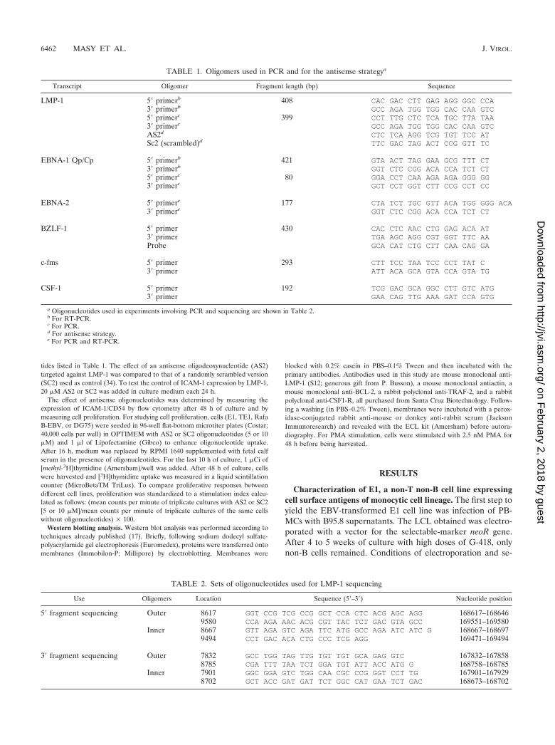
lection were very drastic for B-cell outgrowth. We analyzed thesurviving non-B cells, which grew in permanent cell lines 2months after the beginning of the experiment. As we previ-ously reported, the majority of the cell lines obtained with thisprotocol were naive T cells (17). However one cell line (E1)did not display surface antigens specific for mature T cells(CD3, CD2, and CD28), and no rearrangement of T-cell re-ceptor genes was detected. These cells were also devoid ofB-cell-specific markers (CD19, CD20, light chain of IgG[�] orIgG[�]) or NK cells (CD16, CD56) (Fig. 1 and Table 3). On theother hand, this cell line expressed surface antigens classicallyfound on peripheral monocytes, basically CD14 and HLA-DR,or found after activation or culture in vitro (CD80) (15). Inaddition, E1 was weakly positive for CD4 and CD21 expressionand constitutively negative for CD86 and CD1a expression(Table 3). This pattern of surface antigen expression, exceptfor that aspect related to CD86 (15), argues in favor of theirmonocytic origin. During long-term culture (over 6 months),the E1 cell line was stable, as indicated by constancy in cellmarker expression. This ability to proliferate in long-term cul-tures is characteristic of a transformed phenotype.
Established cell line E1 was injected into nude mice, andtumors were obtained (data not shown), showing that this cellline readily exhibits a malignant phenotype. Cell line TE1 wasderived from one of these tumors. The TE1 cell line displaysthe same pattern of surface marker expression as E1, exceptthat CD20 is weakly expressed on this cell line (Table 3).Nevertheless, no expression of rearranged immunoglobulingenes was detected by nested RT-PCR for E1 and TE1 (datanot shown).
E1 and TE1 express c-fms and its specific ligand. To furtherdemonstrate the monocytic origin of E1 and TE1, we investi-gated by RT-PCR the expression of the receptor for macro-phage colony-stimulating factor/colony-stimulating factor 1(CSF-1) (c-fms or CSF-1 receptor), which is implicated in the
differentiation of monocytic cells. This highly specific markerof the monocytic lineage (46) is expressed in both cell lines(Fig. 2A). The expression of CSF-1 was also detected by RT-PCR in both cell lines (Fig. 2B).
In addition, since the c-myc gene is induced and seemsinstrumental in the proliferative effects that result from thebinding of CSF-1 to its receptor (7, 10), we tested expression ofthis proto-oncogene. Overexpression of c-myc in the E1 andTE1 cell lines was detected by Northern blotting, whereasc-myc was weakly expressed in the NC5 and TC T-cell lines(Fig. 2C).
The E1 and TE1 monocytic cell lines are infected by EBV.To confirm that E1 and TE1 cells were infected by EBV, thepresence of viral DNA in Hirt’s extracts was investigated (8).Thus, we could detect by PCR the LMP-1-, EBNA-1-, andEBNA-2-encoding genes (Fig. 3A). In addition, preparativePCR was performed to determine the sequences of the LMP-1gene (also called BNLF-1) and its promoter on DNA extractedfrom various EBV-infected cells. These included the E1 andTE1 cell lines, as well as the two infected T-cell lines, NC5 andTC, derived from the same donor (17, 37). LMP-1 sequences ofthe corresponding B-LCL (B-EBVneo) and donor PBMCs(PBL25) were also analyzed. Except for the results for PBL25cells, the sequences of 5� and 3� PCR-amplified fragmentsencompassing the LMP-1 coding region were identical to thoseobtained from the prototype B95.8 strain. Sequence analysis ofLMP-1 from PBL25 showed 15 sequence variations and twodeletions compared to the B95.8 strain. However, only nine ofthe variations, found mainly in the transmembrane region,resulted in nonconservative amino acids (Table 4). The twodeletions were found in the region coding for the cytoplasmicdomain of the protein. One corresponds to the 30-bp deletionthat was reported as possibly associated with an increasedtransforming potential of the LMP-1 protein (32). The otheraffects the number of repeats of the 33-bp motif (4 versus 4.5
TABLE 3. Cell surface phenotype of monocytic cell lines E1 and TE1
MarkerResulta for:
Clone Isotype Supplier FluorochromeE1 TE1
CD3 � � UCHT1 IgG1 Immunotech FITCCD2 � � 39C1.5 IgG2a Immunotech FITCCD4 �w �w 13B8.2 IgG1 Immunotech FITCCD8 � � B9.11 IgG1 Immunotech FITCCD25 � � ACT-1 IgG1 Dako FITCCD28 � � CD28.2 IgG1 Immunotech FITCHLA-DR � � B8.12.2 IgG2b Immunotech FITC� chain � � Polyclonal Polyclonal Dako FITC� chain � � Polyclonal Polyclonal Dako PECD19 � � J4.119 IgG1 Immunotech PECD20 � �w B9E9(HRC20) IgG2a Immunotech FITCCD10 � � ALB2 IgG2a Immunotech FITCCD21 �w �w BL13 IgG1 Immunotech FITCCD22 � � 4KB128 IgG1 Dako FITCCD14 � � RMO52 IgG2a Immunotech FITCCD80 � � MAB104 IgG1 Immunotech NoneCD86 � � 2331(FUN-1) IgG1 Pharmingen NoneCD1a � � BL6 IgG1 Immunotech NoneCD16 � � 3G8 IgG1 Immunotech FITCCD34 � � 8G12 IgG1 Becton Dickinson PECD54 � � 84H10 IgG1 Immunotech PE
a �, negative; �, positive; �w, weakly positive. Comparisons are to results for the control antibody.
VOL. 76, 2002 EBV TRANSFORMATION OF A MONOCYTIC CELL LINE 6463
on February 2, 2018 by guest
http://jvi.asm.org/
Dow
nloaded from
repeats in PBL25 and B95.8 sequences, respectively) (1). Thisresult demonstrates the presence of a very divergent residentEBV strain in donor cells but confirms that the viral strainpresent in our monocytic cell lines is derived from the B95.8strain. This last point was corroborated by sequence analysesperformed on the LMP-1 promoter that showed a perfect co-linearity between the B95.8 strain and the strain present in thein vitro-transformed cell lines (data not shown).
EBV-infected monocytic cell lines expressed viral gene prod-ucts characteristic of a type II latency. Since an EBV genomewas clearly detected in E1 and TE1 cells and since both celllines presented a transformed phenotype and oncogenic prop-erties, we decided to evaluate the presence of specific viral
mRNA transcripts in these cells. Thus, we determined theexpression pattern of EBV in E1 and TE1 by performing RT-PCR and blotting experiments using primers and probes spe-cific for a selected set of viral genes. Figure 3B shows that E1and TE1 expressed an LMP-1 transcript with a size similar tothat of the transcript found in B95.8 cells. By contrast, noexpression of EBNA-2 was detected in both cells. In addition,we found that the EBNA-1 transcript is selectively expressedfrom the Qp promoter and that the BZLF-1 mRNA, specificfor the lytic phase, was not present (data not shown). As noBZLF-1 transcript was found in these cells, we did not explorefurther expression of the late lytic transcripts.
Furthermore, the LMP-1 protein product was detected by
FIG. 1. Membrane markers expressed by E1 and analyzed by flow cytometry. Antibodies used for immunophenotyping are listed in Table 3.
6464 MASY ET AL. J. VIROL.
on February 2, 2018 by guest
http://jvi.asm.org/
Dow
nloaded from

Western blotting (data not shown) and by flow cytometry anal-ysis (Fig. 4). The expression levels of LMP-1 in E1 and TE1 arevery similar to that found in Kas cells, a LCL used as a positivecontrol. LMP-1 expression is more homogeneous in E1 than inTE1. Expression of EBNA-1 and LMP-1 together with the lackof EBNA-2 expression specifies type II latency for the virus inboth cell lines.
The E1 and TE1 cell lines display phagocytic activity andmonokine production. The main characteristic of monocytes isthe ability to phagocytose foreign organisms in order topresent antigens to T cells through class II proteins of the
major histocompatibility complex. It has been reported thatinfection of monocytes by EBV alters their phagocytic activity(47). Figure 5 shows that E1 and TE1 cells exhibit a phagocyticactivity when incubated with FITC-labeled E. coli at 37°C.After incubation at 4°C, this activity was strongly decreased toa low level comparable to that obtained at 37°C for Jurkat cells,the negative-control cell line.
Monocytes and macrophages produce a set of cytokines,such as IL-8, IL-6, and TNF-�, which play the main role in thebiological effect of these cells on the immune response, par-ticularly during inflammation. IL-8, the prototypic CXC che-mokine, is necessary for the recruitment of leukocytes in in-flammatory processes. After 40 h of culture, E1 and TE1 cellsproduced IL-8 constitutively. This production was enhancedafter stimulation by PMA. The amounts of IL-8 produced forthese cell lines were higher than those produced for monocyticcontrol cell lines HL60 and U937. IL-6 induces production ofacute-phase proteins by hepatocytes. IL-6 was produced by E1and TE1 cells mainly after activation by PMA. TNF-� plays akey role in the orchestration of inflammation. Its biologicalactivity in L929 cells can be measured by using the cytotoxicityassay. Biologically active TNF-� is still present 40 h after stim-ulation of E1 and TE1 cells but not after stimulation of HL60and U937 cell lines (Table 5). To investigate the basis of thepersistence of this monokine 40 h after the activation, thekinetics of TNF-� production in E1 and TE1 cells and in acontrol monocytic cell line (U937) were analyzed. For U937,the maximum TNF-� production arises between 2 and 4 h afteractivation. For E1 and TE1, the appearance of the peak isdelayed (24 h for E1) and the amounts produced by these twocell lines are higher than that produced by the U937 cell line(Fig. 6). Both these findings can explain the persistence ofdetectable levels of biologically active TNF-� at 40 h. As ex-pected, control Jurkat cells produced TNF-�, IL-6, and smallamounts of IL-8.
Antisense to LMP-1 down-regulates E1 and TE1 cell prolif-eration as well as expression of adhesion molecule ICAM-1and the BCL-2 antiapoptotic protein. Since LMP-1 was dem-onstrated to display oncogenic properties (2, 50), we thenwanted to know whether LMP-1 played a role in the immor-talized and transformed phenotype of these cell lines. As a firstapproach, we investigated the proliferative rate of cells treatedwith LMP-1 antisense oligonucleotides. To this end, we de-signed an antisense oligodeoxynucleotide (AS2) correspondingto the first 20 nucleotides of the LMP-1 mRNA coding se-quence (34). A randomly scrambled version of the LMP-1antisense oligodeoxynucleotide (SC2) was used as a control(Fig. 7A). To show the efficacy of antisense oligonucleotides,the effects on the expression of LMP-1 were determined byimmunoblotting using the S12 anti-LMP-1 monoclonal anti-body. LMP-1 expression was significantly down-regulated inTE1 (Fig. 8B) and Rafa B-LCL (not shown) cells treated withthe antisense oligodeoxynucleotide (AS2) compared to expres-sion in control cells. As shown in Fig. 7, inhibiting LMP-1reduced by 80% the [3H]thymidine incorporation of TE1 an-tisense oligodeoxynucleotide-treated cells. The 5 �M dose wasas efficient as 10 �M, indicating that 5 �M is already a plateaudose. A weaker effect was observed with the E1 cell line (datanot shown). As already reported by others (25), the LMP-1antisense oligonucleotide also significantly reduced the prolif-
FIG. 2. Detection of c-fms, CSF-1, and c-myc transcripts. TotalRNA was isolated from both cells. RNA was reverse transcribed andamplified with pairs of PCR primers specific for the c-fms (A) or CSF-1(B) gene. Specific products were visualized by ethidium bromide stain-ing on a 1.8% agarose gel. (C) Northern blot experiments performedwith total RNA from the cell lines listed and hybridized with c-myc-and GAPDH-specific probes. MW, molecular weight markers.
VOL. 76, 2002 EBV TRANSFORMATION OF A MONOCYTIC CELL LINE 6465
on February 2, 2018 by guest
http://jvi.asm.org/
Dow
nloaded from

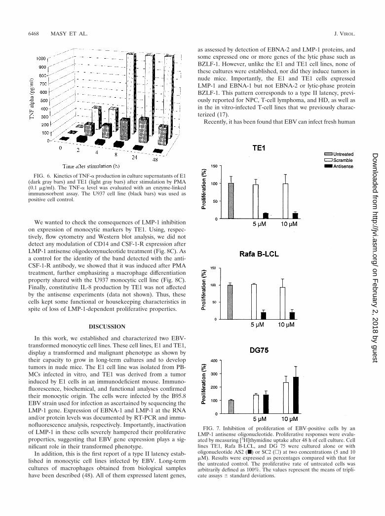
erative rate of LCLs but did not selectively affect the prolifer-ation of DG75, a Burkitt lymphoma cell line negative for EBV.A cytotoxic effect of LMP-1 antisense oligonucleotides can beruled out because of the increase of DG 75 cell proliferationand the noncytotoxic effect observed for all the cell lines testedwith scrambled oligonucleotides. This result indicates thatLMP-1 is essential for the proliferation of E1 and TE1, pin-pointing the critical role played by EBV infection in the phe-notype and the properties of these cells.
Numerous reports using LCLs or transient expression ofLMP-1 in noninfected cells stress the role of this receptor-like
protein in induction of genes involved in cellular proliferationand/or activation (12). This is the case for ICAM-1, a geneencoding an adhesion molecule (CD54) induced or up-regu-lated by LMP-1 partly through induction of a NF-�B pathway(9). Thus, we have investigated by flow cytometry analysismodulation of ICAM-1 expression by LMP-1 in E1 and TE1cells using the antisense strategy. As shown in Fig. 8A, wefound that addition of AS2 to the culture medium reproduciblydown-regulated ICAM-1 expression compared to addition ofthe scrambled control (SC2). The apparently modest down-
FIG. 3. E1 and TE1 expressed the EBV genome. (A) Detection of the EBV genome in infected monocytes. The presence of viral DNA in Hirt’sextracts was evaluated by PCR amplification of three EBV latent genes: LMP-1, EBNA-1, and EBNA-2. The corresponding PCR products (seeprimers in Table 2) were visualized by ethidium bromide staining on a 1.8% agarose gel. An LCL was used as a positive control for detection ofthe viral gene. Lane MW, 100-bp ladder. (B) Detection of viral RNA in E1 and TE1 by RT-PCR and Northern blotting experiments using primersand probes specific for a selected set of viral genes (see primers in Table 2). B95.8 cells were used as a positive control.
FIG. 4. Detection of LMP-1 protein expression in E1 and TE1 celllines. After fixation and permeabilization, cells were first incubatedwith anti-LMP-1 antibodies (CS1-4) and then with an anti-mouse Iglabeled with PE. Cells were then analyzed by flow cytometry. KasB-LCL was used as a positive control. The signal obtained with theisotypic control for E1 is indicated.
TABLE 4. Substitutions and deletions in the amino acid sequenceof LMP-1 identified in EBV strains infecting donor
cells or derived cell lines
Nucleotideposition
B95-8 sequenceMutationa
in PBL25Codon Nucleotideor change
Aminoacid
5� region169183 46 G Asp A/Asn169003 106 T Phe A/Ile168943 126 A Leu C/Phe168934 129 G Met T/Ile168934 132 A Arg G/Gly168889 144 T Phe A/Ile168871 150 A Asp C/Ala168868 151 A Leu C/Ile
3� region168754 189 A Gln C/Pro168265–168294 352–343 30-bp deletion NAb Deletion
a Comparison is to the B95–8 reference strain (GenBank accession no.X66863). The mutated nucleotide and corresponding amino acid are shown. Nomutations in cell lines B-EBVneo, NC5, TC, E1, and TE1 were found.
b NA, not applicable.
6466 MASY ET AL. J. VIROL.
on February 2, 2018 by guest
http://jvi.asm.org/
Dow
nloaded from

regulation of ICAM-1 after antisense oligonucleotide treat-ment is relevant of LMP-1-independent constitutive expressionof this protein in TE1 cells as well as in LCLs.
Similar results were obtained with antisense vectors in co-transfection experiments. A down-regulation of ICAM-1 ex-pression was obtained for EGFP-positive cells after cotrans-fection with an EGFP-expressing vector and a plasmidencoding an antisense transcript of LMP-1 (pSV LMP-1-AS)or a mutated form of I�B� (Fig. 8A).
In LCLs, down-regulation of prototypic antiapoptotic pro-tein BCL-2 was tested as a good indicator of loss of LMP-1expression (24). We thus investigated the effects of antisenseoligonucleotide treatment on expression of BCL-2, as well asTRAF-1 and TRAF-2, well-known mediators of the signalingproperties of LMP-1. Whereas BCL-2 expression was reducedboth in TE1 cells (Fig. 8B) and LCLs (data not shown),TRAF-2 was not affected. TRAF1, which is an LMP-1 targetgene in LCLs and other cell lines (8) (data not shown), was notdetected in TE1 cells (data not shown).
FIG. 5. Demonstration of phagocytic properties for EBV-infected monocytes E1 and TE1. E1 and TE1 were incubated with a suspension ofE. coli labeled with FITC at 4 (dotted lines) and 37°C (solid lines). After two washes with cold PBS, cells were analyzed on a cytometer at 488 nm.Jurkat cells were used as a negative control, and HL60 cells were used as a positive control. In the center, phagocytosis of E. coli labeled with FITCby E1 cells at 37°C is shown by microscopic fluorescence analysis.
TABLE 5. Production of three monokines (TNF-�, IL-6, and IL-8)in cell supernatants after 40 h of culture with (activated) or without
(constitutive) cell activation by PMA and ionomycin
Cell line Expression
Levela of:
TNF-�(U/ml)
IL-6(pg/ml)
IL-8(pg/ml)
Jurkat Constitutive 15Activated 497
E1 Constitutive 10.2 356Activated 14 15.4 20,520
TE1 Constitutive 780Activated 26 18.4 24,740
U937 Constitutive 10.3 75Activated 36.4 5,098
HL60 Constitutive 261Activated 8,053
a IL-6 and IL-8 levels were evaluated by enzyme immunoassay, and TNF-�levels were determined by a cytoxicity test against L929., lower than the sen-sitivity threshold of the test.
VOL. 76, 2002 EBV TRANSFORMATION OF A MONOCYTIC CELL LINE 6467
on February 2, 2018 by guest
http://jvi.asm.org/
Dow
nloaded from
We wanted to check the consequences of LMP-1 inhibitionon expression of monocytic markers by TE1. Using, respec-tively, flow cytometry and Western blot analysis, we did notdetect any modulation of CD14 and CSF-1-R expression afterLMP-1 antisense oligodeoxynucleotide treatment (Fig. 8C). Asa control for the identity of the band detected with the anti-CSF-1-R antibody, we showed that it was induced after PMAtreatment, further emphasizing a macrophage differentiationproperty shared with the U937 monocytic cell line (Fig. 8C).Finally, constitutive IL-8 production by TE1 was not affectedby the antisense experiments (data not shown). Thus, thesecells kept some functional or housekeeping characteristics inspite of loss of LMP-1-dependent proliferative properties.
DISCUSSION
In this work, we established and characterized two EBV-transformed monocytic cell lines. These cell lines, E1 and TE1,display a transformed and malignant phenotype as shown bytheir capacity to grow in long-term cultures and to developtumors in nude mice. The E1 cell line was isolated from PB-MCs infected in vitro, and TE1 was derived from a tumorinduced by E1 cells in an immunodeficient mouse. Immuno-fluorescence, biochemical, and functional analyses confirmedtheir monocytic origin. The cells were infected by the B95.8EBV strain used for infection as ascertained by sequencing theLMP-1 gene. Expression of EBNA-1 and LMP-1 at the RNAand/or protein levels was documented by RT-PCR and immu-nofluorescence analysis, respectively. Importantly, inactivationof LMP-1 in these cells severely hampered their proliferativeproperties, suggesting that EBV gene expression plays a sig-nificant role in their transformed phenotype.
In addition, this is the first report of a type II latency estab-lished in monocytic cell lines infected by EBV. Long-termcultures of macrophages obtained from biological sampleshave been described (48). All of them expressed latent genes,
as assessed by detection of EBNA-2 and LMP-1 proteins, andsome expressed one or more genes of the lytic phase such asBZLF-1. However, unlike the E1 and TE1 cell lines, none ofthese cultures were established, nor did they induce tumors innude mice. Importantly, the E1 and TE1 cells expressedLMP-1 and EBNA-1 but not EBNA-2 or lytic-phase proteinBZLF-1. This pattern corresponds to a type II latency, previ-ously reported for NPC, T-cell lymphoma, and HD, as well asin the in vitro-infected T-cell lines that we previously charac-terized (17).
Recently, it has been found that EBV can infect fresh human
FIG. 6. Kinetics of TNF-� production in culture supernatants of E1(dark gray bars) and TE1 (light gray bars) after stimulation by PMA(0.1 �g/ml). The TNF-� level was evaluated with an enzyme-linkedimmunosorbent assay. The U937 cell line (black bars) was used aspositive cell control.
FIG. 7. Inhibition of proliferation of EBV-positive cells by anLMP-1 antisense oligonucleotide. Proliferative responses were evalu-ated by measuring [3H]thymidine uptake after 48 h of cell culture. Celllines TE1, Rafa B-LCL, and DG 75 were cultured alone or witholigonucleotide AS2 (■) or SC2 (�) at two concentrations (5 and 10�M). Results were expressed as percentages compared with that forthe untreated control. The proliferative rate of untreated cells wasarbitrarily defined as 100%. The values represent the means of tripli-cate assays standard deviations.
6468 MASY ET AL. J. VIROL.
on February 2, 2018 by guest
http://jvi.asm.org/
Dow
nloaded from

FIG. 8. Effect of LMP-1 inhibition on expression of different markers in E1 or TE1 cells. (A) Effect on ICAM-1/CD54 expression on E1 cellsanalyzed by flow cytometry. (Top) The LMP-1 antisense oligonucleotide (AS2) down-regulated membrane expression of LMP-1 molecular targetICAM-1 on E1 cells. Scramble oligomers were used as the control (SC2). (Bottom) The same results were obtained with cells cotransfected withpEGFP-C1 and pSV LMP-1-AS or with pcDNA3 containing the coding sequence for mutated I�B�32/36A. The percentage of ICAM-1-expressingcells and the density of expression (MnI X) in these conditions are indicated. (B) Western blot analysis of several markers after LMP-1 inhibitionexperiments with TE1 cells. Immunoblots were probed with the S12 monoclonal antibody (LMP-1) or antibodies specific for TRAF-2, BCL-2, andactin proteins. Detection was performed after antisense oligonucleotide (AS2) or scramble (SC2) treatment, and results were compared to thosefor untreated cells (U). (C) Western blot and cytometry analyses of monocytic markers. (Left) Detection of CSF1-R after AS2, SC2, or PMAtreatment in TE1 cells. U937 cells untreated or stimulated by PMA were used as the control for CSF1-R induction. (Right) Flow cytometry profilefor CD14 labeling after treatment of TE1 cells. Rafa B-LCL is shown as a negative control.
VOL. 76, 2002 EBV TRANSFORMATION OF A MONOCYTIC CELL LINE 6469
on February 2, 2018 by guest
http://jvi.asm.org/
Dow
nloaded from
monocytes, in which a replicative cycle is observed, withoutviral persistence and without installation of latency. In addi-tion, those EBV-infected monocytes have reduced phagocyticactivity and TNF-� production (47). This inhibitory effect onTNF-� production seems to be triggered by the virus withoutpenetration into the cells (16). These processes could reflect astrategy developed by the virus to escape immune system con-trol in vivo. In contrast, the protocol we used, with a 5-weekselective phase, could favor the appearance of infected cells ina latent phase rather than in a lytic phase. In addition, both E1and TE1 display phagocytic activity and secrete high levels ofproinflammatory cytokines such as TNF-�. Therefore, the viralstatus in the respective monocytic cell lines obtained may dic-tate the functional properties of the cells.
Some human hemophagocytic syndromes are related toEBV infection (22, 26, 27). Related to the pathogenesis of thisEBV-associated syndrome, enhanced phagocytosis and secre-tion of TNF-� by monocytes/macrophages (31, 40) seeminglyresult from an activation of their functions by EBV-infected Tcells. We show here that, consequent to their infection byEBV, monocyte/macrophage cell lines display abnormally highTNF-� production, reminiscent of the effects of hemophago-cytic syndromes. Therefore, our results argue for a carefulreexamination of EBV infection of macrophages in hemoph-agocytic syndromes, which was not precisely investigated.
The two monocytic cell lines described here expressed anonmutated LMP-1 protein. To assess LMP-1 functionality inE1 and TE1 cells, expression of ICAM-1, an LMP-1 targetmolecule, was examined. After 48 h of cell culture with anantisense LMP-1 oligonucleotide or with a plasmid encodingan antisense LMP-1 transcript, down-regulation of ICAM-1,compared to what was found for control cells, was observed inboth cases. This result indicates that, in E1 and TE1 cells,LMP-1 is functional and can modulate cellular genes. As ex-pected (5, 14), a dominant-negative I�B�32/36A, which inhib-its the NF-�B pathway, affected ICAM-1 expression, suggest-ing that ICAM-1 down-regulation following introduction of aLMP-1 antisense oligodeoxynucleotide partially relied on theNF-�B pathway.
The critical proliferative and antiapoptotic roles played byLMP-1 in various cellular models including LCLs is well es-tablished (23, 24, 25). Though more controversial, some re-ports show a correlation between LMP-1 expression and tu-morigenicity in different cell types (28, 38, 44). Both themonocyte E1 cells and the NC5 T-cell line we previously de-scribed (17, 37) expressed LMP-1 and are tumorigenic afterinjection into immunodeficient mice. The fact that this tumor-igenic property is not shared by LCLs seems not related to adifference in the levels of LMP-1 expression, since B95.8-trans-formed E1 cells and the Kas B-LCL expressed similar amountsof the LMP-1 protein. In line with this observation, we did notdetect any mutation in the LMP-1 promoter region of the EBVstrain infecting these cell lines. LMP-1 inactivation using anantisense strategy suggests an instrumental role for LMP-1 inthe proliferative status of the infected monocytes. Indeed, theresults clearly demonstrate that cells with reduced LMP-1 ex-pression were greatly impaired in their proliferation. More-over, the fact that BCL-2 was down-regulated after antisenseoligonucleotide treatment suggests a role for LMP-1 in TE1cell survival, as is the case for LCLs (24).
The role of LMP-1 in establishment and maintenance of theproliferative status of LCLs expressing type III latency is wellknown (43). Lack of relevant experimental models hamperedinvestigation of this role in other EBV-infected cell lines ex-pressing LMP-1. Recently however, Noguchi et al. showed thatproliferation of two LMP-1-positive NK lymphoma cell lineswas insensitive to the LMP-1 antisense strategy used in thiswork (39). Thus, to our knowledge, this is the first report of theinstrumental role of LMP-1 in maintaining proliferation ofEBV-infected cells expressing a type II latency.
Of note, whereas ectopic expression of c-myc renders prolifer-ation of LCLs independent of LMP-1 (41), c-myc overexpressiondid not alleviate the LMP-1 dependence of the growth of EBV-transformed monocytes. This key regulator of cell proliferationcould be targeted by the binding of CSF-1 to its cognate receptor,expressed by both E1 and TE1 cells. Whether this results from apossible autocrine effect of CSF-1 produced by these cells andwhat could be the role of LMP-1 in this case remain to be ad-dressed. As a preliminary result, we showed that, like other mono-cytic markers (CD14 and IL-8 secretion), CSF-1-R expressionwas not affected by LMP-1 inhibition.
A unique infected monocyte cell line was isolated concom-itantly with four T-cell lines. This observation suggests thatinfection and transformation of monocytes by EBV are excep-tional events. Nevertheless, direct infection of T cells by EBVis also very rare, as demonstrated recently with a novel greenfluorescent protein-expressing EBV (49). Thus, the existenceof very few T cells and even fewer monocytes in an overwhelm-ing majority of B cells could explain the isolation of four T-celllines and of a unique monocytic cell line infected by EBV inour model. In agreement with this hypothesis, we were unableto detect CD14-expressing cells in LCLs, due probably to thepaucity of monocytic cells in these cell lines (data not shown).This impeded the screening of additional infected clones byclassical isolation processes. Infection of T cells and monocytesand their immortalization occur in exceptional cases, possiblyinfluenced by the isolation protocol.
In B cells, the initial event for the entry of EBV is theinteraction of the viral outer envelope glycoproteins gp350 andgp220 with CD21, the main receptor for EBV. In contrast, thepath for entry of EBV in monocytes is not known. E1 and TE1weakly expressed CD21 in a small percentage of cells and alsoHLA-DR molecules, known as CD21 coreceptors (18, 49).Alternatively, apoptotic bodies derived from EBV-carrying Blymphocytes can be a source of viral transfer to phagocytosingcells such as monocytes/macrophages (19). Thus, the massiveapoptosis triggered by the electroporation and selection pro-cesses used to yield E1 cells could favor this kind of entry.Finally, cellular fusion of an infected B or T cell with a mono-cytic cell was ruled out, since no rearrangement of Ig or T-cellreceptor genes was detected.
In conclusion, our T and monocytic cell lines can serve astools to elucidate this infection and transformation processesand represent unique in vitro models to study EBV-infectedcells displaying a type II latency.
ACKNOWLEDGMENTS
E.M. and E.A. contributed equally to this work.We thank Philippe Gosset for help in performing monokine pro-
duction assays, Jean Luc Baert, Jean Feuillard, Alain Sergeant, Eve-
6470 MASY ET AL. J. VIROL.
on February 2, 2018 by guest
http://jvi.asm.org/
Dow
nloaded from

lyne Manet for reagents, and Véronique Fafeur for critical reading ofthe manuscript.
This work was supported by the Association pour la Recherche surle Cancer (ARC; grants 9615 and 5455; fellowship for E.A.), CNRS,Lille II, Institut Pasteur de Lille, and INSERM.
REFERENCES
1. Baer, R., A. T. Bankier, M. D. Biggin, P. L. Deininger, P. J. Farrell, T. J.Gibson, G. Hatfull, G. S. Hudson, S. C. Satchwell, C. Seguin, et al. 1984.DNA sequence and expression of the B95–8 Epstein-Barr virus genome.Nature 310:207–211.
2. Baichwal, V. R., and B. Sudgen. 1988. Transformation of Balb/3T3 cells bythe BNLF-1 gene of Epstein-Barr virus. Oncogene 12:1–9.
3. Baumforth, K. R. N., L. S. Young, K. J. Flavell, C. Constandinou, and P. G.Murray. 1999. The Epstein-Barr virus and its association with human can-cers. Mol. Pathol. 52:307–322.
4. Brousset, P., F. Meggetto, S. Chittal, F. Bibeau, J. Arnaud, B. Rubin, and G.Delsol. 1993. Assessment of the methods for the detection of Epstein-Barrnucleic acids and related gene products in Hodgkin’s disease. Lab. Investig.69:483–490.
5. Cahir-McFarland, E. D., D. M. Davidson, S. L. Schauer, J. Duong, and E.Kieff 2000. NF-kappa B inhibition causes spontaneous apoptosis in Epstein-Barr virus-transformed lymphoblastoid cells. Proc. Natl. Acad. Sci. USA97:6055–6060.
6. Chen, C. L., R. H. Sadler, D. M. Walling, I. J. Su, H.-C. Hsieh, and N.Raab-Traub. 1993. Epstein-Barr virus (EBV) gene expression in EBV-pos-itive peripheral T-cell lymphomas. J. Virol. 67:6303–6308.
7. Cheng, M., D. Wang, and M. F. Roussel. 1999. Expression of c-Myc inresponse to colony-stimulating factor requires mitogen-activated protein ki-nase kinase-1. J. Biol. Chem. 274:6553–6558.
8. Chinsky, J., and R. Soeiro. 1981. Fv-1 host restriction of Friend leukemiavirus: analysis of unintegrated proviral DNA. J. Virol. 40:45–55.
9. Devergne, O., E. D. Cahir-McFarland, G. Mosialos, K. M. Izumi, C. F. Ware,and E. Kieff. 1998. Role of the TRAF binding site and NF-�B activation inEpstein-Barr virus latent membrane protein 1-induced cell gene expression.J. Virol. 72:7900–7908.
10. Dey, A., H. She, L. Kim, A. Boruch, D. L. Guris, K. Carlberg, S. M. Sebti,D. T. Woodley, A. Imamoto, and W. Li. 2000. Colony-stimulating factor-1receptor utilizes multiple signalling pathways to induce cyclin D2 expression.Mol. Biol. Cell 11:3835–3848.
11. Domke-Opitz, I., and H. Kirchmer. 1990. Stimulation of macrophages byendotoxin results in the reactivation of a persistent herpes simplex infection.Scand. J. Immunol. 32:69–75.
12. Eliopoulos, A. G., and A. B. Rickinson. 1998. LMP1 masquerades as anactive receptor. Curr. Biol. 8:R196–R198.
13. Faumont, N., T. Al Saati, P. Brousset, C. Offer, G. Delsol, and F. Meggetto.2001. Demonstration by single cell PCR that Reed-Sternberg cells as by-stander B lymphocytes are infected by different Epstein-Barr virus inHodgkin’s disease. J. Gen. Virol. 82:1169–1174.
14. Feuillard, J., M. Schuhmacher, S. Kohanna, M. Asso-Bonnet, F. Ledeur, R.Joubert-Caron, P. Bissières, A. Polack, G. W. Bornkamn, and M. Raphaël.2000. Inducible loss of NF-�B activity is associated with apoptosis and Bcl-2down-regulation in Epstein-Barr virus-transformed B lymphocytes. Blood95:2068–2075.
15. Fleischer, J., E. Soeth, N. Reiling, E. Grage-Griebenow, H. D. Flad, and M.Ernst. 1996. Differential expression and function of CD80 (B7–1) and CD86(B7–2) on human peripheral blood monocytes. Immunology 89:592–598.
16. Gosselin, J., L. Flamand, M. D’Addario, J. Hiscott, I. Stephanescu, D. V.Ablashi, R. C. Gallo, and J. Menezes. 1992. Modulatory effects of Epstein-Barr, herpes simplex and human herpes-6 viral infection and co-infection oncytokine synthesis. J. Immunol. 149:181–187.
17. Groux, H., F. Cottrez, C. Montpellier, B. Quatannens, J. Coll, D. Stehelin,and C. Auriault. 1997. Isolation and characterization of transformed humanT-cell lines infected by Epstein-Barr virus. Blood 89:4521–4530.
18. Haan, K. M., W. W. Kwok, R. Longnecker, and P. Speck. 2000. Epstein-Barrvirus entry utilizing HLA-DP or HLA-DQ as a coreceptor. J. Virol. 74:2451–2454.
19. Holmgren, L., A. Szeles, E. Rajnavölgyi, J. Folkman, G. Klein, I. Ernberg,and K. I. Falk. 1999. Horizontal transfer of DNA by the uptake of apoptoticbodies. Blood 93:3956–3963.
20. Ibanez, C. E., R. Schrier, P. Ghazal, P. Wiley, and J. A. Nelson. 1991. Humancytomegalovirus productively infects primary differentiated macrophages.J. Virol. 65:6581–6588.
21. Kawa, K 2000. Epstein-Barr virus-associated diseases in humans. Int. J. He-matol. 71:108–117.
22. Kawaguchi, H., T. Miyashita, H. Herbst, G. Niedobitek, M. Asada, M.Tsuchida, R. Hanada, A. Kinoshita, M. Sakurai, N. Kobayashi, et al. 1993.Epstein-Barr virus-infected T lymphocytes in Epstein-Barr virus-associatedhemophagocytic syndrome. J. Clin. Investig. 92:1444–1450.
23. Kawanishi, M. 1997. Expression of Epstein-Barr virus latent membrane
protein 1 protects Jurkat T cells from apoptosis induced by serum depriva-tion. Virology 228:244–250.
24. Kenney, J. L., M. E. Guinness, T. Curiel, and J. Lacy. 1998. Antisense to theEpstein-Barr virus (EBV)-encoded latent membrane protein 1 (LMP-1)suppresses LMP-1 and Bcl-2 expression and promotes apoptosis in EBV-immortalized B-cells. Blood 92:1721–1727.
25. Kenney, J. L., M. E. Guinness, M. Reiss, and J. Lacy. 2001. Antisense to theEpstein-Barr virus (EBV)-encoded latent membrane protein 1 (LMP1) sen-sitizes EBV-immortalized B-cells to transforming growth factor-beta andchemotherapeutic agents. Int. J. Cancer 91:89–98.
26. Kikuta, H. 1995. Epstein-Barr virus associated hemophagocytic syndrome.Leukoc. Lymphoma 16:425–429.
27. Kikuta, H., Y. Sakiyama, S. Matsumoto, T. Oh-Ishi, T. Nakano, T. Na-gashima, T. Oka, T. Hironaka, and K. Hirai. 1993. Fatal Epstein-Barr virusassociated hemophagocytic syndrome. Blood 82:3259–3264.
28. Kim, K.-R., T. Yoshizaki, H. Miyamori, K. Hasegawa, T. Horikawa, M.Furukawa, S. Harada, M. Seiki, and H. Sato. 2000. Transformation ofMadin-Darby canine kidney (MDCK) epithelial cells by Epstein-Barr viruslatent membrane protein 1 (LMP1) induces expression of Ets1 and invasivegrowth. Oncogene 19:1764–1771.
29. Kondo, K., T. Kondo, T. Okuno, M. Takahashi, and K. Yamanishi. 1991.Latent human herpesvirus 6 infection of human monocytes/macrophages.J. Gen. Virol. 72:1401–1408.
30. Larochelle, B., L. Flamand, P. Gourde, D. Beauchamp, and J. Gosselin.1998. Epstein-Barr virus infects and induces apoptosis in human neutrophils.Blood 92:291–299.
31. Lay, J. D., C. J. Tsao, J. Y. Chen, M. E. Kadin, and I. J. Su. 1997. Upregu-lation of tumor necrosis factor-alpha gene by Epstein-Barr virus and activa-tion of macrophages in Epstein-Barr virus-infected T cells in the pathogen-esis of hemophagocytic syndrome. J. Clin. Investig. 100:1969–1979.
32. Li, S. N., Y. S. Chang, and S. T. Liu. 1996. Effect of a 10-amino acid deletionon the oncogenic activity of latent membrane protein 1 of Epstein-Barr virus.Oncogene 12:2129–2135.
33. Lindhout, E., A. Lakeman, M. L. C. M. Mevissen, and C. de Groot. 1994.Functionally active Epstein-Barr virus-transformed follicular dendritic cell-like cell lines. J. Exp. Med. 179:1173–1184.
34. Mattia, E., S. Chichiarelli, T. Hickish, A. Gaeta, C. Mancini, D. Cunningham,and J. van Renswoude. 1997. Inhibition of in vitro proliferation of Epstein-Barrvirus infected B cells by an antisense oligodeoxynucleotide targeted againstEBV latent membrane protein LMP1. Oncogene 15:489–493.
35. Mehl, A. M., N. Fischer, M. Rowe, F. Hartmann, H. Daus, L. Trumper, M.Pfreundschuh, N. Muller-Lantzsch, and F. A. Grasser. 1998. Isolation andanalysis of two strongly transforming isoforms of the Epstein-Barr virus(EBV)-encoded latent membrane protein-1 (LMP1) from a single Hodgkin’slymphoma. Int. J. Cancer 76:194–200.
36. Menet, A., C. Speth, C. Larcher, W. M. Prodinger, M. G. Schwendinger, P.Chain, M. Jäger, F. Schwarzmann, H. Recheis, M. Fontaine, and M. P.Dierich. 1999. Epstein-Barr virus infection of human astrocyte cell line.J. Virol. 73:7722–7733.
37. Montpellier, C., P. Crépieux, B. Quatannens, B. Delobel, M. F. Croquette, D.Stehelin, C. Auriault, H. Groux, and J. Coll. 1997. Homologous T and B cellsimmortalized in vitro by the Epstein-Barr virus exhibit differential geneticand functional features. Int. J. Oncol. 11:87–96.
38. Nicholson, L. J., P. Hopwood, I. Johannessen, J. R. Salisbury, J. Codd, D.Thorley-Lawson, and D. H. Crawford. 1997. Epstein-Barr virus latent mem-brane protein does not inhibit differentiation and induces tumorigenicity ofhuman epithelial cells. Oncogene 15:275–283.
39. Noguchi, T., K. Ikeda, K. Yamamoto, I. Yoshida, A. Ashiba, J. Tsuchiyama,K. Shinagawa, T. Yoshino, M. Takata, and M. Harada. 2001. Antisenseoligodeoxynucleotides to latent membrane protein 1 induce growth inhibi-tion, apoptosis and Bcl-2 suppression in Epstein-Barr virus (EBV)-trans-formed B-lymphoblastoid cells, but not in EBV-positive natural killer celllymphoma cells. Br. J. Haematol. 114:84–92.
40. Ohga, S., A. Matsuzaki, M. Nishizaki, T. Nagashima, T. Kai, M. Suda, andK. Ueda. 1993. Inflammatory cytokines in virus hemophagocytic syndrome.Am. J. Pediatr. Hematol. Oncol. 15:291–298.
41. Polack, A., K. Hortnagel, A. Pajic, B. Christoph, B. Baier, M. Falk, J.Mautner, C. Geltinger, G. W. Bornkamm, and B. Kemples. 1996. c-mycactivation renders proliferation of Epstein-Barr virus (EBV)-transformedcells independent of EBV nuclear antigen 2 and latent membrane protein 1.Proc. Natl. Acad. Sci. USA 93:10411–10416.
42. Revoltella, R. P., E. Vigneti, A. Fruscalzo, M. Park, G. Ragona, G. Rocchi,and E. Calef. 1989. Epstein-Barr virus DNA sequences in precursor mono-cyte-macrophage cell lines established from the bone marrow of childrenwith maturation defects of haematopoiesis. J. Gen. Virol. 70:1203–1215.
43. Rickinson, A. B., and E. Kieff. 1996. Epstein-Barr virus, p. 2397–2446. InB. N. Fields, D. M. Knipe, and P. M. Howley (ed.), Fields virology, 3rd ed.,vol. 2. Lippincott-Raven Publishers, Philadelphia, Pa.
44. Roberts, M. L., and N. R. Cooper. 1998. Activation of a Ras-MAPK-depen-dent pathway by Epstein-Barr virus latent membrane protein 1 is essentialfor cellular transformation. Virology 240:93–99.
45. Sambrook, J., E. F. Fritsch, and T. Maniatis. 1989. Molecular cloning: a
VOL. 76, 2002 EBV TRANSFORMATION OF A MONOCYTIC CELL LINE 6471
on February 2, 2018 by guest
http://jvi.asm.org/
Dow
nloaded from

laboratory manual, 2nd ed., p. 7.19–7.22. Cold Spring Harbor LaboratoryPress, Plainview, N.Y.
46. Sariban, E., K. Imamura, M. Sherman, V. Rothwell, P. Pantazis, and D.Kufe. 1989. Downregulation of c-fms gene expression in human monocytestreated with phorbol esters and colony-stimulating factor 1. Blood 74:123–129.
47. Savard, M., C. Bélanger, M. Tardif, P. Gourde, L. Flamand, and J. Gosselin.2000. Infection of primary human monocytes by Epstein-Barr virus. J. Virol.74:2612–2619.
48. Shimakage, M., M. Kimura, S. Yanoma, M. Ibe, S. Yokota, G. Tsujino, T.
Kozuka, T. Dezawa, S. Tamura, A. Ohshima, M. Yutsudo, and A. Hakura.1999. Expression of latent and replicative-infection genes of Epstein-Barrvirus in macrophage. Arch. Virol. 144:157–166.
49. Speck, P., and R. Longnecker. 1999. Epstein-Barr virus (EBV) infectionvisualized by EGFP expression demonstrates dependence on known medi-ators of EBV entry. Arch. Virol. 144:1123–1137.
50. Wang, D., D. Liebowitz, and E. Kieff. 1985. An Epstein-Barr virus membraneprotein expressed in immortalized lymphocytes transforms established ro-dent cells. Cell 43:831–840.
6472 MASY ET AL. J. VIROL.
on February 2, 2018 by guest
http://jvi.asm.org/
Dow
nloaded from