Embed Size (px)
Citation preview

MRI EVALUATION ON
EXIT FORAMINA AND LATERAL RECESS
IN PATIENTS WHO UNDERGO
POSTERIOR LUMBAR INTERBODY FUSION
IN HOSPITAL RAJA PEREMPUAN ZAINAB II
By:
DR. SITI AISAH BINTI HASSAN
Dissertation Submitted In Partial Fulfilment
The Degree Of Master Of Medicine
(Radiology)
UNIVERSITI SAINS MALAYSIA
2011

MAGNETIC RESONANCE IMAGING (MRI) EVALUATION ON LATERAL RECESS
AND EXIT FORAMINA IN PATIENTS WHO UNDERGO POSTERIOR LUMBAR
INTERBODY FUSION (PLIF) IN HOSPITAL RAJA PEREMPUAN ZAINAB II (HRPZ II)
Dr. Siti Aisah Hassan
MMed Radiology
Radiology Department,
School of Medical Sciences, Universiti Sains Malaysia
Health Campus, 16150 Kelantan, Malaysia
Introduction: Magnetic Resonance Imaging (MRI) is the gold standard imaging modality
for investigation of degenerative disc disease. Changes of degenerative discs are well-
demonstrated. Disc dessication and herniations, thickened ligamentum flavum and
osteophytosis cause narrowing of neuroforamina, and further compromise the spinal cord and
exiting nerve roots. Posterior Lumbar Interbody Fusion (PLIF) is a surgical technique of
nerve root decompression using posterior approach. In PLIF procedure, laminectomy is done
to gain access to the intervertebral disc space. Discectomy is then performed and disc
materials are replaced with disc spacer to restore disc height. It was postulated that PLIF
procedure is able to restore foraminal height and therefore relieved nerve root compression
without foraminotomy.

Objectives: To study the improvement of exit foramina and lateral recess stenosis in patients
with back pain who underwent Posterior Lumbar Interbody Fusion (PLIF), using MRI as
diagnostic tool, to correlate the MRI findings with clinical symptoms and to determine
whether disc spacer height is a good predictor for improvement of lateral recess and exit
foramina.
Methodology: This is a cross-sectional, observational study of patients with degenerative
disc disease who underwent Posterior Lumbar Interbody Fusion (PLIF) in Hospital Raja
Perempuan Zainab II from June 2007 till June 2010. Patients’ clinical symptoms were
assessed using Oswestry Disability Index (ODI) pre- and post- procedure. MRI lumbosacral
pre and post PLIF were analysed in axial and sagittal views. The depth of the lateral recess
and exit foramina were measured at mid zone, exit zone and far lateral zone, and compared
pre and post PLIF. The height of the disc spacer was measured at mid sagittal views. Mean
difference of lateral recess and exit foramina size before and after procedure were analysed
using paired t-test, and correlated with ODI score using Pearson’s correlation test. Correlation
test was also used to determine whether disc spacer height is a good predictor for
improvement of lateral recess and exit foramina size.
Results: From 39 patients underwent PLIF in Hospital Raja Perempuan Zainab II, 25
patients fulfilled the inclusion criteria with 43 lumbar segments available for analysis.
Increment of lateral recess and exit foramina measurements post PLIF were observed at all
levels which was statistically significant (p<0.05). Improvement of clinical symptoms based
on ODI score (p<0.05) was also noted. However, there was no significant correlation between
patients clinical improvement and improvement of lateral recess and exit foramen (p>0.05).

It was also noted that the height of the disc spacer was not a predictor for increment of exit
foramen (p>0.05)
Conclusion: Posterior Lumbar Interbody Fusion is proven to restore depth of lateral recess
and exit foramen. Patients underwent PLIF had significant good surgical outcome. The
difference of disc spacer height used in the procedure did not determine the increment of exit
foramina. Therefore, current practice of using disc spacer height according to adjacent disc
height is an acceptable method.
Dr. Juhara Haron: Supervisor
Mr. Ahmad Sabri Omar: Co-Supervisor
Dr. Mohd Ariff Abas: Co-Supervisor
THIS DISSERTATION HAS BEEN PRESENTED AS
POSTER PRESENTATION TITLED
LATERAL RECESS AND EXIT FORAMINA SIZE AND ITS CLINICAL
OUTCOME FOLLOWING POSTERIOR LUMBAR INTERBODY FUSION
IN COLLEGE OF RADIOLOGY CONFERENCE,
RADIOLOGY: BEYOND IMAGE INTERPRETATION
IN LEGEND HOTEL, KUALA LUMPUR
ON 9th and 10th APRIL 2011

ii
To:
my husband, MOHD AZREEN , who have been so patience and encouraging,
my children, LUQMAN HAKIM, ABDUL HAFIZ, MUHAMMAD FATTAH,
and NUR HANNAN , my little sunshine who gave me strength,
my parents HJ HASSAN and HJH SITI KHATIJAH,
for their endless pray,
my brothers and sisters, IDA, HAZMI, ARFAH, MOHAIMIN,
HASBI, HAMZI , hope I have shown you the way....

iii
ACKNOWLEDGEMENTS
The preparation of this dissertation was not an easy task. Without courage and strength,
this dissertation may not be a reality. The time spent, the endless effort and the sacrifice,
makes me a better person. I would like to acknowledge these people, who have
continuously gave their guidance and support for the preparation of this dissertation.
DR. JUHARA HARON, my supervisor, who has always trust me, gives me her support
and always be there when I need her,
MR. AHMAD SABRI OMAR, my co-supervisor, who has initiated this study and gives
me his continuous support,
DR. MOHD ARIFF ABAS, Head of Department of Diagnostic Imaging, HRPZ II, who
is always concern on our knowledge, I believe learning is forever,
DR. KAMARUL IMRAN MUSA, Biostatistician, with his statistics help,
LECTURERS in Department of Radiology, USM, for the guidance and encouragement,
MY COLLEGUES, how we’ve gone through this together, our life full of strategies,
and those inner strength and maternal power that we have, thanks for caring and
sharing.
And, finally, to everyone who has direct or indirectly involved in this study
No words can say my thanks to all of you. May Allah bless you, always

iv
TABLE OF CONTENTS
Acknowledgement iii
Table of contents iv
List of Tables viii
List of Graphs ix
List of Figures x
Abbreviations xi
Abstrak xii
Abstract xvi
1.0 INTRODUCTION 1
2.0 LITERATURE REVIEW
2.1 Background 6
2.2 Anatomy and function 6
2.3 Pathophysiology 14
2.4 Imaging technique 18
2.5 Management 23
2.6 Posterior lumbar interbody fusion 24
2.7 Rationale of study 28
3.0 OBJECTIVES
3.1 General objective 29
3.2 Specific objectives 29

v
3.3 Null Hypothesis 30
4.0 METHODOLOGY
4.1 Study design 31
4.2 Ethical committee approval 31
4.3 Population and sample 32
4.3.1 Reference population 32
4.3.2 Source population 32
4.3.3 Inclusion criteria 32
4.3.4 Exclusion criteria 32
4.3.5 Sampling method 33
4.3.6 Sample size calculation 34
4.4 Research tools 36
4.5 Imaging technique 37
4.6 Image analysis 38
4.7 Validation of technique 43
4.8 Surgical technique 43
4.9 Data collection 44
4.10 Statistical analysis 44
5.0 RESULTS 45
5.1 Demographic data 46
5.1.1 Gender 46
5.1.2 Age 47
5.2 Number of lumbar segments involved in PLIF surgery 48

vi
5.3 MRI changes of lateral recess and exit foramina pre- and
post- PLIF 49
5.3.1 Difference in measurements of lateral recess and
exit foramina pre- and post-PLIF 51
5.4 Correlation of MRI findings with patients’ clinical symptoms 52
5.5 Correlation between height of the disc spacer with increment of
exit foramina post- (PLIF) 55
6.0 DISCUSSION 59
6.1 Demographic data 59
6.1.1 Gender 59
6.1.2 Age 60
6.2 Number of lumbar segments involved in PLIF surgery 61
6.3 MRI evaluation of lateral recess and exit foramina pre-
and post- PLIF 62
6.3.1 Lateral recess 62
6.3.2 Intervertebral foramen 63

vii
6.4 Correlation patients clinical outcome with improvement of
exit foramina 65
6.5 Correlation between height of disc spacer with improvement of
exit foramen 69
7.0 LIMITATION 71
8.0 CONCLUSION 73
REFERENCES 75
APPENDICES

viii
LIST OF TABLES
No Title Page no
Table 5.1 Mean depth of right and left lateral recess and exit
foramina pre and post PLIF 49
Table 5.2 Mean difference of right and left lateral recess and
exit foramina pre and post PLIF 51
Table 5.3 ODI scores pre and post PLIF for each patient 53
Table 5.4 Correlation between improvement of clinical symptoms and
increament in exit foramen 54
Table 5.5 Height of the disc spacer as a predictor for increment of
exit foramen 56
Table 5.6 Comparison of Oswestry Disability Index (ODI) score pre and
post surgical procedures on various studies 66

ix
LIST OF GRAPHS
No Title Page no
Graph 5.1 Number of PLIF cases from June 2007 till June 2010 45
Graph 5.2 Percentage of patients according to gender 46
Graph 5.3 Number and distribution of cases according to age 47
Graph 5.4 Number of lumbar segments operated in PLIF surgery 48
Graph 5.5 Correlation between disc spacer height and increment of
exit foramen 55

x
LIST OF FIGURES
Figure Title Page no
Figure 1 The lumbar vertebra 7
Figure 2 Structure of a lumbar vertebra body 8
Figure 3 The three zones of the intervertebral canal of the lumbar spine 9
Figure 4 The spinal canal zones demonstrated in axial plane 10
Figure 5 Diagram of the spinal canal 12
Figure 6 Changes in degenerative discs 15
Figure 7 Normal MRI lumbosacral spine 20
Figure 8 Sagittal T-2 WI MRI of degenerative lumbosacral spine 21
Figure 9 A graphical diagram of posterior lumbar interbody fusion 25
Figure 10 Multiple views of posterior lumbar interbody fusion allograft
spacer 26
Figure 11 Sample size calculation (1) using G*Power software 34
Figure 12 Sample size calculation (2) using G*Power software 35
Figure 13 Axial T2WI at L3 vertebra body : lateral recess 38
Figure 14 Axial T2WI at L4 vertebra body: intervertebra foramen (mid zone) 39
Figure 15 Axial T2WI at L4 vertebra body: intervertebra foramen (exit zone) 40
Figure 16 Axial T2WI at L4 vertebra body: intervertebra foramen (far lateral
zone) 41
Figure 17 Mid sagittal T2WI. 42
Figure 18 Example of axial image of a patient pre PLIF 50
Figure 19 Example of axial image of same patient post PLIF 50
Figure 20 A mid sagittal view of a patient post PLIF 57
Figure 21 A mid-sagittal image of a patient pre and post PLIF 58

xi
ABBREVIATIONS
AP Anteroposterior
CI Confidence Interval
cm centimeter
CT Computed tomography
IAP Inferior articular process
LSS Lumbar spinal stenosis
MRI Magnetic resonance imaging
MRM Magnetic resonance myelogram
ODI Oswestry Disability Index
PD Proton density
PLIF Posterior lumbar interbody fusion
SAP Superior articular process
SD Standard deviation
TE Time to echo
TR Time to repetition
xii
Abstrak
Bahasa Melayu
Tajuk:
Penilaian Magnetic Resonance Imaging (MRI) kepada liang-liang saraf (lateral recess
dan neuroforamina) terhadap pesakit yang bermasalah sakit tulang belakang
degeneratif dan menjalani Posterior Interbody Lumbar Fusion (PLIF) di Hospital Raja
Perempuan Zainab II (HRPZ II).
Latar Belakang:
Sakit tulang belakang disebabkan proses degeneratif merupakan salah satu
keluhan klinikal yang paling umum di kalangan pesakit di klinik pesakit luar dan
ortopedik. Sementara kebanyakan kes biasanya sembuh dalam tempoh beberapa
minggu, 2% daripada mereka mempunyai tanda-tanda radikulopati dan memerlukan
penyiasatan radiologi yang lebih lanjut. Tanpa mengira usia dan pekerjaan pesakit,
kesakitan tulang belakang merupakan penyebab umum ketidakupayaan sehingga
memberikan kesan besar kepada kehidupan peribadi dan sosial individu dan
keluarga,dan beban ekonomi kepada masyarakat.
Magnetic Resonance Imaging (MRI) adalah modaliti standard untuk penyiasatan
penyakit tulang belakang degeneratif kerana kebolehannya mempamerkan kontras
tinggi terhadap tisu lembut, kemampuan pengimejan multiplanar dan tidak melibatkan
radiasi pengionan. Perubahan imej degeneratif disk intervertebra adalah jelas, di mana
herniasi disk, penebalan ligamentum flavum dan pembentukan tulang baru

xiii
menyebabkan penyempitan kepada neuroforamina, dan selanjutnya memberi kompromi
terhadap akar saraf yang keluar. Oleh itu, penilaian MRI amat bernilai untuk
menentukan segmen tulang belakang yang terlibat dan tahap penyempitan
neuroforamina. MRI juga digunakan sebagai panduan untuk membantu perancangan
pembedahan dekompresi saraf –saraf akar. Posterior Interbody Lumbar Fusion (PLIF)
merupakan satu teknik pembedahan pada disk degeneratif di mana ianya dipostulatkan
boleh mengembalikan kedalaman neuroforamina kepada tahap lebih baik.
Objektif:
Untuk menilai peningkatan kedalaman liang saraf (lateral recess and
neuroforamina) pada pesakit yang mengalami sakit tulang belakang dan menjalani
Posterior Interbody Lumbar Fusion (PLIF), dengan menggunakan MRI sebagai alat
diagnostik, untuk mengkorelasikan penemuan MRI dengan gejala klinikal dan untuk
menentukan sama ada ketinggian alat ganti disk adalah prediktor untuk peningkatan
liang saraf.
Metodologi:
Ini adalah kajian keratan lintang terhadap pesakit yang mengalami sakit tulang
belakang akibat penyakit degeneratif disk yang menjalani Posterior Interbody Lumbar
Fusion (PLIF) di Hospital Raja Perempuan Zainab II dari bulan Jun 2007 hingga Jun
2010. Tahap ketidakupayaan dan kesakitan pesakit diukur menggunakan Oswestry

xiv
Disability Index (ODI). MRI lumbosakral pra dan pasca PLIF dianalisis dalam
pandangan paksi (axial) dan sagital. Kedalaman neuroforamina diukur pada tiga zon:
zon tengah, zon keluar dan zon paling jauh. Ketinggian alat ganti disk diukur pada
pertengahan pandangan sagital.
Keputusan:
Dari 39 pesakit menjalani PLIF di Hospital Raja Perempuan Zainab II, 25
pesakit yang memenuhi kriteria inklusi dengan 43 segmen lumbar sedia untuk dianalisa.
Penilaian lateral reses dan neuroforamina pasca pengukuran PLIF mendapati terdapat
peningkatan kedalaman di semua zon yang secara statistik bermakna (p <0.05).
Perubahan signifikan gejala klinikal berdasarkan skor ODI (p <0.05) juga direkodkan.
Namun, tidak ada hubungan yang signifikan antara pembaikan klinikal pesakit dengan
peningkatan lateral reses dan neuroforamina (p> 0.05). Kajian ini juga mendapati
bahawa ketinggian alat ganti disk bukan merupakan prediktor untuk peningkatan
neuroforamina (p>0.05).
Kesimpulan:
Posterior Lumbar Fusion Interbody terbukti dapat mengembalikan kedalaman
lateral reses dan neuroforamina. Pesakit yang menjalani pembedahan PLIF mempunyai
penyembuhan yang signifikan baik. Perbezaan ketinggian alat ganti disk yang
digunakan dalam prosedur tidak menentukan peningkatan kedalaman neuroforamina .

xv
Oleh kerana itu, amalan kini mengukur ketinggian alat ganti disk berdasarkan
ketinggian disk intervertebra yang berdekatan adalah dianggap kaedah yang boleh
diterima pakai.
xvi
Abstract
English
Title:
Magnetic Resonance Imaging (MRI) Evaluation on Lateral Recess and Exit Foramina
in Patients Who Undergo Posterior Lumbar Interbody Fusion (PLIF) in Hospital Raja
Perempuan Zainab II (HRPZ II)
Background:
Low back pain due to degenerative lumbar spine is one of the most common
presenting complaints in out-patient and orthopaedic clinics. While most cases usually
resolve in few weeks, 2% of them develop radiculopathy and need further radiological
investigations. Regardless of age and occupation of patients, back pain is a common
cause of disability and gives a great personal and social impact to the individual and
families, and economic burden to the society.
Magnetic Resonance Imaging (MRI) is the gold standard imaging modality for
investigation of degenerative disc disease for its superior soft tissue contrast,
multiplanar imaging capability and non-ionizing property. Changes of degenerative
intervertebral discs are well-demonstrated. Disc herniations, thickened ligamentum
flavum and osteophytosis cause narrowing of neuroforamina, and further compromise
the spinal cord and exiting nerve roots. Therefore, MRI is valuable to determine the
degree of stenosis and level of compression, and hence, used as a guide to aid plan of
surgical decompression.

xvii
Posterior Lumbar Interbody Fusion (PLIF) is one of the surgical technique of
nerve root decompression using posterior approach. In PLIF procedure, incision is made
at midline at the back. This is followed by laminectomy to gain access to the
intervertebral disc space. Discectomy is then performed and disc materials are replaced
with disc spacer to restore disc height. It was postulated that PLIF procedure is able to
restore foraminal height and therefore relieved nerve root compression without
foraminotomy.
Objectives:
To study the improvement of exit foramina and lateral recess stenosis in patients
with back pain who underwent Posterior Lumbar Interbody Fusion (PLIF), using MRI
as diagnostic tool, to correlate the MRI findings with clinical symptoms and to
determine whether disc spacer height is a good predictor for improvement of lateral
recess and exit foramina.
Methodology:
It is a cross-sectional, observational study of patients with back pain due to
degenerative disc disease who underwent Posterior Lumbar Interbody Fusion (PLIF) in
Hospital Raja Perempuan Zainab II from June 2007 till June 2010. Patients’ clinical
symptoms were assessed using Oswestry Disability Index (ODI) pre- and post-
procedure. MRI lumbosacral pre and post PLIF were analysed in axial and sagittal
views. The depth of the lateral recess and exit foramina were measured at mid zone, exit
zone and far lateral zone, and compared pre and post PLIF. The height of the disc

xviii
spacer was measured at mid sagittal views. Mean difference of lateral recess and exit
foramina size before and after procedure were analysed using paired t-test, and
correlated with ODI score using Pearson’s correlation test. Correlation test was also
used to determine whether disc spacer height is a good predictor for improvement of
lateral recess and exit foramina size.
Results:
From 39 patients underwent PLIF in Hospital Raja Perempuan Zainab II, 25
patients fulfilled the inclusion criteria with 43 lumbar segments available for analysis.
Increment of lateral recess and exit foramina measurements post PLIF were observed at
all levels which was statistically significant (p<0.05). Improvement of clinical
symptoms based on ODI score (p<0.05) was also noted. However, there was no
significant correlation between patients clinical improvement and improvement of
lateral recess and exit foramen (p>0.05). It was also noted that the height of the disc
spacer was not a predictor for increment of exit foramen (p>0.05)
Conclusion:
Posterior Lumbar Interbody Fusion is proven to restore depth of lateral recess
and exit foramen. Patients underwent PLIF had significant good surgical outcome. The
difference of disc spacer height used in the procedure did not determine the increment
of exit foramina. Therefore, current practice of using disc spacer height according to
adjacent disc height is an acceptable method.

1
1.0 INTRODUCTION
Low back pain is one of the most common presenting complaints in the out-
patient and orthopedic clinics. Most of these cases present with non-specific low back
pain which usually resolves in a few weeks. These patients usually need reassurance,
rest from work and conservative management. However, when back pain is associated
with radiculopathy, it warrants further investigations (Michelle, 2009). A study by
Veerapen et al., (2007) revealed 11.6% out of 2600 populations in a semirural area,
Malaysia were diagnosed with low back pain problem. Another study by Nurul Izzah
Abdul et a.,l (2010) showed that the prevalence of low back pain among primary school
teachers in Malaysia was 40.4%. A study in Canada and North America proved that
low back pain was the leading cause of disability and morbidity in middle-aged person
which was by far the most expensive source of workers' compensation costs (Manga et
al., 1993). Therefore, regardless of age and occupation of patients, back pain gives a
great personal and social impact to the individual and families, and economic burden to
the society.
Plain radiograph still serve as the initial primary investigation in patients with
low back pain (Tan, 2003). It is good in demonstrating bony details and intervertebral
disc space height. The changes of degenerative spine is well-demonstrated on plain
radiograph, this includes osteoporosis, sclerosis of endplates, osteophyte formation,
reduction of disc height, vacuum disc phenomenon, spinal canal and foramina
narrowing and presence of spondylolisthesis. Even though it lacks soft tissue details, it

2
is sufficient in most cases when the patient only need conservative or medical
management. It is also a good imaging modality in follow-up of these patients.
When conservative and medical management failed, patient may then need to be
further evaluated for possibilities of surgical decompression to relieve the symptoms. In
these situations, other radiological investigations then play important roles and are used
to further investigate the lumbar levels involved as well as to evaluate the degree and
severity of the disease.
Myelogram, Computed Tomography (CT) scan, CT myelogram and MRI can be
used in the investigations of degenerative disc disease with nerve roots or spinal cord
involvement (Kenneth and Hesselink, 1997). Depending upon availability of the
resources, examination time, benefits, invasiveness of the procedures, advantages and
disadvantages, as well as indications and contraindications, choice of modality used in
the investigations of patients with low back pain who may benefit from surgical
decompression should be discussed, justified and agreed between the radiologists and
the managing team.
Magnetic Resonance Imaging (MRI) is one of the most important radiological
investigations in patients with low back pain. It is the gold standard modality for
visualizing disc pathology, which is the primary event in degenerative spine leading to
foramina stenosis and nerve root impingement. It is superior compared to other imaging
modalities because of its non-ionizing radiation property, ability to demonstrate
excellent soft tissue contrast and non- invasive. It is also known for its high resolution,

3
multiplanar imaging capability. It is relatively a safe and fast examination provided that
all the precautions are well taken care of.
However, the use of MRI in the investigation of non-specific low back pain is
discouraged and should be reserved only to those with severe or progressive
neurological deficit, or for those cases in which serious underlying pathology is
suspected. The MRI findings of disc bulges or protrusions in people with low back pain
may frequently be coincidental (Jensen et al, 1994). The high prevalence of
asymptomatic disc degenerations must be taken into account when MRI is used for
assessment of spinal symptoms (Powell et al, 1986)
At present, MRI is the primary and preferred radiological investigation in
deciding management plan for patients with degenerative disc disease requiring surgery.
It is the modality of choice in pre-operative assessment of spinal stenosis and nerve root
impingement. It is used to determine the lumbar levels involved and severity of the
disease, and later, as a guide to the managing team in planning the surgical approach
and procedures. Its role in planning surgical management is well acknowledged in cases
with radiculopathy and spinal stenosis (N J Sheehan, 2010)
There are various surgical procedures practiced worldwide to decompress the
nerve roots impingement in degenerative lumbar spine. Posterior Lumbar Interbody
Fusion (PLIF) is a surgical decompression procedure that is been practiced in HRPZ II.
In PLIF surgery, the spine is approached in the midline posteriorly, and laminectomy is
performed to allow visualization of the nerve roots. The nerve roots are then retracted to

4
one side and the disc space is cleaned of the disc material. An interbody spacer, made of
carbon fiber, together with a bone graft, is then inserted into the disc space.
It is postulated that the interbody spacer inserted into the disc space will restore
the disc height, and thereby, indirectly restore the foraminal height and release the nerve
root compression.
Fusion rates in PLIF surgery are higher ( 90% -95%) than posterolateral fusion
rates (Peter F Ullrich, 2004). This is because bone graft is inserted into the anterior
portion of the spine, where there is more surface area. Furthermore, this bone is under
compression, and thus heals better. Non-union rates are higher in patients who have had
prior spine surgery, had multiple level fusion surgery, those who smoke or obese, and
those with history of cancer and being treated with radiation. Other than non-union, the
risks of PLIF procedure include infection and bleeding, which are fairly uncommon,
occurring about 1% - 3%.
Post-operatively, patients are assessed for recovery and success of surgical
procedure based on clinical assessment and plain radiograph. MRI examination is not a
routine assessment in patients who underwent surgical decompression for nerve root
impingement, unless there are symptoms suggestive of complications and failed back
surgery, such as infections or failure of instrumentation.
With the postulation that PLIF procedure is able to restore foraminal height and
therefore, released nerve root compression, this study is aimed to determine the change
in size of exit foramina and lateral recess measurement after PLIF using MRI as

5
imaging tool. These changes will be correlated with clinical outcome. It will try to
determine whether different height of the disc spacer is a good predictor to
improvement of exit foramina and lateral recess stenosis.
6
2.0 LITERATURE REVIEW
2.1 BACKGROUND
About 70% - 85% population will experience low back pain at some time in
their life. Annual incidence of low back pain is estimated at 5%, but only 1% will
develop radiculopathy (Nick Harden et al., 2005). Fortunately, the low back pain
resolves in the vast majority within two to four weeks (McKeon et al., 2006).
For individuals younger than 45 years old, back pain represents the most
common cause of disability and is generally associated with a work-related injury. For
individuals older than 45 years old, back pain is the third common cause of disability.
Determining the cause of low back pain is not easy. It is often multifactorial that
contributes to back pain. Radiological investigations often needed to further reveal the
cause of the pain, whether it is developmental, traumatic, infective or degenerative in
origin. Anatomical abnormalities are also common in spine that it should not necessarily
translate into clinical symptoms. Therefore, a careful clinical history and physical
examination are vital to evaluation, treatment and management of patient.
2.2 ANATOMY AND FUNCTION
Lumbar spine is made up of five vertebrae, referred as L1 to L5 (Figure 1). It
provides skeletal support for the human body and responsible for the distribution of
axial loads. These vertebrae bear much of the body’s weight and related to

7
biomechanical stress. For these reasons too, the lumbar vertebrae are more subjected to
trauma and degenerative diseases. Thus, the lumbar vertebral bodies are taller and
bulkier compared to the rest of the spine.
Figure 1 : The lumbar vertebra
Adopted from Spine Universe, Stewart G, 2010.
Each lumbar vertebra is composed of a vertebra body, two pedicles, two laminae
and a spinous process. The arrangement of these structures formed a bony ring called as
spinal canal. The vertebral structures and intervertebral discs protect the spinal cord
Sacrum
Vertebra body
Intervertebral disc
Neuroforamen/ Intervertebral foramen
Pedicle

8
and spinal nerve. The spinal cord is protected within the spinal canal. The pedicles and
laminae provide protective roof over the spinal nerves. (Figure 2).
(a) Axial view
(a) Axial view
(b) Lateral oblique view
Figure 2 : Structure of a lumbar vertebra body
Adopted from Spine Universe, Stewart G, 2010.
Nucleus pulposus
Annulus fibrosus

9
Rauschning W (1987) and Lee et al (1998) describes three clinically important
part of intervertebral canal: the entrance zone (lateral recess area), the mid zone
(sublamina blind area) and the exit zone (near the intervertebral foramen) (Figure 3).
Figure 3 : The three zones of the intervertebral canal of the lumbar spine
Adopted from the Spine, Rauschning W (1987).

10
The spinal nerve exits the spinal canal through a foramen (figure 4). The
foramen through which the spinal nerves exit the spinal canal is called as intervertebral
foramen or exit foramen or neural foramen. Anatomically, it is bounded by the vertebral
body and intervertebral discs anteriorly, the superior facet of the lower vertebra
posteriorly, and above and below by the pedicles of adjacent vertebrae (Kenneth and
Hesselink, 1997). The normal intervertebral foramen measures more than 4 mm in
anteroposterior (AP) diameter (Stephanie Ryan, 2004).
Figure 4: The spinal canal zones demonstrated in axial plane.
C : central zone, F : foraminal zone. E : extraforaminal zone
Adopted from the Spine, Rauschning W (1987).
C F
E

11
The anterolateral portion of the spinal canal where the descending nerve root lies
is called the lateral recess. Anatomically, the lateral recess is an area bordered anteriorly
by the posterior surface of the vertebral body, posteriorly by the superior articular facet
and laterally by the pedicle. It is funnel-shaped, narrowest at its cephalic portion at the
superior border of the pedicle.
The spinal nerve root leaves the dural tube, descends obliquely downward and
outward through the lateral recess, and emerges under the pedicle via the foramen. The
depth of the lateral recess should be measured between the most anterior portion of the
superior articular facet and the posterior border of the spinal canal at the superior
margin of the pedicle (Figure 5). The normal lateral recess measures more than 5 mm in
its depth (Michael A Mikhael et al, 1981).

12
Figure 5 : Diagram of the spinal canal. The lateral recess (4) is bounded
anteriorly by the posterior part of the vertebral body, posteriorly by the superior
articular facet, and laterally by the pedicle.
( Adopted from Neuroradiological evaluation of lateral recess syndrome. Michael A.
Mikhael, (1981). Radiology.)
1. Anteroposterior (sagittal) diameter
2. Transverse diameter
3. Interfacetal diameter
4. Depth of the lateral recess,
5. Interlaminar diameter

13
The intervertebral discs are fibrocartilagenous cushion serving as the spine’s
shocking absorbing system to protect the vertebrae and nerves. Each is composed of an
outer annulus fibrosus and a central nucleus pulposus. The strength of the lumbar disc is
related to fluid and proteoglycan content of the disc. Proteoglycan (PG) molecules are
important because they attract and retain water. Although both annulus fibrosus and
nucleus pulposus are composed of water, collagen and proteoglycans (PGs), the amount
of fluid (water and PGs) is the greatest in nucleus pulposus. The nucleus pulposus
contains a hydrated gel-like matter that resists compressions.
The spine functions best within a realm of static and dynamic stability. Bony
architecture and associated specialized soft tissue structures, especially the
intervertebral disk, provide static stability. Anterior elements bear over 90% of forces
transmitted through the lumbar spine in sitting; during standing, this portion decreases
to approximately 80%. Posterior elements of the lumbar spinal functional unit typically
bear less weight than anterior elements in all positions.
Dynamic stability, however, is accomplished through a system of muscular and
ligamentous supports acting in concert during various functional, occupational, and
avocational activities.
The discs allow some vertebral motion, ie extension and flexion. Even though
each disc has limited movement, considerable motion is possible when several discs
combine forces.

14
2.3 PATHOPHYSIOLOGY
The incidence of degenerative disc increases with age (Powell et al, 1986).
Lumbar spinal stenosis is one manifestation of the general process of spinal
degeneration that occurs with aging. The distribution of axial load is responsible for the
typical localization of spine degeneration. In the lumbosacral region, the most
frequently degenerated levels are the lower lumbar level, ie L4/5 and L5/S1, because
they are the sites for the highest dynamic and static load. The functional integrity of the
spinal curves also contributes to the degenerative changes. Spinal curves allow optimal
redistribution of axial load. When curves are preserved, the spine is 30 times more
elastic than a straight structure. If correct spine alignment is lost, an asymmetrical load
distribution may cause focal or diffuse spine degeneration. As the degenerative process
progresses, relative anterior-to-posterior force transmission approaches parity.
In degenerative disc disease, there are changes of the intervertebral discs and
structures surrounding it, causing either reduced in disc height, disc bulge or prolapse,
osteophytes formation or ligamentous hypertrophy, of which any of this could cause
spinal canal stenosis, lateral recess stenosis or intervertebral foramen stenosis (Figure
6). These changes would impinge and compress onto the nerve root causing the
symptoms.

15
Figure 6: Changes in degenerative discs
Adopted from Spine Universe, Stewart G, 2010.

16
Spinal canal stenosis is a condition in which part or all of the entire spinal canal
is stenosed (Kenneth and Hesselink, 1997). It results from progressive narrowing of the
central spinal canal and the lateral recesses (Lennard A Nadalo, 2010). The essential
content of the spinal canal includes the spinal cord, the cerebrospinal fluid (CSF) of the
thecal sac, and the dural membranes that enclose the thecal sac. The spinal canal may
become narrowed by bulging or protrusion of the intervertebral disc annulus, herniation
of the nucleus pulposus posteriorly, thickening of the posterior longitudinal ligament,
hypertrophy of the facet joints, hypertrophy of the ligamentum flavum.
In an article by Michael B Furman et al. (2010), the lateral recess stenosis is
further compartmentalized into the entrance zone (lateral recess), mid zone, exit zone,
and far-out (far lateral) stenosis:-
• The entrance zone or lateral recess lies medial to the pedicle and superior
articular process (SAP). Consequently, lateral recess stenosis arises from facet
joint SAP hypertrophy. Other causes include developmentally short pedicle and
facet joint morphology, as well as osteophytosis and herniated nucleus pulposus
(HNP) anterior to the nerve root. The lumbar nerve root compressed below SAP
retains the same segmental number as the involved vertebral level (eg, L5 nerve
root is impinged by L5 SAP).
• The mid zone (intervertebral foramen) extends from the medial to the lateral
pedicle edge. Mid-zone or intervertebra foramen stenosis arises from
osteophytosis under the pars interarticularis and bursal or fibrocartilaginous
hypertrophy at a spondylolytic defect.
• Exit-zone involves an area surrounding the foramen. This area extends
inferiorly till the intervertebral discs level. Exit zone stenosis arises from facet
17
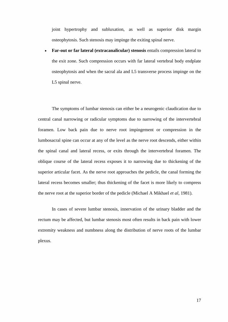
joint hypertrophy and subluxation, as well as superior disk margin
osteophytosis. Such stenosis may impinge the exiting spinal nerve.
• Far-out or far lateral (extracanalicular) stenosis entails compression lateral to
the exit zone. Such compression occurs with far lateral vertebral body endplate
osteophytosis and when the sacral ala and L5 transverse process impinge on the
L5 spinal nerve.
The symptoms of lumbar stenosis can either be a neurogenic claudication due to
central canal narrowing or radicular symptoms due to narrowing of the intervertebral
foramen. Low back pain due to nerve root impingement or compression in the
lumbosacral spine can occur at any of the level as the nerve root descends, either within
the spinal canal and lateral recess, or exits through the intervertebral foramen. The
oblique course of the lateral recess exposes it to narrowing due to thickening of the
superior articular facet. As the nerve root approaches the pedicle, the canal forming the
lateral recess becomes smaller; thus thickening of the facet is more likely to compress
the nerve root at the superior border of the pedicle (Michael A Mikhael et al, 1981).
In cases of severe lumbar stenosis, innervation of the urinary bladder and the
rectum may be affected, but lumbar stenosis most often results in back pain with lower
extremity weakness and numbness along the distribution of nerve roots of the lumbar
plexus.

18
2.4 IMAGING TECHNIQUE
In the evaluation of degenerative spine disease, multiple anatomic sites need to
be imaged, including the intervertebral disk, spinal canal, spinal cord, nerve roots,
neuroforamina, facet joints, and the soft tissues within and surrounding the spine
(Hesselink, 2010). The severity of disc changes, degree of spinal canal and
neuroforamina stenosis and their effect on the spinal cord and exiting nerve roots will
determine the management of the patients.
Plain radiographs have a limited diagnostic value because degenerative changes
are age-related and are equally present in both asymptomatic and symptomatic persons.
However, it still serve as an initial and important radiological investigations in patients
with degenerative spine. It is quick and easy to perform, inexpensive and noninvasive,
and readily available in most clinics. It is able to provide a lot of information on the
severity of the disease. It is good in demonstrating alignment of the lumbar vertebrae,
osteophytes formation, irregularities of the vertebral end plates and sclerosis of the
bones. Another important finding on plain radiographs of patients with degenerative
disc disease is decreased in disc height. This is often accompanied by spondylolisthesis
of the vertebra and subsequent neuroforamina stenosis.
Following surgical decompression, plain radiographs serve as good and reliable
tool in evaluation of bone healing and maturation of surgical fusion.
The gold standard modality for visualizing the herniated disc is magnetic
resonance imaging (MRI), which has been reported to be as accurate as CT
myelography. Many pulse sequences are available, and specific protocols vary among
different MR sites. There is general agreement that the spine needs to be imaged in at
19
least two planes, and surface coils are used almost exclusively. Fast spin-echo (FSE)
techniques allow enormous time savings, and if available, they have replaced
conventional spin-echo for T2
MRI, being a superior imaging modality to demonstrate soft tissue contrast, is
excellent in determining soft tissue causes of stenosis and for determining effect on
neural structures. It also has the ability to demonstrate damage to the intervertebral disc,
including annular tears and edema in the adjacent end plates. As with CT scans, MRI
can reveal bulging and degenerative discs in asymptomatic person. MRI is the technique
most frequently and extensively used in the evaluation of foraminal stenosis. It is also
used to evaluate nerves exiting from the foramen. However, recent studies have shown
that Magnetic resonance myelogram (MRM) is more valuable and reliable in pre-
surgical diagnosis of lumbar foraminal stenosis (Song KS, 2008).
-weighted imaging of the spine.
A normal MRI lumbosacral will demonstrate normal intervertebral disc which is
hypointense on T1WI and hyperintense on T2WI, due to high water content (Figure 7).
The disc height is preserved and no bulge seen. The vertebra body shows homogenous
signal with preserved height and smooth outline.
Changes is degenerative spine is demonstrated as dessicated disc with reduced
height and disc bulges, irregularities of endplates and fatty marrow changes (Figure 8).
MRI changes of disc degeneration should be interpreted with cautions. One third
of patients without low back pain have signs of degenerative discs on MRI. In
symptomatic patients, MRI findings were not correlated with severity of symptoms.
Therefore, abnormalities on MRI must be strictly correlated with age and any clinical
signs and symptoms before operative treatment is contemplated (Boden et al, 1990)

20
(a) (b)
Figure 7 : Sagittal MRI of lumbosacral spine of normal lumbar spine
(a): T-1 weighted image (b): T-2 weighted image
In this figure, the normal lumbar lordosis is preserved. The vertebral body height and
width preserved. The intervertebral disc height preserved with normal signal intensity
on both sequences.
Adopted from Spine Universe, Stewart G, 2010.

21
(c) (d) (e)
Figure 8 : Sagittal T-2 weighted image MRI of degenerative lumbosacral spine
(c): Dessicated disc at L4/L5
(d): Disc protrusion at L5/S1
(e): Multi-level endplate irregularities with reduced disc height and posterior disc bulge
Adopted from Spine Universe, Stewart G, 2010.

22
In patients younger than 50 years old, disc extrusion and sequestration, nerve
root compression, endplates abnormalities, and severe osteoarthritis of the facet joints
may be predictive of low back pain in symptomatic patients. (Weishaupt et al, 1998).
The role of imaging in spinal stenosis is to confirm the clinical diagnosis,
identify the level of stenosis, establish causes, and guide treatment. Any surgical
decisions should be firmly based on the clinical symptoms and corroborating results of
diagnostic testing. The accuracy of MRI for predicting the presence of disk herniations
at surgery is relatively high (varying from 76% to 96%), and thus it has become the
investigation of choice for patients suspected of lumbar disc herniations (Rijn, 2005).
Post-operatively, Magnetic resonance myelography (MRM) is a non-invasive,
efficient and reliable imaging tool in confirming post-operative decompression in
lumbar discectomy patient.
In a study done by Pankaj R Patel et al, (2010), the MRM specificity and
sensitivity was reported as 92% and 33.33% respectively which is better than
conventional MRI. In their study on fifty three patients with single level disc herniation
underwent discectomy, 47 patients (88.7%) showed positive clinico-radiological
correlation.

23
2.5 MANAGEMENT
While most patients with degenerative disc disease only need conservative and
medical management, a small proportion of them may need to undergo surgery. Less
than 2% of symptomatic patients undergo operative treatment. Surgical intervention is
best directed at those with unremitting nerve root symptoms. The purpose of the
surgery is to remove the diseased disc and as such remove the compression of the nerve
or spinal cord so as to relieve the pain and restore function to the nerves.
Among the surgical options usually practiced include :
i) Laminectomy - removal of the laminae overlying the spinal canal and
of the protruding disc
ii) Micro discectomy - removal of fragments of herniated disc through a
smaller incision without doing a laminectomy
iii) Spinal fusion - fusing of vertebrae together with bone grafts or metal
rods .

24
2.6 POSTERIOR LUMBAR INTERBODY FUSION (PLIF)
Previously, lumbar fusions were performed using the intertransverse technique,
necessitating wide exposure and possible use of iliac crest graft. Recent technologic
advances in cage technology, instrumentation and knowledge on bone biology have
widened the scope of fusion options, allowing the surgeon a variety of interbody
devices, and surgical methods to assess disc space, provide anterior column support,
secure rigid fixation and achieve solid fusion. All these goals can be achieved through
the well-known posterior approach (P M Arnold et al., 2009).
Posterior Lumbar Interbody Fusion (PLIF) is one of the surgical techniques that
was used to decompress the spinal cord and nerve roots, at the same time fusing the
vertebrae together in order to restore the alignment and gain spinal stabilization. The
surgical approach was originally advocated by Cloward, later modified by Lin and
others, are now well-known to spine surgeons. This technique is now widely practiced
in Malaysia as well as in Hospital Raja Perempuan Zainab II.
In this technique, the surgery is done using posterior approach. Skin incision is
made at the midline of the back, followed by incision to the muscles at this area.
Laminae are then removed (laminectomy) to gain access to the disc space. Once the disc
space is cleared, an interbody spacer is placed into the disc space with disc distraction.
The spacer remains in disc space and left under compression. The fusion is then
strengthened by adding pedicle screw fixation (Figure 9).