Embed Size (px)
Citation preview
Rom J Leg Med [23] 251-256 [2015]DOI: 10.4323/rjlm.2015.251© 2015 Romanian Society of Legal Medicine
251
Sudden death due to non-traumatic subdural hematoma in a patient with a myeloproliferative disorder
Mihai Ceaușu1, Constantin Dragoteanu2, Sorin Hostiuc3,*, Ionuț Negoi4
_________________________________________________________________________________________ Abstract: Subdural hematomas are known to be almost exclusively traumatic. Spontaneous, non-traumatic subdural hematomas are rarely cited in the scientific literature, being usually associated with oncological pathologies, drug abuse, vascular malformations, or with various treatments. The purpose of this article is to present a case of non-traumatic subdural hematoma with unfavourable course, which appeared in a patient with a newly diagnosed myeloproliferative disorder having a normal platelet count. Histologically the cause of the atraumatic subdural hematoma was proven to be an extramedullary myeloid metaplasia with liver and spleen involvement in the context of an unclassifiable neoplastic leukemic proliferation, which didn’t exclude chronic myeloid leukaemia. A certain diagnosis of Chronic Myeloid Leukemia would have needed bone marrow sampling, which was not performed in our case. However the initial clinical results, associated with the histology investigation were highly suggestive for CML. Key Words: sudden death, subdural hematoma, myloproliferative disorder.
1) (a) Carol Davila University of Medicine and Pharmacy, Dept of Pathology, (b) “Mina Minovici” National Institute of Legal Medicine, Dept of Pathology, Bucharest, Romania2) “Mina Minovici” National Institute of Legal Medicine, Dept of Pathology, Bucharest, Romania3) (a) “Carol Davila” University of Medicine and Pharmacy, Faculty of Medicine, Dept. of Legal Medicine and Bioethics, Department 2 Morphological Sciences, Bucharest, Romania, (b) “Mina Minovici” National Institute of Legal Medicine, Bucharest, Romania* Corresponding author: Sos. Vitan Barzesti 9, 042122, Sector 4 Bucharest, Romania, Tel. +40-723-791072, Email: [email protected], [email protected]) “Carol Davila” University of Medicine and Pharmacy, Dept of Surgery, Bucharest, Romania
Subdural hematomas are known to be almost exclusively traumatic in nature [1], the
main sources of bleeding being cortical haemorrhages (contusion injury, identifiable in 60-70% of all cases), ruptures in bridging veins (2.5-20%), sinusoidal injuries (4-9%), and arterial injuries (often the cortical part of the Sylvian artery – 2.5-10%) [2]. Spontaneous, non-traumatic subdural hematomas are rarely cited in the scientific literature [3], being usually associated with oncological pathologies [3, 4], drug abuse [5], vascular malformations [6], or with various treatments [7, 8]. Most
cases of spontaneous subdural hematomas are identified clinically and are not directly involved in thanatogenesis. The purpose of this article is to present a case of non-traumatic subdural hematoma with unfavourable course, developed at a patient with a newly diagnosed myeloproliferative disorder.
CASe report
A 63 years-old man complained of sever, rapid onset headache and motor deficit on the right side of the
252
Ceaușu M. et al. Sudden death due to non-traumatic subdural hematoma in a patient with a myeloproliferative disorder
body. Almost immediately he entered in a deep coma (GCS=3). The patient was admitted in a neurosurgery ward, where the CT examination revealed a subdural hematoma on the left hemisphere with a thickness of 2.8 cm and a mass effect on the middle line structures. The ventricular system was moved to the right of the sagittal plane. The initial haematological analysis revealed a highly increased white cell count (257.000/µl), a decreased number of red blood cells (2.87*106/µl), and a platelet count within normal range (154.000/µl). Prothrombin time and NIR were slightly increased (19.7 sec, and 1.64 respectively). The subdural hematoma was drained surgically, but the patient died immediately after the surgical intervention. As the patient died immediately after the onset of the symptoms and an initial cause of death was not properly identified, it was considered a sudden death, and a forensic autopsy was requested.
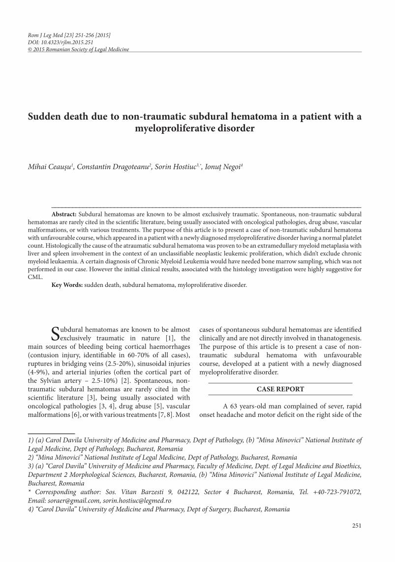
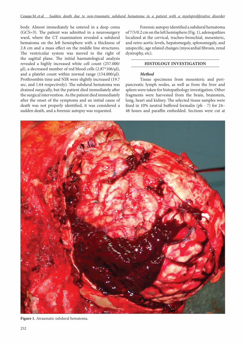
Forensic autopsy identified a subdural hematoma of 7/5/0.2 cm on the left hemisphere (Fig. 1), adenopathies localized at the cervical, tracheo-bronchial, mesenteric, and retro-aortic levels, hepatomegaly, splenomegaly, and unspecific, age related changes (myocardial fibrosis, renal dystrophy, etc).
HiStology inveStigAtion
Method Tissue specimens from mesenteric and peri-pancreatic lymph nodes, as well as from the liver and spleen were taken for histopathology investigation. Other fragments were harvested from the brain, brainstem, lung, heart and kidney. The selected tissue samples were fixed in 10% neutral buffered formalin (ph - 7) for 24–48 hours and paraffin embedded. Sections were cut at
Figure 1. Atraumatic subdural hematoma.
Romanian Journal of Legal Medicine Vol. XXIII, No 4(2015)
253
5 μm and stained with standard HE, PAS and Giemsa. Immunohistochemical analysis (IHC) was done using sections displayed on slides treated first with poly-l-
lysine. IHC was performed on 3 μm thick sections from formalin-fixed paraffin-embedded specimens. The method used was an indirect tristadial avidin-biotin-complex technique, with a Novolink polymer detection system which utilizes a novel control polymerization technology to prepare polymeric hrp-linker antibody conjugates, according to the manufacturer’s specifications (Novocastra, UK). Briefly, the procedure comprised: deparaffination in toluene and rehydration in alcohol series, washing in phosphate buffer saline (PBS), blocking with normal serum, for 5 min., incubation with primary antibody 60 min., incubation with post-primary block 30 min., then with novolink polymer 30 min. Sections are further incubated with the substrate / chromogen 3,3'-DAB and counterstained with Meyers’ Hematoxylin. The antibodies used for ihc were: CD20, UCHL1, CD68, S-100, CD10, CD30, CD34, EMA, Ki-67 (for details, see Table 1). Antigen retrieval techniques (thermal or enzymatic pre-treatment) for some of the aforementioned antibodies were done, according to the producer’s specifications. Both positive and negative controls were used.
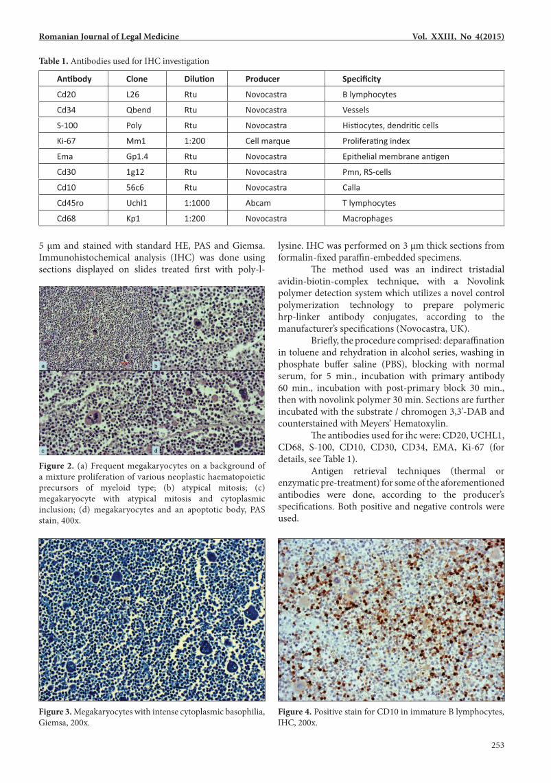
Figure 2. (a) Frequent megakaryocytes on a background of a mixture proliferation of various neoplastic haematopoietic precursors of myeloid type; (b) atypical mitosis; (c) megakaryocyte with atypical mitosis and cytoplasmic inclusion; (d) megakaryocytes and an apoptotic body, PAS stain, 400x.
table 1. Antibodies used for IHC investigation
Antibody Clone Dilution Producer Specificity
Cd20 L26 Rtu Novocastra B lymphocytes
Cd34 Qbend Rtu Novocastra Vessels
S-100 Poly Rtu Novocastra Histiocytes, dendritic cells
Ki-67 Mm1 1:200 Cell marque Proliferating index
Ema Gp1.4 Rtu Novocastra Epithelial membrane antigen
Cd30 1g12 Rtu Novocastra Pmn, RS-cells
Cd10 56c6 Rtu Novocastra Calla
Cd45ro Uchl1 1:1000 Abcam T lymphocytes
Cd68 Kp1 1:200 Novocastra Macrophages
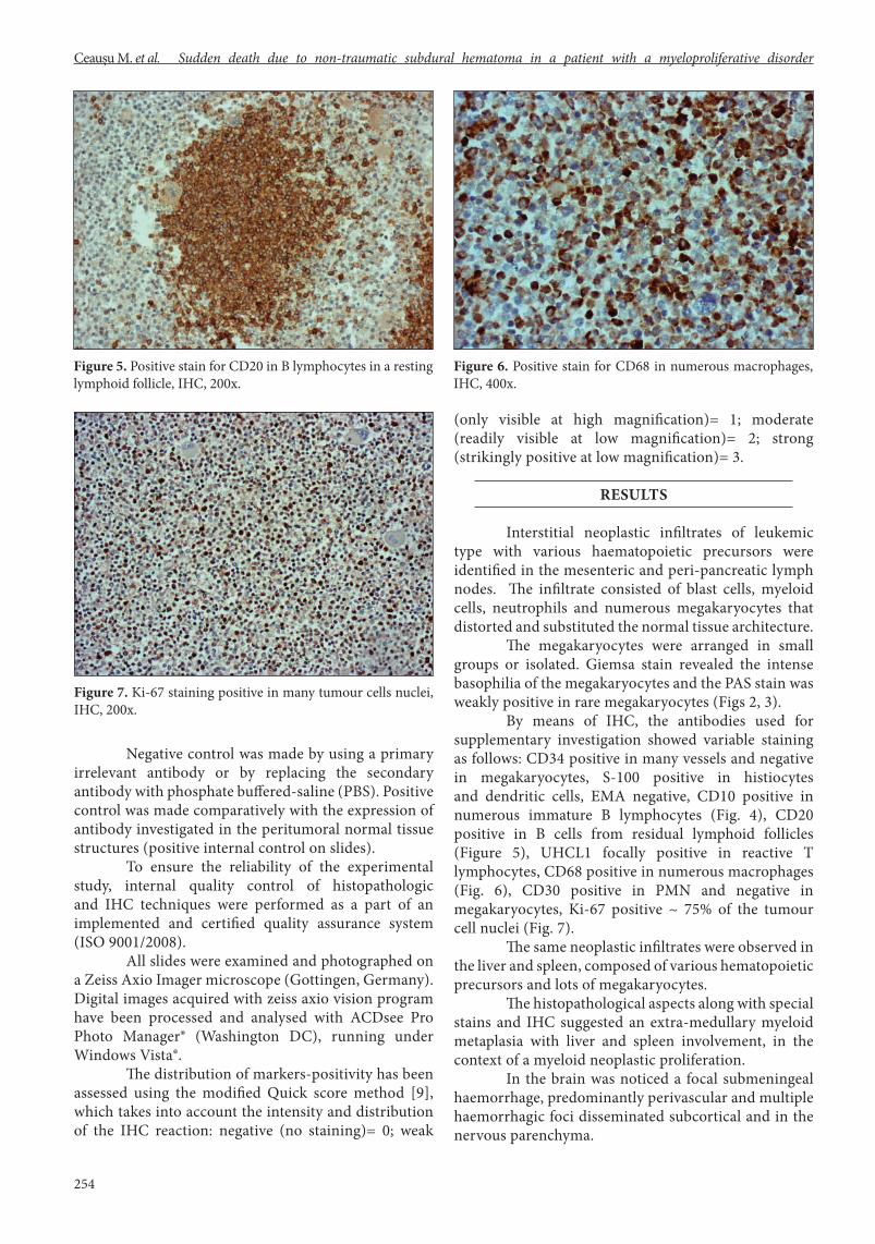
Figure 3. Megakaryocytes with intense cytoplasmic basophilia, Giemsa, 200x.
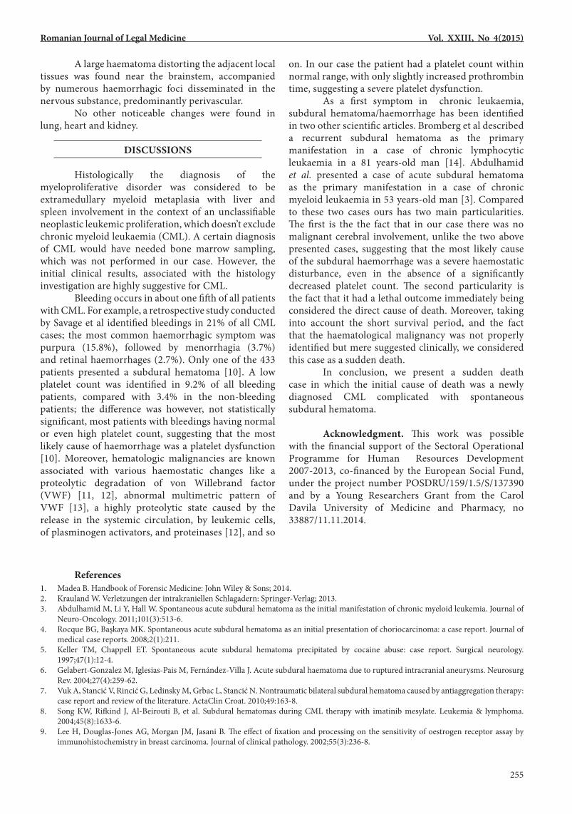
Figure 4. Positive stain for CD10 in immature B lymphocytes, IHC, 200x.
254
Ceaușu M. et al. Sudden death due to non-traumatic subdural hematoma in a patient with a myeloproliferative disorder
Negative control was made by using a primary irrelevant antibody or by replacing the secondary antibody with phosphate buffered-saline (PBS). Positive control was made comparatively with the expression of antibody investigated in the peritumoral normal tissue structures (positive internal control on slides). To ensure the reliability of the experimental study, internal quality control of histopathologic and IHC techniques were performed as a part of an implemented and certified quality assurance system (ISO 9001/2008). All slides were examined and photographed on a Zeiss Axio Imager microscope (Gottingen, Germany). Digital images acquired with zeiss axio vision program have been processed and analysed with ACDsee Pro Photo Manager® (Washington DC), running under Windows Vista®. The distribution of markers-positivity has been assessed using the modified Quick score method [9], which takes into account the intensity and distribution of the IHC reaction: negative (no staining)= 0; weak
(only visible at high magnification)= 1; moderate (readily visible at low magnification)= 2; strong (strikingly positive at low magnification)= 3.
reSultS
Interstitial neoplastic infiltrates of leukemic type with various haematopoietic precursors were identified in the mesenteric and peri-pancreatic lymph nodes. The infiltrate consisted of blast cells, myeloid cells, neutrophils and numerous megakaryocytes that distorted and substituted the normal tissue architecture. The megakaryocytes were arranged in small groups or isolated. Giemsa stain revealed the intense basophilia of the megakaryocytes and the PAS stain was weakly positive in rare megakaryocytes (Figs 2, 3). By means of IHC, the antibodies used for supplementary investigation showed variable staining as follows: CD34 positive in many vessels and negative in megakaryocytes, S-100 positive in histiocytes and dendritic cells, EMA negative, CD10 positive in numerous immature B lymphocytes (Fig. 4), CD20 positive in B cells from residual lymphoid follicles (Figure 5), UHCL1 focally positive in reactive T lymphocytes, CD68 positive in numerous macrophages (Fig. 6), CD30 positive in PMN and negative in megakaryocytes, Ki-67 positive ~ 75% of the tumour cell nuclei (Fig. 7). The same neoplastic infiltrates were observed in the liver and spleen, composed of various hematopoietic precursors and lots of megakaryocytes. The histopathological aspects along with special stains and IHC suggested an extra-medullary myeloid metaplasia with liver and spleen involvement, in the context of a myeloid neoplastic proliferation. In the brain was noticed a focal submeningeal haemorrhage, predominantly perivascular and multiple haemorrhagic foci disseminated subcortical and in the nervous parenchyma.
Figure 7. Ki-67 staining positive in many tumour cells nuclei, IHC, 200x.
Figure 5. Positive stain for CD20 in B lymphocytes in a resting lymphoid follicle, IHC, 200x.
Figure 6. Positive stain for CD68 in numerous macrophages, IHC, 400x.
Romanian Journal of Legal Medicine Vol. XXIII, No 4(2015)
255
A large haematoma distorting the adjacent local tissues was found near the brainstem, accompanied by numerous haemorrhagic foci disseminated in the nervous substance, predominantly perivascular. No other noticeable changes were found in lung, heart and kidney.
DiSCuSSionS
Histologically the diagnosis of the myeloproliferative disorder was considered to be extramedullary myeloid metaplasia with liver and spleen involvement in the context of an unclassifiable neoplastic leukemic proliferation, which doesn’t exclude chronic myeloid leukaemia (CML). A certain diagnosis of CML would have needed bone marrow sampling, which was not performed in our case. However, the initial clinical results, associated with the histology investigation are highly suggestive for CML. Bleeding occurs in about one fifth of all patients with CML. For example, a retrospective study conducted by Savage et al identified bleedings in 21% of all CML cases; the most common haemorrhagic symptom was purpura (15.8%), followed by menorrhagia (3.7%) and retinal haemorrhages (2.7%). Only one of the 433 patients presented a subdural hematoma [10]. A low platelet count was identified in 9.2% of all bleeding patients, compared with 3.4% in the non-bleeding patients; the difference was however, not statistically significant, most patients with bleedings having normal or even high platelet count, suggesting that the most likely cause of haemorrhage was a platelet dysfunction [10]. Moreover, hematologic malignancies are known associated with various haemostatic changes like a proteolytic degradation of von Willebrand factor (VWF) [11, 12], abnormal multimetric pattern of VWF [13], a highly proteolytic state caused by the release in the systemic circulation, by leukemic cells, of plasminogen activators, and proteinases [12], and so
on. In our case the patient had a platelet count within normal range, with only slightly increased prothrombin time, suggesting a severe platelet dysfunction. As a first symptom in chronic leukaemia, subdural hematoma/haemorrhage has been identified in two other scientific articles. Bromberg et al described a recurrent subdural hematoma as the primary manifestation in a case of chronic lymphocytic leukaemia in a 81 years-old man [14]. Abdulhamid et al. presented a case of acute subdural hematoma as the primary manifestation in a case of chronic myeloid leukaemia in 53 years-old man [3]. Compared to these two cases ours has two main particularities. The first is the the fact that in our case there was no malignant cerebral involvement, unlike the two above presented cases, suggesting that the most likely cause of the subdural haemorrhage was a severe haemostatic disturbance, even in the absence of a significantly decreased platelet count. The second particularity is the fact that it had a lethal outcome immediately being considered the direct cause of death. Moreover, taking into account the short survival period, and the fact that the haematological malignancy was not properly identified but mere suggested clinically, we considered this case as a sudden death. In conclusion, we present a sudden death case in which the initial cause of death was a newly diagnosed CML complicated with spontaneous subdural hematoma. Acknowledgment. This work was possible with the financial support of the Sectoral Operational Programme for Human Resources Development 2007-2013, co-financed by the European Social Fund, under the project number POSDRU/159/1.5/S/137390 and by a Young Researchers Grant from the Carol Davila University of Medicine and Pharmacy, no 33887/11.11.2014.
references1. Madea B. Handbook of Forensic Medicine: John Wiley & Sons; 2014.2. Krauland W. Verletzungen der intrakraniellen Schlagadern: Springer-Verlag; 2013.3. Abdulhamid M, Li Y, Hall W. Spontaneous acute subdural hematoma as the initial manifestation of chronic myeloid leukemia. Journal of
Neuro-Oncology. 2011;101(3):513-6.4. Rocque BG, Başkaya MK. Spontaneous acute subdural hematoma as an initial presentation of choriocarcinoma: a case report. Journal of
medical case reports. 2008;2(1):211.5. Keller TM, Chappell ET. Spontaneous acute subdural hematoma precipitated by cocaine abuse: case report. Surgical neurology.
1997;47(1):12-4.6. Gelabert-Gonzalez M, Iglesias-Pais M, Fernández-Villa J. Acute subdural haematoma due to ruptured intracranial aneurysms. Neurosurg
Rev. 2004;27(4):259-62.7. Vuk A, Stancić V, Rincić G, Ledinsky M, Grbac L, Stancić N. Nontraumatic bilateral subdural hematoma caused by antiaggregation therapy:
case report and review of the literature. ActaClin Croat. 2010;49:163-8.8. Song KW, Rifkind J, Al-Beirouti B, et al. Subdural hematomas during CML therapy with imatinib mesylate. Leukemia & lymphoma.
2004;45(8):1633-6.9. Lee H, Douglas-Jones AG, Morgan JM, Jasani B. The effect of fixation and processing on the sensitivity of oestrogen receptor assay by
immunohistochemistry in breast carcinoma. Journal of clinical pathology. 2002;55(3):236-8.

256
Ceaușu M. et al. Sudden death due to non-traumatic subdural hematoma in a patient with a myeloproliferative disorder
10. Savage DG, Szydlo RM, Goldman JM. Clinical features at diagnosis in 430 patients with chronic myeloid leukaemia seen at a referral centre over a 16-year period. British journal of haematology. 1997;96(1):111-6.
11. Hagag AAE, Abdel-Lateef AE, Aly R. Prognostic value of plasma levels of thrombomodulin and von Willebrand factor in Egyptian children with acute lymphoblastic leukemia. Journal of Oncology Pharmacy Practice. 2014;20(5):356-61.
12. Nilsson G, Astermark J, Strandberg K, Wichert S, Sallerfors B, Berntorp E. Coagulation activation and changes during treatment of acute myeloid leukaemia: A prospective cohort study. Journal of Hematological Malignancies. 2012;2(2):p11.
13. Tatewaki W, Takahashi H, Hanano M, Shibata A. Multimeric composition of plasma von Willebrand factor in chronic myelocytic leukaemia. Thrombosis research. 1988;52(1):23-32.
14. Ec Bromberg Wp Vandertop Gh Jansen J. Recurrent subdural haematoma as the primary and sole manifestation of chronic lymphocytic leukaemia. British journal of neurosurgery. 1998;12(4):373-6.