Embed Size (px)
Citation preview
Proc. Nati. Acad. Sci. USAVol. 87, pp. 1725-1729, March 1990Biochemistry
Promoter switching in Epstein-Barr virus during the initial stagesof infection of B lymphocytes
(mutant viral genomes/Si nuclease protection/transfection of B lymphocytes/infection time course)
MAXIMILIAN WOISETSCHLAEGER, CHANDRA N. YANDAVA, LISA A. FURMANSKI, JACK L. STROMINGER,AND SAMUEL H. SPECKDivision of Tumor Virology, Dana-Farber Cancer Institute, Harvard Medical School, 44 Binney Street, Boston, MA 02115
Contributed by Jack L. Strominger, December 26, 1989
ABSTRACT Transcription of the genes encoding the sixviral nuclear antigens present in Epstein-Barr virus latentlyinfected lymphocytes can be initiated from one of two promot-ers (Cp and Wp) mapping near the left end of the viral genome.These promoters are used in a mutually exclusive manner inclonal cell lines established from either Burkitt lymphomatumors or in vitro infection of peripheral B lymphocytes. In thispaper the role of Cp and Wp during viral latency is investi-gated. Cp appears to be the promoter normally employedduring established latent infection. Analysis of two cell linesthat use Wp revealed a deletion spanning Cp in the endogenousviral genomes, suggesting that cell lines exhibiting Wp activityharbor mutated viral genomes with a nonfunctional Cp. How-ever, in contrast to the preferred usage of Cp exhibited byestablished Epstein-Barr virus-infected cell lines, Wp wasshown to be exclusively utilized during the initial stages of viralinfection. In addition to Wp activity, Cp usage was apparent by6 days post-infection. A model is proposed involving B-lym-phocyte differentiation-driven promoter switching during theestablishment of viral latency.
Epstein-Barr virus (EBV) infection of B lymphocytes pre-dominantly results in a latent infection in which little virus isproduced with a concomitant growth transformation of thelymphocytes. Ten viral genes are known to be expressedduring latency, six of which are transcribed from a longcomplex transcriptional unit that spans the lefthand 100kilobases (kb) ofthe viral genome (for review, see ref. 1). Theresulting mRNAs all share common 5' exons and containunique 3' coding exons. Transcription is driven by one oftwopromoters, Cp or Wp, located near the left end of the viralgenome (see Fig. 1). Only one of these promoters is active inany given clonal cell line (2).
In this paper the question of the functional roles of Cp andWp during viral latency is addressed. We show that (i) whenboth functional promoters are present on a reporter con-struct, Cp is preferentially utilized by cell lines that exclu-sively use either Cp or Wp with their endogenous viralgenomes; (ii) Wp is inactive in Cp-utilizing cell lines trans-fected with the same construct; (iii) the region encompassingCp is deleted from the endogenous viral genomes of twoWp-utilizing lymphoblastoid cell lines (LCLs); (iv) deletionof the region containing Cp from a reporter construct con-taining a functional Wp induces Wp activity; and (v) Wp isexclusively used during the initial stages ofEBV infection ofB lymphocytes with activation of Cp apparent by 6 dayspost-infection. A model is proposed to'account for theseresults.
oriPC1 C2 WOW1 W2 W1 W2 W1 W2
3a
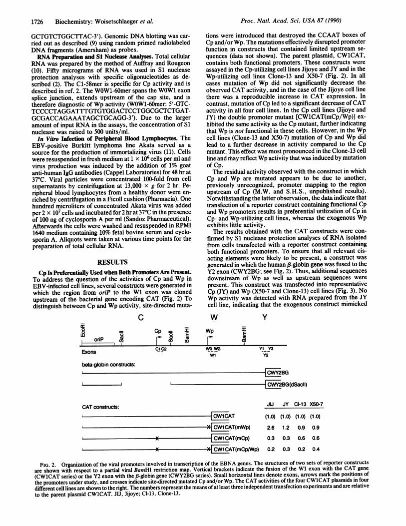
FIG. 1. Transcriptional organization of the EBV nuclear antigen(EBNA) genes. Regions ofthe viral genome encoding the EBNAs areindicated. The outer concentric arc represents the EBNA 1 primarytranscript, and the two arrowheads at the 5' end represent transcrip-tion initiation at either Cp or Wp. The exploded portion of thediagram illustrates the organization of exons present at the 5' end ofall EBNA mRNAs. The proximity of Cp and Wp to the latent originof replication (oriP), which also functions as an EBNA 1-dependentenhancer, is shown. Only the lefthand-most Wp is illustrated. TR,terminal repeats.
MATERIALS AND METHODSCells, Transfections, and Chloramphenicol Acetyltrans-
ferase (CAT) Assays. X50-7 (3), JY (4), and IB4 (5) are humanLCLs transformed with EBV. Jijoye (6) and Clone-13, asubclone of P3HR1 (7), are EBV-positive Burkitt lymphomacell lines. B95-8' is a spontaneously productive marmosetLCL containing EBV from a human patient. All cell lineswere grown at 370C in RPMI 1640 medium supplemented with10% (vol/vol) fetal bovine serum. DNA transfection 'andCAT assays were carried out as described (2).Plasmid Constructions and Site-Directed Mutagenesis. Site-
directed mutations in'CW1CAT were introduced according toKunkel (8) using a commercially available mutagenesis kit(Bio-Rad). Cp and Wp promoter mutants were generated byaltering the respective CCAAT boxes, thereby creating a newEcoRI recognition site, overlined (CP-mut37-mer: 5'-CTT-AGGGGCCGCGAAITCCTTAAGGGCTACATTTTTC-3';Wp-mut38-mer: 5'-CCTCCCTAGAACTGAGAATTCCCT-
Abbreviations: EBV, Epstein-Barr virus; CAT, chloramphenicolacetyltransferase; LCL, lymphoblastoid cell line; EBNA, EBVnuclear antigen.
The publication costs of this article were defrayed in part by page chargepayment. This article must therefore be hereby marked "advertisement"in accordance with 18 U.S.C. §1734 solely to indicate this fact.
1725
1726 Biochemistry: Woisetschlaeger et al.
GCTGTCTGGCTTAC-3'). Genomic DNA blotting was car-ried out as described (9) using random primed radiolabeledDNA fragments (Amersham) as probes.RNA Preparation and Si Nuclease Analyses. Total cellular
RNA was prepared by the method of Auffray and Rougeon(10). Fifty micrograms of RNA was used in S1 nucleaseprotection analyses with specific oligonucleotides as de-scribed (2). The C1-58mer is specific for Cp activity and isdescribed in ref. 2. The WOW1-60mer spans the WOW1 exonsplice junction, extends upstream of the cap site, and istherefore diagnostic of Wp activity (WOW1-60mer: 5'-GTC-TCCCCTAGGATTTGTGTGGACTCCTGGCGCTCTGAT-GCGACCAGAAATAGCTGCAGG-3'). Due to the largeramount of input RNA in the assays, the concentration of S1nuclease was raised to 500 units/ml.In Vitro Infection of Peripheral Blood Lymphocytes. The
EBV-positive Burkitt lymphoma line Akata served as asource for the production of immortalizing virus (11). Cellswere resuspended in fresh medium at 1 x 10⁶ cells per ml andvirus production was induced by the addition of 1% goatanti-human IgG antibodies (Cappel Laboratories) for 48 hr at370C. Viral particles were concentrated 100-fold from cellsupernatants by centrifugation at 13,000 x g for 2 hr. Pe-ripheral blood lymphocytes from a healthy donor were en-riched by centrifugation in a Ficoll cushion (Pharmacia). Onehundred microliters of concentrated Akata virus was addedper 2 x 10⁷ cells and incubated for 2 hr at 370C in the presenceof 100 ng of cyclosporin A per ml (Sandoz Pharmaceutical).Afterwards the cells were washed and resuspended in RPMI1640 medium containing 10%o fetal bovine serum and cyclo-sporin A. Aliquots were taken at various time points for thepreparation of total cellular RNA.
RESULTSCp Is Preferentially Used when Both Promoters Are Present.
To address the question of the activities of Cp and Wp inEBV-infected cell lines, several constructs were generated inwhich the region from oriP to the W1 exon was clonedupstream of the bacterial gene encoding CAT (Fig. 2) Todistinguish between Cp and Wp activity, site-directed muta-
CWE8 =
orip co>Op I
0 E(> co
tions were introduced that destroyed the CCAAT boxes ofCp and/or Wp. The mutations effectively disrupted promoterfunction in constructs that contained limited upstream se-quences (data not shown). The parent plasmid, CW1CAT,contains both functional promoters. These constructs wereassayed in the Cp-utilizing cell lines Jijoye and JY and in theWp-utilizing cell lines Clone-13 and X50-7 (Fig. 2). In allcases mutation of Wp did not significantly decrease theobserved CAT activity, and in the case of the Jijoye cell linethere was a reproducible increase in CAT expression. Incontrast, mutation of Cp led to a significant decrease ofCATactivity in all four cell lines. In the Cp cell lines (Jijoye andJY) the double promoter mutant [CWlCAT(mCp/Wp)] ex-hibited the same activity as the Cp mutant, further indicatingthat Wp is not functional in these cells. However, in the Wpcell lines (Clone-13 and X50-7) mutation of Cp and Wp didlead to a further decrease in activity compared to the Cpmutant. This effect was most pronounced in the Clone-13 cellline and may reflect Wp activity that was induced by mutationof Cp.The residual activity observed with the construct in which
Cp and Wp are mutated appears to be due to another,previously unrecognized, promoter mapping to the regionupstream of Cp (M.W. and S.H.S., unpublished results).Notwithstanding the latter observation, the data indicate thattransfection of a reporter construct containing functional Cpand Wp promoters results in preferential utilization of Cp inCp- and Wp-utilizing cell lines, whereas the exogenous Wpexhibits little activity.The results obtained with the CAT constructs were con-
firmed by Si nuclease protection analyses of RNA isolatedfrom cells transfected with a reporter construct containingboth functional promoters. To ensure that all relevant cis-acting elements were likely to be present, a construct wasgenerated in which the human f3-globin gene was fused to theY2 exon (CWY2BG; see Fig. 2). Thus, additional sequencesdownstream of Wp as well as upstream sequences werepresent. This construct was transfected into representativeCp (JY) and Wp (X50-7 and Clone-13) cell lines (Fig. 3). NoWp activity was detected with RNA prepared from the JYcell line, indicating that the exogenous construct mimicked
wWp
y
Eml
Exons C1C2 WO W2. YI Y3WI Y2
beta-globin constructs:CWY2BG
I CWY2BG(dSacIl)
CAT constructs:I- CwlC~AT
X-4 1CAT(mWp)I-X 4 CAT(mCp)
XI 4[jlAT(mCp/Wp)
JU JY Cl-13 X50-7
(1.0) (1.0) (1.0) (1.0)
2.6 1.2 0.9 0.9
0.3 0.3 0.6 0.6
0.2 0.3 0.2 0.4
FIG. 2. Organization of the viral promoters involved in transcription of the EBNA genes. The structures of two sets of reporter constructs
are shown with respect to a partial viral BamHI restriction map. Vertical brackets indicate the fusion of the W1 exon with the CAT gene(CW1CAT series) or the Y2 exon with the 3-globin gene (CWY2BG series). Small horizontal lines denote exons, arrows mark the positions ofthe promoters under study, and crosses indicate site-directed mutated Cp and/or Wp. The CAT activities of the four CW1CAT plasmids in fourdifferent cell lines are shown to the right. The numbers represent the means of at least three independent transfection experiments and are relativeto the parent plasmid CW1CAT. JIJ, Jijoye; Cl-13, Clone-13.
Proc. Natl. Acad Sci. USA 87 (1990)
Proc. Natl. Acad. Sci. USA 87 (1990) 1727
WO/W1 oligoC1 oligo probe probe
CI-13 X50-7 JY
co cc
O 0(O Ou) (I) Cl)
<scoM co < coc<cnzm <:mm <:}mmQ) Z UJCS Z CS ZCC4NC CI C14C C 4 VJ
0cOU OO 0o
input probe input probe
3protected $ protectedCl oligo - WOW1 oligo
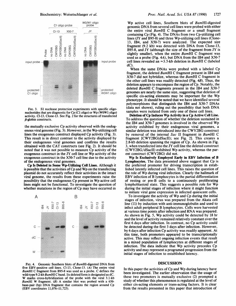
FIG. 3. S1 nuclease protection experiments with specific oligo-nucleotides that are diagnostic for Cp (Cl oligo) or Wp (WOW1 oligo)activity. CI-13, Clone-13. See Fig. 2 for the structures of transfectedp-globin constructs.
the mutually exclusive Cp activity observed with the endog-enous viral genome (Fig. 3). However, in the Wp-utilizing celllines the exogenous construct displayed Cp activity (Fig. 3).This result is in direct contrast to the activity displayed bytheir endogenous viral genomes and confirms the resultsobtained with the CAT constructs (see Fig. 2). It should benoted that it was not possible to measure Cp activity of theexogenous construct in the JY cell line or Wp activity of theexogenous construct in the X50-7 cell line due to the activityof the endogenous viral genomes.Cp Is Deleted in Some Wp-Utilizing Cell Lines. Although it
is possible that the activities ofCp and Wp on the transfectingplasmid do not accurately reflect their activities in the intactviral genome, the results from these experiments raise thepossibility that the endogenous Cp present in Wp active celllines might not be functional. To investigate the question ofwhether mutations in the region of Cp may have occurred in
AOOU
mn >-L)- 0)3 x<C) m
C
Br C-
Icmqr 0 LOm--) 0 m
_w C Q _Q
dC
w
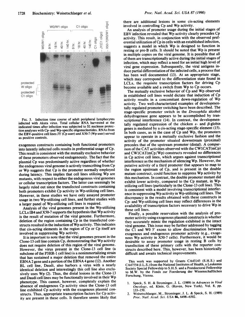
Wp active cell lines, Southern blots of BamHI-digestedgenomic DNA from several cell lines were probed with eitherthe entire viral BamHI C fragment or a small fragmentcontaining Cp (Fig. 4). The DNAs from two Cp-utilizing celllines (JY and B95-8) and three Wp-utilizing cell lines (Clone-13, IB4, and X50-7) were analyzed. The expected sizefragment (9.2 kb) was detected with DNA from Clone-13,B95-8, and JY (although the size of the fragment from JY isslightly smaller), when the entire BamHI C fragment wasused as a probe (Fig. 4A), but DNA from the JB4 and X50-7cell lines revealed an =3.5-kb deletion in BamHI C (labeleddC).When the same DNAs were probed with a labeled Cp
fragment, the deleted BamHI C fragment present in IB4 andX50-7 did not hybridize, whereas the BamHI C fragment inthe other cell lines was readily detected (Fig. 4B). Thus, thedeletion appears to encompass the region of Cp. Notably, thedeleted BamHI C fragments present in the IB4 and X50-7genomes are nearly the same size, suggesting that deletion ofspecific cis-acting elements may be important for the Wpphenotype. It should be noted that we have identified severalpolymorphisms that distinguish the IB4 and X50-7 DNAs(data not shown), ruling out the possibility that both DNAsamples were isolated from only one of these cell lines.
Deletion ofCp Induces Wp Activity in a Cp Active Cell Line.To address the question of whether the deletion sustained inthe IB4 and X50-7 genomes is involved in the observed Wpactivity exhibited by their endogenous viral genomes, asimilar deletion was introduced into the CWY2BG constructby removal of the internal Sac II fragment in BamHI Cfragment [CWY2BG(dSacII); see Fig. 2]. This creates a2.8-kb deletion spanning the region of Cp. As shown in Fig.3, when transfected into the JY cell line the deleted construct(CWY2BG/dSacII) exhibited Wp activity, whereas the par-ent construct (CWY2BG) did not.Wp Is Exclusively Employed Early in EBV Infection of B
Lymphocytes. The data presented above suggest that Cp isthe preferred promoter for driving transcription in estab-lished latently infected cell lines. This raises the question ofthe role ofWp during viral infection. Clearly the hallmark ofEBV infection of B lymphocytes is the partial differentiationof resting or pre-B cells to a continuously proliferatinglymphoblastoid state. This suggests a possible role for Wpduring the initial stages of infection where it might functionto initiate viral gene expression in infected quiescent cells.To investigate the activity of Wp and Cp during the initial
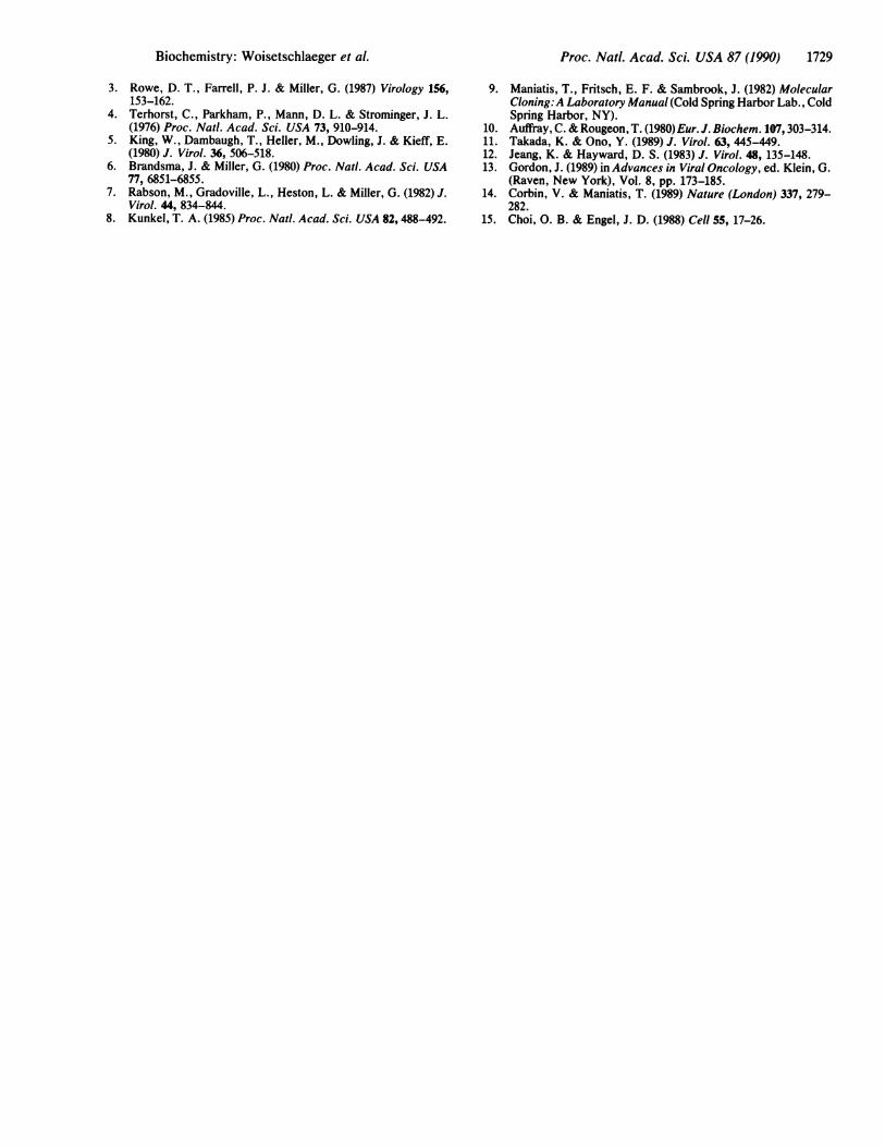
stages of infection, virus was prepared from the Akata cellline (11) by induction with anti-immunoglobulin and used toinfect adult peripheral B lymphocytes. Cells were harvestedat various time points after infection and RNA was prepared.As shown in Fig. 5, Wp activity could be detected by 18 hrand the level of activity remained relatively constant over thefirst 6 days after infection. In contrast, no Cp activity couldbe detected during the first 3 days after infection. However,by 6 days after infection Cp activity was readily apparent. Atthis time, both promoters appeared to be transcriptionallyactive. This may reflect ongoing infection events that resultin a mixed population of lymphocytes at different stages ofinfection. The data indicate that Wp activity precedes Cpactivity and may represent a programed progression from theinitial stages of infection to established latency.
FIG. 4. Genomic Southern blots of BamHI-digested DNA fromfive EBV-positive cell lines. CI-13, Clone-13. (A) The entire viralBamHI C fragment from B95-8 was used as a probe. C defines thewild-type 9.2-kb BamHI C band. Its deleted form is designated as dC.W marks cross-hybridization of the probe with the viral 3.1-kbBamHI W fragment. (B) A similar blot was probed with a 650-base-pair (bp) DNA fragment that contains the region around Cp(EBV coordinates 11,076-11,725).
DISCUSSIONIn this paper the activities of Cp and Wp during latency havebeen investigated. The earlier observation that the usage ofthese two promoters is mutually exclusive (2) prompted astudy of whether this phenomenon is regulated at the level ofeither cis-acting elements or trans-acting factors. It is clearfrom the results presented in this paper that introduction of
Biochemistry: Woisetschlaeger et al.
m
1728 Biochemistry: Woisetschlaeger et al.
WO/Wi oligo Cl oligo
0
0)
a_ eD o q.
I
:0:Mx:1
i0
protected
W Oligo -t
protectedCl Oligo
.0)0.-5 ~>0)
:-
cD To
CT
>- io
i
I
.,,_ _
mW -
FIG. 5. Infection time course of adult peripheral lymphocytesinfected with Akata virus. Total cellular RNA harvested at theindicated times after infection was subjected to S1 nuclease protec-tion analyses with Cp- and Wp-specific oligonucleotides. RNAs fromthe EBV-positive cell lines JY (Cp user) and X50-7 (Wp user) servedas positive controls.
exogenous constructs containing both functional promotersinto latently infected cells results in preferential usage of Cp.This result is consistent with the mutually exclusive behaviorof these promoters observed endogenously. The fact that theplasmid Cp was predominantly active regardless of whetherthe endogenous viral genome is actively transcribing from Cpor Wp suggests that Cp is the promoter normally employedduring latency. This implies that cell lines utilizing Wp are
mutants, with respect to either the endogenous viral genomesor cellular transcription factors. The latter can seemingly belargely ruled out since the transfected constructs containingboth promoters exhibit Cp activity in Wp-utilizing cell lines.
However, in these studies we have only assayed promoterusage in two Wp-utilizing cell lines, and further studies with
a larger panel of Wp-utilizing cell lines is required.Analysis of the viral genomes present in the Wp-utilizing
LCLs IB4 and X50-7 supports the hypothesis that Wp activityis the result of mutation of the viral genome. Furthermore,deletion of the region containing Cp in the transfected con-
structs resulted in the induction ofWp activity. This indicatesthat cis-acting elements in the region of Cp or Cp itself are
involved in suppressing Wp activity.It is important to note that the viral genomes present in the
Clone-13 cell line contain Cp, demonstrating that Wp activitydoes not require deletion of this region of the viral genome.However, the virus present in the Clone-13 cell line (asubclone of the P3HR-1 cell line) is a nonimmortalizing strainthat has sustained a major deletion that removed the entireEBNA 2 gene and a portion ofthe EBNA 4 gene (12). AnotherBL cell line, Daudi, also harbors a virus with a nearlyidentical deletion and interestingly this cell line also exclu-sively uses Wp (2). Thus, the distal lesions in the Clone-13and Daudi cell lines may be functionally involved in their Wpphenotype. This cannot, however, completely explain theabsence of endogenous Cp activity since the Clone-13 cellline exhibited Cp activity with the exogenous plasmid con-
structs. Thus, appropriate transcription factors for Cp activ-
ity are present in these cells. It therefore seems likely that
there are additional lesions in some cis-acting elementsinvolved in controlling Cp and Wp activity.An analysis of promoter usage during the initial stages of
EBV infection revealed that Wp activity clearly precedes Cpactivity. This result, in conjunction with the observed pref-erential utilization ofCp in cells with an established infection,suggests a model in which Wp is designed to function inresting or pre-B cells. It should be noted that Wp is presentin multiple copies on the viral genome. It is possible that allofthem are transcriptionally active during the initial stages ofinfection, which may reflect a need for an initial high level ofviral gene expression. Subsequently, the viral antigens in-duce partial differentiation ofthe infected cells, a process thathas been well documented (13). At an appropriate stage,which may correspond to the differentiation state found inLCLs, the requisite transcription factors for driving Cpbecome available and a switch from Wp to Cp occurs.The mutually exclusive behavior of Cp and Wp observed
in established cell lines would dictate that induction of Cpactivity results in a concomitant down-regulation of Wpactivity. Two well-characterized examples of developmen-tally regulated promoter switching have been described. Thestage-specific promoter switch in the Drosophila alcoholdehydrogenase gene appears to be accomplished by tran-scriptional interference (14). In contrast, the developmen-tally regulated expression of the chicken 6- and f-globingenes is mediated by a cis-acting stage-specific element (15).In both cases, as in the case of Cp and Wp, the promotersappear to operate in a mutually exclusive fashion and theactivity of the promoter situated downstream (proximal)precedes that of the upstream promoter (distal). A compar-ison of the CAT activities observed with the CW1CAT(mCp)and CWlCAT(mCp/Wp) constructs revealed no differencesin Cp active cell lines, which argues against transcriptionalinterference as the mechanism of silencing Wp. However, theputative activity of a third promoter that appears to map tothe region upstream of Cp, and is present in the doublemutant construct, could function to suppress Wp activity bythis mechanism. In contrast, the double promoter mutant didexhibit lower activity, compared to the Cp mutant, in Wp-utilizing cell lines (particularly in the Clone-13 cell line). Thisis consistent with a model involving transcriptional interfer-ence in suppressingWp activity in Wp-utilizing cell lines. Thediscrepancy in the results obtained with these constructs inCp- and Wp-utilizing cell lines may reflect differences in theavailability of transcription factors necessary to drive Wp inthese cell lines.
Finally, a possible reservation with the analysis of pro-moter activity using exogenous plasmid constructs is whetherthey accurately mimic the regulation exhibited in the intactviral genome. This issue may be further addressed by taggingthe C1 and WO 5' exons to allow discrimination betweenexogenous and endogenous promoter activity (e.g., exoge-nous Wp activity in X50-7 cells). Furthermore, it would bedesirable to assay promoter usage in resting B cells bytransfection of these primary cells with the reporter con-
structs described here. This, however, has been historicallydifficult and awaits technical improvements.
This work was supported by Grants CA43143 (S.H.S.) andCA47554 (J.L.S.) from the National Institutes of Health, a LeukemiaSociety Special Fellowship to S.H.S. and a Postdoctoral Fellowshipto M.W. by the Fonds zur Foerderung der WissenschaftlichenForschung, Vienna.
1. Speck, S. H. & Strominger, J. L. (1989) in Advances in ViralOncology, ed. Klein, 6. (Raven, New York), Vol. 8, pp.133-150.
2. Woisetschlaeger, M., Strominger, J. L. & Speck, S. H. (1989)Proc. Natl. Acad. Sci. USA 86, 6498-6502.
Proc. Natl. Acad. Sci. USA 87 (1990)
-;Ij.I .:..... ..:.::
.".4A"',... .,
..f.f:
Biochemistry: Woisetschlaeger et al.
3. Rowe, D. T., Farrell, P. J. & Miller, G. (1987) Virology 156,153-162.
4. Terhorst, C., Parkham, P., Mann, D. L. & Strominger, J. L.(1976) Proc. Nail. Acad. Sci. USA 73, 910-914.
5. King, W., Dambaugh, T., Heller, M., Dowling, J. & Kieff, E.(1980) J. Virol. 36, 506-518.
6. Brandsma, J. & Miller, G. (1980) Proc. Nail. Acad. Sci. USA77, 6851-6855.
7. Rabson, M., Gradoville, L., Heston, L. & Miller, G. (1982) J.Virol. 44, 834-844.
8. Kunkel, T. A. (1985) Proc. Nail. Acad. Sci. USA 82,488-492.
Proc. Nati. Acad. Sci. USA 87 (1990) 1729
9. Maniatis, T., Fritsch, E. F. & Sambrook, J. (1982) MolecularCloning:A Laboratory Manual (Cold Spring Harbor Lab., ColdSpring Harbor, NY).
10. Auffray, C. & Rougeon, T. (1980) Eur. J. Biochem. 107,303-314.11. Takada, K. & Ono, Y. (1989) J. Virol. 63, 445-449.12. Jeang, K. & Hayward, D. S. (1983) J. Virol. 48, 135-148.13. Gordon, J. (1989) in Advances in Viral Oncology, ed. Klein, G.
(Raven, New York), Vol. 8, pp. 173-185.14. Corbin, V. & Maniatis, T. (1989) Nature (London) 337, 279-
282.15. Choi, 0. B. & Engel, J. D. (1988) Cell 55, 17-26.