Embed Size (px)
Citation preview
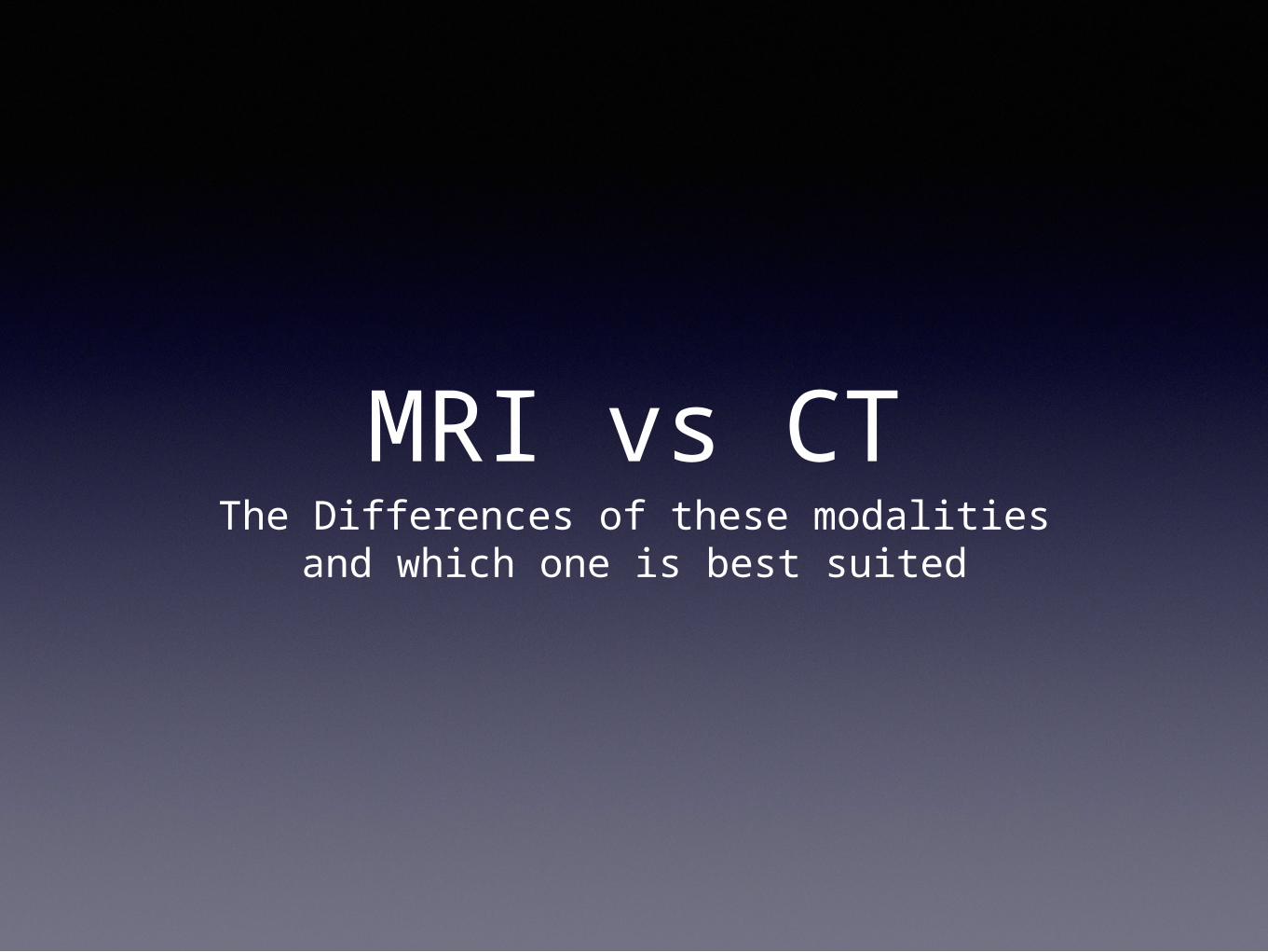
MRI vs CTThe Differences of these modalities
and which one is best suited
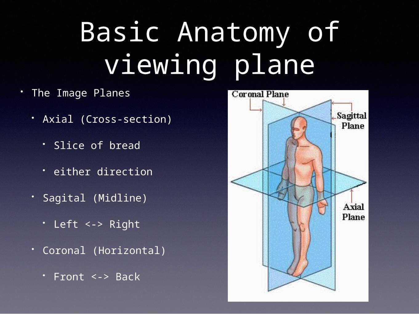
Basic Anatomy of viewing plane
• The Image Planes
• Axial (Cross-section)
• Slice of bread
• either direction
• Sagital (Midline)
• Left <-> Right
• Coronal (Horizontal)
• Front <-> Back
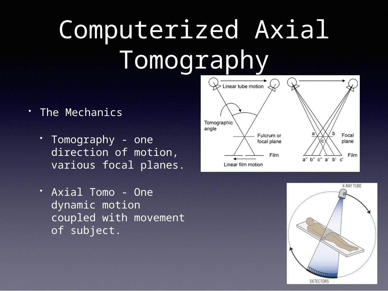
Computerized Axial Tomography
• The Mechanics
• Tomography - one direction of motion, various focal planes.
• Axial Tomo - One dynamic motion coupled with movement of subject.
Computerized Axial Tomography
• Cross-sectional imaging using X-Rays
• Acquires images extremely fast (mSec)
• Accuracy to 1mm (thin slices)
• SNR - low
• Motion sensitivity - low
• Fast Image reconstructions
Computerized Axial Tomography
• Great for imaging:
• Bone
• Lung
• Calcifications
• Heart
• Vessels (Arterial & Venous)
• Soft Tissue (with vessels)
• 3D reconstructions
Computerized Axial Tomography
• Limitations and Risks:
• Not good for Pregnancy, Kids, Sensitive Tissue (Post RTX)
• Contrast needed for imaging vessels, but low probability to cause Nephrological Toxicity
• Weight limit of 400-450 lbs
Magnetic Resonance Imaging
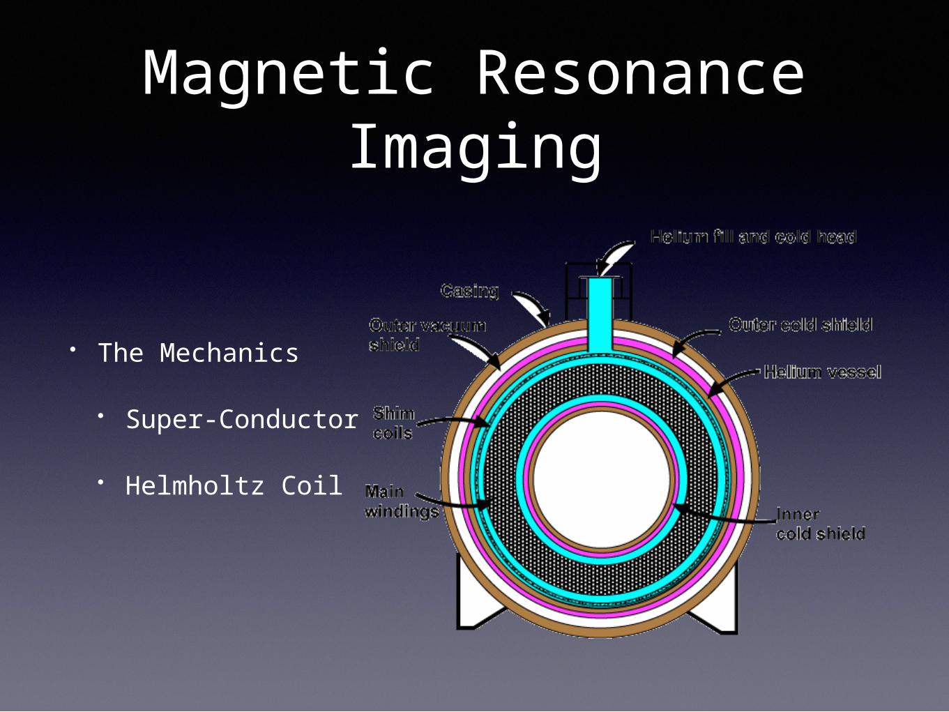
• The Mechanics
• Super-Conductor
• Helmholtz Coil
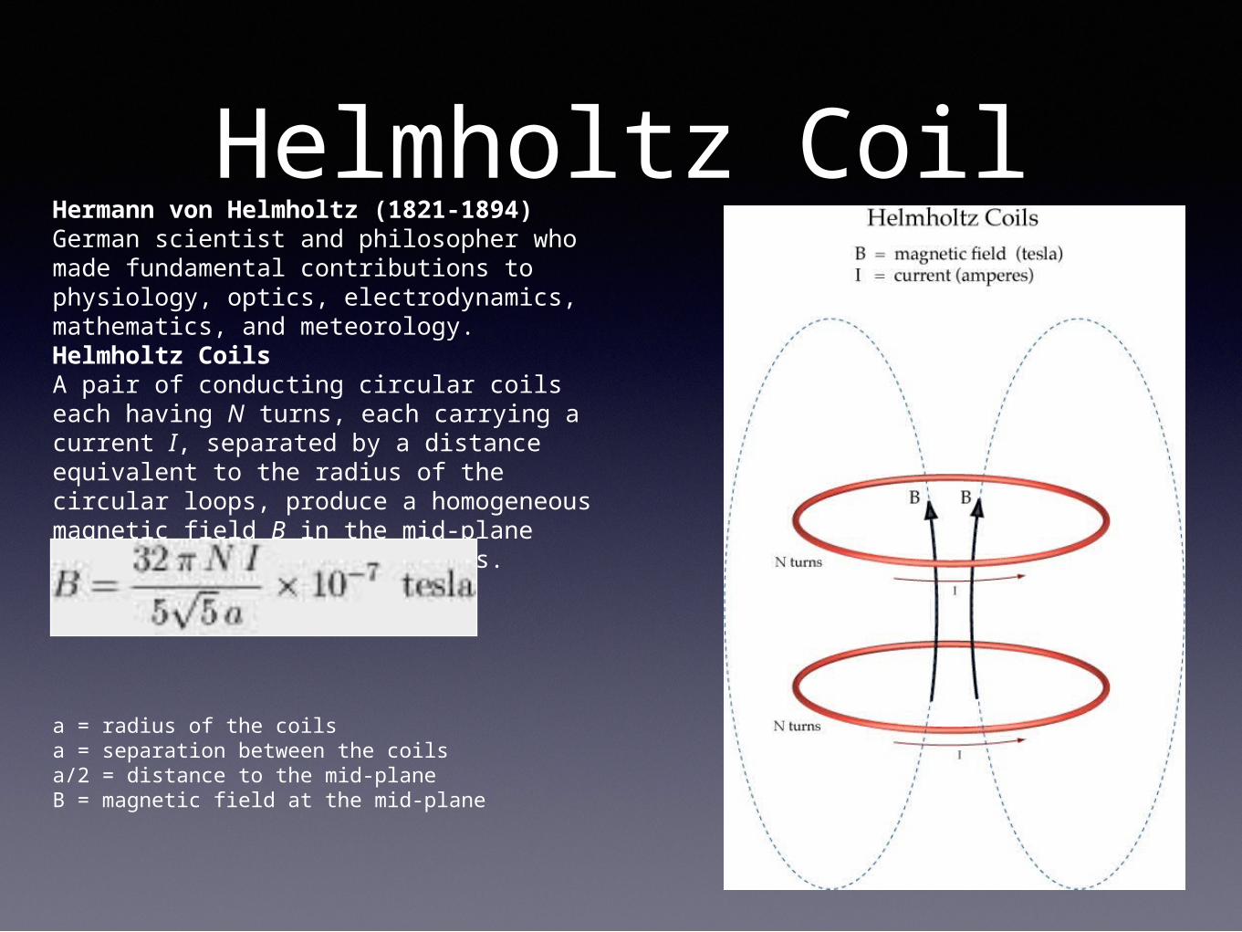
Helmholtz CoilHermann von Helmholtz (1821-1894)German scientist and philosopher who made fundamental contributions to physiology, optics, electrodynamics, mathematics, and meteorology.Helmholtz CoilsA pair of conducting circular coils each having N turns, each carrying a current I, separated by a distance equivalent to the radius of the circular loops, produce a homogeneous magnetic field B in the mid-plane between the two circular coils.
a = radius of the coilsa = separation between the coilsa/2 = distance to the mid-planeB = magnetic field at the mid-plane
Magnetic Resonance Imaging
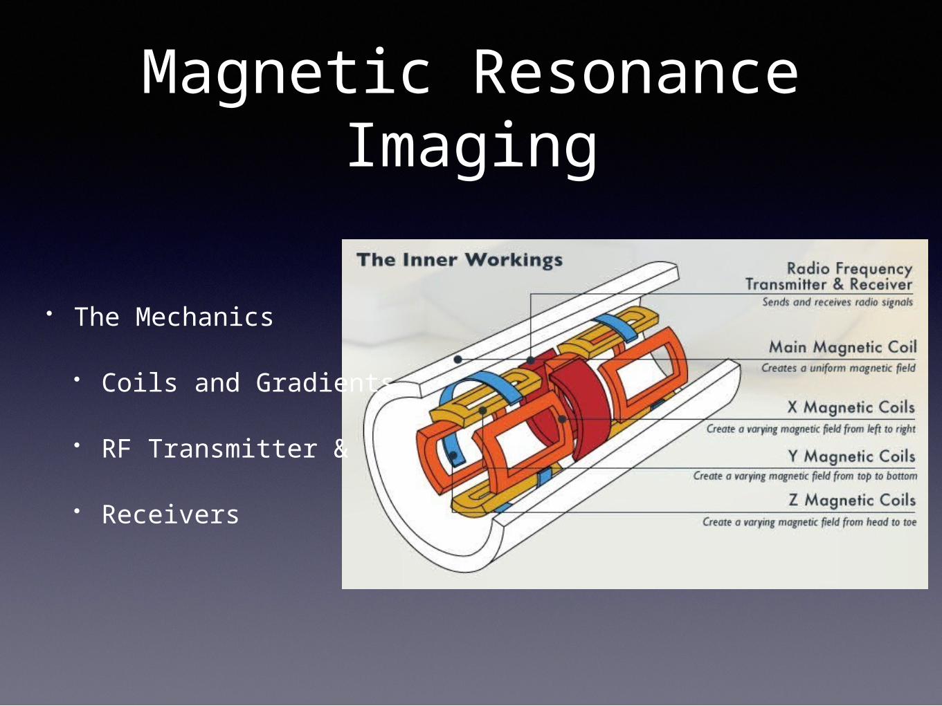
• The Mechanics
• Coils and Gradients
• RF Transmitter &
• Receivers
Magnetic Resonance Imaging
• Uses Magnetic Fields and RadioFrequency devices for acquiring images
• Imaging in any plane (X,Y,Z)
• Image time varies significantly (30sec - 45min)
• Very sensitive to Motion
• Accuracy within 1.5mm (but with many compromises and only on specific Sequences)
• Image reconstruction more time consuming
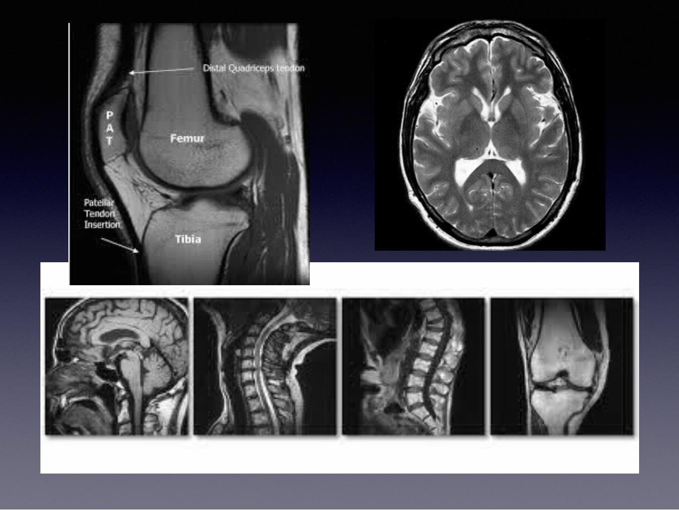
Magnetic Resonance Imaging
• Great for Imaging:
• Soft Tissue (more information than CT)
• Ligaments, Tendons, Spinal Cord
• Bone Tumors & minute abnormalities
• Vessels
Magnetic Resonance Imaging
• Limitations and Risks:
• Not good for Pacemaker, Aneurysm clips, Metal implants, Cochlear implants, etc.
• High Anxiety component
• Very sensitive to motion (time variant)
• Weight limit 300-350 lbs
• Not good for Lung or Heart Imaging
• Contrast not always needed, but higher Nephrological Toxicity
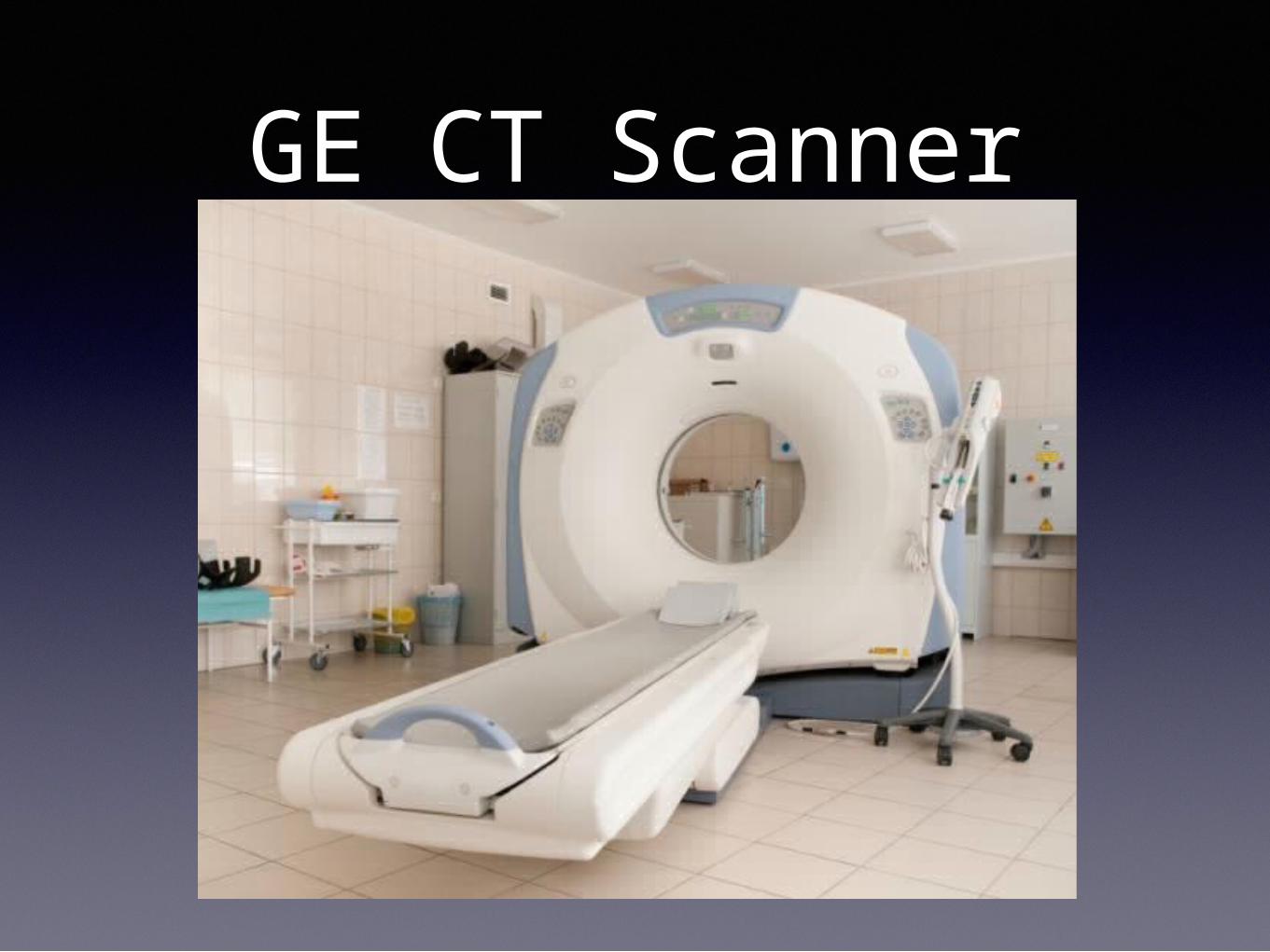
GE CT Scanner

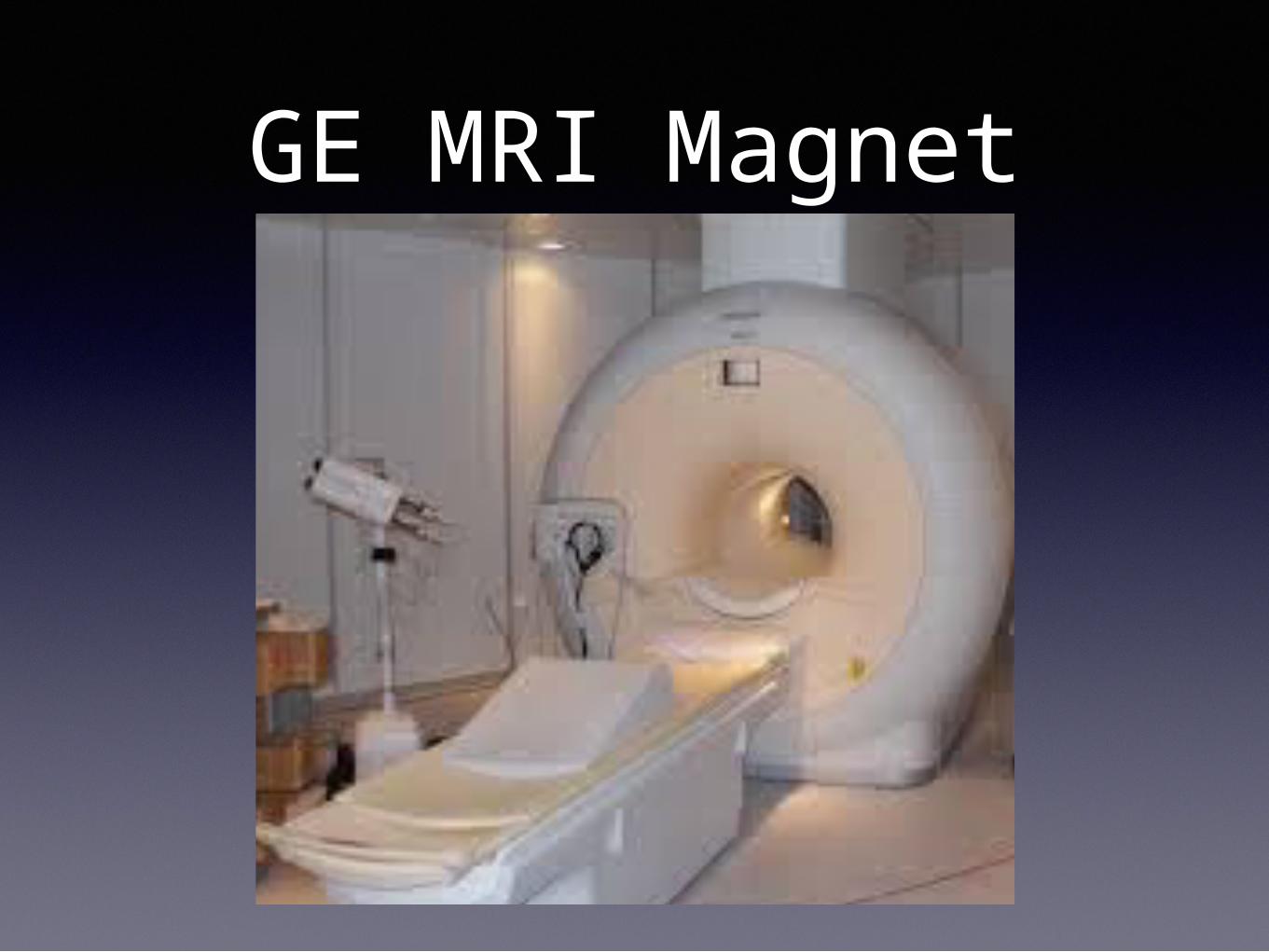
GE MRI Magnet
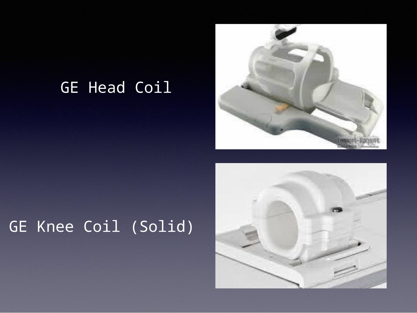
GE Head Coil
GE Knee Coil (Solid)